mini review - jsir.gr.jp
TRANSCRIPT
炎症・再生 Vol.23 No.1 2003198 Mini Review Regenerative Medicine for Spinal Cord Injury
KKKKKey wey wey wey wey wooooordsrdsrdsrdsrds spinal cord injury, neural crest stem cell, chondroitin sulfate proteoglycan,semaphorin 3A, diffusion tensor tractography
Mini Review
Regenerative medicine for spinal cord injury:Current status and open issues
Masaya Nakamura1, *), Narihito Nagoshi1,2), Kanehiro Fujiyoshi1,2),Shinjiro Kaneko1), Yoshiaki Toyama1), and Hideyuki Okano2)
1) Department of Orthopaedic Surgery, Keio University School of Medicine, Tokyo, Japan2) Department of Physiology, Keio University School of Medicine, Tokyo, Japan
Spinal cord injuries result in devastating loss of function, because spinal cord of human beings never regen-
erates after injury. People believed in this dogma for a long time. There is an emerging hope for regeneration-
based therapy of the damaged spinal cord due to the progress of neuroscience and regenerative medicine
including stem cell biology. In this review, we have summarized recent studies aimed at the development of
regeneration-based therapeutic approaches for spinal cord injuries, including therapy with transplantation of
neural crest stem cells and induction of axonal regeneration, and the establishment of new method for evalu-
ating injured and regenerated axonal fibers by MRI.
Rec.12/24/2008, 1/16/2009, pp198-203
*Correspondence should be addressed to:Nakamura M, Department of Orthopaedic Surgery, Keio University School of Medicine, 35 Shinanomachi, Shinjuku, Tokyo160-8582, Japan. Phone: +81-3-5363-3812. Fax: +81-3-3353-6597. e-mail: [email protected]
Introduction Neural stem cell transplantation is a promising regenerative
medicine strategy for the treatment of spinal cord injury (SCI).
We previously investigated the optimum timing of neural stem
cell transplantation from the perspective of microenvironments
within the injured spinal cord1-3), and successfully transplanted
rat neural stem cells into the injured spinal cord of adult rats4)
and human neural stem cells into the injured spinal cord of com-
mon marmosets5), thereby promoting functional recovery. We
believe these findings represent a significant step toward the
clinical application of neural stem cell transplantation. However,
because of various problems, it has not yet been possible to use
neural stem cells clinically. Herein, we present basic studies
that have been conducted to address various barriers against the
realization of regenerative medicine for SCI. These problems
include: (1) ethical issues related to the use of aborted fetal tis-
sues, (2) axonal growth inhibitors within the injured spinal cord,
and (3) insufficient methods for evaluating damaged and regen-
erated axons within the spinal cord.
Ethical issues related to the use of abortedfetal tissues Aborted fetus-derived cells were used in our above-mentioned
studies on neural stem cell transplantation for treating SCI. Ethi-
cal issues related to the clinical use of such cells have long been
discussed at the relevant councils of the Ministry of Health,
199Inflammation and Regeneration Vol.29 No.3 MAY 2009
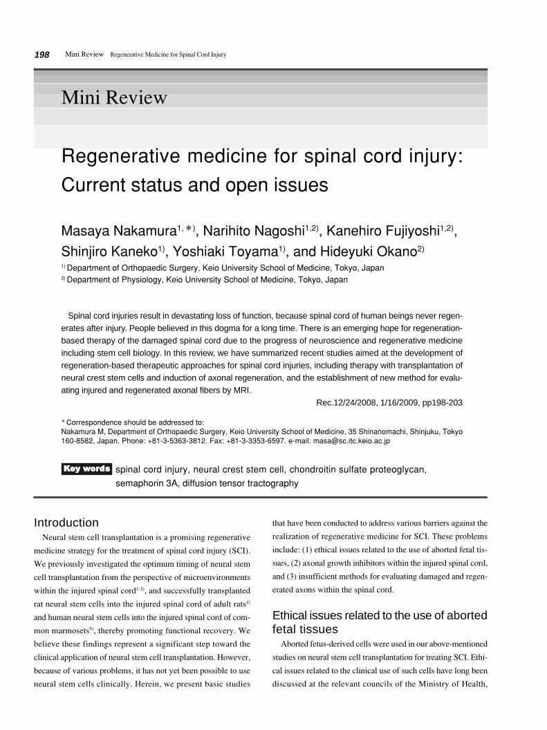
Fig.1 Avoidance of ethical problemsassociated with aborted tissue-derived neural stem cells by us-ing neural crest cells
Labour and Welfare. However, even the guidelines on the use of
human stem cells for clinical research, published in 2006, do not
include a definite stance on the validity of using aborted fetus-
derived cells (Fig.1). Because of these problems and uncertain-
ties, we have recently focused on neural crest cells derived from
the patient's own tissue as a source of somatic stem cells, rather
than on stem cells from aborted fetal tissue.
1) What is a neural crest cell?
Neural crest cells, which are induced at the border of the epi-
dermal ectoderm and neural plate during development, move to
the surrounding tissue immediately after closure of the neural
tube. The migratory neural crest cells have diverse differentiation
potentials, and are able to differentiate into neurons and glia of
the sensory and autonomic systems, adrenal medulla, pigment
cells, cranial skeleton (bone and cartilage), teeth (odontoblasts),
arterial smooth muscle, and other cell types. Neural crest cells
play an important role in many aspects of organ development
and therefore are called the “fourth germ layer”6). Migratory
neural crest cells differentiate into diverse tissues, depending on
their environment. However, a portion of these cells remains
undifferentiated and latent within various tissues while retaining
their multipotential nature, even in adult organs. These neural
crest stem cells have recently been attracting close attention as a
potential cell source for autologous transplantation, because of
their capacities of self-renewal and multipotential7).
2) Isolation and identification of neural crest stem cells
Neural crest stem cells exist in many tissues, including the
skin, intestine, heart, and corneas of adult mice8-11). Using trans-
genic PO-Cre and Wnt1-Cre/FLoxed-EGFP mice12-14), we dem-
onstrated that neural crest stem cells are also present in the dor-
sal root ganglia and bone marrow of adult mice15). Although the
presence of stem cells (serving as a source of neural cells) in
bone marrow has been shown in many reports, their embryo-
logical origin and differentiation potentials have been regarded
as questionable. There are no reports that explain how the bone
marrow-derived cells, of mesodermal origin, can differentiate
into neural cells, which are of ectodermic origin. In addressing
these questions, we found that neural crest stem cells move via
the blood into the bone marrow during early embryonic devel-
opment and remain latent in the bone marrow until adulthood,
when they can produce neurons and glia. Furthermore, when we
compared the properties of neural crest cells derived from dif-
ferent tissues (i.e. dorsal root ganglion, skin, and bone marrow)
of adult mice, we found striking differences in their differentia-
tion potential and gene expression profile . This result indicated
that the neural crest stem cells latent in each adult tissue are not
uniform, but rather retain properties that depend on their tissue
of origin.
3) Transplantation of neural crest stem cells
Several recent transplantation studies have used skin-derived
neural crest stem cells as a cell source. Miller et al. reported that
skin-derived neural crest stem cells transplanted into demyeli-
nated regions of central and peripheral axons differentiate into
Schwann cells, which subsequently engage in remyelination16).
Furthermore, these authors reported that skin-derived neural crest
stem cells transplanted into injured spinal cords also differenti-
ate into Schwann cells, and their differentiation is followed by
axonal growth and the accumulation of endogenous Schwann
cells, leading to a recovery in locomotor function17). However,
in all these reports, the neural crest stem cells were obtained
from neonatal mice. There are significant differences in the prop-
erties of neonatal and adult neural crest stem cells18).
To achieve the goal of clinical application, it is important to
evaluate the potential usefulness of neural crest stem cells de-
rived from various adult tissues. In another study, neural crest
stem cells derived from adult mouse skin were transplanted into
injured mouse spinal cord. Some of the transplanted cells survived,
but no improvement in locomotor function was described19).
Further studies are needed to determine the effectiveness of this
approach. Since neural crest stem cells are found in a variety of
adult tissues, and since their characteristics depend on their
tissue of origin, it will be essential to select the type of neural crest
炎症・再生 Vol.23 No.1 2003200 Mini Review Regenerative Medicine for Spinal Cord Injury
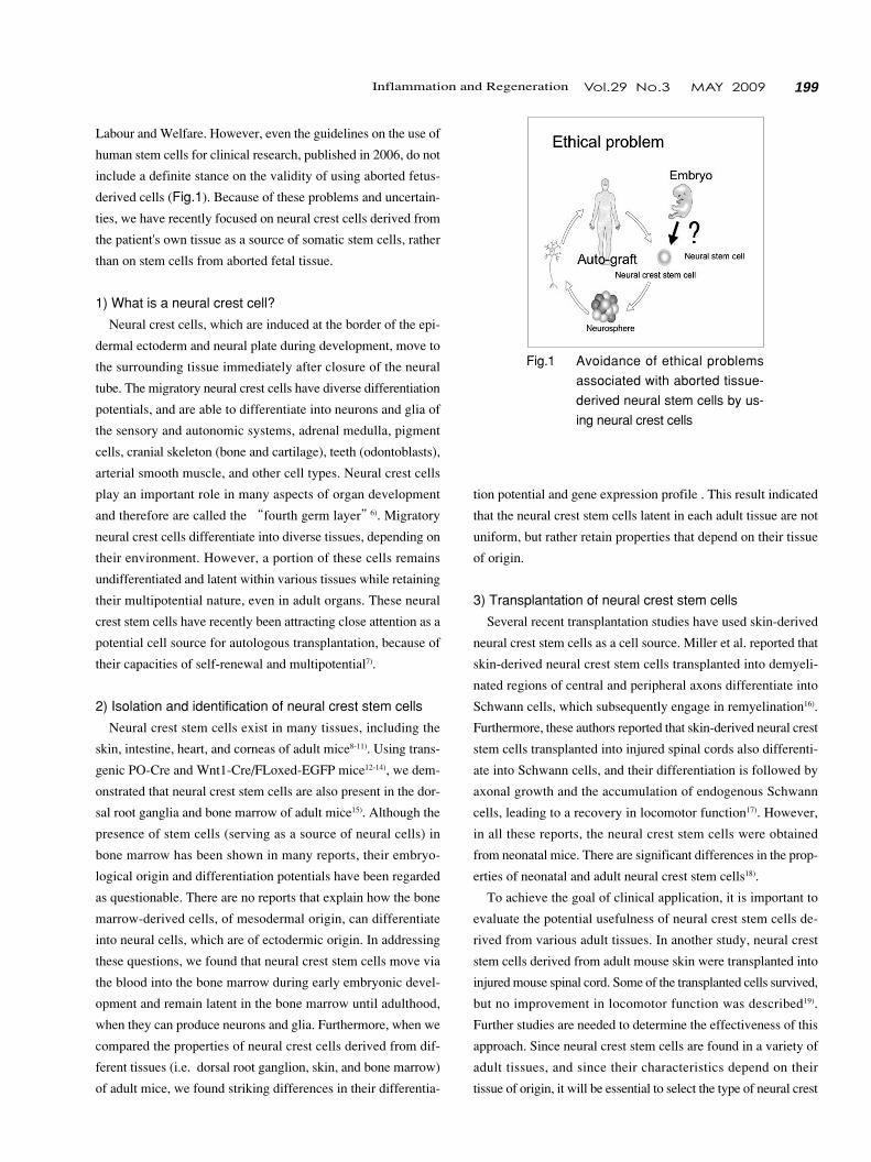
Fig.2 Axonal growth inhibitors in the injured spinalcord
stem cell for transplantation that will yield optimal results. In
any event, neural crest stem cells are somatic stem cells that can
be used for autologous transplantation. Given these features, in
terms of both ethics and safety, neural crest stem cells are a prom-
ising source for transplantation in clinical cases.
Overcoming axonal growth inhibitors Axonal growth does not occur in the injured central nervous
system, although it can take place in the injured peripheral ner-
vous system. One explanation for this is the presence of factors
that inhibit axonal regeneration in the central nervous system.
Even if effective stem cell transplantation for acute or sub-acute
SCI can be achieved, it will still be difficult to establish valid
regenerative treatments for chronic SCI unless the effects of the
axonal growth inhibitors in the central nervous system can be
overcome.
The axonal growth inhibitors found to date in the central ner-
vous system can be roughly divided into myelin-associated pro-
teins present in the myelin sheath (Nogo, MAG, and OMgp), and
extracellular matrix components present in glial scar tissue, such
as chondroitin sulfate proteoglycan (CSPG) and semaphorin 3A
(Sema 3A) (Fig.2). In recent studies, animal models of SCI were
treated with Nogo receptor antagonists (NEP1-40)20), chondroi-
tinase ABC (an enzyme involved in the degradation of CSPG)21),
and Rho signal- suppressing drugs (C3 and Y-27632)22,23). These
methods are anticipated to be of value in treating spinal cord
injuries.
We developed a Sema 3A inhibitor and applied it to the sub-
arachnoid cavity of rats for 4 weeks after complete transection
of the thoracic spinal cord. This agent stimulated axonal regen-
eration, induced vascularization, and promoted the migration of
Schwann cells into the injured area, thus facilitating the recov-
ery of leg locomotor function in the rats24). We also induced a
thoracic contusive SCI in rats and administered chondroitinase
ABC into the subarachnoid cavity of each rat for one week, be-
ginning one week after injury. The CSPG level in the injured
spinal cord decreased to a normal level after this treatment. In
the same study, neural stem cell transplantation, applied in com-
bination with chondroitinase ABC, exerted synergistic effects,
and induced more marked axonal regeneration than either treat-
ment given alone25). These results indicated that the regeneration
of injured axons can be induced by combining the use of axonal
extension inhibitors with neural stem cell transplantation. This
important finding opens the door for effective treatments for
chronic SCI.
Establishment of a method for evaluatingspinal cord regeneration The realization of regenerative medicine for the spinal cord
requires the establishment of an evaluation method. Needless to
say, axonal regeneration in the spinal projection tract is impor-
tant for achieving spinal cord regeneration. However, the ab-
sence of an established method for evaluating axonal regenera-
tion has made it clinically difficult to evaluate the responses of
the injured spinal cord to cell transplantation.
To address this need, we have focused on a particular imag-
ing technique, diffusion weighted imaging (DWI), which yields
images based on the diffusion of water molecules. Two DWI
methods, diffusion tensor imaging (DTI) and diffusion tensor
tractography (DTT), have especially attracted our attention. We
have applied these methods to the visualization of long tracts
within injured spinal cords.
1) Anisotropy and the FA map
How water molecules diffuse in the living body varies de-
pends on the nature of the local environment, and this variation
is called, “anisotropic diffusion.” For example, the white mat-
ter fibers constituting the spinal cord are highly anisotropic, and
visualization of their anisotropy should delineate axonal arrange-
ments. FA (fractional anisotropy) provides an indicator of the
magnitude of anisotropy. FA ranges from 0 to 1, where it is 0 in
cases with isotropic diffusion, and approaches 1 as the diffusion
becomes more anisotropic. An image representing anisotropy
two-dimensionally is called an“anisotropy map”or an“FA map.”
In a color FA map, different colors are assigned to different axes;
thus, fibers can be distinguished from each other by using dif-
201Inflammation and Regeneration Vol.29 No.3 MAY 2009
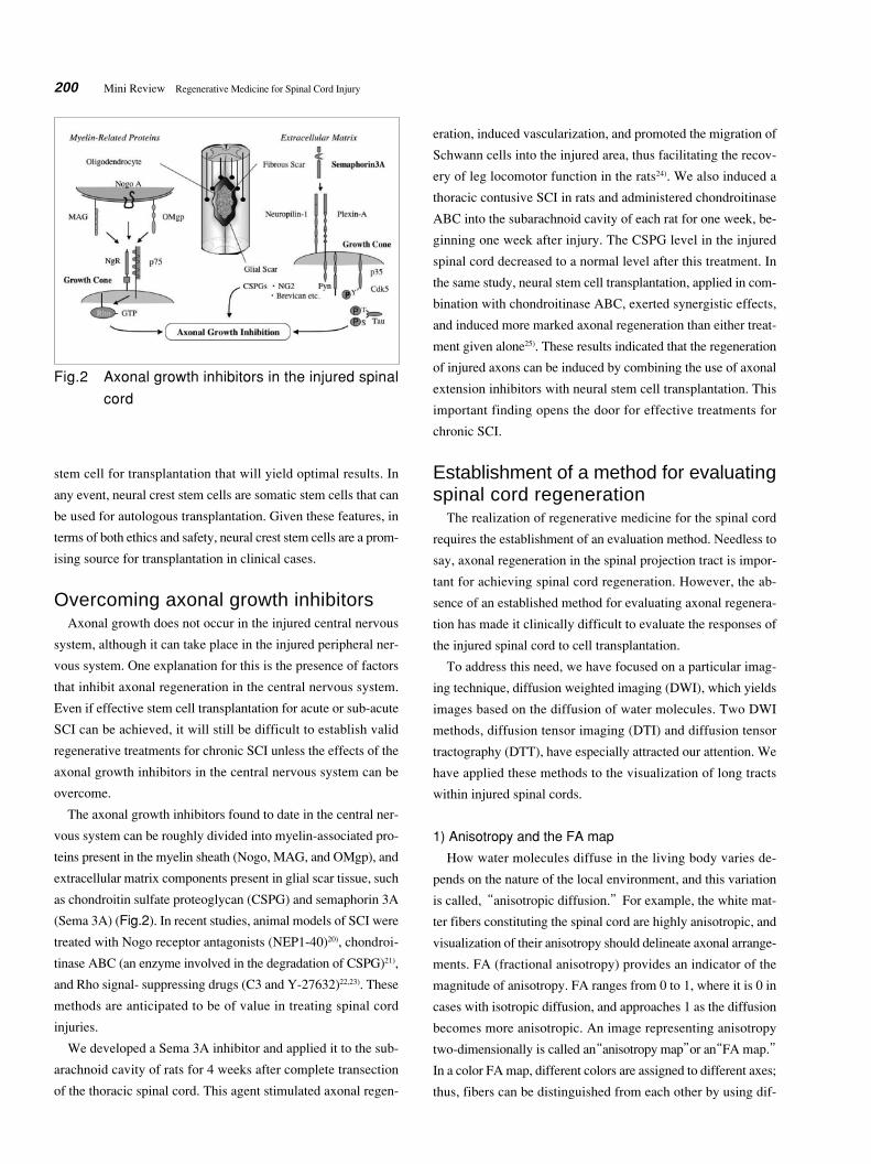
Fig.3 MRI and DTI of the normal spinal cord of acommon marmoset
T2-weighted image (T2WI) and DTI (anisotropy map and col-ored anisotropy map) of the common marmoset spinal cord(cross-section). On the anisotropy map, white matter fibers,which have high anisotropy, are depicted as high signal areas.On the colored anisotropy map, different colors are assignedto different axes. White matter fibers are blue. The image showsthe white matter to be composed of longitudinally arrangedaxonal fibers. (Reproduced from Reference 26) Fig.4 T2WI, DTT, and histological features of a
common marmoset with half-cut spinal cordCervical segment of the common marmoset spinal cord 2weeks after it was cut halfway through at the C5/6 level (apost-mortem model). A) MRI T2WI. B) Interrupted nerve fi-bers of the half-cut spinal cord are visible by DTT. C-F) Spinalcord 2-cm cranial to the half-cut level. G-J) Spinal cord at thehalf-cut level. Histological features of the HE- (F,J) and LFB-(E,I) stained specimens are well reflected by DTT (C,G) andthe colored anisotropy map (D,H). (Reproduced from Refer-ence 26)
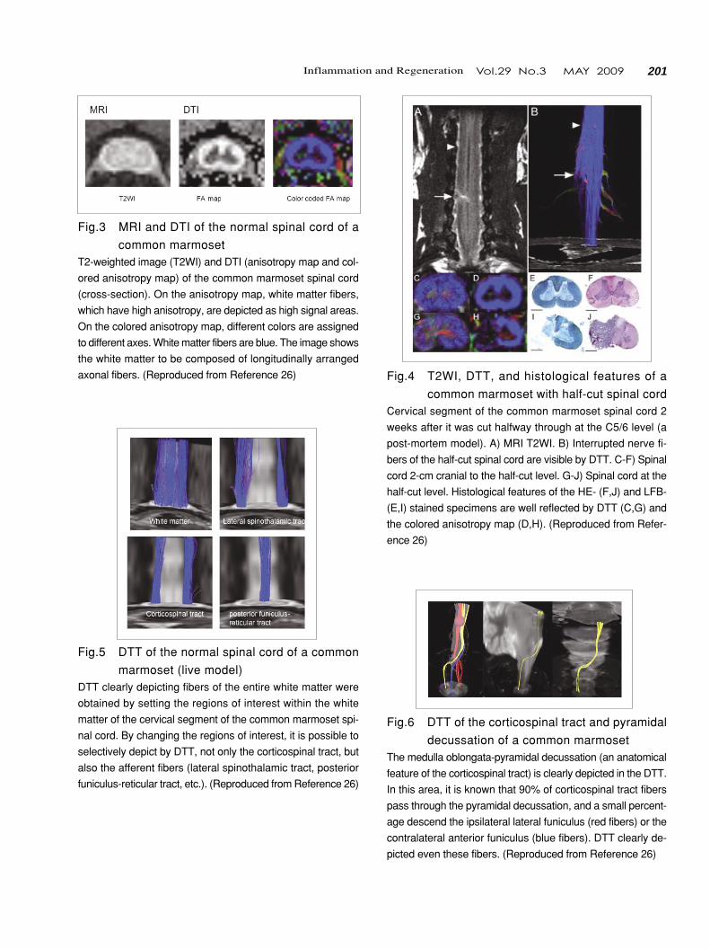
Fig.5 DTT of the normal spinal cord of a commonmarmoset (live model)
DTT clearly depicting fibers of the entire white matter wereobtained by setting the regions of interest within the whitematter of the cervical segment of the common marmoset spi-nal cord. By changing the regions of interest, it is possible toselectively depict by DTT, not only the corticospinal tract, butalso the afferent fibers (lateral spinothalamic tract, posteriorfuniculus-reticular tract, etc.). (Reproduced from Reference 26)
Fig.6 DTT of the corticospinal tract and pyramidaldecussation of a common marmoset
The medulla oblongata-pyramidal decussation (an anatomicalfeature of the corticospinal tract) is clearly depicted in the DTT.In this area, it is known that 90% of corticospinal tract fiberspass through the pyramidal decussation, and a small percent-age descend the ipsilateral lateral funiculus (red fibers) or thecontralateral anterior funiculus (blue fibers). DTT clearly de-picted even these fibers. (Reproduced from Reference 26)
炎症・再生 Vol.23 No.1 2003202 Mini Review Regenerative Medicine for Spinal Cord Injury
ferent colors according to the direction of their arrangement.
Usually, blue is assigned to fibers running longitudinally in the
spinal cord, red to fibers running laterally, and green to fibers
running dorsoventrally. Fig.3 shows a color FA map of the cross-
section of a common marmoset spinal cord. In this figure, the
blue represents white matter fibers running vertically26).
2) Diffusion tensor tractography of the spinal cord
DTT (diffusion tensor tractography) is an imaging technique
in which the direction of maximum anisotropy for each voxel is
traced. Before spinal DTT can be applied clinically, it is indis-
pensable to conduct detailed analyses to determine the extent to
which DTT reflects each tissue type, and the reliability with which
DTT depicts axonal information.
With this purpose in mind, we performed DTT for a common
marmoset after SCI. Our results yielded the first, worldwide,
clear DTT of the spinal cord of an experimental primate. In this
experiment, the cervical segment of the spinal cord of a com-
mon marmoset was cut halfway through at the C5/6 level, and
DTT was performed two weeks later (immediately following the
sacrifice of the animal). Unlike MRI, which depicts the injured
spinal cord only as changes in signal intensity on T1 and T2
weighted images, DTT allows visualization of the injury in the
form of interrupted white matter fibers (Fig.4). The examina-
tion of histological specimens stained with HE and LFB con-
firmed that DTI and DTT precisely reflected the histological
features of the injured tissue. By performing detailed post-mortem
DTT analyses of this animal model, we devised various ways to
minimize artifacts (e.g., movement caused by respiration, the
beating of cerebrospinal fluid, etc.), which enabled us to per-
form spinal cord DTT in live common marmosets. Furthermore,
by changing the regions of interest for DTT on the basis of our
neuroanatomical findings, we obtained clear projection tract-
selective DTT images in live animals (Fig.5). We have also ob-
tained images of the pyramidal decussation, which was previ-
ously considered to be impossible (Fig.6)26). We have thus dem-
onstrated that DTT is a very useful method of fiber tracking that
may replace conventional tracers for monitoring SCI and repair.
Future perspectives Basic research has been steadily advancing and overcoming
the obstacles to the realization of regenerative medicine for SCI.
Recently, induced pluripotent stem cells, developed by Yamanaka
et al27), have been attracting considerable attention as a cell source
and are expected to provide significant advancements in regen-
erative medicine. To promote regenerative medicine in Japan
and advance its techniques worldwide, further basic research
aimed at ascertaining its safety and efficacy, followed by clini-
cal trials, are essential.
References1) Nakamura M, Bregman BS: Difference in neurotrophic fac-
tor gene expression profiles between neonate and adult rat
spinal cord after injury. Exp Neurol, 169: 407-415, 2001.
2) Nakamura M, Houghtling RA, MacArthur L, et al: Differ-
ences in cytokine expression profile between acute and sec-
ondary injury in adult rat spinal cord. Exp Neurol, 184: 313-
325, 2003.
3) Okada S, Ishii K, Yamane J, Iwanami A, et al: In vivo im-
aging of engrafted neural stem cells: its application in evalu-
ating the optimal timing of transplantation for spinal cord
injury. FASEB J, 19: 1839-1841, 2005.
4) Ogawa Y, Sawamoto K, Miyata T, et al: Transplantation of
in vitro-expanded fetal neural progenitor cells results in
neurogenesis and functional recovery after spinal cord con-
tusion injury in adult rats. J Neurosci Res, 69: 925-933, 2002.
5) Iwanami A, Kaneko S, Nakamura M, et al: Transplantation
of Human Neural Stem Cells for Spinal Cord Injury in Pri-
mates. J Neurosci Res, 80: 182-190, 2005.
6) Le Douarin NM, Kalcheim C: The neural crest UK: Cam-
bridge University Press, 1999.
7) Crane JF, Trainor PA: Neural crest stem and progenitor
cells. Annu Rev Cell Dev Biol, 22: 267-286, 2006.
8) Fernandes KJ, McKenzie IA, Mill P, et al: A dermal niche
for multipotent adult skin-derived precursor cells. Nat Cell
Biol, 6: 1082-1093, 2004.
9) Kruger GM, Mosher JT, Bixby S, et al: Neural crest stem
cells persist in the adult gut but undergo changes in self-
renewal, neuronal subtype potential, and factor responsive-
ness. Neuron, 35: 657-669, 2002.
10) Tomita Y, Matsumura K, Wakamatsu Y, et al: Cardiac neu-
ral crest cells contribute to the dormant multipotent stem
cell in the mammalian heart. J Cell Biol, 170: 1135-1146,
2005.
11) Yoshida S, Shimmura S, Nagoshi N, et al: Isolation of
multipotent neural crest-derived stem cells from the adult
mouse cornea. Stem cells, 24: 2714-2722, 2006.
12) Yamauchi Y, Abe K, Mantani A, et al: A novel transgenic
technique that allows specific marking of the neural crest
cell lineage in mice. Dev Biol, 212: 191-203, 1999.
13) Danielian PS, Muccino D, Rowitch DH, et al: Modification
of gene activity in mouse embryos in utero by a tamoxifen-
203Inflammation and Regeneration Vol.29 No.3 MAY 2009
inducible form of Cre recombinase. Curr Biol, 8: 1323-1326,
1998.
14) Kawamoto S, Niwa H, Tashiro F, et al: A novel reporter
mouse strain that expresses enhanced green fluorescent
protein upon Cre-mediated recombination. FEBS Lett, 470:
263-268, 2000.
15) Nagoshi N, Shibata S, Kubota Y, et al: Multipotent Neural
Crest-Derived Stem Cells in Bone Marrow, Dorsal Root
Ganglia and Facial Skin. Cell Stem cell, 2: 392-403, 2008.
16)McKenzie IA, Biernaskie J, Toma JG, et al: Skin-derived
precursors generate myelinating Schwann cells for the in-
jured and dysmyelinated nervous system. J Neurosci, 26:
6651-6660, 2006.
17) Biernaskie J, Sparling JS, Liu J, et al: Skin-derived precur-
sors generate myelinating Schwann cells that promote
remyelination and functional recovery after contusion spi-
nal cord injury. J Neurosci, 27: 9545-9559, 2007.
18) Bixby S, Kruger GM, Mosher JT, et al: Cell-intrinsic dif-
ferences between stem cells from different regions of the
peripheral nervous system regulate the generation of neural
diversity. Neuron, 35: 643-656, 2002.
19) Sieber-Blum M, Schnell L, Grim M, et al: Characterization
of epidermal neural crest stem cell (EPI-NCSC) grafts in
the lesioned spinal cord. Mol Cell Neurosci, 32: 67-81, 2006.
20) GrandPre T, Li S, Strittmatter SM: Nogo-66 receptor an-
tagonist peptide promotes axonal regeneration. Nature, 417:
547-551, 2002.
21) Bradbury EJ, Moon LD, Popat RJ et al: Chondroitinase ABC
promotes functional recovery after spinal cord injury. Na-
ture, 416: 636-640, 2002.
22) Ellezam B, Dubreuil C, Winton M et al: Inactivation of in-
tracellular Rho to stimulate axon growth and regeneration.
Prog Brain Res, 137: 371-380, 2002.
23) Fournier AE, Takizawa BT, Strittmatter SM. Rho kinase
inhibition enhances axonal regeneration in the injured CNS.
J Neurosci, 23: 1416-1423, 2003.
24) Kaneko S, Iwanami A, Nakamura M, et al: Axonal Regen-
eration and Functional Recovery by Administration of Strong
and Selective Semaphorin3A Inhibitor into the Injured Spi-
nal Cord. Nat Med, 12: 1380-1389, 2006.
25) Ikegami T, Nakamura M, Yamane J, et al: Chondroitinase
ABC combined with neural stem/progenitor cell transplan-
tation enhances graft cell migration and outgrowth of GAP-
43-positive fibers after rat spinal cord injury. Euro J Neurosci,
2: 3036-3046, 2005.
26) Fujiyoshi K, Yamada M, Nakamura M, et al: In vivo trac-
ing of neural tracts in the intact and injured spinal cord of
marmosets by diffusion tensor tractography. J Neurosci, 27:
11991-11998, 2007.
27) Takahashi K, Yamanaka S: Induction of pluripotent stem
cells from mouse embryonic and adult fibroblast cultures
by defined factors. Cell, 126: 663-676. 2006.