methods of analysis of the nasal profile: a systematic
TRANSCRIPT

Review ArticleMethods of Analysis of the Nasal Profile: A SystematicReview with Meta-analysis
Agnieszka Jankowska ,1 Joanna Janiszewska-Olszowska ,2 Maciej Jedliński ,2
and Katarzyna Grocholewicz 2
1Private Practice “Dental Clinic Jankowscy” Zary, Poland2Department of Interdisciplinary Dentistry, Pomeranian Medical University in Szczecin, Poland
Correspondence should be addressed to Joanna Janiszewska-Olszowska; [email protected]
Received 14 October 2020; Revised 31 January 2021; Accepted 1 March 2021; Published 16 March 2021
Academic Editor: Cristina Grippaudo
Copyright © 2021 Agnieszka Jankowska et al. This is an open access article distributed under the Creative Commons AttributionLicense, which permits unrestricted use, distribution, and reproduction in any medium, provided the original work isproperly cited.
The nose is the most prominent structure of the face, influencing facial appearance and profile. Orthodontists have an awareness offacial structures, including nasal morphology, when diagnosing and treatment planning. Maxillofacial surgeons influence facialprofile by bimaxillary surgery, improving facial aesthetics and harmony. The aim of this review was to summarize the availablemethods of analysing nasal morphology and profile, and to assess their complexity. A literature search was conducted inPubMed, Scopus, Web of Science, and Embase using the following search terms: “nasal profile analysis”, “nasolabial angle”, and“nasal profile cephalometric” in order to select studies providing knowledge on correlations between occlusion and nasaldevelopment, differences between skeletal classes, ethnic variability, and differences between the sexes. Studies concerninggenetic disorders were excluded. Finally, 17 full-text papers were analysed, which pertained to nasolabial angle, or facial profileincluding the nose. Data concerning methods, ethnic group, reference landmarks used, and measurements made were extractedand placed in tables. Numerous methods of nasal profile analysis can be found in the literature. These methods describe variousnumbers of parameters, which have influence on facial aesthetic. Nasal parameters are correlated to skeletal class and nasolabialangle, positions of upper incisors, and maxillary inclination.
1. Background
The nose is the most prominent element of the face, influenc-ing facial appearance and profile [1–5]. According to thestudy by Ghorbanyjavadpour and Rakhshan [6], there aresome factors associated with the esthetics of the soft-tissueprofile, also associated with the nose, such as less prominentnoses with higher tips and subnasales anterior to the upperlip. Numerous authors deal with analysis of the facial profile[7–15]. The studies take into consideration age, sex, skeletalclass, and ethnic group. Maxillofacial surgeons influencefacial profile by bimaxillary surgery, improving facial aes-thetics and harmony [16], as well as nasal projection andnasolabial angle (NLA) [17]. Nasal growth since early child-hood as well as nasal shape and profile has been subjectedto various analyses by numerous authors [1–3, 7, 9, 14, 18–
22]. Orthodontists have an awareness of facial structures,including nasal morphology, when diagnosing and treatmentplanning in order to achieve good results after treatment ces-sation [11]. The aim of this review was to summarize theavailable methods of analysing nasal morphology and profileand to assess their complexity.
2. Material and Methods
2.1. Search Strategy. The search and the entire review wereperformed according to the PRISMA statement [23] and fol-lowing the guidelines from the Cochrane Handbook for Sys-tematic Reviews of Interventions [24]. All searching wasperformed using a combination of different MeSH termsand free-text terms. After all, the final search strategy wasdetermined by several presearches. The literature search
HindawiBioMed Research InternationalVolume 2021, Article ID 6680175, 18 pageshttps://doi.org/10.1155/2021/6680175

was conducted in following databases: PubMed, Scopus, Webof Science, and Embase using the following search terms:“nasal profile analysis” OR “nasolabial angle” OR “nasal pro-file cephalometry” on 6th December 2020. The papers ini-tially selected were subjected to detailed analysis, regardingthe methods of analysis as well as knowledge on nasal mor-phology and development.
2.2. Eligibility Criteria. The following inclusion criteria wereemployed for this review: (1) randomized clinical trials(RCTs); (2) analytical studies; (3) observational studies; (4)studies on human, healthy subjects; (5) studies published inEnglish.
Then, the following exclusion criteria were employed forthis review: (1) case reports; (2) reviews; (3) abstract andauthor debates or editorials; (4) lack of effective statisticalanalysis; (5) studies concerning congenital deformities; (6)studies evaluating theoretical algorithms, classification sys-tems, or descriptions of protocols. No limitation referringto the year of publication of the studies was imposed.
All papers found were analysed in order to select studiesproviding knowledge on correlations between occlusion andnasal development, differences between skeletal classes, eth-nic variability, and differences between the sexes.
2.3. Data Extraction. Titles and abstracts were selected inde-pendently by two authors (MJ and AJ), following the inclu-sion criteria. The full text of each identified primarilyincluded article was then analysed to find out whether itwas appropriate for inclusion. Disagreements were resolvedthrough discussion with the team supervisor (JJO). Author-ship, year of publication, data concerning methods, ethnicgroup, reference landmarks used, and measurements takenwere independently extracted by two authors (AJ and MJ)and examined by the third author (JJO).
2.4. Risk of Bias. According to the PRISMA statements, theevaluation of methodological quality gives an indication ofthe strength of evidence provided by the study because meth-odological flaws can result in bias [23]. For studies based onthe observation of structures found in radiological examina-tions, a specific scale for Clinical Studies of Radiologic Exam-inations should be applied. For this reason, it was decided touse the Arrive´ Scale [25]. It consists of 15 components, i.e.,study design, study purpose, reference standard, inclusioncriteria, indeterminate results, exclusion criteria, spectrumof patients, analysis method, analysis criteria, avoidedwork-up bias, avoided diagnostic-review bias, avoided test-review bias, intraobserver reliability, interobserver reliability,and statistical analysis, that accurately assess the bias risk,and due to their complexity, they provide detailed analysisof the results. One point is given for the compliance of thetest characteristics with the required characteristics listed inthe scale. In the event of a defect in the methodology, theresearch receives 0 points. The more points the researchreceived, the better the methodology it has.
2.5. Meta-analysis. Meta-analysis was performed usingrandom-effects model via metafor and compute.es R pack-ages [26] with Standardized Mean Differences (SMD) and
95% confidence intervals (95% CI) being calculated as effectestimates. Heterogeneity was assessed quantitatively usingI2-statistics and Cochran’s Q. [27]. The meta-analysisincluded studies that examined the values of nasiolabial angleseparately for women and men and provided SD values forboth groups.
3. Results
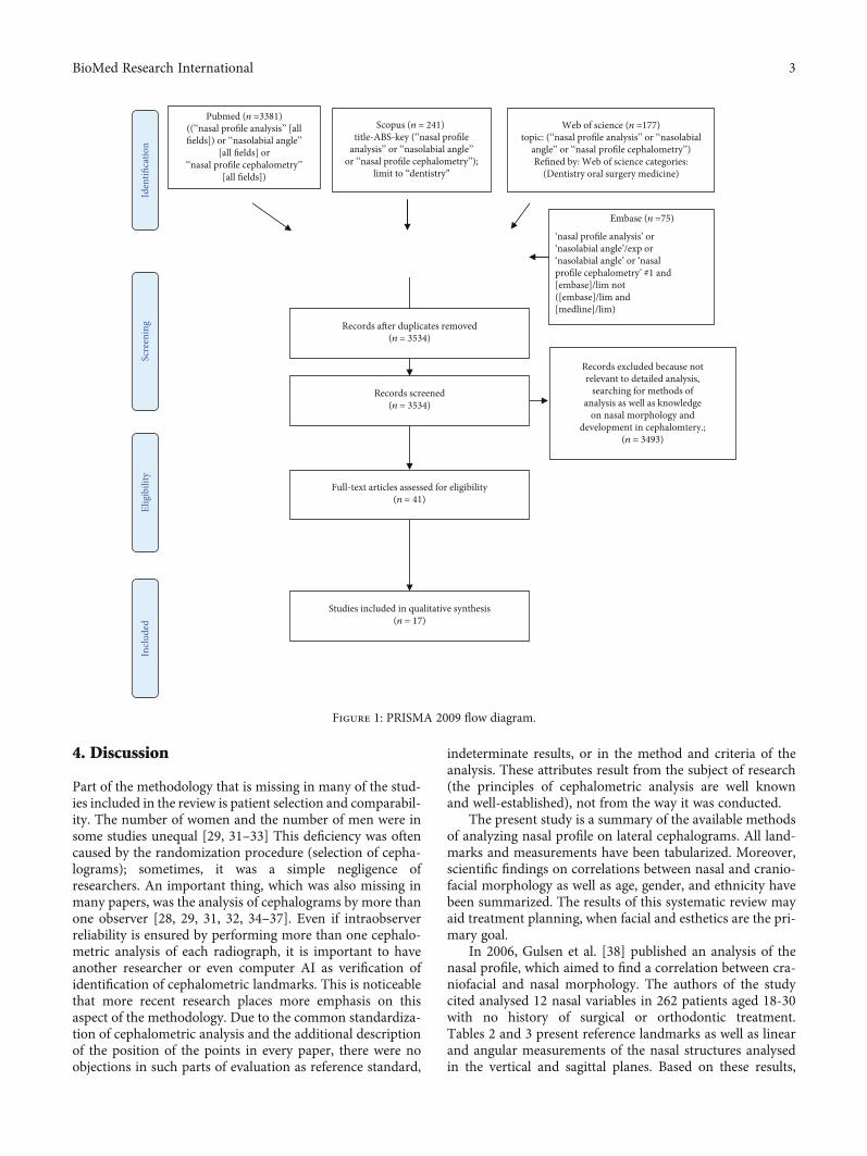
The search strategy identified 3874 potential articles: 3381from PubMed, 241 from Scopus, 177 from Web of Science,and 75 from Embase. After duplicates had been removed,3534 articles were screened. After that, 3493 papers wereexcluded because they did not correspond with the topic ofthis review. Of the remaining 41 papers, 24 were excludedbecause they were not relevant to the eligibility criteria.Finally, 17 full-text papers were included into qualitativeanalysis (Figure 1 PRISMA 2009 Flow Diagram). All ofincluded studies pertained to nasolabial angle, or facial pro-file, including the nose.
The study material of the studies included is presented inTable 1. Table 2 presents nasal and cephalometric landmarksfrom the literature. Angular and linear variables from thestudies included have been described in Table 3.
3.1. Risk of Bias. The Arrive Scale was chosen in order tounify quality assessment of all studies included in this sys-tematic review. If a decision was made to choose less specific,more popular scales, such as Newcastle-Ottawa scale orJadad scale, it would cause chaos due to overdivision throughvarious types of research, so the results of the risk of biasassessment would not be transparent. (Table 4).
3.2. Meta-analysis. Many of the studies included in thereview leave the question open as to whether gender influ-ences the nasiolabial angle. It was concluded that it isworth performing metanalysis in order to unify the resultsincluded in the review of studies and draw a common,consistent conclusion. There were 8 included studies inmetanalysis. The values and SD of NLA that were reportedare presented in Table 5.
The results by Hwang et al. [28], Paradowska–Stolarzand Kawala [29], and Kumar et al. [30] are presented sep-arately (in 2 separate groups) because of the significantfactors differing study groups. The results are shown inFigure 2. SMD should be treated as measure of genderinfluence on the value of nasiolabial angle. Positive valueof SMD indicates greater angle in male patients, negati-ve—in female patients.
Forest plot of 11 studies on gender influence on the valueof nasiolabial angle has been presented in Figure 2. Positivevalue of SMD indicates a higher angle in male patients, nega-tive—in female patients. Gender has an insignificant(p = 0:671) negative effect size. Study results are consis-tent—heterogeneity is insignificant (p = 0:228); only 18.5%of the variability come from heterogeneity. Funnel plot(Figure 3) does not reveal publication bias.
2 BioMed Research International
4. Discussion
Part of the methodology that is missing in many of the stud-ies included in the review is patient selection and comparabil-ity. The number of women and the number of men were insome studies unequal [29, 31–33] This deficiency was oftencaused by the randomization procedure (selection of cepha-lograms); sometimes, it was a simple negligence ofresearchers. An important thing, which was also missing inmany papers, was the analysis of cephalograms by more thanone observer [28, 29, 31, 32, 34–37]. Even if intraobserverreliability is ensured by performing more than one cephalo-metric analysis of each radiograph, it is important to haveanother researcher or even computer AI as verification ofidentification of cephalometric landmarks. This is noticeablethat more recent research places more emphasis on thisaspect of the methodology. Due to the common standardiza-tion of cephalometric analysis and the additional descriptionof the position of the points in every paper, there were noobjections in such parts of evaluation as reference standard,
indeterminate results, or in the method and criteria of theanalysis. These attributes result from the subject of research(the principles of cephalometric analysis are well knownand well-established), not from the way it was conducted.
The present study is a summary of the available methodsof analyzing nasal profile on lateral cephalograms. All land-marks and measurements have been tabularized. Moreover,scientific findings on correlations between nasal and cranio-facial morphology as well as age, gender, and ethnicity havebeen summarized. The results of this systematic review mayaid treatment planning, when facial and esthetics are the pri-mary goal.
In 2006, Gulsen et al. [38] published an analysis of thenasal profile, which aimed to find a correlation between cra-niofacial and nasal morphology. The authors of the studycited analysed 12 nasal variables in 262 patients aged 18-30with no history of surgical or orthodontic treatment.Tables 2 and 3 present reference landmarks as well as linearand angular measurements of the nasal structures analysedin the vertical and sagittal planes. Based on these results,
Scre
enin
gIn
clud
edEl
igib
ility
Iden
tifica
tion
Records screened(n = 3534)
Full-text articles assessed for eligibility(n = 41)
Records after duplicates removed(n = 3534)
Embase (n =75)
Studies included in qualitative synthesis(n = 17)
Pubmed (n =3381)((‘‘nasal profile analysis’’ [allfields]) or ‘‘nasolabial angle’’
[all fields] or‘‘nasal profile cephalometry’’
[all fields])
Scopus (n = 241) title-ABS-key (‘‘nasal profile
analysis’’ or ‘‘nasolabial angle’’or ‘‘nasal profile cephalometry’’);
limit to “dentistry”
Web of science (n =177)topic: (‘‘nasal profile analysis’’ or ‘‘nasolabial
angle’’ or ‘‘nasal profile cephalometry’’)Refined by: Web of science categories:
(Dentistry oral surgery medicine)
Records excluded because notrelevant to detailed analysis,
searching for methods ofanalysis as well as knowledge
on nasal morphology anddevelopment in cephalomtery.;
(n = 3493)
‘nasal profile analysis’ or‘nasolabial angle’/exp or‘nasolabial angle’ or ‘nasalprofile cephalometry’ #1 and[embase]/lim not([embase]/lim and[medline]/lim)
Figure 1: PRISMA 2009 flow diagram.
3BioMed Research International

Table1:Characteristics
ofinclud
edstud
ies.
Autho
rand
year
ofpu
blication
Typeof
stud
yStud
yobjective
Ethnic
grou
pNum
berof
subjects
Age
range
(years)
Testgrou
pCephalometric
characteristics
includ
edin
analysis
Verification
Results
Gulsenetal.
(2006)
[38]
RCT
Toevaluatethe
relation
ship
betweenthefacial
skeletalandthe
nasalp
rofile.
Anatolian
Turkish
adults
262(167
F,95
M)
18-30
Subjectswithno
prior
historiesof
trauma,
orthod
ontictreatm
ent
orfacialsurgery.
27parameters:15
facialskeletal,10
nasalsofttissue,2
nasalskeletal.
100rand
omlyselected
ceph
alogramswere
retracted2weeks
later
bythesame
orthod
ontist
The
anteriop
osterior
andvertical
classification
swere
foun
dto
besignificant
withanalysisof
NLA
,NMA,and
SFC.
Sexwas
significant
for
nasallength,
nasal
depths,h
ump,
inaddictionto
SFC.
Facialheights,lengths,
the
anteropo
sterior/vertical
position
ofthemaxilla
andmandiblewere
foun
dtobe
correlated
tonasallengthandform
.
Arshadetal.
(2013)
[31]
Analytical
stud
y
Todeterm
ineany
significant
difference
innasal
profi
lesam
ongst
subjectsin
sagittal
andverticalskeletal
patterns,and
todeterm
inegend
erdimorph
ism
ifany.
Pakistani
origin
119(81F,
38M)
18-40
Subjectswithno
prior
historiesof
orthod
ontictreatm
ent,
nocranioskeletal
synd
romes,ano
malies
andskeletal
asym
metries
12measurementsof
nasalp
rofile
20lateral
ceph
alogramswerere-
evaluatedafteron
eweekby
theprincipal
investigator.
Significant
differences
betweenskeletalclasses
I,II,and
IIIforNLA
,NMA,and
SFC.
Significant
differences
betweenmales
and
females
fornasallength,
nasald
epth,colum
ella,
convexity,andnasal
bone
length
was
foun
d.
Nehra
and
Sharma
(2009)
[34]
RCT
Toinvestigate
relation
ship
betweennasal
morph
ologyand
verticalmaxillary
skeletalpattern.
Indian
adults
190(103
F,87
M)
18-27
Subjectswho
had
undergon
eorthod
ontictreatm
ent.
Any
congenital
anom
alyor
prior
historyof
orthod
ontic
treatm
ent,surgeryor
traumato
theface.
7verticalfacial
skeletaland6nasal
softtissue
parameters
35rand
omlyselected
lateralradiographs
weretraced
twiceby
Karan
Nehra
atan
intervalof
1mon
th.
There
was
asignificant
correlationbetween
verticalmaxillary
skeletalandsofttissue
parameters.
Nasallength
significantlycorrelated
withup
peranterior
facialheight
and
inclinationof
palatal
plane.
Upw
ardnasaltip
inclinationshow
eda
4 BioMed Research International

Table1:Con
tinu
ed.
Autho
rand
year
ofpu
blication
Typeof
stud
yStud
yobjective
Ethnic
grou
pNum
berof
subjects
Age
range
(years)
Testgrou
pCephalometric
characteristics
includ
edin
analysis
Verification
Results
significant
negative
correlationwith
inclinationof
thepalatal
plane.
Skinazietal.
(1994)
[35]
Analytical
stud
y
Toexam
inethe
relative
sizesof
the
compo
nent
partsof
thesofttissue
profi
lein
onepo
pulation
ofyoun
gadults.
French
white
adults
66(21F,45M)
18-26
Subjectswithangle
classIocclusionwith
nohistoryof
orthod
ontictreatm
ent.
Twolin
es:R
ickett’s
E-line
Juanitalin
eNodata
The
meanfemaleprofi
lewas
moreconvex
and
themeanmaleprofi
lewas
relativelystraighter.
Mengetal.
(1988)
[39]
Analytical
stud
y
Todeterm
ine
grow
thchangesin
nasald
imension
sandmorph
ology
relative
tothe
pterygom
axillary
vertical(PMV)
plane.
Caucasian
origin
305lateral
ceph
alograms
(23F,
17M)
7-18
Noorthod
ontic
treatm
entperformed,
classIor
end-to-end
molar
relation
ship
withno
rmaloverbite
andoverjetat
theages
of7or
8years;classI
molar
relation
ship
withno
rmaloverbite
andoverjetat
17or
18yearsof
age.Arelaxed
lippo
sture.Sixor
moreceph
alograms
perindividu
aldistribu
tedevenly
during
theage7to
18years.
4lin
earvariablesand
2angular
measurements
54lateral
ceph
alograms
belongingto
three
maleandthreefemale
subjectsweretraced
anddigitized.
After
aperiod
oftwoweeks,
theseceph
alograms
wereretraced
and
redigitized.
Increamentsin
nose
height,d
epth
aninclinationarecomplete
ingirlsby
16yearsof
age,whilecontinuing
toincrease
inmales
upto
andbeyond
18years.
Inboth
sexestheratioof
upperto
lower
nose
heightsremains
approxim
ately3:1.
The
ratioof
theno
sedepthto
sagittaldepth:
1:2
at7yearsin
both
sexes
1:1,5(M
)and1:1,6(F)
at18
years
Upp
erno
seinclination
was
similarforboth
sexes,lower
nose
inclinationwas
slightly
larger
infemale.
Persons
withgreater
increm
entsin
nose
depththan
inno
sehight/sagittaldepth
developlarger
upper
nose
inclinations.
5BioMed Research International

Table1:Con
tinu
ed.
Autho
rand
year
ofpu
blication
Typeof
stud
yStud
yobjective
Ethnic
grou
pNum
berof
subjects
Age
range
(years)
Testgrou
pCephalometric
characteristics
includ
edin
analysis
Verification
Results
Hwangetal.
(2002)
[28]
Case-
control
stud
y
The
purposeof
this
stud
ywas
tocompare
thesoft
tissue
profi
les
obtained
from
Koreanand
Europ
ean-American
adults,inorderto
understand
the
ethn
icdifferences.
Europ
ean-
American
origin
42(27F,15M)
18-34
Subjectswithbalanced
facialestheticsand
norm
alocclusion,
nohistoryof
orthod
ontic
treatm
entor
extensive
restorativedentistry.
10angular
measurementsfor
facialform
,7lin
ear
andangular
measurementsforlip
position
Onthebasisof
anun
traced
lateral
ceph
alogram,
orthod
ontists(three
American
orthod
ontistsin
Europ
ean-American
origin
andthree
Koreanorthod
ontists
inKoreanorigin)
unanim
ouslyagreed
thateach
subjectinthe
subsam
plehadawell-
balanced
face.O
neinvestigator
traced
the
lateralcephalogram
.
Acomparisonof
the
slop
eof
theforehead
show
edno
significant
differencesbetweenthe
twogrou
ps.
Korean
origin
60(30F,
30M)
17-23
Subjectswithno
rmal
occlusion,
noprevious
orthod
ontictreatm
ent
orprosthetic
replacem
entof
teeth.
Subjectswho
show
edclassImolar
and
canine
relation
ship
withno
orminim
alcrow
ding
were
selected.
Cephalogram
swiththe
lipat
rest.
Genecov
etal.(1990)
[36]
RCT
1.Todeterm
inethe
course
ofnasal
developm
ent
2.Toevaluatethe
relative
position
oftheno
sein
relation
totherestof
thesoft
tissue
profi
ledu
ring
maturation
3.Todeterm
inethe
size,shape
and
position
ofthelip
sdu
ring
maturation
4.Tosearch
for
associations
betweenthepatterns
ofhard
andsoft
tissue
developm
ent.
Caucasian
origin
64(classI—
16F,16
M)(class
II—16
F,16
M)
7-18
Subjectswithno
historyof
orthod
ontic
treatm
ent,allergiesor
airw
ayproblems.
Lateralcephalogram
swereevaluatedatthree
timeperiod
s:1.The
mixed
dentition7-9
years;2.The
early
perm
anentdentition
11-13years;3.The
earlyadulthood16-
18years.
25lin
ear
measurementsand4
angular
measurements
10rand
omlyselected
tracings
were
redigitizedto
evaluate
measurementerror
which
was
determ
ined
tobe
insignificant.
Anteroposterior
grow
thandsubsequent
increased
anterior
projectionof
the
nose
continuedin
both
sexesafterskeletal
grow
thhadsubsided.
Females
hadalarge
proportionof
softtissue
developm
entb
yage12
whilein
males
continued
grow
thun
tilage17
resulting
greatersoft
tissuedimensions.
Duringthe
developm
entalperiod,
theangularshapeand
positionalrelationshipof
theno
se,lipsandchin
remainedrelatively
constant
forboth
sexes
andwas
relatively
independ
ento
fthe
underlying
hard
tissues.
6 BioMed Research International

Table1:Con
tinu
ed.
Autho
rand
year
ofpu
blication
Typeof
stud
yStud
yobjective
Ethnic
grou
pNum
berof
subjects
Age
range
(years)
Testgrou
pCephalometric
characteristics
includ
edin
analysis
Verification
Results
Robinson
etal.(1986)
[37]
RCT
Toinvestigatethe
relation
ship
skeletal
facialpatternand
softtissue
nasal
form
.
White
American
females
123F
11-20,6
Subjectswithno
historiesof
pathology,
trauma,surgical
intervention
,or
orthod
ontictreatm
ent.
3lin
ear
measurementsand2
angular
measurements
Nodata
Patientswithstraight
profi
lestend
edto
have
straight
noses.
Con
vexprofi
les
accompanied
convex
nasalshapes.
Con
cave
profi
leswere
foun
dwithconcave
nasalshapes.
Buschang
etal.(1993)
[40]
Case-
control
stud
y
Toqu
antify
the
child
hood
and
adolescent
grow
thchangesof
theup
per
andlower
nasal
dorsum
andto
evaluateaspectsof
nasalstructure.
French
–Canadian
females
111
ceph
alograms
(37girls)
6-14
Alongitud
inalsample
of37
girls,each
having
ceph
alogramsat
6,10
and14
yearsof
age.
1referencelin
e,3
linearmeasurements
and4angular
measurements
The
ceph
alograms
(madein
6,10
and14
yearsof
age)
were
traced
and
superimpo
sedby
one
well-trained
ceph
alom
etrist.
Metho
derrorsrange
between0.2-0.4mm.
The
upperdo
rsum
rotatesup
wardand
forw
ard
(cou
nterclockw
ise)
approxim
ately10
0
between6to
14yearsof
age.The
resultsclearly
indicatethat
changesin
thenasald
orsum
are
mostcloselyrelatedto
angulation
changesof
thelower
dorsum
,particularlydu
ring
adolescence.Rotational
changesof
thelower
dorsum
aremostclosely
relatedwithvertical
changesat
pron
asale.
Posen
(1967)
[41]
Analytical
stud
y
Toinvestigatethe
grow
thpatternof
theno
sein
norm
alperson
sfrom
infancythrough
adulthoodand
sexualdifference
innasalgrowth
patterns.
Caucasian
child
ren
477
cefalogram
s(15girlsand
15boys)
0,25-18
Subjectswithno
rmal
skeletalprofi
le.
6lin
ear
measurements
Nodata
After
theageof
14years
theno
setipdidno
tgrow
forw
ardto
the
sameextent
asdidthe
nasalb
ones.T
hese
resulted
ina
straighteningor
humping
ofthenasal
dorsum
.Nasalgrow
thchanges
both
size
andform
were
significant
aftertheage
of13
years.
7BioMed Research International

Table1:Con
tinu
ed.
Autho
rand
year
ofpu
blication
Typeof
stud
yStud
yobjective
Ethnic
grou
pNum
berof
subjects
Age
range
(years)
Testgrou
pCephalometric
characteristics
includ
edin
analysis
Verification
Results
The
nose
tipbecame
moreprom
inentwithin
thetotalfacialp
rofile
after2to
3yearsof
age
inboth
grou
ps.
Boyshadlarger
nasal
compo
nent
dimension
sthan
girls.
Girlshadgreaterdegree
ofmaturityin
nasaland
facialform
than
did
boys
inthat
age.
Magnani
etal.(2004)
[43]
Case-
control
stud
y
Toassessaverage
values
forthe
nasolabialanglein
youn
gBrazilian
individu
alsand
assessthe
occurrence
ofsexual
dimorph
ism.
You
ngBrazilian
black
subjects
3610-14
Subjectswithno
rmal
occlusionup
onclinical
exam
inationandno
historyof
orthod
ontic
treatm
ent.
The
nasolabialangle
The
caph
alom
etric
land
marks
weretraced
bysingleresearcher.
The
nasolabialangle
was
significantlysm
aller
infemales.
Fitzgerald
etal.(1992)
[32]
RCT
Todevelopa
consistent
and
reprod
ucible
metho
dof
constructing
anasolabialangle.
Americans
104(24F,
80M)
22-32
SubjectswithclassI
occlusions
withgood
facialbalance,no
historyof
orthod
ontic
treatm
entor
facial
surgery.
3nasolabial
parametersand6
skeletalangular
measurements
Allceph
alom
etric
radiograph
swere
retraced
atrand
omby
theprim
aryexam
iners
andredigitizedaftera
7-dayperiod
.
Areliablemetho
dof
constructing
the
nasolabialanglehas
been
devisedthat
includ
esangulation
sof
thelower
border
ofthe
nose
andtheup
perlip
.There
was
nostatistically
significant
difference
betweenmen
andwom
en.
Nocorrelationbetween
thesofttissue
profi
lemeasuresandthesix
skeletalmeasurements
inthewell-balanced
profi
le.
8 BioMed Research International

Table1:Con
tinu
ed.
Autho
rand
year
ofpu
blication
Typeof
stud
yStud
yobjective
Ethnic
grou
pNum
berof
subjects
Age
range
(years)
Testgrou
pCephalometric
characteristics
includ
edin
analysis
Verification
Results
Bagwan
etal.
(2014)
[33]
RCT
Toevaluatesoft
tissue
parameters
foradults,and
applying
anew
metho
dforsoft
tissue
analysisto
providegood
diagno
sisand
treatm
entplanning.
Egyptian
popu
lation
100(30F,
70M)
18-25
Subjectswithaccepted
facialprop
ortion
sand
norm
aloverjetand
overbite,A
ngle’sclassI
occlusion,
full
complem
entof
perm
anentteeth,
nohistoryof
orthod
ontic
treatm
entor
any
orthognathicor
plastic
surgery.
6lip
position
parameters,7facial
form
parameters
includ
ednasolabial
angle
Nodata
Egyptianpopu
lation
groupwerefoun
dtohave
moreconvex
faces,
protrusive
lipsandacute
nasolabialangles.
Males
hadmoreconvex
facesandprotrusive
lips
than
females.
Egyptianpopu
lation
grouphadsignificant
deviations
from
thewhite
standard
softtissue.
Paradow
ska-
Stolarzand
Kaw
ala
(2015)
[29]
Case-
control
stud
y
Tocompare
the
nasolabialangle
betweenthegrou
psof
patientswithtotal
cleftsof
thelip
,alveolar
bone
and
palateandhealthy
individu
als.
Polish
child
ren
118withcleft
(45F,
73M)
Nodata
Subjectswithclefts(27
withthebilateraland
91withtheun
ilateral
type
ofdeform
ity)
The
nasolabialangle
Nodata
Inpatientswithcleft
deform
ities,the
nasolabialanglevalues
weresm
allerthan
inhealthyindividu
als.
Amon
gthepatients
withclefts,the
oneswith
abilateraltypeof
deform
ityare
characterizedby
highest
meanvalues
ofnasolabialangle.
101healthy
(69F,32M)
The
controlgroup
was
healthypatientswith
orthod
ontictreatm
ent
needs.
Kum
aretal.
(2019)
[30]
Cross-
sectional
stud
y
Toevaluateand
compare
thesoft
tissue
grow
thchangesbetween
malesandfemalesof
twogrou
psfrom
8to
16years.
Nodata
160(group
I—40
F,40
M;
grou
pII—40
F,40
M)
8-16
(group
I—8-12
years;
grou
pII—12-
16years)
Subjectswithskeletal
classI,no
historyof
priororthod
ontic
treatm
ent,no
history
ofbone
deform
ities,or
bone
diseases,and
major
illnessin
the
past,n
ocongenital
abno
rmalitiesaffecting
grow
thand
developm
ent.
8lin
earparameters
and4angular
parametersinclud
ednasolabialangle
Nodata
Alltheparameters
increasedin
their
dimension
whileangleof
totalfacialconvexity
includingno
seand
nasolabialangle
decreases.
Amon
gthelinear
variables,no
seheight,lip
thicknessatBpoint,soft
tissuechin
thicknessand
measurementsof
lipsto
E-planewerefoun
dsignificant
forboth
subgroup
s.Malesshow
edalarger
valueofallthe
parameters
inrelationto
females.
9BioMed Research International

Table1:Con
tinu
ed.
Autho
rand
year
ofpu
blication
Typeof
stud
yStud
yobjective
Ethnic
grou
pNum
berof
subjects
Age
range
(years)
Testgrou
pCephalometric
characteristics
includ
edin
analysis
Verification
Results
Aljabaa
(2019)
[48]
RCT
Toestablishno
rmal
values
forthenasal
form
andits
relation
ship
tothe
othercranial
structures
amon
gmaleandfemale
skeletalclassI.
Saud
iadults
62(32F,
30M)
20-24
Subjectswithpleasant
facialprofi
le,class
Imolar
andcanine
relation
ship,n
ormal
overjetandoverbite,
nocrow
ding,
competent
lips,no
previous
orthod
ontic
treatm
ent,no
significant
medical
history,no
trauma
history,andno
craniofacial
deform
ities.
3nasalsize
measurements,3
nasalshape
angles,3
angularparameters
ofthenasalp
osition,
6lin
ear(horizon
tal
andvertical)
parameters
20ceph
alom
etric
radiograph
swere
rand
omlyselected
and
traced
twiceby
the
author.
There
werestatistically
significant
differences
betweentheSaud
imales
andfemales
inthenasal
length,n
asolabialangle,
horizontaldistance
from
theno
setipto
the
incisaledgeof
themost
prom
inentup
per
centralincisor,and
chin.
Malehadlonger
dorsa
andincreasedvertical
distancesfrom
the
pron
asaleto
thechin
than
females.
Females
hadlonger
verticaldistancesfrom
thepron
asaleto
the
upperlip
andlarger
nasolabialangles
than
males.
Tahaand
Ahm
ed(2020)
[49]
RCT
Toinvestigate
differencesin
the
nasalp
rofilewith
differentskeletal
classgrou
ps.
Iraqi
adults
90(classI—
15F,
15M)(class
II—15
F,15
M)(class
III—
15F,
15M)
18-25
Subjectswithno
previous
historyof
facialtrauma,
congenitaldefect,
orthod
ontictreatm
ent
oranyorthognathicor
plasticsurgeryin
the
face.
3nasalsize
measurements,3
nasalshape
angles,3
angularparameters
ofthenasalp
osition,
6lin
ear(horizon
tal
andvertical)
parameters,3angle
determ
inatethe
skeletalclass
10rand
omlyselected
patients,w
hose
ceph
alom
etric
radiograph
swere
redigitizedand
remeasuredby
the
sameinvestigator
one
mon
thlaterto
help
elim
inatemem
ory
bias.
Significant
male-female
differenceswerefoun
din
themeasurementsof
nasolabialangleandthe
horizontaldistances
relating
theno
seto
the
incisaledgeof
themost
prom
inentmaxillary
centralincisor
andto
thechin.
The
angular
measurementsof
the
nasaltip
projection
angle,naso-m
ental
angleandnaso-facial
anglewerealso
considerablevaried
amon
gthethreeskeletal
classes.
10 BioMed Research International

Gulsen et al. [38] defined numerous nasal parametersdescribing shape, size, the presence of dorsal curvatures,and nasal length or depth. The same method of nasal analysiswas later used by Arshad et al. [31] on cephalometric radio-graphs of 119 subjects aged 18-40, with no congenital defor-mities and no history of orthodontic treatment, in order toassess nasal profile in the sagittal and vertical planes and ana-lyse sexual dimorphism. The variables measured on cephalo-metric radiographs are presented in Table 3.
Nehra and Sharma [34] analysed correlation betweenvertical skeletal pattern of the maxilla and nasal morphologyon 190 pretreatment lateral cephalometric radiographs of
patients aged 18-27 years. The cephalometric variables usedcan be found in Table 3. They found a correlation betweennasal parameters (length, depth, tip angle, and nasolabialangle), maxillary and mandibular inclinations, and anteriorand posterior facial heights. They found significant correla-tions between nasal values (length and nose tip angle) andposition of the maxilla.
Changes in nasal growth, size, and morphology referringto the vertical pterygomaxillary plane (PMV) were describedby Meng et al. [39] based on 305 cephalometric radiographsof 23 females and 17 males aged 7-18 years. The referencelandmarks, lines, and angles are presented in Tables 2 and
Table 2: Nasal and cephalometric landmarks in the literature (included in the paper nos. 28-30, 32).
LandmarksDefinition
Abbreviation Name
G′ Soft tissue Glabella The most prominent point of the forehead
N′ [28–30] Soft tissue Nasion The point of greatest concavity in the midline between the forehead and the nose
N′ [32] Projected NasionThe point of intersection of the soft-tissue profile by a line drawn perpendicular to the PMV
plane through the nasion
Mn Midnasale The halfway point on nasal length (N′- Pr) that divides the dorsum into the upper and lowerdorsum
St Supratip The point constructed between midnasal and pronasal on the lower third of the nasal dorsum
Pr Pronasale The tip of the nose (nasal tip)
Prn PronasaleThe point constructed with a line drawn parallel to the PMV plane and tangential to the
anterior profile of the nose
Prn′ Projected PronasaleThe point of intersection of a line drawn parallel to the PMV plane from the projected nasion
(N′) with a line drawn perpendicular to the PMV plane through the pronasale (Prn)
Cm Columella The most convex point on the columellar-lobular junction
PCm Posterior Columella pointThe most posterior point of the lower border of the nose at which it begins to turn inferiorly to
merge with the philtrum of the upper lip
Sn Subnasale The deepest point at which the columella merges with the upper lip in the midsagittal plane
Ls Labrale superior The point indicating the mucocutaneous border of the upper lip
Ac Alar curvature point The most convex point on the nasal alar curvature
Pg′ Soft-tissue Pogonion The most anterior point on the chin in the midsagittal plane
N Nasion The intersection of the frontal and nasal bones
N1 Nasion 1 The most concave point of the nasal bone
N2 Nasion 2 The most convex point of the nasal bone
R Rhinion The most anterior and inferior point on the tip of the nasal bone
S Sella Centre of the Sella turcica
ANS Spina nasalis anterior The most prominent point of the nasal spine
Ans′ Anterior nasal spineprojected to soft tissue
The point of intersection of the soft-tissue profile with a line drawn perpendicular to the PMVplane through the anterior nasal spine (Ans)
Ans″ Projected anterior nasalspine
The point of intersection of a line drawn parallel to the PMV plane through projected nasion(N′) with a line drawn perpendicular to the PMV plane through anterior nasal spine (Ans)
PNS Spina nasalis posterior The most posterior point of the nasal spine
Go Gonion The most posterior and inferior point of the branch of the mandible
Me Menton The most inferior bony point of the mandible
Gn Gnation The most inferior and anterior point of the mandible
Se Sphenoethmoid pointThe intersection of the shadows of the greater wings of the sphenoid with the floor of the
anterior cranial fossa
Ptm Pterygomaxillary point The most inferior and posterior point on the pterygomaxillary fissure
11BioMed Research International

Table 3: Angular and linear variables (included in the papers of Ref nos. 28-30, 32, 35).
Abbreviation(unit)
Name Definition Interpretation
N′-St The axis of the dorsumDistance between the soft tissue nasion point and the
supratip pointLength of the nasal dorsum
N′-Pr Nasal length Distance between the N′ point and the Pr point Total nasal length
N LTh Nasal length Distance between the N′ point and the Pr point Total nasal length
(1) Nasal depth (1) Perpendicular distance between Pr and the N′-Sn lineSagittal position of the nose tip referring to
the face
N Dpt Nasal depth Perpendicular distance between Pr and the N′-Sn lineSagittal position of the nose tip referring to
the face
Al.-Pr Nasal depth (2) Distance between points Al and PrSagittal position of the nose tip referring to
the alar base
Prn′-Prn Nasal depthPerpendicular to the PMV plane distance between
points Prn′ and Prn on the tip of the noseThe frontal depth of the nose
PMV-Prn′ Sagittal nasal depth Distance between PMV plane and point Prn′ Sagittal depth of skeleton underlying thepronasale
N′-Prn′ Upper nose height Distance between points N′ and Prn′ Upper nose height to the point Prn′Prn′-Ans″ Lower nose height Distance between points Prn′ and Ans″ Lower nose height to the point Ans″
Hump HumpPerpendicular distance between the axis of dorsum and
its most prominent soft tissue pointConvexity of nasal dorsum
NBA Nasal base angle Angle between the G′-Sn line and the long axis of thenostril
Inclination of the nasal base referring tothe face
NMA Nasomental angleAngle between the axis of the dorsum and the Pr-WPg
lineRelation between nasal dorsum inclination
and chin position
SFCSoft tissue facial
convexity Angle between the lines G′-Sn and Sn-WPg line Profile convexity
NTP Nasal tip angleThe angle formed by the axis of the dorsum and PCm
tangentThe angle of the tip of the nose
PMV-N′-Prn Upper noseinclination
The angle formed by the line between PMV plane andpoint N′ and nasal length (N′-Prn) The angle of the upper height of the nose
PMV-Ans′-Prn
Lower noseinclination
The angle formed by the line between PMV plane andpoint Ans′ and Ans′-Prn line
The angle of the lower height of the nose
DconvLower dorsum
convexityPerpendicular distance between the Mn-Pr line and its
most prominent pointConvexity of the lower part of the nasal
dorsum
Cconv Columella convexityPerpendicular distance between the Pr-Sn line and themost anterior point on the convexity of the columella
Convexity of the nasal base
NboneL Nasal bone lengthThe line constructed between the N point and the R
pointLength of the long axis of the nasal bone
NboneA Nasal bone angle The posterior angle between the lines N1-N2 and N2-R Curvature of the nasal bone
NLA Nasolabial angle Angle between the points ctg, Sn, ULRelationship between the upper lip and the
columella
UNLANasal upward tip
angle
The posterio-inferior angle formed when the PCmtangent is extended anteriorly to intersect the
Frankfurt horizontal plane
The posterio-inferior angle formed bylower border of the nose to the Frankfurt
horizontal plane
LNLA Upper lip inclinationThe anterio-inferior angle formed by the PCm-Ls line
extended superiorly to intersect the Frankfurthorizontal plane
Inclination of the upper lip to theFrankfurt horizontal plane
GoGn-SNInclination of the
mandibular plane tothe cranium
Inclination the lines S-N to the Go-GnThe mandibular plane inclination to the
anterior cranial fossa
S-Go Posterior facial height Distance between points S and Go Posterior facial height
N-Me Anterior facial height Distance between points N and Me Anterior facial height
N-ANS Distance between point N and point ANS Upper front of the facial height
12 BioMed Research International

3. Between the ages of 7-18 years, an increase of both theupper and lower nasal height was found. However, at theage of 7, upper nasal height achieved 80% of its final measure.Moreover, girls had 90% of their final nasal height already atthe age of 7, boys after the age of 17. Nasal depth was 70% ofits final measure in girls at the age of 7, in boys at the age of11. Nasal measurements were always lower in girls than inboys.
A single study by Buschang et al. [40] was found describ-ing a growth analysis of the upper and lower parts of thenasal dorsum in children and adolescents. The nasal dorsumappeared to grow on average by 10° between 6 and 14 years ofage. Its development is correlated with nasal tip growth. Insubjects with a horizontal skeletal growth pattern, the dor-sum moves upwards and forwards. In the case of verticalgrowth, rotations occur directed downwards and backwards.
The correlation between skeletal patterns and nasal shapewas analysed by Robinson et al. [37] in 123 women, based ontwo angles and three linear measurements (Table 3). Astraight nasal dorsum was more prevalent in skeletal class I,convex (nasal hump)—in class II, whereas concave—in classIII. Nasal length and depth were strongly correlated with age.
Skinazi et al. [35] focused on analyzing nasal surface areain a French population. They drew Rickett’s Esthetic Line(from nasal tip to the most prominent chin point) and Jua-nita line (drawn from Sn (the depth of the nasolabial sulcus)to the deepest point of the labiomental sulcus) on facial pro-file and calculated surface areas of the nose, lips, and chin.Mean nasal surface area was 246:14 ± 65:51mm2 in womenand 235:4 ± 59:16mm2 in men.
Eight papers were found pertaining to NLA. The authorsof the studies cited [28, 31–33] analysed variability in nasalmorphology. The data extracted are presented in Table 5.
Both Arshad et al. [31] and Gulsen et al. [38] found a sig-nificant correlation between nasomental angle (NMA) andconvexity of facial soft tissue (SFC) and skeletal class. NMAvalues are higher in skeletal class III, lower in class II [31,38]. A higher SFC angle is found in class II, a lower in classIII [31, 38]. Moreover, Gulsen et al. [38] found correlationbetween NLA and skeletal class. Arshad et al. [31] indicatedthat concavity of the lower part of the nasal dorsum (Dconv)is strongly dependent on skeletal class. Numerous authorsproved significant sexual dimorphism concerning individualnasal variables. The results indicate that both nasal length[31, 37–39, 41, 42] and nasal depths (Ndepth1, Ndepth2)[31, 37, 38] have higher values in men than in women. Menare also characterized by higher values of the variabledescribing nasal dorsum convexity (Hump) [31, 38]. On theother hand, no significant correlations were found betweenskeletal class and the size of the nasal dorsum hump [31,38]. Gulsen et al. [38] stated that NLA in class II is higherthan in classes I and III.
Numerous authors [7–10, 28, 29, 31–34, 38, 43–47] haveused NLA, enabling them to assess nasal position in the facialprofile and, indirectly, the position of maxillary anteriorteeth. NLA is very important for orthodontic treatment plan-ning and is easy to measure. Table 5 shows that the values ofNLA differ between ethnic groups: they are lowest in Koreanand higher in European-American adults. NLA is similar inboth sexes.
The following correlations were found between nasalstructures and facial skeleton [38]: Ndepth1—positive corre-lation with mandibular length, posterior facial height, andhump; Nlength—positive correlation with anterior and pos-terior face height, maxillary and mandibular length, hump,and nasal bone length; Ndepth2—positive correlation with
Table 3: Continued.
Abbreviation(unit)
Name Definition Interpretation
Anterior maxillaryheight
ANS-Me:LAFH
Lower front of thefacial height
Ratio between the middle Height of the lower facial part
SN-PpInclination of palatal
planeThe angle between the Sella-nasion plane and the ANS-
PNS lineInclination of the palatal plane to the
anterior cranial fossa
Angle ofinclination
Angle of inclinationThe angle between the perpendicular drawn from N′
on Se-N′ line and the palatal planeThe angle between entry of the Sella soft
tissue nasion and the palatal plane
NF Nasofacial angleFormed by the intersection of the soft-tissue glabella tothe soft pogonion with the plane of the bridge of the
nose
Protrusion of the nose from the facialbones
CLColumellar to the lipangle (nasolabial
angle)
Formed by the intersection of the line from the upperlip to the glabella and the lower border of the nose
The vertical angulation of the nose tip
UN Upper nose lengthHorizontal distance from the nasion to the tip of the
noseUpper length of the nose
ANS Nose depth Horizontal distance from ANS to nose tip Depth of the nose
BN Lower nose lengthHorizontal distance from soft-tissue point A to the tip
of the noseLower length of the nose
13BioMed Research International

Table4:Characteristics
ofthestud
iesinclud
ed—accordingto
ArriveScale[25].
Autho
rsand
year
ofpu
blication
Stud
ydesign
Stud
ypu
rpose
Reference
standard
Inclusion
criteria
Indeterm
inate
results
Exclusion
criteria
Spectrum
ofpatients
Analysis
metho
dAnalysis
criteria
Avoided
work-up
bias
Avoided
diagno
stic-
review
bias
Avoided
test-
review
bias
Intraobserver
reliability
Interobserver
reliability
Statistical
analysis
Gulsenetal.
(2006)
[38]
11
11
11
01
11
11
10
1
Arshadetal.
(2013)
[31]
11
11
11
01
11
11
10
1
Nehra
and
Sharma(2009)
[34]
11
11
10
11
11
11
10
1
Skinazietal.
[35]
11
11
10
01
11
11
00
1
Mengetal.
(1988)
[39]
11
11
11
11
11
11
11
1
Hwangetal.
(2002)
[28]
11
11
11
11
11
11
11
1
Genecov
etal.
(1990)
[36]
11
11
10
11
11
11
10
1
Robinsonetal.
(1986)
[37]
11
11
10
11
10
01
0(not
prop
erly
described)
01
Buschangetal.
(1993)
[40]
11
11
11
11
11
11
11
1
Posen
(1967)
[41]
11
11
11
11
10
01
11
1
Magnani
etal.
[28],2004
11
11
11
01
11
11
10
1
Bagwan
etal.
(2014)
[33]
11
11
10
01
11
11
10
1
Fitzgeraldetal.
(1992)
[32]
11
11
10
01
11
11
11
1
Paradow
ska-
Stolarzand
Kaw
ala(2015)
[29]
11
10
10
01
11
11
0(not
prop
erly
described)
01
Kum
aretal.
(2019)
[30]
11
11
10
11
11
11
11
1
Aljabaa(2019)
[48]
11
11
10
11
11
11
11
1
Tahaand
Ahm
ed(2020)
[49]
11
11
11
01
11
11
11
1
14 BioMed Research International

maxillary length and columella convexity; Hump—positivecorrelation with anterior and posterior face height, colu-mella convexity, and nasal bone length; NLA—positivecorrelation with SFC, NBA; NMA—weak positive correla-
tion with mandibular length and position; SFC—weak pos-itive correlation with maxillary position, mandibularinclination, and strong positive correlation with facialconvexity.
Table 5: Nasolabial angle in various ethnic groups.
Authors, year of publicationNaso-labial angle (degrees) Group size Study groupFemale Male Female Male
Fitzgerald JP et al. [32], 1992 116:19 ± 9:76 113:55 ± 9:44 24 80 Americans
Hwang HS et al. [28], 2002 109:71 ± 7:60 112:05 ± 9:86 27 15 European-American origin adults
Hwang HS et al. [28], 2002 92:00 ± 9:55 91:11 ± 8:12 30 30 Korean origin adults
Arshad T et al. [31], 2013 98:87 ± 15:76 100:55 ± 14:52 81 38 Pakistani origin
Bagwan AA et al. [33], 2015 96:46 ± 11:30 94:40 ± 10:23 30 70 Egyptian adults
Paradowska-Stolarz AM and Kawala B [29], 2015 112:77 ± 13:17 116:60 ± 11:58 69 32 Poles - control groups
Paradowska- Stolarz AM and Kawala B [29], 2015 101:14 ± 17:51 100:36 ± 18:13 45 73 Poles with cleft lip and palate
Kumar A et al. [30], 2019 103:93 ± 6:78 104:25 ± 6:02 40 40 Indian origin (8-12 years)
101:05 ± 1:96 101:90 ± 4:67 40 40 Indian origin (12-16 years)
Aljabaa AH [48], 2019 104:19 ± 11:92 96:23 ± 12:74 30 30 Saudi subjects
Taha and Ahmed [49], 2020 101:73 ± 12:15 97:93 ± 9:75 15 15 Iraqi origin, skeletal class I
Total
−1.5 −1 −0.5 0 0.5 1Standardized mean difference
Taha and Ahmed
Aljabaa
Kumar et al. Teen.
Kumar et al. Child.
Paradowska-Stolarz AM Clef.
Paradowska-Stolarz AM Contr.
Bagwan AA
Arshad T
Hwang HS Korean
Hwang HS American
Fitzgerald JP
15/15
30/30
40/40
40/40
73/45
32/69
70/30
38/81
30/30
15/27
80/24
−0.34 [−1.06, 0.39]
−0.64 [−1.16, −0.12]
0.24 [−0.20, 0.67]
0.05 [−0.39, 0.49]
−0.04 [−0.41, 0.33]
0.30 [−0.12, 0.72]
−0.19 [−0.62, 0.23]
0.11 [−0.28, 0.49]
−0.10 [−0.61, 0.41]
0.27 [−0.36, 0.91]
−0.28 [−0.73, 0.18]
−0.03 [−0.19, 0.12]
Study SMD [95% CI]NM/NF
I2 = 18.5%, Q =12.92, P = 0.228
Figure 2: Forest plot of 8 studies on gender influence on the value of nasiolabial angle.
15BioMed Research International

Arshad et al. [31] reported the following findings: skeletalclasses I, II, and III are characterized by different nasal pro-files due to different values of NLA, soft tissue convexity,and low convexity of the nasal dorsum and significant differ-ences exist between women and men concerning nasal pro-files in terms of nasal length, nasal depth, hump, convexitycolumella, and nasal bone length.
In the studies by Gulsen et al. [38] and Arshad et al. [31],significantly higher values of the nasal length (Nlength),nasal depths 1 and 2 (Ndepth1 and Ndepth2), and nasalhump (Hump) were reported in men. The convexity of thelower part of the nasal dorsum (Cconv) and nasal bonelength (NboneL) were higher in men in the study by Arshadet al. [31], whereas SFC was higher in men in the study byGulsen et al. [38].
The results reported by Nehra and Sharma [34] indicate asignificant correlation between nasal length and upper fron-tal facial height, inclination of the hard palate vault, andupper facial height. An upturned nose in adults is signifi-cantly correlated with maxillary anterior rotation [34]. Thisreport is contrary to the study by Gulsen et al. [38], whofound no significant correlation between the nasolabial angle(which is strongly correlated to the upturned nose) and facialskeletal parameters. Moreover, nasal length is correlated withpalatal inclination NL (maxillary inclination) [34], similar tofindings by Gulsen et al. [38], who observed an associationbetween the nasal base angle and the inclination of the palatalplane.
Meng et al. [39] in their study on young Americansnoticed that the proportion between upper and lower nasalheight (3 : 1) is stable between the ages of 7-18 and is the samein both sexes. In males, continuous changes in nasal growthwere observed between ages 7, 13, and 18. The nose grewmore forwards than downwards [1, 39, 42]. Between the ages13 and 18, the nose moved forwards more by the increasingnasal depth (Prn′-Prn) than by increasing the distancePMV-Prn′. The highest increase in females was noticed fromage 7 to age 16. The nose grows more forwards than down-wards, similarly as in men. In comparison, Buschang et al.
[40] reported that in French-Canadian children, the nasaldorsum growth was upwards and forwards, but also in verti-cal growth, the nose directed downwards and backwards.These findings are consistent with the study by Robinsonet al. [37], who noticed the presence of nasal hump in classII. However, in females, a lower increase in nasal depth wasfound than in males [11, 39]. Meng et al. [39] concluded thatnasal growth in males is still present after the age of 18. How-ever, in females, the nose grows until the age of 16 [34, 39, 41,42].
Skinazi et al. [35] used Rickett’s E-line and the JuanitaLine. A sandwich is formed by these two lines andencloses the soft tissue profile in two thirds. Based onthese measurements, Skinazi et al. [35] concluded thatthe mean upper lip, lower lip, chin, and total area wereall statistically larger in men. They found no sexual dimor-phism in the size of the nose [35]. Referring to nasaldimensions, similar results have been reported by Scavoneet al. [8] (Japanese-Brazilian population), and contraryones—by Anić- Milošević et al. [7] (investigation in Croa-tian and American population).
5. Conclusions
(1) Numerous methods of nasal profile analysis can befound in the literature. These methods describe vari-ous numbers of parameters, which have influence onthe facial aesthetic
(2) The methods by Gulsen et al. as well as by Arshadet al. consider the highest number of variables andthus provide lots of information of potential clinicalsignificance
(3) Nasal parameters are correlated to skeletal class andnasolabial angle, positions of upper incisors, andmaxillary inclination
(4) Nasolabial angle has no sexual dimorphism, irrespec-tive of ethnic group
Funnel plot
Standardized mean difference
Stan
dard
erro
r
0.368
0.276
0.184
0.092
0
−0.5 0 0.5
Figure 3: Funnel plot of 8 studies does not reveal publication bias.
16 BioMed Research International

Abbreviations
NLA: Nasolabial anglePMV: Vertical pterygomaxillary planeTVL: True vertical lineSn: Subnasale pointNMA: Nasomental angleSFC: Convexity of facial soft tissueDconv: Concavity of lower part of nasal dorsumNdepth1: Nasal depth 1Ndepth2: Nasal depth 2Hump: Nasal dorsum convexityPrn′-Prn: Nasal depthTVL-NT: True vertical line-nasal tipTVL-B: True vertical line-point BNlength: Nasal lengthCconv: Convexity of lower part of nasal dorsumNboneL: Nasal bone length.
Data Availability
All data is a part of the manuscript.
Conflicts of Interest
The authors declare that they have no conflicts of interest.
References
[1] S. J. Chaconas, “A statistical evaluation of nasal growth,”American Journal of Orthodontics, vol. 56, no. 4, pp. 403–414, 1969.
[2] L. G. Farkas, J. C. Kolar, and I. R. Munro, “Geography of thenose: a morphometric study,” Aesthetic Plastic Surgery,vol. 10, no. 1, pp. 191–223, 1986.
[3] J. F. Manera and J. D. Subtelny, “A cephalometric study of thegrowth of the nose,” American Journal of Orthodontics, vol. 47,pp. 703–705, 1961.
[4] C. J. Burstone, “The integumental profile,” American Journalof Orthodontics, vol. 44, no. 1, pp. 1–25, 1958.
[5] O. Van Schijndel, A. J. Tasman, and R. Litschel, “The noseinfluences visual and personality perception,” Facial PlasticSurgery, vol. 31, no. 5, pp. 439–445, 2015.
[6] F. Ghorbanyjavadpour and V. Rakhshan, “Factors associatedwith the beauty of soft-tissue profile,” American Journal ofOrthodontics and Dentofacial Orthopedics, vol. 155, no. 6,pp. 832–843, 2019.
[7] S. Anić-Milošević, S. Meštrović, M. Lapter-Varga,J. Dumancic, and M. Šlaj, “Analysis of the soft tissue profilein Croatians with normal occlusions and well-balanced faces,”European Journal of Orthodontics, vol. 33, no. 3, pp. 305–310,2011.
[8] H. Scavone Jr., H. Trevisan Jr., D. G. Garib, and F. V. Ferreira,“Facial profile evaluation in Japanese-Brazilian adults withnormal occlusions and well-balanced faces,” American Journalof Orthodontics and Dentofacial Orthopedics, vol. 129, no. 6,pp. 721.e1–721.e5, 2006.
[9] P. Fernández-Riveiro, E. Smyth-Chamosa, D. Suárez-Quinta-nilla, and M. Suárez-Cunqueiro, “Angular photogrammetricanalysis of the soft tissue facial profile,” European Journal ofOrthodontics, vol. 25, no. 4, pp. 393–399, 2003.
[10] C. Tanikawa and K. Takada, “Objective classification of nose-lip-chin profiles and their relation to dentoskeletal traits,”Orthodontics & Craniofacial Research, vol. 17, no. 4, pp. 226–238, 2014.
[11] S. J. Chaconas and J. D. Bartroff, “Prediction of normal soft tis-sue facial changes,” The Angle Orthodontist, vol. 45, no. 1,pp. 12–25, 1975.
[12] L. D. Garner, “Soft-tissue changes concurrent with orthodon-tic tooth movement,” American Journal of Orthodontics,vol. 66, no. 4, pp. 367–377, 1974.
[13] G. E. Marchiori, L. O. Sodré, T. C. R. da Cunha, F. C. Torres,H. D. Rosário, and L. R. Paranhos, “Pleasantness of facial pro-file and its correlation with soft tissue cephalometric parame-ters: perception of orthodontists and lay people,” EuropeanJournal of Dentistry, vol. 9, no. 3, pp. 352–355, 2019.
[14] K. Lopatienė, A. Šidlauskas, A. Vasiliauskas, L. Čečytė,V. Švalkauskienė, and M. Šidlauskas, “Relationship betweenmalocclusion, soft tissue profile, and pharyngeal airways: acephalometric study,” Medicina, vol. 52, no. 5, pp. 307–314,2016.
[15] C. N. Bronfman, G. Janson, A. Pinzan, and T. L. Rocha, “Ceph-alometric norms and esthetic profile preference for the Japa-nese: a systematic review,” Dental Press Journal ofOrthodontics, vol. 20, no. 6, pp. 43–51, 2015.
[16] G. I. Marşan, N. Cura, and U. Emekli, “Soft and hard tissuechanges after bimaxillary surgery in Turkish female class IIIpatients,” Journal of Cranio-Maxillo-Facial Surgery, vol. 37,no. 1, pp. 8–17, 2009.
[17] P. Galzignato, D. Bertossi, M. Albanese et al., “Variations ofthe profile of the nose of the upper lip,” Minerva Stomatolo-gica, vol. 68, no. 3, pp. 105–111, 2019.
[18] C. N. Stephan, M. Henneberg, and W. Sampson, “Predictingnose projection and pronasale position in facial approxima-tion: a test of published methods and proposal of new guide-lines,” American Journal of Physical Anthropology, vol. 122,no. 3, pp. 240–250, 2003.
[19] B. S. Clements, “Nasal imbalance and the orthodontic patient,”American Journal of Orthodontics, vol. 55, no. 3, pp. 244–264,1969.
[20] B. S. Clements, “Nasal imbalance and the orthodontic patient,”American Journal of Orthodontics, vol. 55, no. 4, pp. 329–352,1969.
[21] A. F. Ridel, F. Demeter, J. Liebenberg, E. N. L’Abbé,D. Vandermeulen, and A. C. Oettlé, “Skeletal dimensions aspredictors for the shape of the nose in a South African sample:a cone-beam computed tomography (CBCT) study,” ForensicScience International, vol. 289, pp. 18–26, 2018.
[22] R. L. Crumley and M. Lanser, “Quantitative analysis of nasaltip projection,” Laryngoscope, vol. 98, no. 2, pp. 202–208, 1988.
[23] D. Moher, A. Liberati, J. Tetzlaff, D. G. Altman, and ThePRISMA Group, “Preferred reporting items for systematicreviews and meta-analyses: the PRISMA statement,” PLoSMedicine, vol. 6, no. 7, article e1000097, 2009.
[24] J. P. T. Higgins and S. Green, Cochrane Handbook for System-atic Reviews of Interventions Version 5.1.0 [updated March2011]2011, http://www.handbook.cochrane.org/.
[25] L. Arrivé, R. Renard, F. Carrat et al., “A scale of methodologicalquality for clinical studies of radiologic examinations,” Radiol-ogy, vol. 217, no. 1, pp. 69–74, 2000.
[26] G. Wells, B. Shea, D. O’Connell, J. E. Peterson, and V. Welch,“The Newcastle-Ottawa Scale (NOS) for assessing the quality
17BioMed Research International

of case-control studies in meta-analyses,” European Journal ofEpidemiology, vol. 25, pp. 603–605, 2011.
[27] J. P. Higgins and S. G. Thompson, “Quantifying heterogeneityin a meta-analysis,” Statistics in Medicine, vol. 21, no. 11,pp. 1539–1558, 2002.
[28] H. S. Hwang, W. S. Kim, and McNamara JAJr., “Ethnic differ-ences in the soft tissue profile of Korean and European-American adults with normal occlusions and well-balancedfaces,” The Angle Orthodontist, vol. 72, no. 1, pp. 72–80, 2002.
[29] A. M. Paradowska-Stolarz and B. Kawala, “The nasolabialangle among patients with total cleft lip and palate,” Advancesin Clinical and Experimental Medicine, vol. 24, no. 3, pp. 481–485, 2015.
[30] A. Kumar, P. Tandon, G. K. Singh, and G. P. Singh, “Soft tissuegrowth changes from 8 to 16 years of age: A cross-sectionalstudy,” National Journal of Maxillofacial Surgery, vol. 10,no. 2, pp. 161–167, 2019.
[31] T. Arshad, A. Shaikh, and M. Fida, “Comparison of nasal pro-files in various skeletal patterns,” Journal of Ayub Medical Col-lege, Abbottabad, vol. 25, pp. 1-2, 2013.
[32] J. P. Fitzgerald, R. S. Nanda, and G. F. Currier, “An evaluationof the nasolabial angle and the relative inclinations of the noseand upper lip,” American Journal of Orthodontics and Dento-facial Orthopedics, vol. 102, no. 4, pp. 328–334, 1992.
[33] A. A. Bagwan, M. I. al-Shennawy, and M. M. Alskhawy, “Eval-uation of soft tissue parameters for adults with accepted occlu-sion using Legan and Burstone analysis,” Tanta DentalJournal, vol. 12, no. 1, pp. 1–6, 2015.
[34] K. Nehra and V. Sharma, “Nasal morphology as an indicatorof vertical maxillary skeletal pattern,” Journal of Orthodontics,vol. 36, no. 3, pp. 160–166, 2014.
[35] G. L. Skinazi, S. J. Lindauer, and R. J. Isaacson, “Chin, nose,and lips. Normal ratios, in young men and women,” AmericanJournal of Orthodontics and Dentofacial Orthopedics, vol. 106,no. 5, pp. 518–523, 1994.
[36] J. S. Genecov, P. M. Sinclair, and P. C. Dechow, “Developmentof the nose and soft tissue profile,” The Angle Orthodontist,vol. 60, no. 3, pp. 191–198, 1990.
[37] J. M. Robison, D. J. Rinchuse, and T. G. Zullo, “Relationship ofskeletal pattern and nasal form,” American Journal of Ortho-dontics, vol. 89, no. 6, pp. 499–506, 1986.
[38] A. Gulsen, C. Okay, B. I. Aslan, O. Uner, and R. Yavuzer, “Therelationship between craniofacial structures and the nose inAnatolian Turkish adults: a cephalometric evaluation,” Amer-ican Journal of Orthodontics and Dentofacial Orthopedics,vol. 130, no. 2, pp. 131.e15-132.e25, 2006.
[39] H. P. Meng, J. Goorhuis, S. Kapila, and R. S. Nanda, “Growthchanges in the nasal profile from 7 to 18 years of age,” Ameri-can Journal of Orthodontics and Dentofacial Orthopedics,vol. 94, no. 4, pp. 317–326, 1988.
[40] P. H. Buschang, R. De La Cruz, A. D. Viazis, and A. Demirjian,“Longitudinal shape changes of the nasal dorsum,” AmericanJournal of Orthodontics and Dentofacial Orthopedics,vol. 104, no. 6, pp. 539–543, 1993.
[41] J. M. Posen, “A longitudinal study of the growth of the nose,”American Journal of Orthodontics, vol. 53, no. 10, pp. 746–756,1967.
[42] J. D. Subtelny, “A longitudinal study of soft tissue facial struc-tures and their profile characteristics, defined in relation tounderlying skeletal structures,” American Journal of Ortho-dontics, vol. 45, no. 7, pp. 481–507, 1959.
[43] M. B. Magnani, D. F. Nouer, P. R. Nouer, J. S. Pereira Neto,I. U. Garbui, and E. M. Böeck, “Assessment of the nasolabialangle in young Brazilian black subjects with normal occlu-sion,” Brazilian Oral Research, vol. 18, no. 3, pp. 233–237,2004.
[44] G. W. Arnett, J. S. Jelic, J. Kim et al., “ORIGINAL ARTICLE:Soft tissue cephalometric analysis: Diagnosis and treatmentplanning of dentofacial deformity,” American Journal ofOrthodontics and Dentofacial Orthopedics, vol. 116, no. 3,pp. 239–253, 1999.
[45] C. J. Burstone, “Lip posture and its significance in treatmentplanning,” American Journal of Orthodontics, vol. 53, no. 4,pp. 262–284, 1967.
[46] A. H. Owen 3rd, “Diagnostic block cephalometrics. Part 1,”Journal of clinical orthodontics: JCO, vol. 18, no. 6, pp. 400–422, 1984.
[47] J. A. McNamara Jr., “A method of cephalometric evaluation,”American Journal of Orthodontics, vol. 86, no. 6, pp. 449–469, 1984.
[48] A. H. Aljabaa, “Lateral cephalometric analysis of the nasalmorphology among Saudi adults,” Clinical, Cosmetic andInvestigational Dentistry, vol. Volume 11, pp. 9–17, 2019.
[49] A. A. Taha and A. S. Ahmed, “A comprehensive assessment ofthe nasal profile among Iraqi adults with different skeletal clas-ses: a retrospective study,” Journal of Oral Biology and Cranio-facial Research, vol. 10, no. 2, pp. 175–179, 2020.
18 BioMed Research International