mesenchymal stem cells with overexpression of midkine enhance
TRANSCRIPT

Zhao et al. Stem Cell Research & Therapy 2014, 5:37http://stemcellres.com/content/5/2/37
RESEARCH Open Access
Mesenchymal stem cells with overexpression ofmidkine enhance cell survival and attenuatecardiac dysfunction in a rat model of myocardialinfarctionShu-Li Zhao1,2†, Yao-Jun Zhang1,3†, Ming-Hui Li1, Xin-Lei Zhang1 and Shao-Liang Chen1*
Abstract
Introduction: Elevated midkine (MK) expression may contribute to ventricular remodeling and ameliorate cardiacdysfunction after myocardial infarction (MI). Ex vivo modification of signaling mechanisms in mesenchymal stemcells (MSCs) with MK overexpression may improve the efficacy of cell-based therapy. This study sought to assess thesafety and efficacy of MSCs with MK overexpression transplantation in a rat model of MI.
Methods: A pLenO-DCE vector lentivirus encoding MK was constructed and infected in MSCs. MSC migrationactivity and cytoprotection was examined in hypoxia-induced H9C2 cells using transwell insert in vitro. Rats wererandomized into five groups: sham, MI plus injection of phosphate buffered saline (PBS), MSCs, MSCs-greenfluorescent protein (MSCs-GFP) and MSCs-MK, respectively. Survival rates were compared among groups using log-rank test and left ventricular function was measured by echocardiography at baseline, 4, 8 and 12 weeks.
Results: Overexpression of MK partially prevented hypoxia-induced MSC apoptosis and exerted MSC cytoprotectionto anoxia induced H9C2 cells. The underlying mechanisms may be associated with the increased mRNA and proteinlevels of vascular endothelial growth factor (VEGF), transformation growth factor-β (TGF-β), insulin-like growth factor1 (IGF-1) and stromal cell-derived factor 1 (SDF-1a) in MSCs-MK compared with isolated MSCs and MSCs-GFP. Consistentwith the qPCR results, the culture supernatant of MSCs-MK had more SDF-1a (9.23 ng/ml), VEGF (8.34 ng/ml) and TGF-β1(17.88 ng/ml) expression. In vivo, a greater proportion of cell survival was observed in the MSCs-MK group than in theMSCs-GFP group. Moreover, MSCs-MK administration was related to a significant improvement of cardiac functioncompared with other control groups at 12 weeks.
Conclusions: Therapies employing MSCs with MK overexpression may represent an effective treatment for improvingcardiac dysfunction and survival rate after MI.
IntroductionMyocardial dysfunction after acute myocardial infarction(MI) is a progressive condition, which is clearly associ-ated with a poor prognosis and results in heart failureand cardiac death [1-4]. Over the past decade, a numberof studies have documented that mesenchymal stem cell(MSC) therapy may have a favorable impact on cardiacfunction in experimental animal models or patients after
* Correspondence: [email protected]†Equal contributors1Department of Cardiology, Nanjing First Hospital, Nanjing MedicalUniversity, No. 68 Changle Road, Nanjing 210006, ChinaFull list of author information is available at the end of the article
© 2014 Zhao et al.; licensee BioMed Central LCommons Attribution License (http://creativecreproduction in any medium, provided the or
MI [5,6]. Our previous studies reported that autologousbone marrow MSCs transplantation improved cardiacfunction in 96 patients with acute MI who experienced per-cutaneous coronary intervention and the cardioprotectiveeffect remained six months after the procedure [7,8].Recently, studies from several preclinical experimentssuggested that genetic strategies may play a critical rolein improving MSC survival and differentiation [9-13].However, insight into the mechanistic issues underlyingthe effect of genetically altered MSC transplantationremains unsettled, especially for finding a gene or a set ofgenes that potentially have both autocrine and paracrineeffects in advancing MSC-directed myocardial repair.
td. This is an Open Access article distributed under the terms of the Creativeommons.org/licenses/by/2.0), which permits unrestricted use, distribution, andiginal work is properly cited.
Zhao et al. Stem Cell Research & Therapy 2014, 5:37 Page 2 of 13http://stemcellres.com/content/5/2/37
Midkine (MK) is a heparin-binding growth factor witha molecular weight of 13 kDa, first isolated as a productof the retinoic acid-responsive gene in the embryonalcarcinoma cell differentiation system [14]. MK has variousbiological activities, which promote neurite outgrowth,the survival of embryonic neurons, and angiogenic action[15]. In an experimental study comparing cardiac functionafter ischemia/perfusion (I/R) in wild-type mice and MK-deficient mice, there was a significant increased infarctarea and adverse left ventricular (LV) fractional shortening(FS) in MK−/− mice [16]. Interestingly, supplemental ap-plication of MK protein to the Mk−/− mice at the time ofI/R significantly reduced the infarcted size [16,17]. Alter-natively, the studies from the H Takenaka and S Fukuigroups showed that MK prevented the cardiac remodelingof mice after MI through an enhancement of angiogenesisand subsequently improved the survival rate. Apart fromangiogenesis, additional research found that MK promo-ted the growth of mouse embryonic stem cells by inhi-biting apoptosis through the PI3K/Akt signaling pathway[18]. Therefore, MK application is recently regarded as anew therapeutic strategy for the treatment of ischemicheart failure [19,20].In the present study, we tested the hypothesis that the
combination of MSC transplantation and MK overex-pression is superior to MSC transplantation in the treat-ment of rat MI models with decreased infarct size andimproved cardiac function. LV function and angiogenesiswere separately evaluated by echocardiography and im-munohistochemistry staining after transplantation. Thebiological activities of MSCs were also examined.
MethodsAnimalsHealthy female Sprague-Dawley rats (weighing 60 to80 g for isolation of MSCs and 200 to 220 g for an MImodel) were obtained from the Vital River LaboratoryAnimal Co., Ltd., Beijing Laboratory Animal ResearchCenter (Beijing, China); and were housed in specificpathogen-free conditions at Nanjing First Hospital AnimalCenter (Nanjing, China), in a room controlled for tem-perature (21 ± 2°C), humidity (55 ± 5%), and light (12-hlight/dark cycle). Water was available to the rats ad libi-tum. After acclimation for two weeks, the rats were usedfor the study. All procedures involving animals were ap-proved by the Ethics Committee for Animal Research ofNanjing Medical University.
Isolation, culture and characterization of bone marrowmesenchymal stem cellsBone marrow (BM) from the femur cavity was flushedusing α-MEM medium (Invitrogen Corporation, Paisley,UK) containing 10% FCS (Hyclone Laboratories, PerbioScience, Cheshire, UK), 1% L-glutamine and 1% penicillin/
streptomycin [21]. The cell suspension was centrifuged(350 g, seven minutes), and cells were plated in cultureflasks (200,000 cells/cm2). Non-adherent cells were re-moved after 72 h. MSCs were recovered based on theircapacity to strongly adhere to plastic culture dishes with-out cell sorting. MSCs were characterized by flow cytome-try before recombinant lentiviral vector transfection usinganti-rat CD90, CD29 FITC-conjugated antibodies andanti-rat CD44, CD34, CD31, CD86 PE-conjugated [22].The antibodies and isotype-matched negative control anti-bodies labeled with FITC and PE were purchased fromBD PharMingen (San Diego, California, USA) and used inall the experiments. Immediately before in vivo injection,the adherent MSCs and the genetically modified MSCswere detached with trypsin-EDTA, centrifuged for oneminute at 1,200 g, and resuspended in PBS-BSA (0.1%).
Construction of recombinant lentiviral vectorsA lentiviral vector system was selected because lentiviru-ses exhibit limited toxicity to infected cells. To constructthe lentivirus encoding MK plasmid (pLenO-DCE-MK),the cDNA-encoding rat MK (NM_030859, 423-bp cDNA)was synthesized and cloned into the EcoRI and BamHI re-striction endonuclease sites of the pLenO-DCE vector(cat. No. 26208-1, Invabio, Shanghai, China), a mamma-lian expression vector containing green fluorescent pro-tein (GFP) and puromycin resistance genes. After thecorrect sequence was confirmed, lentiviral vector particleswere produced in accordance with the manufacturer’s in-structions (Invitrogen, Carlsbad, CA, USA). pRsv-REV,pMDlg-pRRE, pMD2G and pLenO-DCE-MK (or pLenO-DCE) were co-transfected into 293 T cells, and viral super-natants were harvested 48 and 72 hours after transfection,passed through 0.45-μm filters (Millipore corp., Bedford,Massachusetts, USA) and concentrated using four roundsof ultracentrifugation [23]. The viral pellet was resus-pended in serum-free Dulbecco’s modified Eagle’smedium to obtain a 10,000-fold concentrate, and thedebris was spun down. Viral stocks were stored at -80°Cuntil use for transduction; functional viral titers were mea-sured using QuickTiter Lentivirus Quantitation Kit (CellBiolabs, San Diego, California, USA) and were deter-mined by infection of 293 T cells [24].
Transfection of cells with lentivirus encoding rat midkineThe MSCs were transfected with pLenO-DCE-MK vec-tors and pLenO-DCE control vectors as previously de-scribed [25]. Briefly, primary MSCs (4 × 10⁵ cells/well)were seeded in six-well plates (Costar, Corning, NY, USA)in complete culture medium. Twenty-four hours after see-ding, MSCs were infected with recombinant lentiviruspLenO-DCE-MK vectors in multiples of 10, 20, 50 or100 pfu/cell (MSCs-MK). The recombinant lentivirusencoding green fluorescent protein (pLenO-DCE) was
Zhao et al. Stem Cell Research & Therapy 2014, 5:37 Page 3 of 13http://stemcellres.com/content/5/2/37
used as a control (MSCs-GFP). The cells were incu-bated with the virus for at least 4 h in minimal cul-ture medium, with shaking every 15 minutes. After 4 h oftransfection, unbound virus was removed and replacedwith fresh medium. The cells were incubated for another48 h before treatment. After determination of the effect ofinfection, multiplicity of infection = 10 were chosen forthe other experiment with sufficient overexpression ofMK and minimum harm to the infected cells.
Western blot analysisMK overexpression was confirmed by Western blot ana-lysis as previously described [26]. The MSCs, MSCs-GFPand MSCs-MK were lysed in lysis buffer (50 mmol/L Tris–Cl, pH 8.0, 150 mmol/L NaCl, 0.02% NaN3, 0.1% SDS,100mg/L phenylmethylsulfonyl Xuoride, 1mg/L aprotinin,and 1% Triton). Lysates were centrifuged at 12,000 rpm for15 minutes. The supernatant was collected and denatured.Proteins were separated in 10% SDS-PAGE gels and blot-ted onto polyvinylidene difluoride membranes (PVDF)(Merck Millipore, Darmstadt, Germany). The blot wasblocked for 1.5 h at room temperature in 5% BSA,followed by overnight incubation at 4°C with anti-rat MKantibodies (Abcam, Cambridge, UK, clone # EP1143Y).Membranes were rinsed and incubated for 1 h with thecorresponding peroxidase-conjugated secondary anti-bodies. MK protein was detected using an enhancedchemiluminescent reaction.
Cytoprotective effects of MK overexpression to MSCsTo determine the cytoprotective effects of MK overex-pression for MSCs, the oxygen and glucose deprivation(OGD) model was established in vitro [27]. After cultur-ing for 24 h in six-well culture plates, the cell culturemedia from groups of MSCs, MSCs-DCE and MSCs-MKwere replaced with glucose and serum-free Dulbecco’smodified Eagle’s medium (DMEM) (Gibco industries,Oklahoma, USA). The plates were then placed in a 37°Canoxia chamber saturated with 95% N2/5% CO2. At 12 hafter incubation, apoptosis was analyzed using a flow cyt-ometer to detect Annexin V-PE/7AAD staining (KeyGENBiotech, Nanjing, China).
Analysis of MSCs for growth factors and cytokinesTo determine whether MK affected the paracrine factorsecretions of MSCs, qPCR and ELISA were performedfor the mRNA and protein levels of pro-angiogenesis fac-tors (vascular endothelial growth factor (VEGF), trans-forming growth factor beta (TGF-β), fibroblast growthfactor (FGF)2 and FGF7) and stem cell factors (stromalcell-derived factor (SDF)-1a, insulin-like growth factor(IGF)-1, granulocyte-macrophage colony-stimulating fac-tor (GM-CSF) and stem cell factor (SCF)), respectively.Briefly, after culturing 1 × 10⁶/ml MSCs, MSCs-GFP and
MSCs-MK cells for 48 h in six-well culture plates, the cellsand the culture media were collected.Total RNA was isolated directly from the MSCs, MSCs-
GFP and MSCs-MK using TRIzol reagent (Invitrogen LifeTechnologies, Paisley, UK) and reverse-transcribed usinga Super Script III reverse transcriptase kit (Invitrogen LifeTechnologies) according to standard protocols. Quantita-tive RT-PCR analysis was performed using a SYBR GreenqPCR Master kit (Takara, Otsu, Shiga, Japan) and300 mM primers (Table 1). After an initial 95°C (30 sec-onds) hot start, 40 cycles of 95°C (5 seconds) and 55°C(34 seconds) were performed using an ABI 7500 Real-Time PCR System (Life Technologies, NY, USA).The paracrine and secretion functions of MSCs, MSCs-
GFP and MSCs-MK cells were evaluated by measuringthe protein levels of IGF-1, SDF-1a, VEGF and TGF-β intheir culture supernatants using the ELISA kits. After a48-hour incubation of 1 × 10⁶/ml cells (fourth passage) inserum-free culture medium, the media was collected andcentrifuged at 10,000 g at 4°C for five minutes, and super-natants were stored at -20°C. Then, 100 μL of the super-natants were assayed for IGF-1 (Genway, Canada, GWB-ZZD062 sensitivity: <62.5 pg/ml), VEGF (Abcam, UKab100786, sensitivity: <0.82 pg/ml), TGF-β1 (Abcam, UKab119558, sensitivity: 31.3 pg/ml) and SDF-1a (Mybio-source, USA, MBS162802) using ELISA according to themanufacturer’s instructions.
Cytoprotective effects of MSCs on H9C2 cellsThe H9C2 cell line, an embryonic rat heart-derived cellline, was obtained from the Institute of Biochemistryand Cell Biology, the Chinese Academy of Sciences andmaintained in DMEM supplemented with 100% v/v fetalbovine serum and 100 mg/ml penicillin/streptomycin at37°C in a humidified atmosphere containing 5% CO2. Toinduce hypoxia, 5 × 10⁵ H9C2 cells were planted in asix-well Transwell co-culture plate and placed in a 37°Canoxia chamber saturated with 95% N2/5% CO2 for eighthours and incubated with serum free and glucose freeDMEM as described above.To observe the impact of MK on MSCs migration and
cytoprotection, MSCs-GFP and MSCs-MK were col-lected and seeded in the top well of a Transwell insert(Millipore, USA) at a density of 2 × 10⁵ cells/well in400 μL DMEM containing 10% FCS. For inhibition ex-periments, all MSCs were pre-incubated with CXCR4 an-tagonist AMD3100 (10 μg/mL, Sigma-Aldrich, Shanghai,China), the VEGFR inhibitors Sorafenib (20 nM, SelleckChemicals, Houston, Texas, USA) and the same concen-tration vehicle control for 30 minutes before seeding.The hypoxic H9C2 cells were added to the bottom wells
of the Transwell plates. DMEM containing 10% FCS wasused as a migration control. After being cultured at 37°Cin a humidified atmosphere of 5% CO2 for 12 h, the

Table 1 The primers used for qPCR assays
Genes Forward Reverse cDNA size (bp)
VEGF CAGCTATTGCCGTCCAATTGA CCAGGGCTTCATCATTGCA 131
FGF2 GGCTCTACTGCAAGAACGGC GAAACAGTATGGCCTTCTGTC 353
FGF7 TTTGGAAAGAGCGACGACTT GGCAGGATCCGTGTCAGTAT 209
TGF-β TACAGGGCTTTCGCTTCAGT TGGTTGTAGAGGGCAAGGAC 238
SDF-1a TTTCACTCTCCGTCCACCTC ATCTGAAGGGCACAGTTTGG 251
SCF TTCGCTTGTAATTGGCTTTGC TTCAACTGCCCTTGTAAGACTTGA 296
IGF-1 AAGCCTACAAAGTCAGCTCG GGTCTTGTTTCCTGCACTTC 166
GM-CSF GCTCACCCAACCCTGTCACCCG CCTCATTTCTGGACCGGCTTCC 376
β-actin AGGGAAATCGTGCGTGACAT AACCGCTCATTGCCGATAGT7 149
Zhao et al. Stem Cell Research & Therapy 2014, 5:37 Page 4 of 13http://stemcellres.com/content/5/2/37
cells in the bottom wells were collected, the propor-tion of GFP(+) cells was determined to evaluate theMSC migration activity, the apoptosis ratio was measuredwith a flow cytometer using Annexin V-PE/7AAD stainingand the caspase-3 activity was measured by Western blot-ting. Each assay was carried out in triplicate.
Myocardial infarction modelAll animals received humane care in compliance withthe ‘Guide for the Care and Use of Laboratory Animals’prepared by the Institute of Laboratory Animal Re-sources, the National Research Council, and publishedas the ‘Guide to the Care and Use of Experimental Ani-mals’ by the Chinese Council on Animal Care. All pro-cedures involving animals were approved by the EthicsCommittee for Animal Research of Nanjing MedicalUniversity. MI was induced in 70 experimental rats byligating the left anterior descending (LAD) coronary ar-tery as previously described [28], with some modifications.Briefly, rats were anesthetized with sodium pentobarbital(50 mg/kg intraperitoneally) and intubated and ventilated.A left lateral thoracotomy in the fourth intercostal spacewas performed to expose the anterior surface of the heart.The proximal LAD coronary artery was ligated with a 6.0polypropylene snare (Ethicon Inc., Somerville, New Jersey,USA). The area displaying tissue blanching and wall mo-tion akinesis was identified as the infarct. Rats in the shamgroup underwent the same procedure except for ligationof LAD.
Implantation of MSCsSeventy rats were used to establish the infarcted model.Sixty rats were selected from the 62 surviving ligatedanimals, 15 of which were randomly re-selected as themodel-assessment group for baseline evaluation of theheart infarcted size. Two weeks after the ligation, the 60model rats were equally randomized to one of fourgroups: (1) the MSCs group (n = 15), in which MSCs insuspension were injected intramuscularly at the left
anterior free wall using a 30-gauge needle; (2) theMSCs-GFP group (n = 15), in which the animals wereinjected intramuscularly with pLenO-DCE transfectedMSCs suspension; (3) the MSCs-MK group (n = 15), inwhich the animals were injected intramuscularly withpLenO-DCE-MK transfected MSCs suspension; and (4)the PBS group (n = 15), where the animals were injectedwith PBS. PBS or cell solutions were injected at three in-jection sites into anterior and lateral aspects of the viablemyocardium bordering the infarction (total 5.0 × 10⁶ cellsin 0.1 mL). After injection, the chest was closed and theanimals were allowed to recover.
Echocardiography measurementsEchocardiography measurements in a blinded fashionwere performed one day before MI induction (baseline)and 4, 8 and 12 weeks after implantation in anesthetizedrats (2% isoflurane inhalation) using a Vevo 770 cardiacsystem (VisualSonics Inc., Toronto, ON, Canada). Left ven-tricular end-diastolic diameter (LVEDD), LV end-systolicdiameter (LVESD), and LVFS were recorded from theparasternal long-axis M-mode images using averagedmeasurements from three to five consecutive cardiaccycles in accordance with the American Society of Echo-cardiography guidelines. Left ventricular end-diastolic andend-systolic volumes (LVEDV and LVESV) were calcu-lated from bidimensional long-axis parasternal viewstaken through the infarcted area by means of the single-plane area-length method [17]. The LV ejection fraction(LVEF) was calculated as follows: LVEF = (LVEDV-LVESV)/LVEDV) × 100%.
Histological analysisWe performed PCR to further confirm the promotion byquantifying the mRNA expression of GFP in the LV freewall of rat hearts. The primers of 5′-GAGCTGAAGGGCATCGACTT-3′ and 5′-CTTGTGCCCCAGGATGTTG-3′ were used in PCR amplification to detect GFP inMSCs-GFP and MSCs-MK.

Zhao et al. Stem Cell Research & Therapy 2014, 5:37 Page 5 of 13http://stemcellres.com/content/5/2/37
Capillary density was determined by immunohistoche-mical staining with anti-CD31 antibody after cell ther-apy, as previously described. The tissue sections (5 μm)were stained for CD31 (Santa Cruz Biotechnology Inc.,TX, USA) to identify capillaries. Immunohistochemicalstaining was performed using a two-step immunohisto-chemical technique with DAB (Maixin Bio, Fuzhou,China), as described in the manufacturer’s specifications.After being restained with hematine, the samples werecover-slipped and photographed. The cytoplasm of theendothelial cells was stained red. The capillaries werecounted with a × 200 microscopic objective in 10 ran-domly selected fields in two sections per animal and av-eraged. The criteria for being counted consisted ofhaving diameters of less than 50 μm and including sin-gle or tiny vascular endothelial cells.To detect fibrosis and apoptosis in cardiac muscle, the
hearts were excised, cut transversely, embedded in paraf-fin, stained with Masson’s trichrome and the TUNEL kit(Boster Bio, Wuhan, China), photographed and analyzed.The blue area was regarded as fibrotic tissue, and thebrown area was regarded as apoptosis tissue.
Statistical analysisData are presented as the mean ± standard deviation.Statistical analysis was performed using Student’s t-testor ANOVA, as appropriate. Differences in echo param-eter changes from baseline for each measured variablewere assessed with repeated-measures, one-way analysisof variance (ANOVA) followed by Dunnett’s multiple-range test to evaluate changes of the measured variablefrom the corresponding baseline within the same animal.Differences between groups in echo parameters were de-termined by two-way ANOVA followed by Bonferronimultiple comparison post-tests when the ANOVA indi-cated significant differences in echo parameters. P <0.05was considered statistically significant. All statisticalanalyses were performed using SPSS 20.0 (IBM Corp.,Armonk, NY, USA).
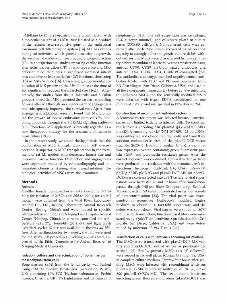
ResultsCharacterization of MSCs-MKAfter three passages, the adherent MSC cells were sym-metric with phenotypic surface antigens as reported,including positivity for CD29, CD44 and CD90 and ne-gativity for CD34, CD31 and CD86 (>90%, Figure S1 inAdditional file 1) [22,29,30]. We successfully developedthe high-titer lentiviral vectors that drive expression of ratMK/GFP (pLenO-DCE-MK, 1.5 × 10⁹ TU/ml) and GFP(pLenO-DCE, 2.0 × 10⁹ TU/ml) (Figures S2 and S3,Additional file 1). After infection with recombinantlentivirus, the efficiency of the gene transduction ofMSCs to overexpress MK was similar to that of mocklentivirus (95.2 vs. 95.3%) (Figure 1A); moreover, above
six phenotypic surface antigens did not change (datanot shown).
Overexpression of MK improved MSCs survivalLentivirus-mediated transduction and expression of MKwere confirmed with real-time PCR and Western blot-ting. Quantitative real-time PCR data indicated thatexpression of MK was 54.2-fold higher in MSCs-MK(Figure 1B). MSCs-MK also exhibited higher levels ofMK protein (Figure 1C); moreover, the midkine wassecreted (Figure 1D). To investigate the resistance ofMSCs-MK to anoxia, MSCs, MSCs-GFP and MSCs-MKwere exposed to oxygen and glucose deprivation and ana-lyzed with the Annexin V-PE/7ADD kit. MSCs displayedmorphological changes of apoptosis and necrosis after an-oxia. Overexpression of MK, however, partially preventedapoptosis induced by anoxia. The numbers of apoptoticand necrotic cells (17.9 ± 4.2%) decreased significantly inthe MSCs-MK group compared with the MSCs andMSCs-GFP groups (28.4 ± 3.7%, p< 0.05; 32.6 ± 4.9%,p< 0.05; Figure 1E).
MK enhanced the paracrine effects of MSCsIt is known that the main mechanisms by which MSCscan increase repair on myocardial infarction are car-diogenic differentiation (direct contribution), autocrineeffects (affecting the MSCs themselves) and paracrineeffects (affecting the wounded/infracted tissue) [31]. Inthe present study, we did not find any evidence that MKcould promote MSC differentiation into cardiomyocytes(data not shown). To determine whether MK impactsthe expression of pro-angiogenesis and some stem cell-related factors, we measured mRNA of VEGF, TGF-β,FGF2, FGF7, SDF-1a, IGF-1, GM-CSF and SCF in MSCs,MSCs-MK and MSCs-GFP respectively (Figure 2A,B), andthe protein levels of IGF-1, SDF-1a, VEGF and TGF-β1were also quantified by ELISA (Figure 2C). The mRNAlevels of VEGF, TGF-β, IGF-1 and SDF-1a were ele-vated 5.85-, 2.78-, 3.7- and 2.01-fold, but no elevationin the mRNA levels of FGF-2, FGF-7, GM-CSF andSCF was observed in MSCs-MK compared with MSCsand MSCs-GFP, indicating that this elevation was spe-cific for MK overexpression. Consistent with the qPCRresults, MSCs-MK secreted more SDF-1a (9.23 ng/ml),VEGF (8.34 ng/ml) and TGF-β1 (17.88 ng/ml) thanMSCs and MSCs-GFP; this was not the case for IGF-1(1.89 ng/ml).
MK increased the protective effects of MSCs oncardiomyocytesThe secreted factors of MSCs exert biological activitiesby binding to their corresponding receptors on cardiaccells. To determine the ability of MSC homing to the
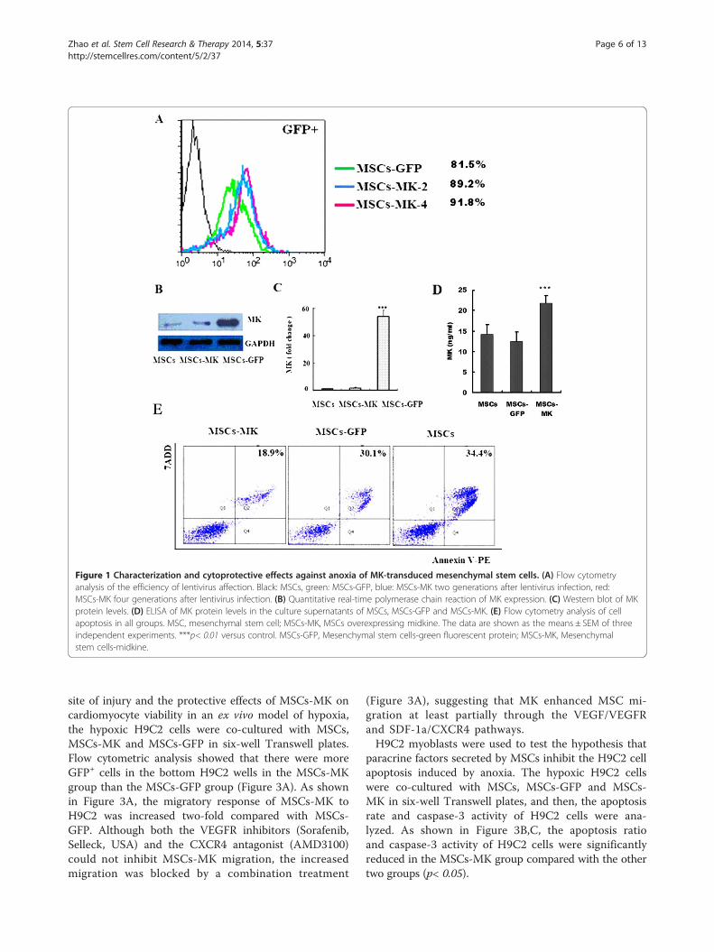
Figure 1 Characterization and cytoprotective effects against anoxia of MK-transduced mesenchymal stem cells. (A) Flow cytometryanalysis of the efficiency of lentivirus affection. Black: MSCs, green: MSCs-GFP, blue: MSCs-MK two generations after lentivirus infection, red:MSCs-MK four generations after lentivirus infection. (B) Quantitative real-time polymerase chain reaction of MK expression. (C) Western blot of MKprotein levels. (D) ELISA of MK protein levels in the culture supernatants of MSCs, MSCs-GFP and MSCs-MK. (E) Flow cytometry analysis of cellapoptosis in all groups. MSC, mesenchymal stem cell; MSCs-MK, MSCs overexpressing midkine. The data are shown as the means ± SEM of threeindependent experiments. ***p< 0.01 versus control. MSCs-GFP, Mesenchymal stem cells-green fluorescent protein; MSCs-MK, Mesenchymalstem cells-midkine.
Zhao et al. Stem Cell Research & Therapy 2014, 5:37 Page 6 of 13http://stemcellres.com/content/5/2/37
site of injury and the protective effects of MSCs-MK oncardiomyocyte viability in an ex vivo model of hypoxia,the hypoxic H9C2 cells were co-cultured with MSCs,MSCs-MK and MSCs-GFP in six-well Transwell plates.Flow cytometric analysis showed that there were moreGFP+ cells in the bottom H9C2 wells in the MSCs-MKgroup than the MSCs-GFP group (Figure 3A). As shownin Figure 3A, the migratory response of MSCs-MK toH9C2 was increased two-fold compared with MSCs-GFP. Although both the VEGFR inhibitors (Sorafenib,Selleck, USA) and the CXCR4 antagonist (AMD3100)could not inhibit MSCs-MK migration, the increasedmigration was blocked by a combination treatment
(Figure 3A), suggesting that MK enhanced MSC mi-gration at least partially through the VEGF/VEGFRand SDF-1a/CXCR4 pathways.H9C2 myoblasts were used to test the hypothesis that
paracrine factors secreted by MSCs inhibit the H9C2 cellapoptosis induced by anoxia. The hypoxic H9C2 cellswere co-cultured with MSCs, MSCs-GFP and MSCs-MK in six-well Transwell plates, and then, the apoptosisrate and caspase-3 activity of H9C2 cells were ana-lyzed. As shown in Figure 3B,C, the apoptosis ratioand caspase-3 activity of H9C2 cells were significantlyreduced in the MSCs-MK group compared with the othertwo groups (p< 0.05).

Figure 2 MK increases the paracrine effects of MSCs. A and B: Real-time PCR analysis of pro-angiogenesis factors (VEGF, TGF-β, FGF2 andFGF7) and stem cell factor (SDF-1, IGF-1, GM-CSF and SCF) mRNAs. The relative expression of VEGF (5.85-fold), TGF-β (2.78-fold), IGF-1 (3.7-fold)and SDF-1 (2.01-fold) mRNA in MSCs-MK were higher than in control cells (***p< 0.01). C: ELISA of IGF-1, SDF-1a, VEGF and TGF-β1 protein levelsin the culture supernatants of MSCs, MSCs-GFP and MSCs-MK. MSCs-MK secreted more SDF-1a (9.23 ng/ml), VEGF (8.34 ng/ml) and TGF-β1(17.88 ng/ml) than MSCs (6.35, 4.75 and 9.18 ng/ml) and MSCs-GFP (6.92, 4.55 and 9.88 ng/ml); this was not the case for IGF-1 (MSCs-MK, 1.89 vs.MSCs, 1.51 and MSCs-GFP 1.40 ng/ml). FGF, fibroblast growth factor; GM-CSF, Granulocyte-macrophage colony-stimulating factor; IGF-1,Insulin-like growth factor 1; MSCs-GFP, Mesenchymal stem cells-green fluorescent protein; MSCs-MK, Mesenchymal stem cells-midkine; SCF, Stem cellfactor; SDF-1, Stromal cell-derived factor 1; TGF-β, Transforming growth factor beta; VEGF, Vascular endothelial growth factor.
Zhao et al. Stem Cell Research & Therapy 2014, 5:37 Page 7 of 13http://stemcellres.com/content/5/2/37
Injection of MSCs-MK prevented cardiac dysfunctionafter MIThe effect of MK overexpression on the efficiency ofcell-based therapy with MSCs was evaluated in a ratmodel of MI. Two weeks after ligation of the coronaryartery, the MI models of rat were confirmed by electro-cardiogram, and MSCs, MSCs-GFP and MSCs-MK wereinjected into the border zone myocardium. We preparedfive groups of mice, namely sham and MI plus injectionof PBS, MSCs, MSCs-GFP and MSCs-MK. All sham-operated rats survived, whereas only 42% of the PBS-injected MI mice survived at 12 weeks. Although therewas no significant difference in survival rate among theMSCs (50%), MSCs-GFP (42%) and saline groups, 75%of rats injected with MSCs-MK survived (Figure 4A).To evaluate changes in LV dimensions and contracti-
lity, echocardiographic analyses were conducted beforethe surgery and after 4, 8 and 12 weeks (Figure 4B).In the PBS-injected MI rats, a significant increase inLVEDD and LVESD and a significant decrease in
fractional shortening (%FS) were observed comparedwith the sham-operated rats at four weeks and there-after, suggesting that the LV is dilated and systolicfunction is compromised. Although both LVEDDand LVESD increased progressively in theMSCs-, MSCs-GFP- and MSCs-MK-injected MI rats(p< 0.05) compared with those in sham-operated Ratsat 12 weeks (Figure 4C,D), there were no significant differ-ences (p> 0.05) among the three groups, suggesting thatinjection of MSCs, MSCs-GFP and MSCs-MK retard LVdilation but with no difference among them.The EF and FS in the MSCs and MSCs-GFP groups
significantly and progressively decreased from four weeksonwards, suggesting that injection of MSCs and MSCs-GFP prevented LV dysfunction. Furthermore, the FS andEF in the MSCs-MK-injected MI rats gradually decreased,but these values were still significantly higher than othergroups (although only EF, p> 0.05) (Figure 4E,F). Theseresults suggest that injection of MSCs or MSCs-GFP alonedoes not have long-term therapeutic effects, but ex vivo
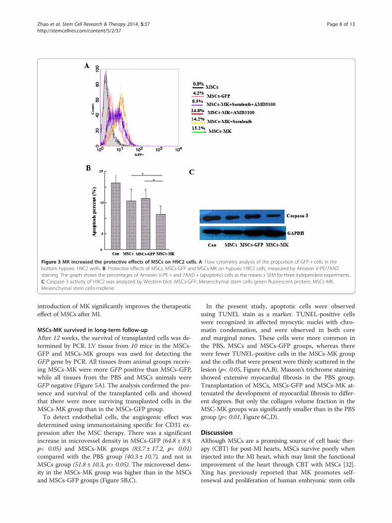
Figure 3 MK increased the protective effects of MSCs on H9C2 cells. A: Flow cytometry analysis of the proportion of GFP + cells in thebottom hypoxic H9C2 wells. B: Protective effects of MSCs, MSCs-GFP and MSCs-MK on hypoxic H9C2 cells, measured by Annexin V-PE/7AADstaining. The graph shows the percentages of Annexin V-PE + and 7AAD + (apoptotic) cells as the means ± SEM for three independent experiments.C: Caspase-3 activity of H9C2 was analyzed by Western blot. MSCs-GFP, Mesenchymal stem cells-green fluorescent protein; MSCs-MK,Mesenchymal stem cells-midkine.
Zhao et al. Stem Cell Research & Therapy 2014, 5:37 Page 8 of 13http://stemcellres.com/content/5/2/37
introduction of MK significantly improves the therapeuticeffect of MSCs after MI.
MSCs-MK survived in long-term follow-upAfter 12 weeks, the survival of transplanted cells was de-termined by PCR. LV tissue from 10 mice in the MSCs-GFP and MSCs-MK groups was used for detecting theGFP gene by PCR. All tissues from animal groups receiv-ing MSCs-MK were more GFP positive than MSCs-GFP,while all tissues from the PBS and MSCs animals wereGFP negative (Figure 5A). The analysis confirmed the pre-sence and survival of the transplanted cells and showedthat there were more surviving transplanted cells in theMSCs-MK group than in the MSCs-GFP group.To detect endothelial cells, the angiogenic effect was
determined using immunostaining specific for CD31 ex-pression after the MSC therapy. There was a significantincrease in microvessel density in MSCs-GFP (64.8 ± 8.9,p< 0.05) and MSCs-MK groups (83.7 ± 17.2, p< 0.01)compared with the PBS group (40.3 ± 10.7), and not inMSCs group (51.8 ± 10.3, p> 0.05). The microvessel dens-ity in the MSCs-MK group was higher than in the MSCsand MSCs-GFP groups (Figure 5B,C).
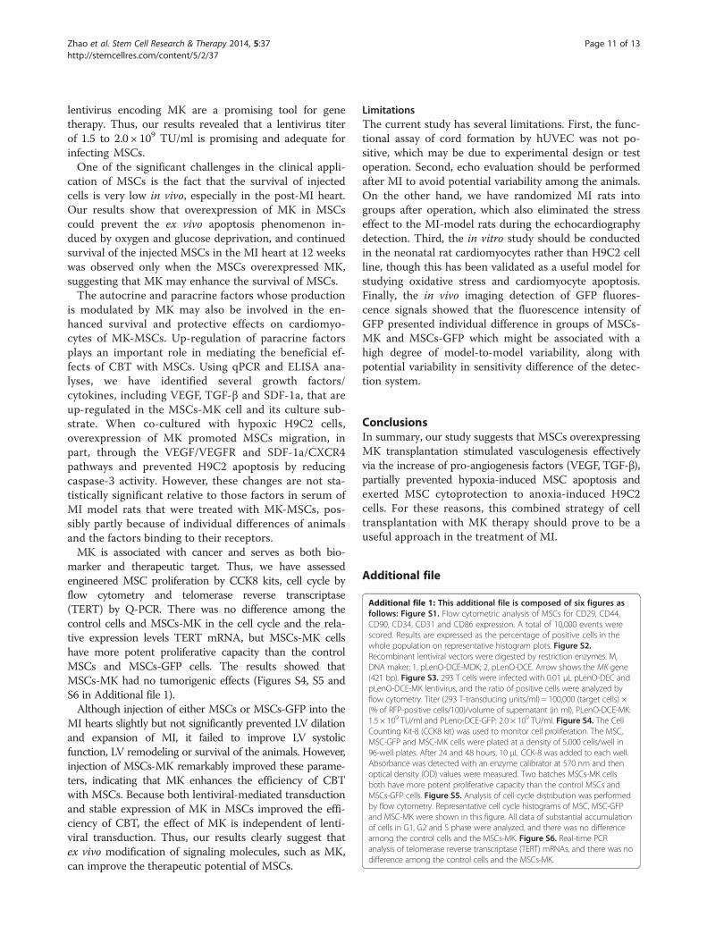
In the present study, apoptotic cells were observedusing TUNEL stain as a marker. TUNEL-positive cellswere recognized in affected myocytic nuclei with chro-matin condensation, and were observed in both coreand marginal zones. These cells were more common inthe PBS, MSCs and MSCs-GFP groups, whereas therewere fewer TUNEL-positive cells in the MSCs-MK groupand the cells that were present were thinly scattered in thelesion (p< 0.05, Figure 6A,B). Masson’s trichrome stainingshowed extensive myocardial fibrosis in the PBS group.Transplantation of MSCs, MSCs-GFP and MSCs-MK at-tenuated the development of myocardial fibrosis to differ-ent degrees. But only the collagen volume fraction in theMSC-MK groups was significantly smaller than in the PBSgroup (p< 0.01, Figure 6C,D).
DiscussionAlthough MSCs are a promising source of cell basic ther-apy (CBT) for post-MI hearts, MSCs survive poorly wheninjected into the MI heart, which may limit the functionalimprovement of the heart through CBT with MSCs [32].Xing has previously reported that MK promotes self-renewal and proliferation of human embryonic stem cells

Figure 4 Curative effect evaluation of MSCs-MK by echocardiography in rats after myocardial infarction. A: Survival rate estimated by theKaplan-Meier method in sham rats (sham; n = 12, none died), MI rats treated by PBS (PBS; n = 12, 7 died), MI rats treated with MSCs (MSCs; n = 12,6 died), with MSCs-GFP (MSCs-GFP; n = 12, 7 died) and with MSCs-MK (MSCs-MK; n = 12, 3 died). Survival rates of MSCs-MK were significantlyhigher than for MSCs and MSCs-GFP. B: Representative echocardiography findings in the MSCs-MK group before MI (pre-operation) and 4, 8 and12 weeks after MI (post-treatment). C, D, E and F: Measurements were performed at baseline before MI and 4, 8 and 12 weeks after MI, asindicated. (C) Left ventricular end-diastolic diameter (LVEDD). (D) Left ventricular end-systolic diameter (LVESD). (E) Fractional shortening (FS).(F) Ejection fraction (EF). *indicates p< 0.05 vs. the PBS group at the same time-point, one-way ANOVA. All values represent mean ± SEM. MI,Myocardial infarction; MSCs-GFP, Mesenchymal stem cells-green fluorescent protein; MSCs-MK, Mesenchymal stem cells-midkine.
Zhao et al. Stem Cell Research & Therapy 2014, 5:37 Page 9 of 13http://stemcellres.com/content/5/2/37
(hESCs) by inhibiting apoptosis while accelerating theprogression toward the S phase and that MK enhan-ces mouse embryonic stem cells (mESCs) self-renewalthrough the PI3K/Akt signaling pathway [33]. Further-more, exogenous MK has a protective effect in models ofMI of mice and rats. In this present study, we show thatex vivo up-regulation of MK in MSCs significantly
increases both survival rate and paracrine signaling of theMSCs after injection into the MI heart, thereby signifi-cantly enhancing the efficiency of the CBT.Lentiviruses are promising vectors for delivery of MK
transgenes due to their ability to integrate and transduceboth dividing and non-dividing cells, which are less con-strained by the size of the MK transgene and facilitate
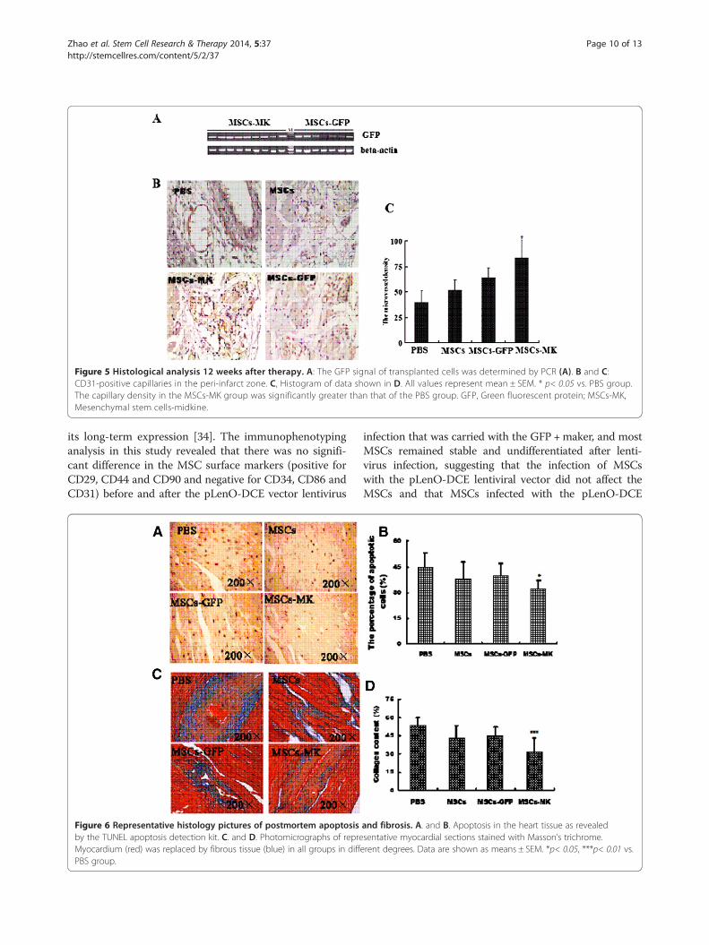
Figure 5 Histological analysis 12 weeks after therapy. A: The GFP signal of transplanted cells was determined by PCR (A). B and C:CD31-positive capillaries in the peri-infarct zone. C, Histogram of data shown in D. All values represent mean ± SEM. * p< 0.05 vs. PBS group.The capillary density in the MSCs-MK group was significantly greater than that of the PBS group. GFP, Green fluorescent protein; MSCs-MK,Mesenchymal stem cells-midkine.
Zhao et al. Stem Cell Research & Therapy 2014, 5:37 Page 10 of 13http://stemcellres.com/content/5/2/37
its long-term expression [34]. The immunophenotypinganalysis in this study revealed that there was no signifi-cant difference in the MSC surface markers (positive forCD29, CD44 and CD90 and negative for CD34, CD86 andCD31) before and after the pLenO-DCE vector lentivirus
Figure 6 Representative histology pictures of postmortem apoptosisby the TUNEL apoptosis detection kit. C. and D. Photomicrographs of repreMyocardium (red) was replaced by fibrous tissue (blue) in all groups in diffPBS group.
infection that was carried with the GFP +maker, and mostMSCs remained stable and undifferentiated after lenti-virus infection, suggesting that the infection of MSCswith the pLenO-DCE lentiviral vector did not affect theMSCs and that MSCs infected with the pLenO-DCE
and fibrosis. A. and B. Apoptosis in the heart tissue as revealedsentative myocardial sections stained with Masson’s trichrome.erent degrees. Data are shown as means ± SEM. *p< 0.05, ***p< 0.01 vs.
Zhao et al. Stem Cell Research & Therapy 2014, 5:37 Page 11 of 13http://stemcellres.com/content/5/2/37
lentivirus encoding MK are a promising tool for genetherapy. Thus, our results revealed that a lentivirus titerof 1.5 to 2.0 × 10⁹ TU/ml is promising and adequate forinfecting MSCs.One of the significant challenges in the clinical appli-
cation of MSCs is the fact that the survival of injectedcells is very low in vivo, especially in the post-MI heart.Our results show that overexpression of MK in MSCscould prevent the ex vivo apoptosis phenomenon in-duced by oxygen and glucose deprivation, and continuedsurvival of the injected MSCs in the MI heart at 12 weekswas observed only when the MSCs overexpressed MK,suggesting that MK may enhance the survival of MSCs.The autocrine and paracrine factors whose production
is modulated by MK may also be involved in the en-hanced survival and protective effects on cardiomyo-cytes of MK-MSCs. Up-regulation of paracrine factorsplays an important role in mediating the beneficial ef-fects of CBT with MSCs. Using qPCR and ELISA ana-lyses, we have identified several growth factors/cytokines, including VEGF, TGF-β and SDF-1a, that areup-regulated in the MSCs-MK cell and its culture sub-strate. When co-cultured with hypoxic H9C2 cells,overexpression of MK promoted MSCs migration, inpart, through the VEGF/VEGFR and SDF-1a/CXCR4pathways and prevented H9C2 apoptosis by reducingcaspase-3 activity. However, these changes are not sta-tistically significant relative to those factors in serum ofMI model rats that were treated with MK-MSCs, pos-sibly partly because of individual differences of animalsand the factors binding to their receptors.MK is associated with cancer and serves as both bio-
marker and therapeutic target. Thus, we have assessedengineered MSC proliferation by CCK8 kits, cell cycle byflow cytometry and telomerase reverse transcriptase(TERT) by Q-PCR. There was no difference among thecontrol cells and MSCs-MK in the cell cycle and the rela-tive expression levels TERT mRNA, but MSCs-MK cellshave more potent proliferative capacity than the controlMSCs and MSCs-GFP cells. The results showed thatMSCs-MK had no tumorigenic effects (Figures S4, S5 andS6 in Additional file 1).Although injection of either MSCs or MSCs-GFP into the
MI hearts slightly but not significantly prevented LV dilationand expansion of MI, it failed to improve LV systolicfunction, LV remodeling or survival of the animals. However,injection of MSCs-MK remarkably improved these parame-ters, indicating that MK enhances the efficiency of CBTwith MSCs. Because both lentiviral-mediated transductionand stable expression of MK in MSCs improved the effi-ciency of CBT, the effect of MK is independent of lenti-viral transduction. Thus, our results clearly suggest thatex vivo modification of signaling molecules, such as MK,can improve the therapeutic potential of MSCs.
LimitationsThe current study has several limitations. First, the func-tional assay of cord formation by hUVEC was not po-sitive, which may be due to experimental design or testoperation. Second, echo evaluation should be performedafter MI to avoid potential variability among the animals.On the other hand, we have randomized MI rats intogroups after operation, which also eliminated the stresseffect to the MI-model rats during the echocardiographydetection. Third, the in vitro study should be conductedin the neonatal rat cardiomyocytes rather than H9C2 cellline, though this has been validated as a useful model forstudying oxidative stress and cardiomyocyte apoptosis.Finally, the in vivo imaging detection of GFP fluores-cence signals showed that the fluorescence intensity ofGFP presented individual difference in groups of MSCs-MK and MSCs-GFP which might be associated with ahigh degree of model-to-model variability, along withpotential variability in sensitivity difference of the detec-tion system.
ConclusionsIn summary, our study suggests that MSCs overexpressingMK transplantation stimulated vasculogenesis effectivelyvia the increase of pro-angiogenesis factors (VEGF, TGF-β),partially prevented hypoxia-induced MSC apoptosis andexerted MSC cytoprotection to anoxia-induced H9C2cells. For these reasons, this combined strategy of celltransplantation with MK therapy should prove to be auseful approach in the treatment of MI.
Additional file
Additional file 1: This additional file is composed of six figures asfollows: Figure S1. Flow cytometric analysis of MSCs for CD29, CD44,CD90, CD34, CD31 and CD86 expression. A total of 10,000 events werescored. Results are expressed as the percentage of positive cells in thewhole population on representative histogram plots. Figure S2.Recombinant lentiviral vectors were digested by restriction enzymes. M,DNA maker; 1, pLenO-DCE-MDK; 2, pLenO-DCE. Arrow shows the MK gene(421 bp). Figure S3. 293 T cells were infected with 0.01 μL pLenO-DEC andpLenO-DCE-MK lentivirus, and the ratio of positive cells were analyzed byflow cytometry. Titer (293 T-transducing units/ml) = 100,000 (target cells) ×(% of RFP-positive cells/100)/volume of supernatant (in ml), PLenO-DCE-MK:1.5 × 10⁹ TU/ml and PLeno-DCE-GFP: 2.0 × 10⁹ TU/ml. Figure S4. The CellCounting Kit-8 (CCK8 kit) was used to monitor cell proliferation. The MSC,MSC-GFP and MSC-MK cells were plated at a density of 5,000 cells/well in96-well plates. After 24 and 48 hours, 10 μL CCK-8 was added to each well.Absorbance was detected with an enzyme calibrator at 570 nm and thenoptical density (OD) values were measured. Two batches MSCs-MK cellsboth have more potent proliferative capacity than the control MSCs andMSCs-GFP cells. Figure S5. Analysis of cell cycle distribution was performedby flow cytometry. Representative cell cycle histograms of MSC, MSC-GFPand MSC-MK were shown in this figure. All data of substantial accumulationof cells in G1, G2 and S phase were analyzed, and there was no differenceamong the control cells and the MSCs-MK. Figure S6. Real-time PCRanalysis of telomerase reverse transcriptase (TERT) mRNAs, and there was nodifference among the control cells and the MSCs-MK.
Zhao et al. Stem Cell Research & Therapy 2014, 5:37 Page 12 of 13http://stemcellres.com/content/5/2/37
AbbreviationsCBT: cell basic therapy; FCS: fetal calf serum; FGF: fibroblast growth factor;FS: fractional shortening; GFP: Green fluorescent protein; GM-CSF:Granulocyte-macrophage colony-stimulating factor; IGF-1: Insulin-like growthfactor 1; LAD: Ligating the left anterior descending coronary artery; LVEDD:Left ventricular end-diastolic diameter; LVEDV and LVESV: Left ventricularend-diastolic and end-systolic volumes; LVEF: The LV ejection fraction;LVESD: LV end-systolic diameter; MI: Myocardial infarction; MK: Midkine;MSCs: Mesenchymal stem cells; OGD: oxygen and glucose deprivation;PBS: phosphate-buffered saline; pLenO-DCE-MK: pLenO-DCE vector encodingMK; SCF: Stem cell factor; SDF-1: Stromal cell-derived factor 1; TGF-β:Transforming growth factor beta; VEGF: Vascular endothelial growth factor.
Competing interestsThe authors have no conflicts of interest to declare.
Authors’ contributionsSLZ participated in the design of experiments, carried out the molecularanalysis of cells, cell transplantation into animals, interpretation and analysisof in vitro and in vivo data, and helped to draft the manuscript. MHL and XLZwere involved in drafting the manuscript and participated in all experimentsinvolving animals, including histological analyses. YJZ and SLC have beeninvolved in all aspects of the study, including experimental design, analysisand interpretation of data, and manuscript writing. All authors read andapproved the final manuscript.
AcknowledgmentsThis work was supported by the Medical Science and TechnologyDevelopment Foundation of Nanjing (Department of Health, Grant #: YKK12076and QRX11243) and the National Natural Science Foundation of China(grant number: 81201598).
Author details1Department of Cardiology, Nanjing First Hospital, Nanjing MedicalUniversity, No. 68 Changle Road, Nanjing 210006, China. 2State KeyLaboratory of Reproductive Medicine, Nanjing Medical University, Nanjing,China. 3Thoraxcenter, Erasmus Medical Center, Rotterdam, The Netherlands.
Received: 29 July 2013 Revised: 30 November 2013Accepted: 11 March 2014 Published: 17 March 2014
References1. Herzog CA, Ma JZ, Collins AJ: Poor long-term survival after acute
myocardial infarction among patients on long-term dialysis. N Engl J Med1998, 339:799–805.
2. Sabbah HN, Sharov VG: Apoptosis in heart failure. Prog Cardiovasc Dis1998, 40:549–562.
3. Mill JG, Stefanon I, dos Santos L, Baldo MP: Remodeling in the ischemicheart: the stepwise progression for heart failure. Braz J Med Biol Res 2011,44:890–898.
4. Takemura G, Fujiwara H: Role of apoptosis in remodeling after myocardialinfarction. Pharmacol Ther 2004, 104:1–16.
5. Vassalli G, Moccetti T: Cardiac repair with allogeneic mesenchymal stemcells after myocardial infarction. Swiss Med Wkly 2011, 141:w13209.
6. Ohnishi S, Ohgushi H, Kitamura S, Nagaya N: Mesenchymal stem cells forthe treatment of heart failure. Int J Hematol 2007, 86:17–21.
7. Chen S, Fang W, Ye F, Liu YH, Qian J, Shan SJ, Zhang JJ, Chunhua RZ,Liao LM, Lin S, Sun JP: Effect on left ventricular function of intracoronarytransplantation of autologous bone marrow mesenchymal stem cell inpatients with acute myocardial infarction. Am J Cardiol 2004, 94:92–95.
8. Chen SL, Fang WW, Qian J, Ye F, Liu YH, Shan SJ, Zhang JJ, Lin S, Liao LM,Zhao RC: Improvement of cardiac function after transplantation ofautologous bone marrow mesenchymal stem cells in patients with acutemyocardial infarction. Chin Med J (Engl) 2004, 117:1443–1448. Erratum inChin Med J (Engl) 2005, 118:88.
9. Alfaro MP, Young PP: Lessons from genetically altered mesenchymalstem cells (MSCs): candidates for improved MSC-directed myocardialrepair. Cell Transplant 2012, 21:1065–1074.
10. Verbeek R: Generation of mesenchymal stem cells as a medicinal productin organ transplantation. Curr Opin Organ Transplant 2013, 18:65–70.
11. Williams AR, Suncion VY, McCall F, Guerra D, Mather J, Zambrano JP,Heldman AW, Hare JM: Durable scar size reduction due to allogeneicmesenchymal stem cell therapy regulates whole-chamber remodeling.J Am Heart Assoc 2013, 2:e000140.
12. Houtgraaf JH, de Jong R, Kazemi K, de Groot D, van der Spoel TI, Arslan F,Hoefer I, Pasterkamp G, Itescu S, Zijlstra F, Geleijnse ML, Serruys PW, DuckersHJ: Intracoronary infusion of allogeneic mesenchymal precursor cellsdirectly after experimental acute myocardial infarction reduces infarctsize, abrogates adverse remodeling, and improves cardiac function.Circ Res 2013, 113:153–166.
13. Forest VF, Tirouvanziam AM, Perigaud C, Fernandes S, Fusellier MS, Desfontis JC,Toquet CS, Heymann MF, Crochet DP, Lemarchand PF: Cell distribution afterintracoronary bone marrow stem cell delivery in damaged and undamagedmyocardium: implications for clinical trials. Stem Cell Res Ther 2010, 1:4.
14. Kadomatsu K, Muramatsu T: Midkine and pleiotrophin in neuraldevelopment and cancer. Cancer Lett 2004, 204:127–143.
15. Horiba M, Kadomatsu K, Nakamura E, Muramatsu H, Ikematsu S, Sakuma S,Hayashi K, Yuzawa Y, Saito H, Muramatsu T: Neointima formation in arestenosis model is suppressed in midkine-deficient mice. J Clin Invest2000, 87:489–495.
16. Obama H, Biro S, Tashiro T, Tsutsui J, Ozawa M, Yoshida H, Tanaka H,Muramatsu T: Myocardial infarction induces expression of midkine, aheparin-binding growth factor with reparative activity. Anticancer Res1998, 18:145–152.
17. Horiba M, Kadomatsu K, Yasui K, Lee J-K, Takenaka H, Sumida A, Kamiya K,Chen S, Sakuma S, Muramatsu T, Kodama I: Midkine plays a protective roleagainst cardiac ischemia/reperfusion injury through a reduction ofapoptotic reaction. Circulation 2006. 114:1713–1720.
18. Takenaka H, Horiba M, Ishiguro H, Sumida A, Hojo M, Usui A, Akita T,Sakuma S, Ueda Y, Kodama I, Kadomatsu K: Midkine preventsventricular remodeling and improves long-term survival after myo-cardial infarction. Am J Physiol Heart Circ Physiol 2009, 296:462–469.
19. Fukui S, Kitagawa-Sakakida S, Kawamata S, Matsumiya G, Kawaguchi N,Matsuura N, Sawa Y: Therapeutic effect of midkine on cardiac remodelingin infarcted rat heart. Ann Thorac Surg 2008, 85:562–570.
20. Lee SH, Suh HN, Lee YJ, Seo BN, Ha JW, Han HJ: Midkine preventedhypoxic injury of mouse embryonic stem cells through activation of Aktand HIF-1α via low-density lipoprotein receptor-related protein-1. J CellPhysiol 2012, 227:1731–1739.
21. Tarnowski M, Szydło A, Anioł J, Koryciak-Komarska H, Lesiak M, Gutmajster E,Sieroń AL, Kusz D: Optimization of genetic engineering and homologousrecombination of collagen type I genes in rat bone marrow mesenchymalstem cells (MSC). Cell Reprogram 2010, 12:275–282.
22. Mathieu E, Lamirault G, Toquet C, Lhommet P, Rederstorff E, Sourice S,Biteau K, Hulin P, Forest V, Weiss P, Guicheux J, Lemarchand P:Intramyocardial delivery of mesenchymal stem cell seeded hydrogelpreserves cardiac function and attenuates ventricular remodeling aftermyocardial infarction. PLoS One 2012, 7:e51991.
23. Liu RS, Hsieh YJ, Ke CC, Chen FD, Hwu L, Wang FH, Hwang JJ, Chi CW,Lee CH, Yeh SH: Specific activation of sodium iodide symporter gene inhepatoma using alpha-fetoprotein promoter combined with hepatitis Bvirus enhancer (EIIAPA). Anticancer Res 2009, 29:211–222.
24. Sanchez-Antequera Y, Mykhaylyk O, van Til NP, Cengizeroglu A, de Jong JH,Huston MW, Anton M, Johnston IC, Pojda Z, Wagemaker G, Plank C:Magselectofection: an integrated method of nanomagnetic separationand genetic modification of target cells. Blood 2011, 117:e171–e181.
25. Xiang J, Tang J, Song C, Yang Z, Hirst DG, Zheng QJ, Li G: Mesenchymalstem cells as a gene therapy carrier for treatment of fibrosarcoma.Cytotherapy 2009, 11:516–526.
26. Zhao S, Wang H, Nie Y, Mi Q, Chen X, Hou Y: Midkine upregulates MICA/Bexpression in human gastric cancer cells and decreases natural killer cellcytotoxicity. Cancer Immunol Immunother 2012, 61:1745–1753.
27. Renic M, Kumar SN, Gebremedhin D, Florence MA, Gerges NZ, Falck JR,Harder DR, Roman RJ: Protective effect of 20-HETE inhibition in a modelof oxygen-glucose deprivation in hippocampal slice cultures. Am JPhysiol Heart Circ Physiol 2012, 302:H1285–H1293.
28. Shim TJ, Bae JW, Kim YJ, Kim DJ, Hwang KK, Kim DW, Cho MC:Cardioprotective effects of 3-phosphoinositide-dependent proteinkinase-1 on hypoxic injury in cultured neonatal rat cardiomyocytes andmyocardium in a rat myocardial infarct model. Biosci Biotechnol Biochem2012, 76:101–107.
Zhao et al. Stem Cell Research & Therapy 2014, 5:37 Page 13 of 13http://stemcellres.com/content/5/2/37
29. Khan M, Mohsin S, Khan SN, Riazuddin S: Repair of senescent myocardiumby mesenchymal stem cells is dependent on the age of donor mice.J Cell Mol Med 2011, 15:1515–1527.
30. Mantovani C, Raimondo S, Haneef MS, Geuna S, Terenghi G, Shawcross SG,Wiberg M: Morphological, molecular and functional differences of adultbone marrow- and adipose-derived stem cells isolated from rats ofdifferent ages. Exp Cell Res 2012, 318:2034–2048.
31. Mathieu E, Lamirault G, Toquet C, Lhommet P, Rederstorff E, Sourice S,Biteau K, Hulin P, Forest V, Weiss P, Guicheux J, Lemarchand P:Intramyocardial delivery of mesenchymal stem cell seeded hydrogelpreserves cardiac function and attenuates ventricular remodeling aftermyocardial infarction. PLoS One 2012, 7:e51991.
32. Gnecchi M, Danieli P, Cervio E: Mesenchymal stem cell therapy for heartdisease. Vascul Pharmacol 2012, 57:48–55.
33. Mangi AA, Noiseux N, Kong D, He H, Rezvani M, Ingwall JS, Dzau VJ:Mesenchymal stem cells modified with Akt prevent remodeling andrestore performance of infarcted hearts. Nat Med 2003, 9:1195–1201.
34. Yao X, Tan Z, Bin G, Rong-rong W, Liu Y, Dai L, Zhang M: Promotion ofself-renewal of embryonic stem cells by midkine. Acta Pharmacol Sin2010, 31:629–637.
35. Naldini L, Blömer U, Gallay P, Ory D, Mulligan R, Gage FH, Verma IM,Trono D: In vivo gene delivery and stable transduction of nondividingcells by a lentiviral vector. Science 1996, 272:263–267.
doi:10.1186/scrt425Cite this article as: Zhao et al.: Mesenchymal stem cells withoverexpression of midkine enhance cell survival and attenuate cardiacdysfunction in a rat model of myocardial infarction. Stem Cell Research &Therapy 2014 5:37.
Submit your next manuscript to BioMed Centraland take full advantage of:
• Convenient online submission
• Thorough peer review
• No space constraints or color figure charges
• Immediate publication on acceptance
• Inclusion in PubMed, CAS, Scopus and Google Scholar
• Research which is freely available for redistribution
Submit your manuscript at www.biomedcentral.com/submit