medical image denoising using adaptive spatial … image denoising using adaptive spatial ... many...
TRANSCRIPT
Journal of Babylon University/Pure and Applied Sciences/ No.(9)/ Vol.(24): 2016
4539
Medical Image Denoising using Adaptive Spatial Domain Schemes with Additive Noise
Osama Qasim Jumah Al-Thahab
University of Babylon, Department of Electric
Hanaa mohsin ali
University of Babylon, Department of Electric
Abstract Image denoising is one of the most significant tasks in medical image processing due to the
significant information obtained by these images related to the human body or the tissues of the body’s
organs. So, many methods have been proposed for removing the noise that affects the medical images.
In this research a new algorithm has been proposed for denoisning medical images work in spatial
domain. A new algorithm depends on the idea that combine between characteristic of different filters
which work in spatial domain with adaptive sizes of windows for reaching to the acceptance results in
remove noise from medical images. This algorithm called Adaptive Window Wiener Filter (AWWF).
Two types of medical images and noise that corrupt these medical image used in this research. The first
type is Poisson noise which corrupts X-ray medical images and the second type is Rician noise which
corrupts MRI medical images. The algorithm begins with using a median filter on a noisy image to get
the blurred version of the image. Then using an edge detection algorithm, the edges detection of the
resulted blurred image is found by using the Prewitt operator. Then Wiener filter of variable size
windows is applied throughout the noisy image to suppress the noise. The window size is made bigger
in homogenous and smooth regions and is made smaller in edge and complex regions.
Keywords: Image processing, Image Denoising, Additive noise.
الخالصة من المستخلصة مةھالم المعلومات بسبب الطبیة الصور معالجة مجال في امھالم ابرز من الصور من الضوضاء إزالة عملیة تعد
الصوور مون الضوضواء إلزالوة طور عودة اقترحوت فقود السوبب ذاھل .الجسوم أعضواء أنسوجة أو البشور الجسوم تخو الصوور والتوي ذهھالخوارزمیوة الجديودة .ألحيوز تعمو فوي المجوال التوي الضوضواء مون الصوور الطبیوة إلزالوة جديودةطريقوو في ىذا البحث اقترحوت .الطبیة
النتوائ الوى للوصوول النوافوذ أحجوامالمختلفة التوي تعمو فوي المجوال ألحيوز موع تنویوع المرشحاتتعتمد على فكرة الجمع بين خصائ فووي ىووذا اسووتخدم .wienerالنافووذة المحد ووة لمرشوو خوارزمیووة دعىتوو الطريقووةه . ىووذالضوضوواء موون الصووور الطبیووة إزالووةفووي المطلوبووة التوووي Poissonمووون الضوضووواء ىوووو ضوضووواء األولالنوووو .نوعوووان مووون الصوووور الطبیوووة والضوضووواء التوووي تصووويب ىوووذه الصوووور البحوووث
باسوتخدام الخوارزمیوة تبودأ ة.الطبیو MRIصوورالتوي تصويب Rician والنوو ال واني ىوو ضوضواء تصيب صور األشوعة السوينیة الطبیوة ديوود تحتحديوود الحافووات . خوارزمیووةالوسووی علووى الصووور المشوشووة للحصووول علووى نسووخة ضووبابیة موون الصووورة .بعوودىا نسووتخدم المرشوو
رة مختلفوة علوى الصوو بأحجوامموع نوافوذ wiener. م يتم تطبيق مرش Prewittباستخدام معام أوجد الحافات للصورة الضبابیة الناتجة المشوشة للتخل من التشویش. حجم النافذة یكون كبير في المناطق الناعمة والمتشابية ویكون اصغر في مناطق الحافة والمعقدة.
, تحلي الصور, الضوضاء المضافة.معالجة الصور -الكممات المفتاحية:
1. Introduction The field of digital image processing refers to processing digital images by means
of a digital computer. A digital image can be considered as a discrete representation
of data possessing both spatial (layout) and intensity (color) information. It is
composed of a finite number of elements, each of which has a particular location and
value. These elements are called picture elements, image elements, pels, and pixels.
Pixel is the term used most widely to denote the elements of a digital image. Digital
image processing techniques began in the late 1960s and early 1970s to be used in
medical imaging, remote Earth resources observations, and astronomy. Medical
imaging has been undergoing a revolution in the past decade with the advent of faster,
Journal of Babylon University/Pure and Applied Sciences/ No.(9)/ Vol.(24): 2016
453:
more accurate, and less invasive devices. This has driven the need for corresponding
software development, which in turn has provided a major impetus for new
algorithms in signal and image processing. Medical images typically suffer from one
or more of the following imperfections:
a) Low resolution (in the spatial and spectral domains).
b) High level of noise.
c) Low contrast.
d) Geometric deformations.
e) Presence of imaging artifacts.
These imperfections can be inherent to the imaging modality (e.g., X-rays offer
low contrast for soft tissues, ultrasound produces very noisy images, and metallic
implants will cause imaging artifacts in Magnetic Resonance Imaging MRI) or the
result of a deliberate trade-off during acquisition. Several tasks can be performed
semi-automatically to support the eye brain system of medical practitioners.
Smoothing is the problem of simplifying the image while retaining important
information. Registration is the problem of fusing images of the same region acquired
from different modalities or putting in correspondence images of one patient at
different times or of different patients. Finally, segmentation is the problem of
isolating anatomical structures for quantitative shape analysis or visualization. The
ideal clinical application should be fast, robust with regards to image imperfections,
simple to use, and as automatic as possible. The ultimate goal of artificial vision is to
imitate human vision, which is intrinsically subjective (Rashid Ismael 2011)
Digital images can be corrupted by noise during the process of acquisition and
transmission, degrading their quality. A major challenge is to remove as much as
possible of the noise without eliminating the most representative characteristics of the
image, such as edges, corners and other sharp structures. Several approaches have
been proposed to suppress the presence of noise in digital images, many of them
based on spatial filters. These filters usually smooth the data to reduce noise effects;
however, this process can cause image blurring or edge removal (Zhang, 2009). Many
techniques for improving spatial filters have been developed by removing the noise
more effectively while preserving edges in the data. Some of these techniques are
based on partial differential equations and computational fluid dynamics such as level
set methods, total variation (TV) methods, non-linear isotropic and anisotropic
diffusion, and essentially non-oscillatory schemes (Rodrigo and Pedrini 2012).
2. Noise in Medical Image
Noise will be inevitably introduced in the image acquisition process and de-
noising is an essential step to improve the image quality. As a primary low-level
image processing procedure ,noise removal has been extensively studied and many
de-noising schemes have been proposed, from the earlier smoothing filters and
frequency domain de-noising methods to the lately developed wavelet, curvelet and
ridge let based methods, sparse representation and K-SVD methods, shape-adaptive
transform, bilateral filtering, non-local mean based methods and non-local
collaborative filtering. With the rapid development of modern digital imaging devices
and their increasingly wide applications in our daily life, there are increasing
requirements of new de noising algorithms for higher image quality (Zhang 2009).
Digital images may be contaminated by different sources of noise. Noise may
be generated due to imperfect instruments used in image processing, problems with
the data acquisition process, and interference, all of which can degrade the data of
interest. Furthermore, noise can be introduced by transmission errors and compression
also. Different types of noises are introduced by different noise sources like dark
Journal of Babylon University/Pure and Applied Sciences/ No.(9)/ Vol.(24): 2016
453;
current noise is due to the thermally generated electrons at sensor sites. It is
proportional to the exposure time and highly dependent on the sensor temperature.
Shot noise, which has the characteristics of Poisson distribution, is due to the quantum
uncertainty in photoelectron generation. Amplifier noise and quantization noise occur
during the conversion of number of electrons to pixel intensities. The overall noise
characteristic in an image depends on many factors, which include sensor type, pixel
dimensions, temperature, exposure time, and ISO speed.
Noise is also channel dependent. Typically, green channel is the least noisy
while blue channel is the noisiest channel. That means noise is in general not white.
Noise in a digital image has low as well as high frequency components. Though the
high-frequency components can easily be removed, it is challenging to eliminate low
frequency noise as it is difficult to distinguish between real signal and low-frequency
noise. Most of the natural images are assumed to have additive random noise, which
is modeled as Gaussian type. Speckle noise is observed in ultrasound images, whereas
Rician noise affects MRI images. Thus, denoising is often a necessary and the first
step to be considered before the image data is analyzed. It is necessary to apply an
efficient denoising technique to compensate for any data corruption. The goal of
denoising is to remove the noise while preserving the important image information as
much as possible (Roy et al., 2010).
3. Medical Imaging Technology Medical imaging systems detect different physical signals arising from a patient
and produce images. An imaging modality is an imaging system which uses a
particular technique. Some of these modalities use ionizing radiation with sufficient
energy to ionize atoms and molecules within the body, and others use non-ionizing
radiation . Appendix A contains test images of these modalities which include:
3.1. X-ray Radiography X-rays are among the oldest sources of Electromagnetic radiation used for imaging.
The best known use of X-rays is medical diagnostics, but they also are used
extensively in industry and other areas, like astronomy (Gonzalez et al., 2002). In
projection or planar X-ray radiography the image is a simple two dimensional
projection or shadow gram of a three-dimensional object, the part of the patient in the
field of view; X-ray film is the detector. Projection radiography includes (Dougherty,
2009):
Film-screen radiography, including chest radiography, abdominal radiography,
angiography (studies of blood vessels) and mammography.
Fluoroscopy, in which images are produced in real time using an image
intensifier tube to detect the X-rays.
Computed radiography, in which a re-usable imaging plate containing storage
phosphors replaces the film as the detector.
Digital radiography, which uses semiconductor sensors.
3.2. Magnetic Resonance Imaging Magnetic resonance imaging (MRI) is a non-ionizing technique that uses radio
frequency (200 MHz–2 GHz) electromagnetic radiation and large magnetic fields
(around 1–2 tesla, compared with the Earth’s magnetic field of about 0.5 × 10⁻⁴
tesla) (Dougherty, 2009). This technique relies on the relaxation properties of
magnetically excited hydrogen nuclei of water molecules in the body. Images are
created from the difference in relaxation rates in different tissues (Angenent et al.,
2006). It has several advantages over other imaging techniques enabling it to provide
Journal of Babylon University/Pure and Applied Sciences/ No.(9)/ Vol.(24): 2016
4542
three-dimensional (3-D) data with high contrast between soft tissues (Zhang et al.,
2001).
4. Medical image denoising Image denoising is a procedure in digital image processing aiming at the removal
of noise, which may corrupt an image during its acquisition or transmission, while
retaining its quality. Medical images obtained from MRI are the most common tool
for diagnosis in Medical field. These images are often affected by random noise
arising in the image acquisition process. The presence of noise not only produces
undesirable visual quality but also lowers the visibility of low contrast objects. Noise
removal is essential in medical imaging applications in order to enhance and recover
fine details that may be hidden in the data (Satheesh1 et al., 2011). Linear filtering
techniques, such as Wiener filter or match filter, have been used for this purpose for
many years. But linear filters may result in some problems, such as blurring the sharp
edges, destroying lines and other finer image details. They generally fail to effectively
remove heavy tailed noise. Due to these facts, an alternative filtering technique like
nonlinear filtering is necessary. Many works have been reported on image denoising
using nonlinear filters. Thresholding algorithm in an orthogonal transform domain,
such as subband or wavelet transform, is a nonlinear filter. Sub band transform with
orthogonal perfect reconstruction filter-banks is an orthogonal transform. It is known
that the sub-band filters act as a set of discrete time based functions in a vector space
and the decomposition of signal is just to project the signal onto these base functions.
As for a signal with noise, there are some differences between the coefficients of
original signal and noise because of their different features (Roy et al., 2010).
5. Noise Models in Medical Images Medical images are generally of poor contrast and get complex types of noise due
to various acquisitions, transmission, storage and display devices, and also because of
application of different types of quantization, reconstruction and enhancement
algorithms (Rashid Ismael, 2011). A common misconception in image processing is
to assume noise to be additive with a zero-mean, constant-variance Gaussian
distribution or to be Poisson distributed. This assumption simplifies image filtering
and deblurring, but the poor quality of the results generally indicates that a better
understanding of the noise properties is required. The noise on MRI images was found
to have a Rician probability density function instead of a Gaussian one whereas the
noise on computed Satheesh1 et al., 2011). The main disadvantage of medical
ultrasonography is the poor quality of images, which are affected by multiplicative
speckle noise (Rashid Ismael, 2011). Then the common types of noise found in
medical imaging are (Gravel et al., 2004):
5.1 Gaussian Noise Gaussian noise takes the bell-shaped curve distribution, which can analytically be
described as (Abdulmunim, 2004):
P(x) = 22)(
22/
2
1
x …………………………………….. (1)
Where (x) is the gray level, (μ) is the mean and (σ) is the standard deviation and (σ2 is
the variance).Approximately 70% of its pixel values are in the range [(x − μ), (x + μ)].
Gaussian noise comes from many natural sources, such as the thermal vibrations of
atoms in antennas (referred to as thermal noise or Johnson noise) and black body
radiation from warm objects (Dougherty, 2009).
Journal of Babylon University/Pure and Applied Sciences/ No.(9)/ Vol.(24): 2016
4543
5.2 Poisson Noise Photon noise results from the statistical nature of electromagnetic waves, which
include visible light, X-rays and γ-rays: all are emitted as packets of energy, photons,
with a probability distribution that is a Poisson distribution (Dougherty, 2009).
Typical examples are found in standard X-ray films, Charge-Coupled Device
cameras, and infrared photometers (Gravel et al., 2004).
5.3 Rician Noise MRI image has two images (X1 and X2) acquired in quadrature. Each image is
degraded by a zero-mean Gaussian noise of standard deviation (σ) (which is defined
as the noise level). The two images are then combined into a magnitude image (X)
and the Gaussian noise pdf is transformed into a Rician noise pdf described by:
P(x) = ).
(1
20
)2
(
2
2
22
SXI
SX
…………………………………………. (2)
Where (I) is the modified Bessel function of the first kind with order zero, (S) is the
image pixel intensity in the absence of noise, and (X) is the measured pixel intensity.
When S σ (high SNR) equation (2.5) is described as:
P(X) ≈2
22
2
2222
2
)(
2
)2
)(
2 2
1
2
1
SXSX
………. (3)
This equation shows that for image regions with high intensities the noisy data
distribution can be considered as a Gaussian distribution with variance (σ2) and mean
(μ = S). A special case of the Rician distribution in image regions where only noise is
present (S = 0) is known as the Rayleigh distribution and is described by:
P(X) = 2
2
2
2
XX
…………………………………………………………. (4)
Rician noise differs greatly from Gaussian and Poisson noises. The variance of a
Gaussian noise is constant whereas the variance of a Poisson noise is proportional to
the noise mean. Rician noise is such that the noise variance depends non-linearly on
the noise mean (Rashid Ismael, 2011).
6. Medical image denoising using filtering Mean and Wiener filters suppress additive white Gaussian noise from an image
very effectively under low and moderate noise conditions. But, these distort and blur
the edges unnecessarily. Lee filter and non-local means (NL-Means) filter work well
under very low noise condition. The method noise (Buades et al., 2005) for these
filters is low as compared to other spatial-domain filters. The computational
complexity of simple mean filter is low whereas that of NL-Means filter is very high.
Mean, Wiener, Lee and NL-means filters are incapable of suppressing the Gaussian
noise quite efficiently under high noise conditions. Therefore, some efficient spatial-
domain filters should be designed with the following ideal characteristics.
i) Suppressing Gaussian noise very well under low, moderate and high noise
conditions without distorting the edges and intricate details of an image;
ii) Having low method noise; and
iii) Having less computational complexity.
Journal of Babylon University/Pure and Applied Sciences/ No.(9)/ Vol.(24): 2016
4544
3 In this research, spatial-domain image denoising schemes is proposed. The
Adaptive Window Wiener filter (AWWF) developed here is a very good scheme to
suppress Rician` and Poisson noises under moderate noise conditions. In this work
used the median and wiener filter to improve filter behavior with high noise
conditions.
7. Development of Adaptive Window Wiener Filter An Adaptive Window Wiener Filter (AWWF) is developed for suppressing
Gaussian noise under low (the noise standard deviation, σn ≤ 10) and moderate noise
(10 < σn≤ 30) conditions very efficiently. But, it does not perform well under high
noise (30<σn ≤ 50) conditions. This filter is a modified version of Wiener filter (I. H.
Jang and N. C. Kim 1997) where the size of the window varies with the level of
complexity of a particular region in an image and the noise power as well. A smooth
or flat region (also called as homogenous region) is said to be less complex as
compared to an edge region. The region containing edges and textures are treated as
highly complex regions. The window size is increased for a smoother region and also
for an image with high noise power. Since the edges in an image are specially taken
care of in this algorithm, the proposed filter is found to be good in edge preservation.
The work begins by using a mean filter on a noisy image to get the blurred
version of the image. Using the edge extraction operator, the edges of the resulted
blurred image is found out. The Wiener filter of variable size is applied throughout
the noisy image to suppress the noise. The window size is made larger in smooth
regions and is kept smaller in the regions where edges are located. This scheme is
adopted not to blur a complex or edge region too much. It is a fact that a noise-free
sample can be estimated with better accuracy from a large number of noisy samples.
Similarly, in order to estimate a true pixel in a particular region from a noisy 2-D
image, a large number of pixels in the neighborhood surrounding the noisy pixel are
required. In other words, a larger-sized window, surrounding the pixel to be filtered,
can be considered for better estimation.
In a homogenous region, the correlation amongst the pixels is high. Hence, a
larger sized window can be taken if the pixel to be filtered belongs to a homogenous
region. On the other hand, a pixel that belongs to a non-homogenous region or the
region containing edges has got less number of correlated pixels in its neighborhood.
In such a case, smaller-sized window has to be taken for denoising a pixel belonging
to a non-homogenous region. However, a little bit of noise will still remain in the non
homogenous or edge region even after filtration. But human eye is not so sensitive to
noise in any edge region. Hence, a variable sized window may be a right choice for
efficient image denoising. In the proposed adaptive window Wiener filter, the window
is made adaptive i.e. the size of the window varies from region to region. In a flat or
homogenous region, the size of the window taken is large enough. The size of
window is small in the regions containing edges. The problem here is to distinguish
the edge and smooth regions. The edges and smooth regions are easily distinguished if
the edge extraction operators are used. Many edge extraction operators such as Sobel,
Canny, Roberts, Prewitt etc. are proposed in the literature (Chaira, and Ray, 2008),
But, finding the true edges in a noisy environment is not so easy.
The edge extraction operator works well on noise free images. So, it is important
to make the noisy image a little bit blurred before edge extraction. In the proposed
filter, the mean filter of window size 5×5 is used when the noise level is low and
moderate to get the blurred version of the noisy image, whereas a 7×7 window is
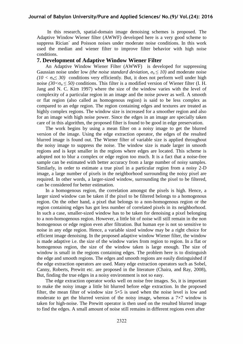
taken for high-noise. The Prewitt operator is then used on the resulted blurred image
to find the edges. A small amount of noise still remains in different regions even after
Journal of Babylon University/Pure and Applied Sciences/ No.(9)/ Vol.(24): 2016
4545
blured image with medain filter noizy image with poisson noise
Prewittblured image with medain filter
passing the noisy image through the mean filter. The Prewitt operator is less
sensitive to isolated high intensity point variations since the local averaging over sets
of three pixels tends to reduce this. In effect, it is a 'small bar' detector, rather than a
point detector. Secondly, it gives an estimate of edge direction as well as edge
magnitude at a point which is more informative.
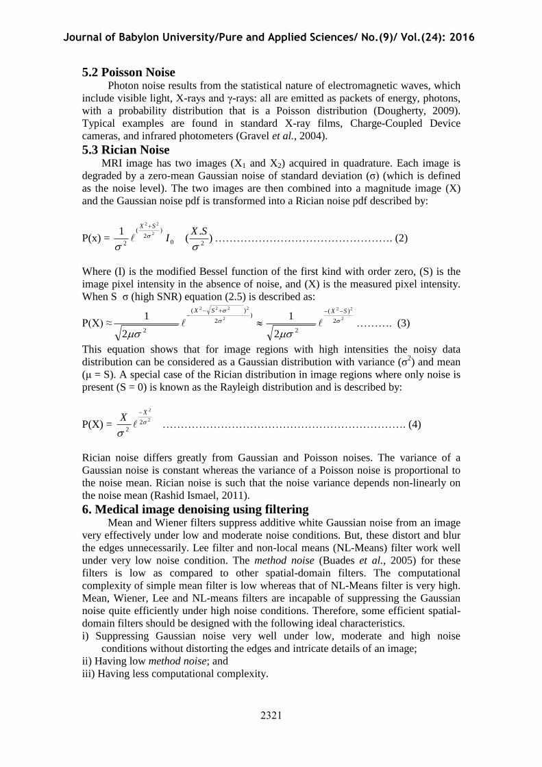
Noisy image Blurred image
Median filter
Fig (1): Blurred Image resulted from median filter
Blurred Image Edge Image
Fig (2) Edge Image using ‘Prewitt’ operator
Journal of Babylon University/Pure and Applied Sciences/ No.(9)/ Vol.(24): 2016
4546
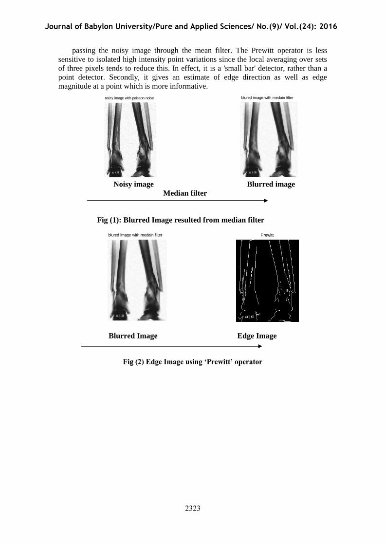
P1 or q1
Fig (3) Filtering operation of AWWF for the pixels 'p' (belonging to smooth
region) and 'q' (belonging to edge region)
The pixels 'p1' and 'q1' are the filtered pixels for the corresponding pixels 'p' and
'q' respectively.
7.1 The proposed Algorithm The proposed algorithm is given below.
Step-1:
Read the original medical image (in this work used two types of medical image
X-ray and MRI images),this images with variant size, and then add selective type of
additive noises which are explained in this research of selected image to produced
noisy image
Step-2:
The noisy image is passed through a median filter, as shown in Fig. (1), to get a
blurred version of the image.
Step-2:
Edge operator (Prewitt operator) is applied on the blurred image, obtained in
Step-2 to get the edge image. The pixels belong to smooth region and edge region are
identified as ‘p’ and ‘q’, respectively. This operation is shown in Fig. (2).
p
q
Wiener
Filtering
noizy image with poisson noise
Journal of Babylon University/Pure and Applied Sciences/ No.(9)/ Vol.(24): 2016
4547
Step-3:
Adaptive window Wiener filter is applied on the noisy image. The size of the
window is varied with the following concepts.
If the center pixel is an edge pixel, then the size of the window is small;
If the center pixel belongs to smooth region, the size of the window is large.
If the noise power is low (σn ≤ 10 ), then the size of the window is small;
If the noise power is moderate (10 <σn ≤30 ), then the size of the window is
medium;
If the noise power is high (30< σn ≤50), then the size of the window is large.
This adaptive filtering concept is depicted in Fig. (3)
The noise power can be determined by using the value of the (σ2
n) Noise Variance of
the noisy image.
Step-4:
All the filtered pixels are united together to obtain the denoised (filtered) image. The
exact window sizes taken for various conditions are presented in the next sub-section.
7.2 Results and discussion In this research the proposed algorithm has been applied on different medical
images with variant sizes in a general N x M image and modalities affected by the
appropriate noise and its performance was compared with other denoising methods.
The performance of the adaptive window wiener algorithm (AWWA) was
compared with the other type of filters (wiener filter). The selection of window in this
work is based on the level of noise present in the noisy image. If the noise level is
unknown, a robust median estimator may be applied to predict the level of noise.
When the noise level is low (σn ≤ 10) a 3×3 window is selected for filtering the noisy
pixels belonging to homogenous regions; the pixel is unaltered if the noisy pixels
belong to edges. When the noise level is moderate (10 <σn ≤30 ), a 5×5 window is
chosen for filtration of noisy pixels of flat regions; the window size is 3×3 if the noisy
pixels to be filtered are identified as edge pixels. When the noise level is high (30< σn
≤50), a 7×7 window is used for filtration of noisy pixels of flat regions; if the noisy
pixels are identified as edge pixels the window size used is 5×5.
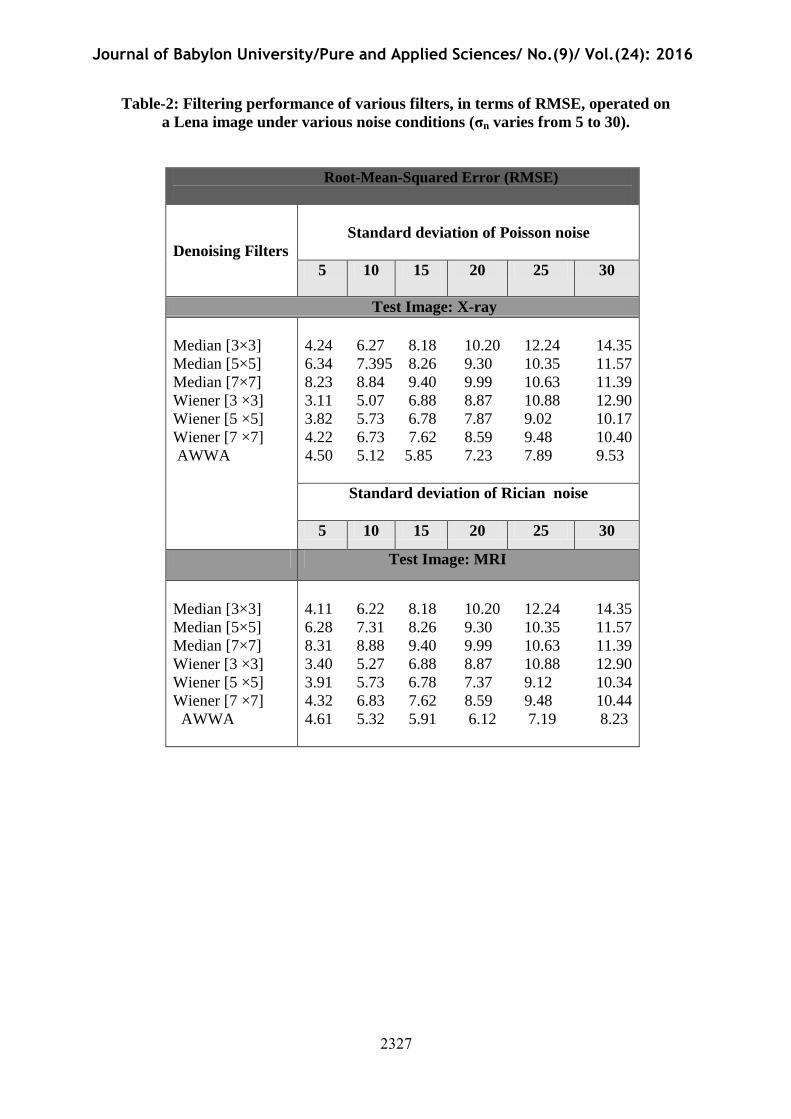
In this work The Wiener filter was implemented using the adaptive filter,
wiener2, in the MATLAB tool box function with a window of size 5 x 5 pixels. Fig.
(4) Illustrates the visual comparison between the proposed algorithm and the other
methods that were applied for denoising "X-Ray" image corrupted by Poisson noise.
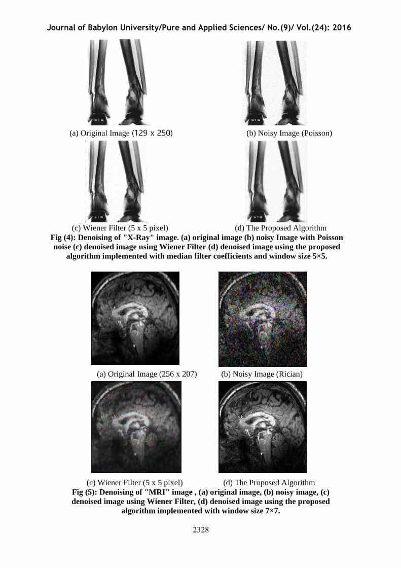
Fig. (5) Illustrates the visual comparison between the denoising methods applied on
"MRI" image of corrupted by Rician noise.
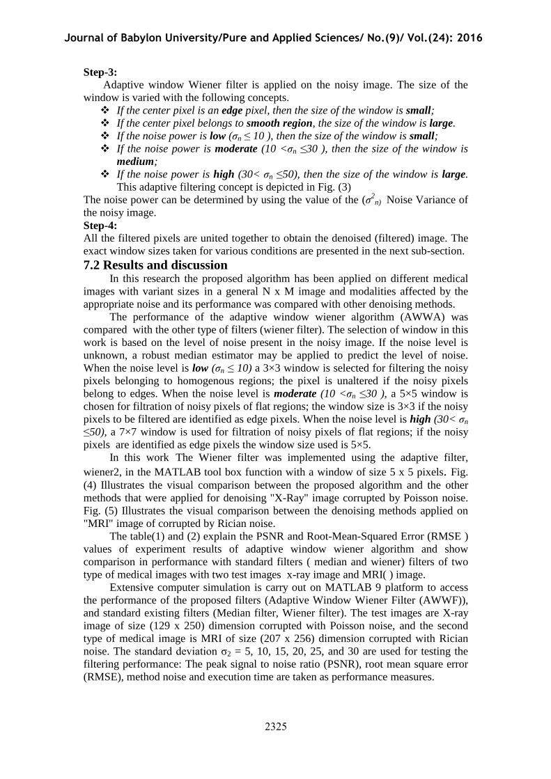
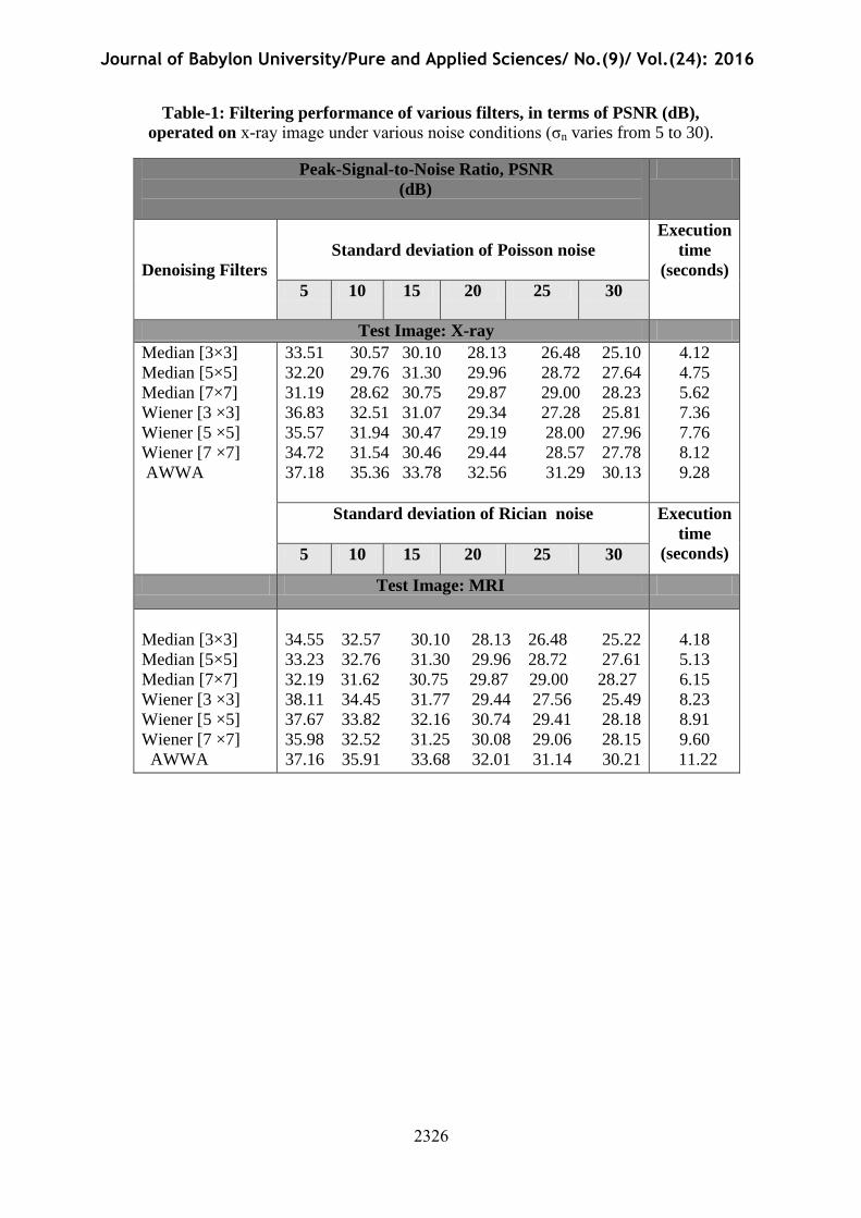
The table(1) and (2) explain the PSNR and Root-Mean-Squared Error (RMSE )
values of experiment results of adaptive window wiener algorithm and show
comparison in performance with standard filters ( median and wiener) filters of two
type of medical images with two test images x-ray image and MRI( ) image.
Extensive computer simulation is carry out on MATLAB 9 platform to access
the performance of the proposed filters (Adaptive Window Wiener Filter (AWWF)),
and standard existing filters (Median filter, Wiener filter). The test images are X-ray
image of size (129 x 250) dimension corrupted with Poisson noise, and the second
type of medical image is MRI of size (207 x 256) dimension corrupted with Rician
noise. The standard deviation σ2 = 5, 10, 15, 20, 25, and 30 are used for testing the
filtering performance: The peak signal to noise ratio (PSNR), root mean square error
(RMSE), method noise and execution time are taken as performance measures.
Journal of Babylon University/Pure and Applied Sciences/ No.(9)/ Vol.(24): 2016
4548
Table-1: Filtering performance of various filters, in terms of PSNR (dB),
operated on x-ray image under various noise conditions (σn varies from 5 to 30).
Peak-Signal-to-Noise Ratio, PSNR
(dB)
Execution
time
(seconds)
Standard deviation of Poisson noise
Denoising Filters
30 25 20 15 10 5
Test Image: X-ray
4.12
4.75
5.62
7.36
7.76
8.12
9.28
33.51 30.57 30.10 28.13 26.48 25.10
32.20 29.76 31.30 29.96 28.72 27.64
31.19 28.62 30.75 29.87 29.00 28.23
36.83 32.51 31.07 29.34 27.28 25.81
35.57 31.94 30.47 29.19 28.00 27.96
34.72 31.54 30.46 29.44 28.57 27.78
37.18 35.36 33.78 32.56 31.29 30.13
Median [3×3]
Median [5×5]
Median [7×7]
Wiener [3 ×3]
Wiener [5 ×5]
Wiener [7 ×7]
AWWA
Execution
time
(seconds)
Standard deviation of Rician noise
30 25 20 15 10 5
Test Image: MRI
4.18
5.13
6.15
8.23
8.91
9.60
11.22
34.55 32.57 30.10 28.13 26.48 25.22
33.23 32.76 31.30 29.96 28.72 27.61
32.19 31.62 30.75 29.87 29.00 28.27
38.11 34.45 31.77 29.44 27.56 25.49
37.67 33.82 32.16 30.74 29.41 28.18
35.98 32.52 31.25 30.08 29.06 28.15
37.16 35.91 33.68 32.01 31.14 30.21
Median [3×3]
Median [5×5]
Median [7×7]
Wiener [3 ×3]
Wiener [5 ×5]
Wiener [7 ×7]
AWWA
Journal of Babylon University/Pure and Applied Sciences/ No.(9)/ Vol.(24): 2016
4549
Table-2: Filtering performance of various filters, in terms of RMSE, operated on
a Lena image under various noise conditions (σn varies from 5 to 30).
Root-Mean-Squared Error (RMSE)
Standard deviation of Poisson noise
Denoising Filters
30 25 20 15 10 5
Test Image: X-ray
4.24 6.27 8.18 10.20 12.24 14.35
6.34 7.395 8.26 9.30 10.35 11.57
8.23 8.84 9.40 9.99 10.63 11.39
3.11 5.07 6.88 8.87 10.88 12.90
3.82 5.73 6.78 7.87 9.02 10.17
4.22 6.73 7.62 8.59 9.48 10.40
4.50 5.12 5.85 7.23 7.89 9.53
Median [3×3]
Median [5×5]
Median [7×7]
Wiener [3 ×3]
Wiener [5 ×5]
Wiener [7 ×7]
AWWA
Standard deviation of Rician noise
30 25 20 15 10 5
Test Image: MRI
4.11 6.22 8.18 10.20 12.24 14.35
6.28 7.31 8.26 9.30 10.35 11.57
8.31 8.88 9.40 9.99 10.63 11.39
3.40 5.27 6.88 8.87 10.88 12.90
3.91 5.73 6.78 7.37 9.12 10.34
4.32 6.83 7.62 8.59 9.48 10.44
4.61 5.32 5.91 6.12 7.19 8.23
Median [3×3]
Median [5×5]
Median [7×7]
Wiener [3 ×3]
Wiener [5 ×5]
Wiener [7 ×7]
AWWA
Journal of Babylon University/Pure and Applied Sciences/ No.(9)/ Vol.(24): 2016
454:
(a) Original Image (129 x 250) (b) Noisy Image (Poisson)
(c) Wiener Filter (5 x 5 pixel) (d) The Proposed Algorithm
Fig (4): Denoising of "X-Ray" image. (a) original image (b) noisy Image with Poisson
noise (c) denoised image using Wiener Filter (d) denoised image using the proposed
algorithm implemented with median filter coefficients and window size 5×5.
(a) Original Image (256 x 207) (b) Noisy Image (Rician)
(c) Wiener Filter (5 x 5 pixel) (d) The Proposed Algorithm
Fig (5): Denoising of "MRI" image , (a) original image, (b) noisy image, (c)
denoised image using Wiener Filter, (d) denoised image using the proposed
algorithm implemented with window size 7×7.
Journal of Babylon University/Pure and Applied Sciences/ No.(9)/ Vol.(24): 2016
454;
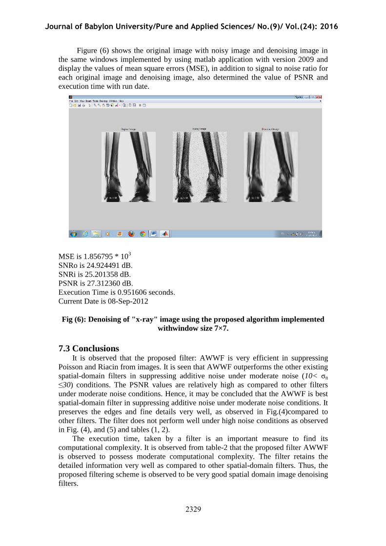
Figure (6) shows the original image with noisy image and denoising image in
the same windows implemented by using matlab application with version 2009 and
display the values of mean square errors (MSE), in addition to signal to noise ratio for
each original image and denoising image, also determined the value of PSNR and
execution time with run date.
MSE is 1.856795 * 103
SNRo is 24.924491 dB.
SNRi is 25.201358 dB.
PSNR is 27.312360 dB.
Execution Time is 0.951606 seconds.
Current Date is 08-Sep-2012
Fig (6): Denoising of "x-ray" image using the proposed algorithm implemented
withwindow size 7×7.
7.3 Conclusions It is observed that the proposed filter: AWWF is very efficient in suppressing
Poisson and Riacin from images. It is seen that AWWF outperforms the other existing
spatial-domain filters in suppressing additive noise under moderate noise (10< σn
≤30) conditions. The PSNR values are relatively high as compared to other filters
under moderate noise conditions. Hence, it may be concluded that the AWWF is best
spatial-domain filter in suppressing additive noise under moderate noise conditions. It
preserves the edges and fine details very well, as observed in Fig.(4)compared to
other filters. The filter does not perform well under high noise conditions as observed
in Fig. (4), and (5) and tables (1, 2).
The execution time, taken by a filter is an important measure to find its
computational complexity. It is observed from table-2 that the proposed filter AWWF
is observed to possess moderate computational complexity. The filter retains the
detailed information very well as compared to other spatial-domain filters. Thus, the
proposed filtering scheme is observed to be very good spatial domain image denoising
filters.
Journal of Babylon University/Pure and Applied Sciences/ No.(9)/ Vol.(24): 2016
4552
According to the experiment results in Table-1and Table-2 the values of PSNR
decreasing when the values of standard deviation (σ2) increasing. Otherwise the values
of RMSE increasing when the values of standard deviation (σ2) decreasing.
Reference Abdulmunim, M.E. 'Color Image Denoising Using Discrete Multiwavelet
Transform', Ph.D. Thesis, University of Technology, Department of Computer
Science, May 2004.
Achim, A.; A. Bezerianos, and P. Tsakalides, 'Novel Bayesian Multiscale Method
for Speckle Removal in Medical Ultrasound Images', IEEE Transactions on
Medical Imaging, Vol. 20, No. 8, pp. 772-783, August 2001.
Angenent, S. ; E. Pichon, and A. Tannenbaum, 'Mathematical Methods in Medical
Image Processing', Bulletin, (New Series) of the American Mathematical
Society, Volume 43, Number 3, pp. 365- 396, July 2006.
Buades, A.; B. Coll, and J. Morel, 'A non-local algorithm for image denoising',
Proc. IEEE international conference on computer vision and pattern recognition,
pp. 60-65, 2005.
Chaira, T. and A.K. Ray, 'A new measure using intuitionist fuzzy set theory and
Its application to edge detection', Applied Soft Computing, vol. 8, no. 2, pp.
919-927, 2008.
Chang, S.; B. Yu, and M. Vetterli, 'Adaptive wavelet thresholding for image
denoising and compression', IEEE Transactions on Image Processing, vol. 9,
no. 9, pp. 1532-1546, 2000.
Dougherty, G. 'Digital Image Processing for Medical Applications', Cambridge
University Press Cambridge, UK, 2009.
Gonzalez, R.C. and R. E. Woods, 'Digital Image Processing', Prentice Hall Inc.,
2002.
Gravel, P.; G. Beaudoin, and J. A. De Guise, 'A Method for Modeling Noise in
Medical Images', IEEE Transactions On Medical Imaging, Vol. 23, No. 10, pp.
1221-1232, October 2004.
Jang I. H. and N. C. Kim, 'Locally adaptive Wiener filtering in wavelet domain for
image restoration', Proc. IEEE R-10 Conference on Speech and Image
Technologies for Computing and Telecommunications, TENCON-97, vol. 1, pp.
25-28, 1997.
Rashid Ismael, M. 'MEDICAL IMAGE DENOISING BASED ON THE DUAL
TREE COMPLEX DISCRETE WAVELET TRANSFORM', Submitted to
the Department of Electrical and Electronic Engineering at the University of
Technology, August 2011 A.D
Rodrigo R. and S. Pedrini, ' Adaptive Edge-Preserving Image Denoising Using
Wavelet Transforms', Institute of Computing, University of Campinas,
Campinas- SP, Brazil, 2012
Roy, S.; N. Sinha and A. K. Sen,'A NEW HYBRID IMAG DENOISING
METHOD', International Journal of Information Technology and Knowledge
Management July-December 2010, Volume 2, No. 2, pp. 491-497
Satheesh1, S. and K. Prasad, ' MEDICAL IMAGE DENOISING USING
ADAPTIVE THRESHOLD BASED ON CONTOURLET TRANSFORM',
An International Journal ( ACIJ ), Vol.2, No.2, March 2011
Zhang, L.; W. Dong, D. Zhang, and G. Shi,' Two-stage image denoising by
principal component analysis with local pixel grouping', paper IEEE, 2009
Elsevier Ltd. All rights reserved.
Journal of Babylon University/Pure and Applied Sciences/ No.(9)/ Vol.(24): 2016
4553
Zhang, Y.; M. Brady, and S. Smith, 'Segmentation of Brain MR Images through a
Hidden Markov Random Field Model and the Expectation-Maximization
Algorithm', IEEE Transactions on Medical Imaging, Vol. 20, No. 1, pp. 45-57,
January 2001.