mechanism of action of hormones
TRANSCRIPT
The hormone is “substances released from
ductless or endocrine glands directly to the
blood”.
A more modern definition of a hormone is
that it is synthesized by one type of cells &
transported through blood to act on another
type of cells.
Signal Transduction through G protein:
Action is through G protein coupled
receptors (GPCR).
Action of several hormones is effected
through this mechanism.
The GPCRs are transmembrane proteins with
7 helical segments spanning the membrane.
When any ligand binds, GPCRs activate heterotrimeric
GTP binding regulatory proteins (G-proteins).
The G-protein will interact with effector proteins
which may be enzymes or ion channel proteins, which
result in the desired effect.
Different types of G proteins are present in the cells
that are coupled with different receptors & activating
different effector proteins.
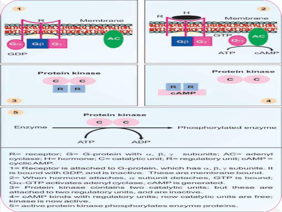
The extracellular messenger, the hormone (H)
combines with the specific receptor (R) on the
plasma membrane.
The H-R complex activates the regulatory
component of the protein designated as G-protein or
nucleotide regulatory protein.
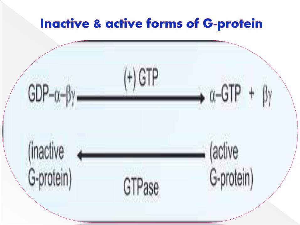
G proteins – they can bind GTP & GDP.
The G-protein is a membrane protein consisting of α,
β and γ subunits.
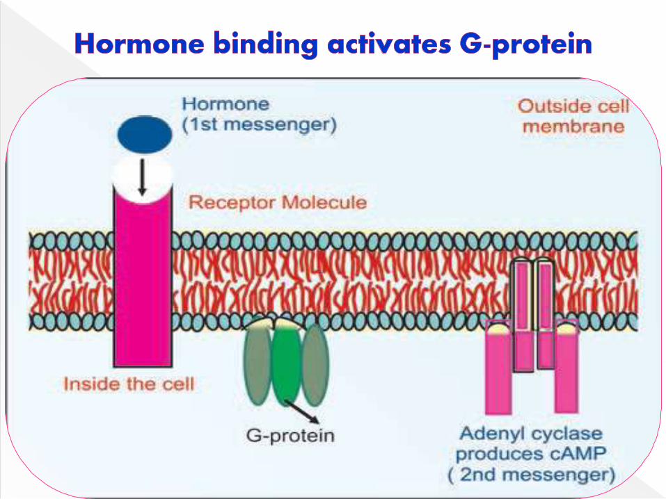
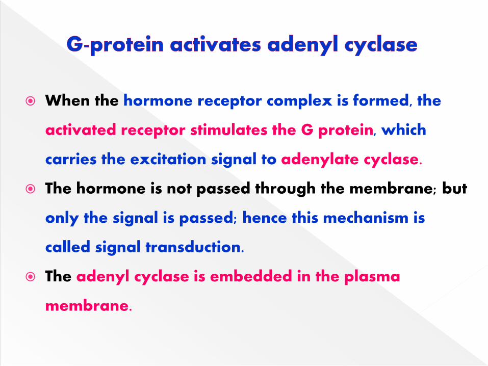
When the hormone receptor complex is formed, the
activated receptor stimulates the G protein, which
carries the excitation signal to adenylate cyclase.
The hormone is not passed through the membrane; but
only the signal is passed; hence this mechanism is
called signal transduction.
The adenyl cyclase is embedded in the plasma
membrane.
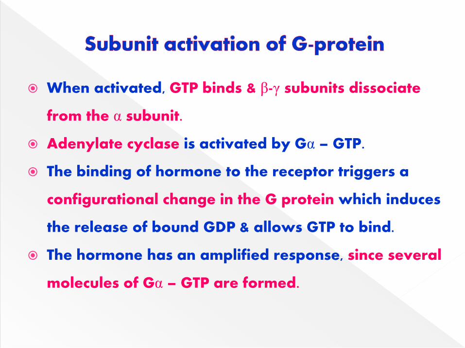
When activated, GTP binds & β-γ subunits dissociate
from the α subunit.
Adenylate cyclase is activated by Gα – GTP.
The binding of hormone to the receptor triggers a
configurational change in the G protein which induces
the release of bound GDP & allows GTP to bind.
The hormone has an amplified response, since several
molecules of Gα – GTP are formed.
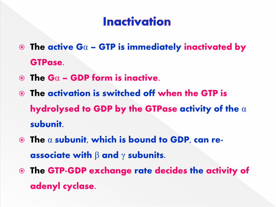
The active Gα – GTP is immediately inactivated by
GTPase.
The Gα – GDP form is inactive.
The activation is switched off when the GTP is
hydrolysed to GDP by the GTPase activity of the α
subunit.
The α subunit, which is bound to GDP, can re-
associate with β and γ subunits.
The GTP-GDP exchange rate decides the activity of
adenyl cyclase.
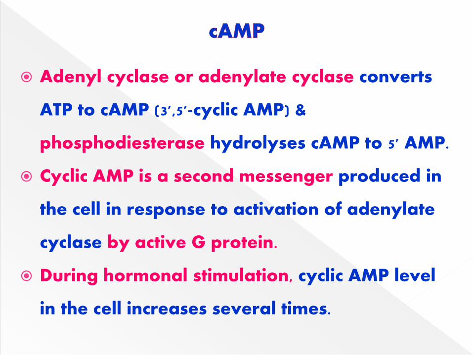
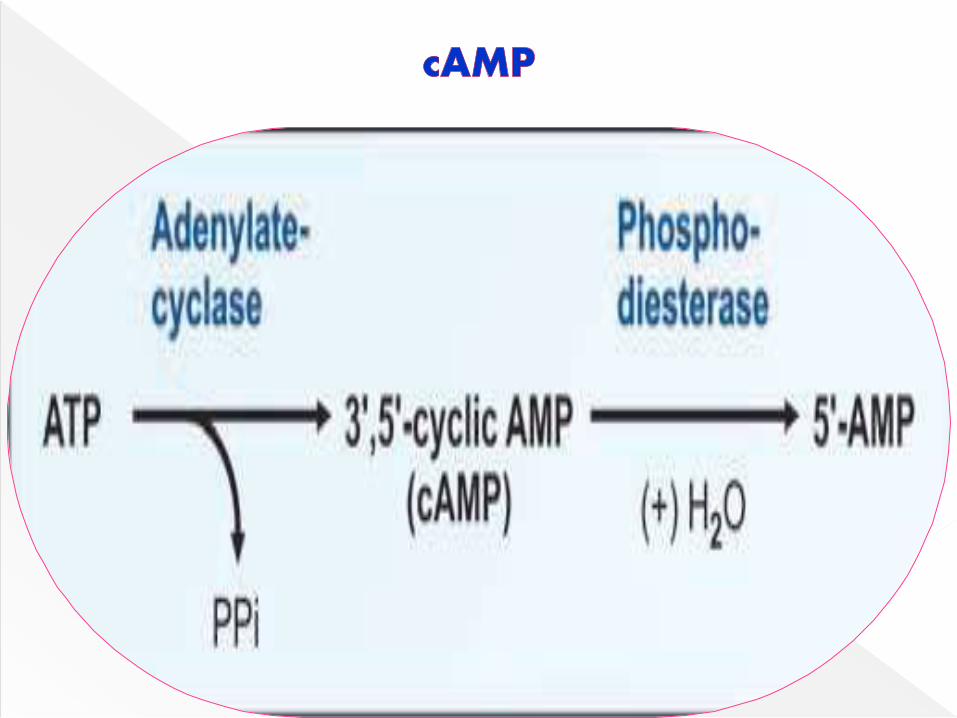
Adenyl cyclase or adenylate cyclase converts
ATP to cAMP (3',5'-cyclic AMP) &
phosphodiesterase hydrolyses cAMP to 5' AMP.
Cyclic AMP is a second messenger produced in
the cell in response to activation of adenylate
cyclase by active G protein.
During hormonal stimulation, cyclic AMP level
in the cell increases several times.
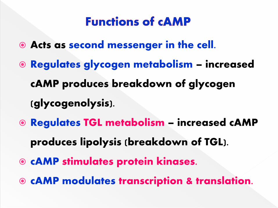
Acts as second messenger in the cell.
Regulates glycogen metabolism – increased
cAMP produces breakdown of glycogen
(glycogenolysis).
Regulates TGL metabolism – increased cAMP
produces lipolysis (breakdown of TGL).
cAMP stimulates protein kinases.
cAMP modulates transcription & translation.
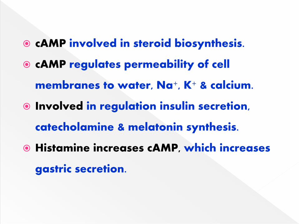
cAMP involved in steroid biosynthesis.
cAMP regulates permeability of cell
membranes to water, Na+, K+ & calcium.
Involved in regulation insulin secretion,
catecholamine & melatonin synthesis.
Histamine increases cAMP, which increases
gastric secretion.
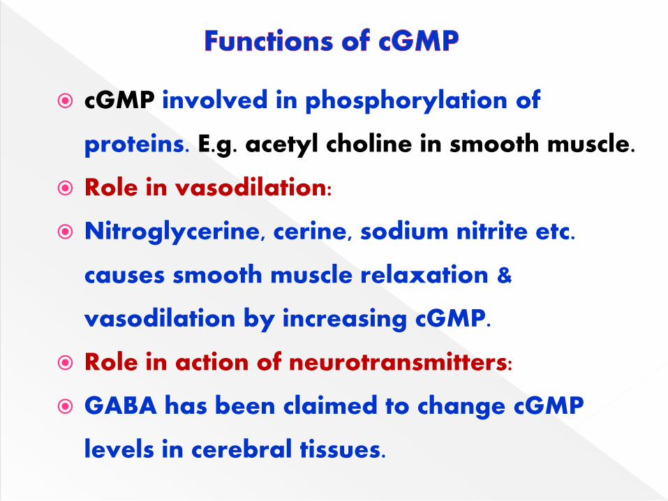
cGMP involved in phosphorylation of
proteins. E.g. acetyl choline in smooth muscle.
Role in vasodilation:
Nitroglycerine, cerine, sodium nitrite etc.
causes smooth muscle relaxation &
vasodilation by increasing cGMP.
Role in action of neurotransmitters:
GABA has been claimed to change cGMP
levels in cerebral tissues.
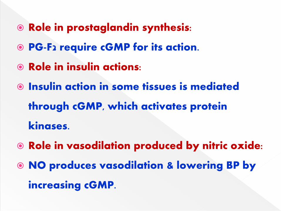
Role in prostaglandin synthesis:
PG-F2 require cGMP for its action.
Role in insulin actions:
Insulin action in some tissues is mediated
through cGMP, which activates protein
kinases.
Role in vasodilation produced by nitric oxide:
NO produces vasodilation & lowering BP by
increasing cGMP.
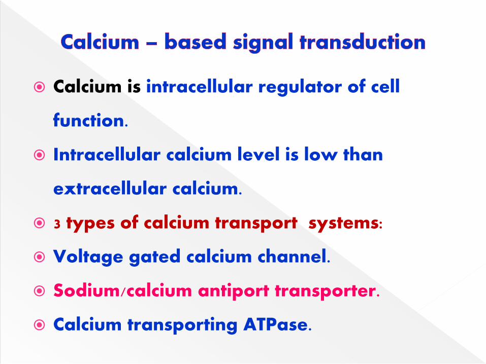
Calcium is intracellular regulator of cell
function.
Intracellular calcium level is low than
extracellular calcium.
3 types of calcium transport systems:
Voltage gated calcium channel.
Sodium/calcium antiport transporter.
Calcium transporting ATPase.
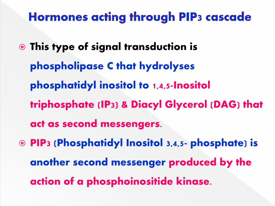
This type of signal transduction is
phospholipase C that hydrolyses
phosphatidyl inositol to 1,4,5-Inositol
triphosphate (IP3) & Diacyl Glycerol (DAG) that
act as second messengers.
PIP3 (Phosphatidyl Inositol 3,4,5- phosphate) is
another second messenger produced by the
action of a phosphoinositide kinase.
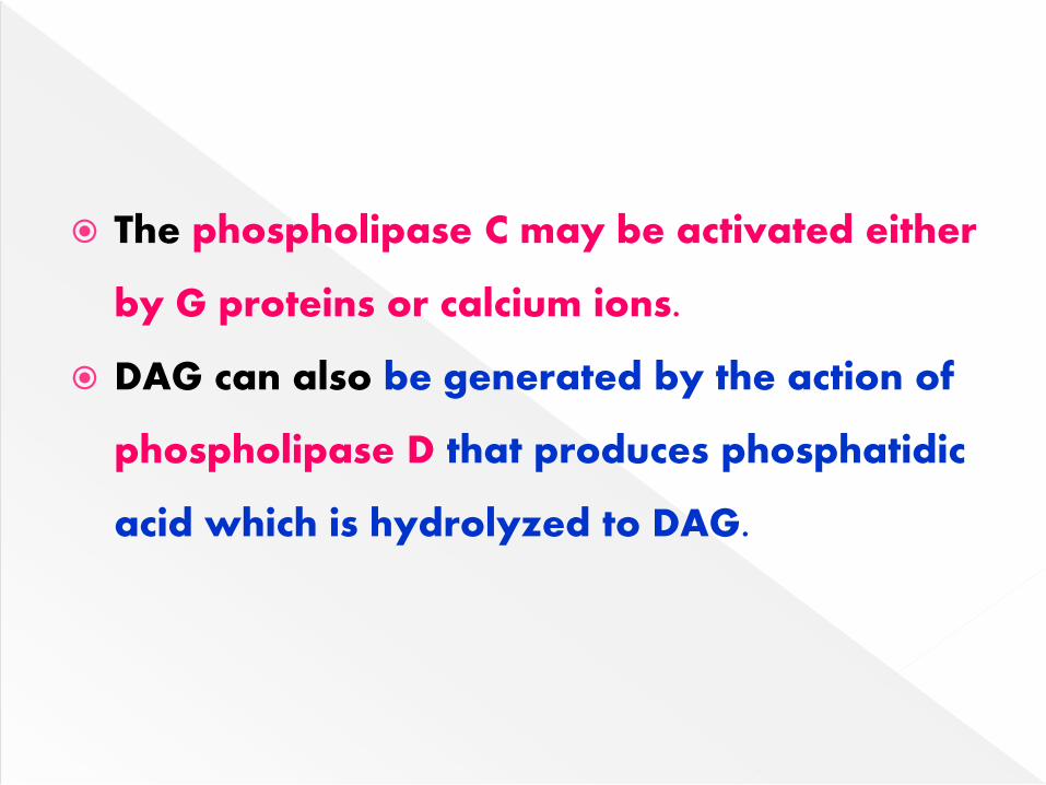
The phospholipase C may be activated either
by G proteins or calcium ions.
DAG can also be generated by the action of
phospholipase D that produces phosphatidic
acid which is hydrolyzed to DAG.
The steroid & thyroid hormones are included
in this group.
They diffuse through plasma membrane &
bind to the receptors in the cytoplasm.
The hormone receptor (HR) complex is
formed in the cytoplasm.
The complex is then translocated to the
nucleus.
Steroid hormone receptor proteins have a
molecular weight of about 80-100 kD.
Each monomer binds to a single steroid
molecule at a hydrophobic site, but on
binding to genes they dimerise.
The HR complex binds to HRE (hormone
responsive element).
HRE increase transcriptional activity.
Newly formed mRNA is translated to specific
protein, which brings metabolic effects.
Steroid hormones influence gene expression
& rate of transcription is also increased.
Textbook of Biochemistry – DM Vasudevan
Textbook of Biochemistry – U Satyanarayana
Textbook of Biochemistry – MN Chatterjea