mapping the cerebral monoamine oxidase type a : positron...
TRANSCRIPT
JPET/2002/46953 1/1
Mapping the Cerebral Monoamine Oxidase Type A : Positron
Emission Tomography Characterisation of the Reversible Selective
Inhibitor [11C]Befloxatone.
Michel BOTTLAENDER, Frédéric DOLLÉ, Ilonka GUENTHER, Dimitri ROUMENOV,
Chantal FUSEAU, Yann BRAMOULLE, Olivier CURET#, Jamir JEGHAM#,
Jean-Louis PINQUIER#, Pascal GEORGE#, Heric VALETTE.
CEA, Service Hospitalier Frédéric Joliot, DSV/DRM, 4, place du général Leclerc 91406 ORSAY, France. #Sanofi-Synthelabo Recherche, 92225 BAGNEUX, France
Copyright 2003 by the American Society for Pharmacology and Experimental Therapeutics.
JPET Fast Forward. Published on February 11, 2003 as DOI:10.1124/jpet.102.046953This article has not been copyedited and formatted. The final version may differ from this version.JPET Fast Forward. Published on February 11, 2003 as DOI: 10.1124/jpet.102.046953
at ASPE
T Journals on January 3, 2020
jpet.aspetjournals.orgD
ownloaded from
JPET/2002/46953 2/2
Running title: Befloxatone in vivo by PET Address for correspondence:
Michel Bottlaender
4, place du général Leclerc
91401 ORSAY, France
Tel : 33 1 69 86 77 12
Fax : 33 1 69 86 77 49
email : [email protected]
Text pages : 21 pages (including abstract and references)
Tables : 2
Figures :6
References : 38
Abstract : 236 words
Introduction : 624 words
Discussion : 1220 words
Neuropharmacology section.
Abbreviations:
PET: Positron Emission Tomography; MAO: Monoamine Oxidase; MAOI: Monoamine
Oxidase Inhibitor; MRI: Magnetic Resonance Imaging; ROI: Region of Interest; DV:
Distribution Volume; ID: Injected dose; TAC: time activity curve.
This article has not been copyedited and formatted. The final version may differ from this version.JPET Fast Forward. Published on February 11, 2003 as DOI: 10.1124/jpet.102.046953
at ASPE
T Journals on January 3, 2020
jpet.aspetjournals.orgD
ownloaded from
JPET/2002/46953 3/3
ABSTRACT
Befloxatone is a competitive and reversible inhibitor of MAO-A (MAOI-A). The aim of
the study was to characterize the in vivo properties of [11C]befloxatone, and to validate its
use as a ligand for the study of MAO-A by positron emission tomography (PET).
PET studies were performed in baboons after i.v. injection of [11C] befloxatone (551 ±
70 MBq, i.e.14.9 ± 1.9 mCi). [11C]befloxatone enters rapidly in the brain with a maximum
uptake at 30 min. Brain concentration of the tracer is high in thalamus, striatum, pons and
cortical structures (1.5-1.8 % of injected dose per 100 ml of tissue) and lower in
cerebellum (1.07 % ID/100ml). Non saturable uptake, obtained after a pre-treatment with a
high dose of non-labelled befloxatone (0.4 mg/kg), is very low and represents only 3% of
the total uptake. Brain uptake of [11C]befloxatone is not altered by a pre-treatment of a
high dose with lazabemide (0.5 mg/kg i.v.), a selective MAOI-B, but is completely
blocked by a pre-treatment with moclobemide (MAOI-A, 10 mg/kg). This confirms, in
vivo, the selectivity of befloxatone for type A MAO. [11C]befloxatone brain radioactivity
was displaced by administration of unlabelled befloxatone (30 min after the tracer
injection). The displacement of the tracer from its binding sites is dose dependent with an
ID50 of 0.02 mg/kg for all studied structures.
These results indicate that [11C]befloxatone will be an excellent probe for the study of
MAO-A in humans using PET.
Key Words : Monoamine oxidase; Positron Emission Tomography; in vivo; befloxatone;
monkey, pharmacology.
This article has not been copyedited and formatted. The final version may differ from this version.JPET Fast Forward. Published on February 11, 2003 as DOI: 10.1124/jpet.102.046953
at ASPE
T Journals on January 3, 2020
jpet.aspetjournals.orgD
ownloaded from

JPET/2002/46953 4/4
Monoamine oxidases (EC 1.4.3.4.; MAO) are flavoproteins integrated to outer
mitochondrial membranes. They oxidatively deaminate neurotransmitters and xenobiotic
amines. The enzyme exists in two isoforms, MAO-A and MAO-B which differ in primary
structure, substrate specificity and inhibitor sensitivity. In the brain MAO-A oxidises mainly
serotonin, norepinephrine and dopamine and is found primarily in catecholaminergic
neurones, whereas MAO-B oxidises phenethylamine and dopamine and is mainly localised in
serotoninergic neurones and glial cells. Both forms are important for regulation of
monoaminergic transmission. Fluctuation in functional MAO activity may be associated with
human diseases such as Parkinson’s and Alzheimer’s diseases, depression and certain
psychiatric disorders (Brunner et al., 1993). Moreover, it was shown that heavy smokers
possess lower brain MAO-A and MAO-B levels (Fowler et al., 1996a; Fowler et al., 1996b)
and that their inhibition facilitates smoking cessation (Berlin et al., 1995).
Positron Emission Tomography (PET) is a non invasive imaging technique allowing the
study of metabolism, receptors and, in the present case, enzymes in living human brain
(Fowler et al., 1987a; Pappata et al., 1996). Few radiotracers have been developed for MAO-
A PET studies such as [11C]clorgyline and [11C]harmine (Fowler et al, 1987a; 1996b; 2000;
Bergström et al., 1997a; Bergström et al., 1997b; Bergström et al., 1997c). However, both
tracers present some drawbacks. A recent study show that [11C]clorgyline present an
unexplained species difference in that [11C]clorgyline was not retained in baboon brain in
contrast to results in human (Fowler et al., 2001). [11C]harmine is extensively metabolised in
plasma therefore the input function determination (unchanged compound) is subject to large
errors (Bergström et al. 1997a). Brofaromine was the first reversible MAO-A inhibitor
labelled for PET (with 11C), but the brain uptake could not be significantly reduced by a pre-
treatment with either irreversible (clorgyline) or reversible (moclobemide) MAO-A inhibitors
(Ametamey et al., 1996).
This article has not been copyedited and formatted. The final version may differ from this version.JPET Fast Forward. Published on February 11, 2003 as DOI: 10.1124/jpet.102.046953
at ASPE
T Journals on January 3, 2020
jpet.aspetjournals.orgD
ownloaded from
JPET/2002/46953 5/5
Befloxatone, an oxazolidinone derivative, is a selective MAO-A reversible inhibitor with
potential antidepressant properties (Curet et al., 1994; Rovei et al., 1994; Wouters et al.,
1999). The biochemical and pharmacological profiles of befloxatone were extensively studied
in rats and in human tissues (Curet et al., 1996;Caille et al., 1996). These studies revealed
that befloxatone is a selective, competitive, specific and reversible MAO-A inhibitor in rat as
well in human tissues. Befloxatone is very potent at inhibiting in vitro MAO-A activity (Ki =
2 nM vs 150 nM for MAO-B), more potent than the other reversible MAO-A inhibitors
(including moclobemide, brofaromine, harmaline). The specificity of befloxatone for MAO-A
was confirmed against other neurotransmitter and transporter systems. In vivo, befloxatone
increases brain levels of norepinephrine, dopamine and serotonin and decreases the levels of
corresponding deaminated metabolites (dihydroxyphenylacetic acid and 5-hydroxyindolacetic
acid (Curet et al., 1996). Befloxatone is much more potent (10 to 500 fold) than other
reversible or irreversible MAO-A inhibitors in classical anti-depressant tests in rodents
(Caille et al., 1996). Befloxatone displays a higher activity in test involving MAO-A (with
ED50 of about 0.1 to 0.2 mg/kg p.o. or 0.07 mg/kg i.v.) than in test involving MAO-B (ED50
of about 58 mg/kg p.o.) (Caille et al., 1996).
Due to its selective and reversible MAO-A inhibition, befloxatone possesses a good safety
profile particularly regarding the observation of tyramine induced pressor effect and the drug
interactions (Caille et al., 1996; Rosenzweig et al., 1998).
All the biochemical and pharmacological characteristics of the drug suggest that
befloxatone is an excellent candidate for the PET study of the MAO-A in human. Moreover,
the structure of the molecule allows to label it with carbon-11, a positron emitter isotope,
using phosgene.
This article has not been copyedited and formatted. The final version may differ from this version.JPET Fast Forward. Published on February 11, 2003 as DOI: 10.1124/jpet.102.046953
at ASPE
T Journals on January 3, 2020
jpet.aspetjournals.orgD
ownloaded from
JPET/2002/46953 6/6
This work aimed to characterise the properties of the drug in living brain and to determine
if [11C]befloxatone is a suitable radioligand to study the MAO-A by PET in vivo. This
preclinical study was performed in non-human primate using PET.
This article has not been copyedited and formatted. The final version may differ from this version.JPET Fast Forward. Published on February 11, 2003 as DOI: 10.1124/jpet.102.046953
at ASPE
T Journals on January 3, 2020
jpet.aspetjournals.orgD
ownloaded from
JPET/2002/46953 7/7
METHODS
Animals
All animal use procedures were in strict accordance with the recommendations of the EEC
(86/609/CEE) and the French National Committee (décret 87/848) for the care and use of
laboratory animals. Twelve PET experiments were carried out on 4 male Papio anubis
baboons (mean weight 14.4 ± 5 kg). Each animal was allowed to rest for a period of at least 2
weeks between PET studies.
Drugs
Befloxatone ((5R)-5-(methoxymethyl)-3-[4-[(3R)-4,4,4-trifluoro-3-
hydroxybutoxy]phenyl] -2-oxazolidinone), moclobemide and lazabemide were provided by
Synthélabo, Bagneux, France. All drugs were dissolved in a mixture of physiological saline /
EtOH / propanediol (98 / 2 / 2, v:v:v) and were injected intravenously.
Befloxatone was labelled with carbon-11 (t1/2 : 20.4 minutes) using [11C]phosgene
(Landais and Crouzel 1987; Link et al., 1997) and the corresponding ring-opened precursor
(R)-1-methoxy-3-[[4-[(3R)-4,4,4-trifluoro-3-hydroxybutoxy]phenyl]amino]-2-propanol
(provided by Synthélabo, Bagneux, France). Details of the radiosynthesis have been
published elsewhere (Dolle et al., 1999; 2003). Typically, 5.55-9.25 GBq (150 to 250 mCi )
of [11C]Befloxatone with a radiochemical- and chemical purity of more than 99% were
routinely obtained within 25 minutes of radiosynthesis (including HPLC purification). Final
formulation as an i.v. injectable pharmaceutical was performed as follows : (1) HPLC
solvent removal by evaporation ; (2) taking up the residue in 5 ml of physiological saline ;
(3) filtration on a 0.22 µm Millipore filter.
This article has not been copyedited and formatted. The final version may differ from this version.JPET Fast Forward. Published on February 11, 2003 as DOI: 10.1124/jpet.102.046953
at ASPE
T Journals on January 3, 2020
jpet.aspetjournals.orgD
ownloaded from
JPET/2002/46953 8/8
Magnetic Resonance Imaging
A magnetic resonance imaging (MRI) examination was performed for each baboon in
order to provide detailed anatomical images corresponding to the PET slices. The
examinations were performed with a 1.5 Tesla SIGNA system (General Electric, Milwaukee,
WI) and a custom-made receive-only coil was used in proximity to the baboon’s head to
provide a high sensitivity. The animal was anaesthetised by i.m. injection of a mixture of
ketamine (15 mg/kg, Ketalar, Park-Davis, France) and xylazine (1.5 mg/kg, Rompun, Bayer,
France) (Banknieder et al., 1978) and positioned using a stereotaxic headholder so that the
stereotaxic reference plane was aligned with a laser beam figuring the axial reference plane of
the MR scanner. The imaging protocol used a T1-weighted inversion-recovery sequence in 3D
mode and a 256x192 matrix over 124 slices of 1.5 mm in thickness.
Positron Emission Tomography data acquisition
PET studies were performed with a high resolution tomograph (ECAT 953B/31; CTI PET
system, Knoxville, TN), which allowed reconstruction of 31 slices every 3.3 mm with spatial
and axial resolution of 5.7 mm and 5.0 mm, respectively (Bendriem, 1991). Animals were
anaesthetised using isoflurane/nitrous-oxide mixture (1%/66%), controlled by an Ohmeda
ventilator (Ohmeda OAV 7710, Madison, WI) with 33% oxygen. The tidal volume was
adjusted to achieve stable end-tidal carbon dioxide tension between 38-40 mmHg. The
baboon’s head was fixed in a headholder and positioned in the scanner gantry for axial plane
acquisition. A transmission scan (68Ge rods, 15 minutes) was recorded to correct for γ-ray
attenuation.
The image acquisition started (T0 min) at the intravenous injection of [11C]befloxatone
(551 ± 70 MBq, i.e.14.9 ± 1.9 mCi; specific radioactivity : 12.5 ± 3.3 GBq/µmol, i.e. 339 ±
89 mCi/µmol) and lasted 120 min (T120 min). Thirty-three images were acquired with scan
This article has not been copyedited and formatted. The final version may differ from this version.JPET Fast Forward. Published on February 11, 2003 as DOI: 10.1124/jpet.102.046953
at ASPE
T Journals on January 3, 2020
jpet.aspetjournals.orgD
ownloaded from
JPET/2002/46953 9/9
duration starting from 30 sec and increasing up to 10 min during the experiment.Three types
of PET experiments were designed. Control experiments were performed to define the
cerebral time activity curves (TAC) of [11C]befloxatone (n=2). Saturation experiments, in
which all MAO-A or MAO-B sites were saturated, were performed by administration of a
treatment dose of appropriate cold drug before the tracer injection. MAO-A sites were
saturated by befloxatone (0.4 mg/kg; n=2) or moclobemide (10 mg/kg; n=1), MAO-B sites
were saturated by lazabemide (0.5 mg/kg; n=1). Displacement experiments were performed
by i.v. administration, 30 min after the tracer injection (T30 min), of a dose of unlabelled
befloxatone. Six displacement experiments were performed with increasing doses of
befloxatone (0.004, 0.02, 0.04, 0.04, 0.1, 0.4 mg/kg i.v.).
Input function and metabolite studies
During each PET acquisition, arterial blood samples (n=30) were withdrawn from a
femoral artery at designated times. Blood and plasma radioactivity were measured in a γ-
counter and the blood-plasma time-activity curves were corrected for [11C] decay from the
beginning of the PET acquisition (T0 min).
In the control experiments, 8 plasma samples were collected (1, 2, 7, 10, 15, 20, 22.5, 27.5
min after tracer injection) for metabolites analysis, using HPLC. After deproteinisation using
acetonitrile the samples were centrifuged and the supernatant removed and used directly for
the chromatographic evaluation. The data acquisition and analysis were carried out using
Winflow software (version 1.21, JMBS Developments, Grenoble, France). The column
(reverse phase Waters µ Bondapak C18 column, 300 x 7.8 mm, 10 µm; Waters, Milford,
MA) was eluted applying a gradient from 20% acetonitrile in 0.01 M phosphoric acid up to
80% in 5.5 minutes, up to 90% in 7.5 minutes and back to 20% at 7.6 minutes with a total
run length of 10 minutes. The flow rate of the eluent as well as the flow rate of the liquid
This article has not been copyedited and formatted. The final version may differ from this version.JPET Fast Forward. Published on February 11, 2003 as DOI: 10.1124/jpet.102.046953
at ASPE
T Journals on January 3, 2020
jpet.aspetjournals.orgD
ownloaded from
JPET/2002/46953 10/10
scintillator was maintained at 6 ml/min. Under this conditions befloxatone elutes with a
retention time of 6 minutes while desmethylbefloxatone elutes before with a retention time of
5.3 minutes.
PET data analysis
Regions of interest (ROI) for anatomical structures such as cortex, cerebellum, pons,
thalamus and striata were delineated on MRI images of each baboon and then transferred to
the PET studies of the corresponding animal. The concentration of radioactivity in each ROI
was determined on each sequential scan and expressed as percent of the injected dose per 100
ml of tissue (% ID / 100 ml). Distribution volumes for individual ROI (DV) were calculated
using the graphic analysis developed by Logan et al.(1990) for reversible tracers. This
computation required the uptake PET data from a ROI versus time and and input function
which is the unchanged tracer concentration in arterial plasma.
As the plasma input function depends on the amount of befloxatone administered (see
Results section), the ratio [brain TACs] to [plasma AUC0→120 min] were calculated to allow
comparison between PET experiments.
Brain radioactivity in control experiments correspond to [11C]befloxatone total uptake.
Values from saturation experiments with pre-treatment of high amount of unlabelled
befloxatone represent [11C]befloxatone non-specific uptake. The [11C]befloxatone regional
specific uptake was estimated by subtracting the regional non-specific uptake from the
regional total uptake (Pappata et al., 1988; Persson et al., 1988; Brouillet et al., 1990).
In our experiments, the injected tracer mass was not higher than 61 nmol (47 ± 14 nmol,
n=12), and the tracer uptake in the brain was always less than 0.02 %ID/ml of tissue
(maximal 0.0179 %ID/ml in putamen, see Table I). As, in vitro, MAO-A Bmax is about 400 to
700 pmol/ml of brain tissue in human and monkey (Saura et al., 1992; May et al., 1991), the
This article has not been copyedited and formatted. The final version may differ from this version.JPET Fast Forward. Published on February 11, 2003 as DOI: 10.1124/jpet.102.046953
at ASPE
T Journals on January 3, 2020
jpet.aspetjournals.orgD
ownloaded from
JPET/2002/46953 11/11
[11C]befloxatone occupied always less than 5% of the MAO-A binding sites. This allows the
11C-labelled tracer to act as a true tracer. Therefore, the percent of [11C]befloxatone
displacement can be considered to represent an index of the MAO-A sites occupied by
befloxatone.
In displacement experiment, the percent [11C]befloxatone displacement was calculated at
T95 min, when the equilibrium between brain and plasma is reached (ratio brain/plasma
radioactivities is constant), using the following equation:
Displacement (%) = 100 × (BC - BD) / BC
where BC represents the specific uptake in control experiments (as defined above). BD is
the specific uptake after displacement, calculated by subtracting the regional non-specific
uptake from the regional residual uptake in displacement experiment.
The logdose-displacement curves were fitted using the logistic model (Microcalc
Origin). The results are expressed as mean ± standard deviation.
This article has not been copyedited and formatted. The final version may differ from this version.JPET Fast Forward. Published on February 11, 2003 as DOI: 10.1124/jpet.102.046953
at ASPE
T Journals on January 3, 2020
jpet.aspetjournals.orgD
ownloaded from
JPET/2002/46953 12/12
RESULTS
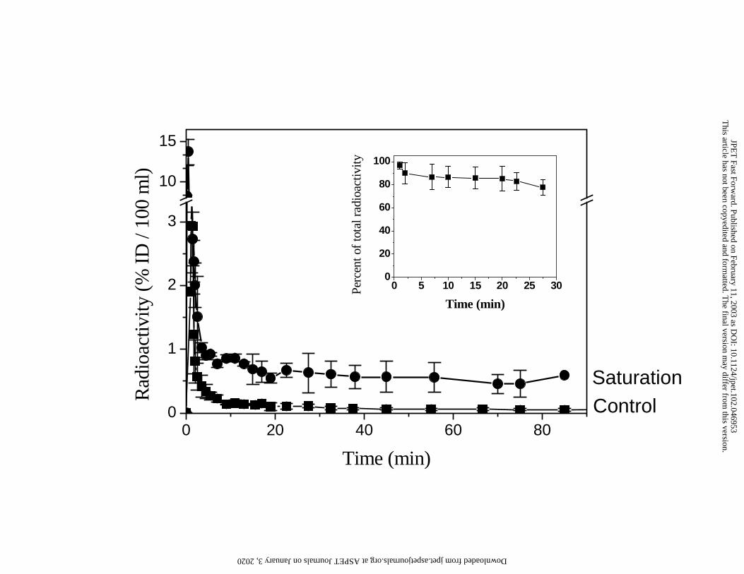
Blood and plasma time activity curves: After [11C]befloxatone a rapid phase of
distribution in blood (about 5 min), the decline in radiotracer plasma concentration was fitted
by a monoexponential function (Intercept = 0. 14 % ID / 100 ml plasma, T½ = 56.8 min in
control experiment and Intercept = 0.83 % ID / 100 ml plasma, T½ = 86.6 min in saturation
experiments). As shown in Figure 1, the [11C]befloxatone plasma TACs showed a marked
difference between control experiments and saturation experiments (pre-injection of high
amount of unlabelled befloxatone). After the distribution phase, the plasma radioactive
concentration was 8-fold higher in the saturation experiments than in the control experiments
(0.6 ± 0.2 vs 0.07 ± 0.03 % ID / 100 ml respectively at T30 min). Similarly, in displacement
experiments, the administration of unlabelled befloxatone at T30 min induced an increase of
radiotracer plasma concentration. In these experiments, the plasma radioactive level,
measured at 90 min (one hour after cold drug administration), was correlated with the amount
of injected cold drug (r=0.994; p<0.001).
In all experiments (n=12), the blood to plasma concentration ratio was constant during the
two hours of the experiments (0.9 ± 0.06) consistent with a similar kinetic of
[11C]befloxatone in both blood and plasma.
Metabolite analysis: [11C]befloxatone was extracted from plasma with an extraction
efficiency of 96%. After tracer injection, [11C]befloxatone was relatively stable in vivo, since
78% of the radioactivity in plasma at T30 min represented unchanged befloxatone (inset in
Figure 1). The detectable metabolite has a shorter retention time (2.1 min) indicating a more
hydrophilic compound. No peak was detectable at the retention time of the
desmethylbefloxatone (5.3 min).
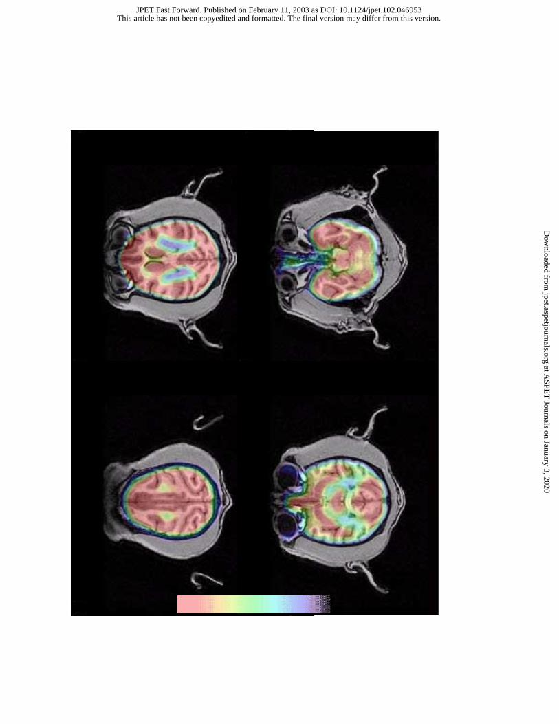
Cerebral distribution of [11C]befloxatone : Tomographic images obtained after i.v.
[11C]befloxatone display an heterogeneous distribution of radioactivity (Figure 2). A high
This article has not been copyedited and formatted. The final version may differ from this version.JPET Fast Forward. Published on February 11, 2003 as DOI: 10.1124/jpet.102.046953
at ASPE
T Journals on January 3, 2020
jpet.aspetjournals.orgD
ownloaded from
JPET/2002/46953 13/13
uptake was found in basal ganglia, thalamus, pons and cortical structures, while cerebellum
and white matter present lower uptake (Table I). Similar results were found using the
calculated DV in the different structures : high DV values (19-23 mL/g) in he basal ganglia,
thalamus and cortex and low values (10-11 mL/g) in cerebellum and white matter (Table I).
The DV values in the different brain structures are strongly correlated with the uptake values
obtained directly from the PET image (30 min after tracer injection) (r = 0.83, p<0.001).
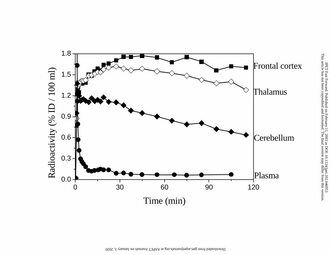
Cerebral time activity curves : After i.v. [11C]befloxatone, radioactivity was detected early
in the brain (in the first 30-sec PET image), and increased rapidly. In brain structures with a
high uptake, the radioactivity reached maximal values at 30 min (Table I), and then decreased
slowly until the end of the experiments (Figure 3). The tracer TACs was slightly different in
structures with lower uptake such as cerebellum and white matter. In these structures the
maximal uptake was reached in less than 5 min and the washout was faster (Figure 3).
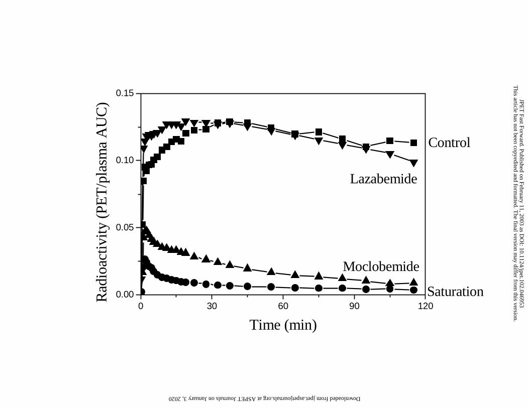
Saturability of [11C]befloxatone brain uptake : Pre-injection of unlabelled befloxatone
(0.4 mg/kg) before the radiotracer prevents the tracer from accumulating in the brain (Figure
4). The radioactive concentrations were very low and identical in all structures (0.25 ± 0.035
% DI/100ml). After correction of the plasma input function, the non-specific uptake
represents less than 5 % (from T45 min) of the total uptake.
Selectivity of [11C]befloxatone brain uptake : Brain uptake of [11C]befloxatone was
blocked by pre-treatment with the MAO-A specific inhibitor moclobemide (10 mg/kg; Figure
4). Pre-injection of the MAO-B specific inhibitor lazabemide (0.5 mg/kg) did not induce
significant change of the [11C]befloxatone brain uptake compared to that observed in control
experiments (Figure 4).
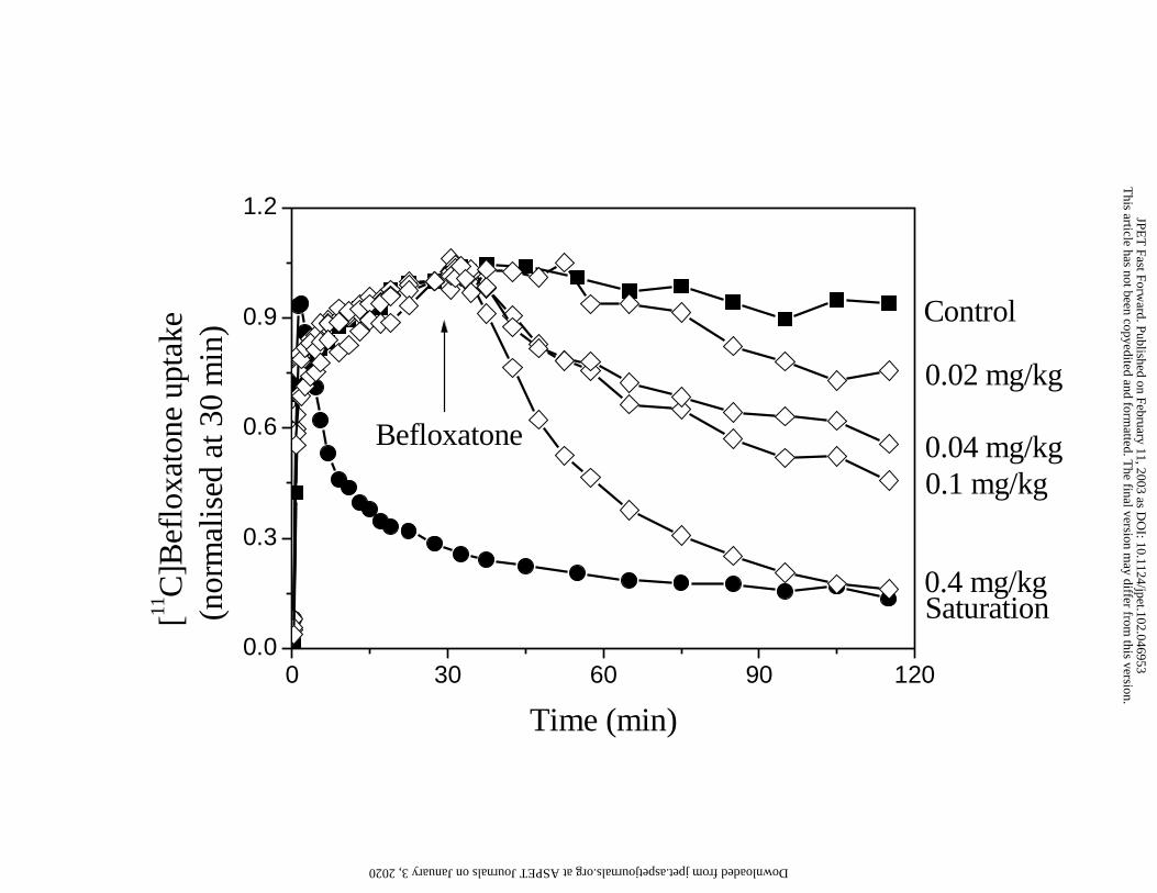
Reversibility of [11C]befloxatone brain uptake : Increasing doses of unlabelled
befloxatone were administered 30 min after the tracer injection (T30 min) in separate
experiments. Figure 5 shows that, compared with control cerebral TACs of [11C]befloxatone,
This article has not been copyedited and formatted. The final version may differ from this version.JPET Fast Forward. Published on February 11, 2003 as DOI: 10.1124/jpet.102.046953
at ASPE
T Journals on January 3, 2020
jpet.aspetjournals.orgD
ownloaded from
JPET/2002/46953 14/14
administration of befloxatone produced a wash-out of the radioactivity. The new equilibrium
between brain and plasma was reached at T95 min i.e. 60 min after the unlabelled
befloxatone administration.
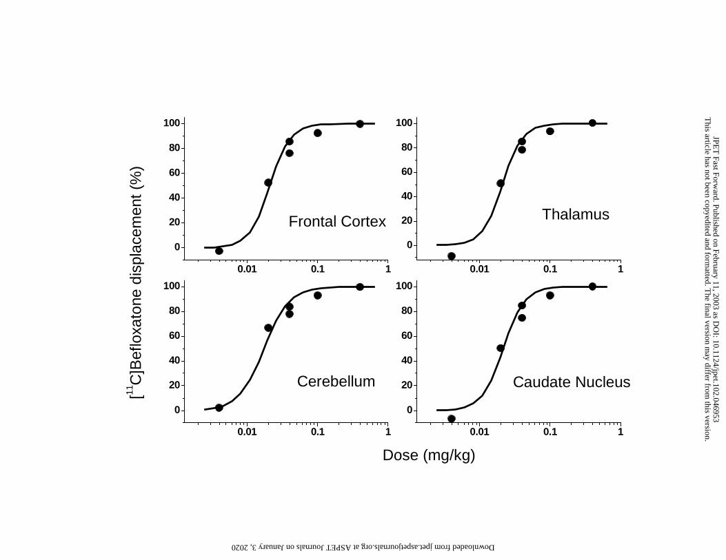
The displacement measured at T95 min was dose-dependent, the lowest dose (0.004
mg/kg) produced non-detectable displacement, the highest dose (0.4 mg/kg) completely
displaced the specifically bound [11C]befloxatone (Table II). The relationship between log-
doses of befloxatone and [11C]befloxatone displacements was roughly sigmoidal (Figure 6).
After non-linear fitting of the dose-displacement data, ID50 could be estimated for each brain
structures studied and was about 0.020 mg/kg.
This article has not been copyedited and formatted. The final version may differ from this version.JPET Fast Forward. Published on February 11, 2003 as DOI: 10.1124/jpet.102.046953
at ASPE
T Journals on January 3, 2020
jpet.aspetjournals.orgD
ownloaded from
JPET/2002/46953 15/15
DISCUSSION
The present study shows that, befloxatone, labelled with a positron emitter (Carbon 11) is
an excellent tool for PET assessment of MAO-A binding sites as demonstrated by
displacement and saturation experiments. In vivo, [11C]befloxatone penetrates and
accumulates rapidly in the brain, displays a high specific uptake which can be rapidly and
dose-dependently reversed. [11C]befloxatone presents a high selectivity for the isoform A of
MAO. These results, obtained in vivo, confirm the biochemical and pharmacological profile
of befloxatone found in rodent and in human tissues (Curet et al., 1996; Caille et al., 1996).
[11C]befloxatone was rapidly distributed in the body after i.v. administration, as shown by
the distribution phase of the radioactivity in plasma. During the elimination phase, the
[11C]befloxatone plasma concentration was very low when a tracer dose is administered. This
concentration was dramatically increased (Figure 1) when a high amount of MAO-A inhibitor
(befloxatone or moclobemide) was administered before the tracer. This is due to the presence
of high level of MAO-A sites in several peripheral organs such as the liver, kidney,
myocardium and duodenum (Saura et al., 1996). Indeed, the saturation of theses sites by the
pre-treatment dose of MAO-A inhibitors, reduces the distribution volume of the
[11C]befloxatone injected and subsequently increase its plasma concentration. This feature
was also found in human using an irreversible MAO-A specific ligand [11C]harmine, after a
seven days treatment with a MAO-A inhibitors (Bergström et al., 1997c). It is therefore
obvious that brain TACs of [11C]befloxatone, which are highly dependent of the plasma input
function, have to be corrected by this parameter (see Methods section).
[11C]Befloxatone at a tracer dose, was relatively stable in vivo. The only radiolabelled
metabolite detected at T30 min in the plasma accounted for about 20% of the total plasma
radioactivity. The short retention time of this compound indicates a low lipophilicity
suggesting a poor brain penetration of this compound.
This article has not been copyedited and formatted. The final version may differ from this version.JPET Fast Forward. Published on February 11, 2003 as DOI: 10.1124/jpet.102.046953
at ASPE
T Journals on January 3, 2020
jpet.aspetjournals.orgD
ownloaded from
JPET/2002/46953 16/16
The distribution of radioactivity measured in vivo by PET in different brain regions
(Figure 2) paralleled the areas of MAO-A concentration determined in vitro in primate brain
either by histochemistry (Westlund et al., 1985) or autoradiography (Saura et al., 1992).
Similar results were found in man, using the irreversible MAO-A PET ligand, [11C]clorgyline
(Fowler et al., 1987). As in in vitro studies, we did not find any region that was devoid of
MAO-A and significant radioactivity was even detectable in white matter.
The noradrenergic system is thought to be the main compartment of MAO-A in most
species examined including monkey, and human brain. Autoradiographical, histochemical
studies and transcript expression of MAO-A has been clearly imaged most abundant in the
human locus cœruleus (3 to 5 times higher than in cortex) and, to a lesser extend in the
interpedoncular nuclei (Luque et al., 1996; Saura et al., 1992; Willoughby et al., 1988;
Westlund et al., 1988). In PET studies, due to the small size of these structures and the
limited resolution of the technique, it is not possible to clearly identify these nuclei within the
pons. Our so called "pons" region which contains these nuclei displayed a high radioactivity.
However, compared to the values we obtained in the cortex, the activity in the pons was not
as high as expected from in vitro studies. This can be explained by the fact that, due to their
small size, compared to the resolution of the PET camera, the nuclei are more sensitive than
larger structures to partial volume effect. Thus PET measured activity in these nuclei is
underestimated when compared to structures such as cortical regions and even basal ganglia.
The brain uptake of [11C]befloxatone showed that the tracer freely crosses the blood-brain
barrier and binds rapidly to its target sites with a maximum at 30 min. This is consistent with
studies in rodents showing that maximal inhibition of MAO-A brain activity is obtained at
30-60 min after befloxatone administered p.o.(Curet et al., 1996). The very low residual
[11C]befloxatone uptake (less than 5% of the total uptake) obtained after injection of a high
dose of unlabelled befloxatone indicates that the most part of the brain radioactivity is
This article has not been copyedited and formatted. The final version may differ from this version.JPET Fast Forward. Published on February 11, 2003 as DOI: 10.1124/jpet.102.046953
at ASPE
T Journals on January 3, 2020
jpet.aspetjournals.orgD
ownloaded from
JPET/2002/46953 17/17
specifically bound to MAO-A. Moreover, pre-treatment with another selective MAO-A
inhibitor, moclobemide (Da Prada et al., 1989), has the same effect. Only a low residual
radioactivity was observed, confirming that [11C]befloxatone binds in vivo with a high
specificity to the MAO-A.
To test, in vivo, the MAO isoform selectivity of [11C]befloxatone, a high dose of
lazabemide, a selective MAO-B inhibitor (Haefely et al., 1990), was injected before
[11C]befloxatone. The lazabemide dose (0.5 mg/kg) was chosen high enough to saturate
MAO-B isoform (Bench et al., 1991). In our study, in spite of this high dose of lazabemide,
the brain uptake of [11C]befloxatone was not decreased (Figure 4). This results demonstrates
that in vivo [11C]befloxatone binds with a high specificity and selectivity to MAO-A. This
confirms the high selectivity of befloxatone in vivo for MAO-A as found in the in vitro and
ex vivo enzyme inhibition studies of befloxatone (Curet et al., 1996) as well as in the in vivo
pharmacological profile(Caille et al., 1996).
Biochemical studies have shown that befloxatone binds to MAO-A in a reversible and
competitive manner in rat and human tissues (Curet et al., 1996). To verify these
characteristics in vivo, competition experiments were performed with increasing doses of
unlabelled befloxatone administered after the tracer injection (at T30 min). We showed that
befloxatone displaces the specifically bound [11C]befloxatone in all brain structures. The
radioactivity washout was very rapid, in that a new equilibrium between brain and plasma
was achieved after 30 min. This confirms that, in vivo, the MAO-A befloxatone binding is
rapidly reversible.
[11C]Befloxatone was displaced in a dose dependent manner confirming the reversible
interaction of befloxatone with MAO-A demonstrated in vitro (Curet et al., 1996; Wouters et
al., 1999). In our study we found that the estimated dose of befloxatone needed to displace
half of the specifically bound radioactivity in brain is very low (about 0.02 mg/kg). The high
This article has not been copyedited and formatted. The final version may differ from this version.JPET Fast Forward. Published on February 11, 2003 as DOI: 10.1124/jpet.102.046953
at ASPE
T Journals on January 3, 2020
jpet.aspetjournals.orgD
ownloaded from
JPET/2002/46953 18/18
affinity of befloxatone for MAO-A (Ki = 2.5 nM), associated with an extensive brain
penetration may explain these results. Befloxatone was shown to be very potent in inhibiting
the rat brain MAO-A activity (increase brain concentration of monoamines ED50 = 0.06
mg/kg, p.o.; decrease monoamine metabolites ED50 = 0.03 mg/kg, p.o.)(Curet et al., 1996).
In conclusion, the in vivo properties of befloxatone may bring new insights for the
therapeutic use of befloxatone. The knowledge of the brain pharmacokinetics of befloxatone
may be of great interest for a better control of the intensity and the duration of the enzyme
inhibition. This, together with the determination of the dose of befloxatone that inhibits its
target, may lead to a better definition of the therapeutical doses. Moreover, the competitivity
of befloxatone binding to its target, demonstrated in vivo, tend to minimise the interaction
with tyramine absorbed with the food and thus to avoid the cheese effect in patients, a major
side effect of irreversible MAO inhibitors. All these features may provide a significant
improvement in the therapeutic use of befloxatone.
The present report shows that in vivo [11C]befloxatone enters readily the brain and binds to
MAO-A in a potent, selective, and reversible manner. Moreover, [11C]befloxatone present a
very low non-saturable uptake and is metabolised rather slowly. All these biochemical
characteristics suggest that [11C]befloxatone is a unique tool for the in vivo study of MAO-A
brain.
AKNOWLEDGMENTS:
We thank C. Jouy and F. Sergent for their help and outstanding care of the non-human
primate colony.
This article has not been copyedited and formatted. The final version may differ from this version.JPET Fast Forward. Published on February 11, 2003 as DOI: 10.1124/jpet.102.046953
at ASPE
T Journals on January 3, 2020
jpet.aspetjournals.orgD
ownloaded from
JPET/2002/46953 19/19
REFERENCE
Ametamey SM, Beer H-F, Guenther I, Antonini A, Leenders KL, Waldmeier PC, and
Schubiger PA (1996) Radiosynthesis of [11C]brofaromine, a potential tracer for imaging
monoamine oxidase A. Nucl.Med.Biol. 23:229-234.
Banknieder AR, Phillips JM, Jackson KT, and Vinal SI (1978) Comparison of ketamine with
the combination of ketamine and xylazine for effective anesthesia in the rhesus monkey
(Macaca mulata). Lab.Anim.Sci. 28:742-745.
Bench CJ, Price GW, Lammertsma AA, Cremer JC, Luthra SK, Turton D, Dolan RJ, Kettler
R, Dingemanse J, Da Prada M, Biziere K, McClelland GR, Jamieson VL, Wood ND, and
Frackowiak RSJ (1991) Measurement of human cerebral monoamine oxidase type B (MAO-
B) activity with positron emission tomography (PET): a dose ranging study with the
reversible inhibitor Ro 19-6327. Eur.J.Clin.Pharmacol 40:173.
Bendriem, B (1991) Quantitative evaluation of a new brain tomograph the ECAT 953B/31.
J.Nucl.Med. 32:1063. 1991.
Bergström, M., Westerberg, G., Kihlberg, T., and Langström, B. (1997a) Synthesis of some
11C-labelled MAO-A inhibitors and their in vivo uptake kinetics in rhesus monkey brain.
Nucl.Med.Biol. 24:381-388.
Bergström, M., Westerberg, G., and Langström, B. (1997b) 11C-harmine as a tracer for
monoamine oxidasse A (MAO-A): In vitro and in vivo studies. Nucl.Med.Biol. 24:287-293.
Bergström M, Westerberg G, Németh G, Traut M, Gross G, Greger G, Müller-Peltzer H, Safer
A, Eckernäs S-A, and Grahner A (1997c) MAO-A inhibition in brain after dosing with
This article has not been copyedited and formatted. The final version may differ from this version.JPET Fast Forward. Published on February 11, 2003 as DOI: 10.1124/jpet.102.046953
at ASPE
T Journals on January 3, 2020
jpet.aspetjournals.orgD
ownloaded from
JPET/2002/46953 20/20
esuprone, moclobemide and placebo in healthy volunteers: In vivo studies with positron
emission tomography. Eur.J.Clin.Pharmacol 52:121-128.
Berlin I, Saïd S, Spreux-Varoquaux O, Launay J-M, Olivares R, Millet V, Lecrubier Y, and
Puech A (1995) A reversible monoamine oxidase A inhibitor (moclobrmide) facilitates
smoking cessation and abstinence in heavy, dependent smokers. Clin.Pharmacol.Ther.
58:444-452.
Brouillet E, Chavoix C, Khalili-Varasteh M, Bottlaender M, Hantraye Ph, Yorke J-C, and
Mazière M (1990) Quantitative evaluation of benzodiazepine receptors in live Papio papio
baboons using Positron Emission Tomography. Mol.Pharmacol. 38:445-451.
Brunner HG, Nelen M, Breakefield XO, Ropers HH, and Van Oost BA (1993) Abnormal
behavior associated with a point mutation in the structural gene for monoamine oxidase A.
Science 262:578-580.
Caille D, Bergis O-E, Fankhausser C, Gardes A, Adam R, Charieras T, Grosset A, Rovei V,
and Jarreau F-X (1996) Befloxatone, a new reversible and selective monoamine oxidase-A
inhibitor. II. Pharmacological profile. J.Pharmacol.Exp.Ther. 277:265-277.
Curet, O.; Damoiseau, G.; Labaune, J.-P.; Rovei, V. and Jarreau, F.-X. (1994) Effects of
befloxatone, a new potent reversible MAO-A inhibitor, on cortex and striatum monoamines
in freely moving rats. J. Neural. Transm. 41:349-355.
Curet O, Damoiseau G, Aubin N, Sontag N, Rovei V, and Jarreau F-X (1996) Befloxatone, a
new reversible and selective monoamine oxidase-A inhibitor. I. Biochemical profile.
J.Pharmacol.Exp.Ther. 277:253-264.
This article has not been copyedited and formatted. The final version may differ from this version.JPET Fast Forward. Published on February 11, 2003 as DOI: 10.1124/jpet.102.046953
at ASPE
T Journals on January 3, 2020
jpet.aspetjournals.orgD
ownloaded from
JPET/2002/46953 21/21
Da Prada M, Kettler R, Keller HH, Burkard WP, Muggli-Maniglio D, and Haefely W (1989)
Neurochemical profile of moclobemide, a short-acting and reversible inhibitor of monoamine
oxidase type A. J.Pharmacol.Exp.Ther. 248:400-412.
Dollé F, Bramoullé Y, Bottlaender M, Guenther I, Valette H, Fuseau C, Jegham S,
George P, Curet O, Pinquier J-L and Crouzel C (1999) [11C]Befloxatone, a Novel
Highly Potent Radioligand for in Vivo Imaging Mono-Amine Oxidase-A. J. Label.
Compounds Radiopharm. 42:608-609.
Dollé F, Valette H, Bramoullé Y, Guenther I, Fuseau C, Coulon C, Lartizien C, Jegham
S, Curet O, Pinquier J-L, George P and Bottlaender M. (2003) Synthesis and In Vivo
Imaging Properties of [11C]Befloxatone : a Novel Highly Potent Positron Emission
Tomography Ligand for Mono-Amine Oxidase-A. Bioorg. Med. Chem. Letters
submitted
Fowler, J. S., Ding, Y. S., Logan, J., MacGregor, R. R., Shea, C., Garza, V., Gimi, R.,
Volkow, N. D., Wang, G-J., Schlyer, D., Ferrieri, R. A., Gatley, S. J., Alexoff, D. L.,
Carter, P., King, P., and Pappas, N. (2001) Species differences in [11C]clorgyline
binding in brain. Nucl.Med.Biol. 28:779-785.
Fowler JS, MacGregor RR, Wolf AP, Arnett CD, Dewey SL, Schlyer D, Christman DR,
Logan J, Smith M, Sachs H, Aquilonius SM, Bjurling P, Halldin C, Hartvig P, Leenders KL,
Lundquist H, Oreland L, Stalnacke C-G, and Langström B (1987) Mapping human brain
monoamine oxidase A and B with 11C-labeled suicide inactivators and PET. Science
235:481-485.
This article has not been copyedited and formatted. The final version may differ from this version.JPET Fast Forward. Published on February 11, 2003 as DOI: 10.1124/jpet.102.046953
at ASPE
T Journals on January 3, 2020
jpet.aspetjournals.orgD
ownloaded from
JPET/2002/46953 22/22
Fowler JS, Volkow ND, Wang G-J, Pappas N, Logan J, MacGregor RR, Alexoff DL, Shea C,
Schlyer D, Wolf AP, Warner D, Zezulkova I, and Cilento R (1996a) Inhibition of monoamine
oxidase B in the brains of smokers. Nature 379:733-736.
Fowler JS, Volkow ND, Wang G-J, Pappas N, Logan J, Shea C, Alexoff DL, MacGregor RR,
Schlyer D, Zezulkova I, and Wolf AP (1996b) Brain monoamine oxidase A inhibition in
cigarette smokers. Proc.Natl.Acad.Sci.USA 93:14065-14069.
Fowler, J. S., Wang, G-J., Volkow, N. D., Logan, J., Franceschi, D., Franceschi, M.,
MacGregor, R. R., Shea, C., Garza, V., Liu, N., and Ding, Y. S. (2000) Evidence that gingko
biloba extract does not inhibit MAO-A and B in living human brain. Life Sci. 66 :PL141-
146.
Haefely W, Kettler R, Keller HH, and Da Prada M (1990) Ro 19-6327, a reversible and
highly selective monoamine oxidase B inhibitor: A novel tool to explore the MAO-B function
in humans, in Parkinson’s Disease: Anatomy, Pathology, and Therapy (Streifler MB,
Korczyn AD, Melamed E, and Youdim MBH eds) pp 505-512, Raven Press, New York.
Landais P and Crouzel C (1987) A new synthesis of carbon-11 labelled phosgene. Appl.
Radiat. Isot. 38:297-300.
Link J.M and Krohn K.A (1997) A simplified production of high specific activity
[11C]labelled phosgene. J. Label. Compds. Radiopharm. 40: 306-308
Logan J, Fowler JS, Volkow ND, Wolf AP, Dewey SL, Schlyer D, MacGregor RR,
Hitzemann R, Bendriem B, Gatley SJ, and Christman DR (1990) Graphical analysis of
reversible radioligand binding time-activity measurements applied to [N-11C-methyl]-(-)-
cocaine PET studies in human subjects. J.Cereb.Blood Flow Metab. 10:740-747.
This article has not been copyedited and formatted. The final version may differ from this version.JPET Fast Forward. Published on February 11, 2003 as DOI: 10.1124/jpet.102.046953
at ASPE
T Journals on January 3, 2020
jpet.aspetjournals.orgD
ownloaded from
JPET/2002/46953 23/23
Luque JM, Bleuel Z, Hendrickson A, and Richards JG (1996) Detection of MAO-A and
MAO-B mRNAs in monkey brainstem by cross-hybridization with human oligonucleotide
probes. Molec.Brain Res. 36:357-360.
May T, Pawlik M, and Rommelspacher H (1991) [3H]Harman binding experiments. II:
Regional and subcellular distribution of specific [3H]Harman binding and monoamine
oxidase subtypes A and B activity in Marmoset and rat. J.Neurochem. 56:500-508.
Pappata S, Samson Y, Chavoix C, Prenant C, Mazière M, and Baron J-C (1988) Regional
specific binding of (11C)-Ro15-1788 to central type benzodiazepine receptors in human brain:
Quantitative evaluation by Positron Emission Tomography. J.Cereb.Blood Flow Metab.
8:304-313.
Pappata S, Tavitian B, Traykov L, Jobert A, Dalger A, Mangin JF, Crouzel C, and
DiGiamberardino L (1996) In vivo imaging of human cerebral acetylcholinesterase.
J.Neurochem. 67:876-879.
Persson A, Pauli S, Halldin C, Stone-Elander S, Farde L, Sjögren I, and Sedvall G (1988)
Saturation analysis of specific [11C] Ro15-1788 binding to the human neocortex using
Positron Emission Tomography. Human Psychopharmacol. 4:21-31.
Rosenzweig P, Patat A, Curet O, Durrieu G, Dubruc C, Zieleniuk I, and Legangneux E
(1998) Clinical pharmacology of befloxatone : a brief review. Journal of Affective Disorders
51:305-312.
Rovei, V.; Caille, D.; Curet, O.; Ego, D. and Jarreau, F.-X. (1994) Biochemical pharmacoly
of befloxatone (MD370503), a new potent reversible MAO-A inhibitor. J. Neural. Transm.
41:339-347.
This article has not been copyedited and formatted. The final version may differ from this version.JPET Fast Forward. Published on February 11, 2003 as DOI: 10.1124/jpet.102.046953
at ASPE
T Journals on January 3, 2020
jpet.aspetjournals.orgD
ownloaded from
JPET/2002/46953 24/24
Saura J, Kettler R, Da Prada M, and Richards JG (1992) Quantitative enzyme
radioautography with 3H-Ro41-1049 and 3H-Ro19-6327 in vitro: Localization and abundance
of MAO-A and MAO-B in rat CNS, peripheral organs and human brain. J.Neurosci.
12:1977-1999.
Saura J, Nadal E, van den Berg B, Vila M, Bombi JA, and Mahy N (1996) Localization of
monoamine oxidases in human peripheral tissues. Life Sci. 59:1341-1349.
Westlund KN, Denney RM, Kochersperger LM, Rose RM, and Abell CW (1985) Distinct
monoamine oxidase A and B population in primate brain. s 230:181-183.
Westlund KN, Denney RM, Rose RM, and Abell CW (1988) Localization of distinct
monoamine oxidase A and monoamine oxidase B cell population in human brainstem.
Neurosci. 25:439-456.
Willoughby J, Glover V, and Sandler M (1988) Histochemical localisation of monoamine
oxidase A and B in rat brain. J.Neural Transm. 74:29-42.
Wouters J, Moureau F, Evrard G, Koenig J-J, Jegham S, George P, and Durant F (1999) A
reversible monoamine oxidase a inhibitor, Befloxatone: Structural approach of its
mechamism of action. Bioorg.Med.Chem. 7:1683-1693.
This article has not been copyedited and formatted. The final version may differ from this version.JPET Fast Forward. Published on February 11, 2003 as DOI: 10.1124/jpet.102.046953
at ASPE
T Journals on January 3, 2020
jpet.aspetjournals.orgD
ownloaded from
JPET/2002/46953 25/25
LEGENDS
Figure 1: [11C]befloxatone plasma TACs in control (n ; n=2) and saturation experiments
with befloxatone (l ; n=2). Values are expressed in % ID / 100ml of plasma. The
inset represent the percent of non metabolised [11C]befloxatone in plasma during
the control experiments.
Figure 2: PET axial brain slices obtained 30 min after i.v. [11C]befloxatone and
superimposed with the MRI image. Radioactive concentration is reported in color.
A high uptake was found in basal ganglia, thalamus, pons and cortical structures,
while cerebellum and white matter present lower uptake
Figure 3: [11C]befloxatone TACs in several brain structures, in a control experiment
(radiotracer is injected i.v. at T0 min). Radioactivity in plasma is also displayed.
Values are expressed in % ID / 100ml of tissue.
Figure 4: [11C]befloxatone TACs in the frontal cortex obtained in the control (n ; n=2) and
saturation with befloxatone l ; n=2) experiments and compared to TACs obtained
after pre-treatment with the MAO-A specific inhibitor moclobemide (10 mg/kg; �
) and the MAO-B specific inhibitor lazabemide (0.5 mg/kg; ����9DOXHV�DUH�3(7�
data corrected by the plasma AUC0→120.
Figure 5: [11C]befloxatone TACs in the frontal cortex obtained in 4 separates displacement
experiments (open diamonds) as compared to the control (n) and saturation with
befloxatone (l) experiments. To make the figure more readible, data are
normalised to the value at 30 min
Figure 6: . Dose-displacement relationship in four brain structures. Experimental data (l)
are fitted using the logistic model (line).
This article has not been copyedited and formatted. The final version may differ from this version.JPET Fast Forward. Published on February 11, 2003 as DOI: 10.1124/jpet.102.046953
at ASPE
T Journals on January 3, 2020
jpet.aspetjournals.orgD
ownloaded from
0 20 40 60 800
1
2
3
10
15
SaturationControl
Rad
ioac
tivity
(%
ID
/ 10
0 m
l)
Time (min)
0 5 10 15 20 25 300
20
40
60
80
100
Perc
ent o
f to
tal r
adio
activ
ity
Time (min)
This article has not been copyedited and form
atted. The final version m
ay differ from this version.
JPET
Fast Forward. Published on February 11, 2003 as D
OI: 10.1124/jpet.102.046953
at ASPET Journals on January 3, 2020 jpet.aspetjournals.org Downloaded from
This article has not been copyedited and formatted. The final version may differ from this version.JPET Fast Forward. Published on February 11, 2003 as DOI: 10.1124/jpet.102.046953
at ASPE
T Journals on January 3, 2020
jpet.aspetjournals.orgD
ownloaded from
0 30 60 90 1200.0
0.3
0.6
0.9
1.2
1.5
1.8
Frontal cortex
Thalamus
Cerebellum
PlasmaRad
ioac
tivity
(%
ID
/ 10
0 m
l)
Time (min)
This article has not been copyedited and form
atted. The final version m
ay differ from this version.
JPET
Fast Forward. Published on February 11, 2003 as D
OI: 10.1124/jpet.102.046953
at ASPET Journals on January 3, 2020 jpet.aspetjournals.org Downloaded from
0 30 60 90 1200.00
0.05
0.10
0.15
Moclobemide
Lazabemide
Saturation
Control
Rad
ioac
tivity
(PE
T/p
lasm
a A
UC
)
Time (min)
This article has not been copyedited and form
atted. The final version m
ay differ from this version.
JPET
Fast Forward. Published on February 11, 2003 as D
OI: 10.1124/jpet.102.046953
at ASPET Journals on January 3, 2020 jpet.aspetjournals.org Downloaded from
0 30 60 90 1200.0
0.3
0.6
0.9
1.2
Saturation0.4 mg/kg
0.1 mg/kg0.04 mg/kg
0.02 mg/kg
Control
Befloxatone
[11
C]B
eflo
xato
ne u
ptak
e(n
orm
alis
ed a
t 30
min
)
Time (min)
This article has not been copyedited and form
atted. The final version m
ay differ from this version.
JPET
Fast Forward. Published on February 11, 2003 as D
OI: 10.1124/jpet.102.046953
at ASPET Journals on January 3, 2020 jpet.aspetjournals.org Downloaded from
0.01 0.1 1
0
20
40
60
80
100
Frontal Cortex
[11C
]Bef
loxa
tone
dis
plac
emen
t (%
)
0.01 0.1 1
0
20
40
60
80
100
Thalamus
0.01 0.1 1
0
20
40
60
80
100
Dose (mg/kg)
Cerebellum
0.01 0.1 1
0
20
40
60
80
100
Caudate Nucleus
This article has not been copyedited and form
atted. The final version m
ay differ from this version.
JPET
Fast Forward. Published on February 11, 2003 as D
OI: 10.1124/jpet.102.046953
at ASPET Journals on January 3, 2020 jpet.aspetjournals.org Downloaded from
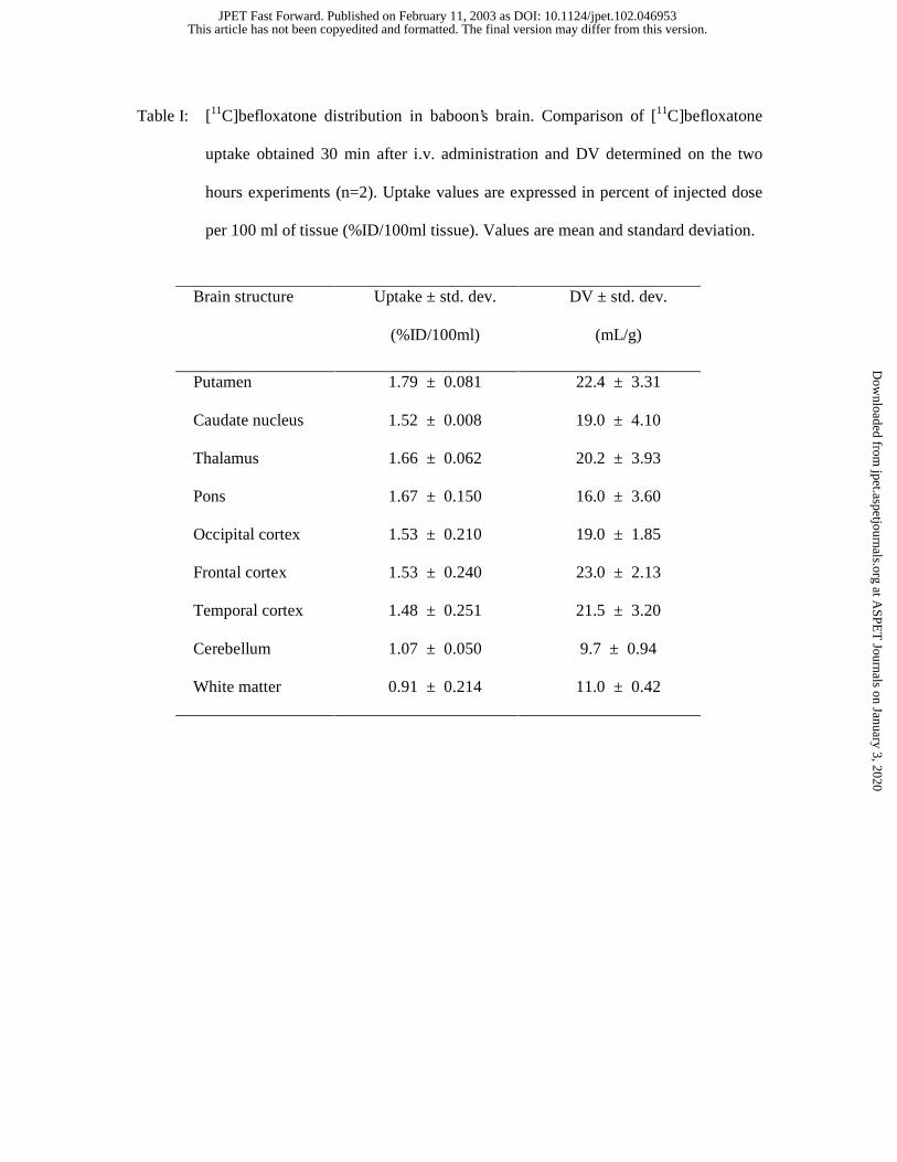
Table I: [11C]befloxatone distribution in baboon’s brain. Comparison of [11C]befloxatone
uptake obtained 30 min after i.v. administration and DV determined on the two
hours experiments (n=2). Uptake values are expressed in percent of injected dose
per 100 ml of tissue (%ID/100ml tissue). Values are mean and standard deviation.
Brain structure Uptake ± std. dev.
(%ID/100ml)
DV ± std. dev.
(mL/g)
Putamen 1.79 ± 0.081 22.4 ± 3.31
Caudate nucleus 1.52 ± 0.008 19.0 ± 4.10
Thalamus 1.66 ± 0.062 20.2 ± 3.93
Pons 1.67 ± 0.150 16.0 ± 3.60
Occipital cortex 1.53 ± 0.210 19.0 ± 1.85
Frontal cortex 1.53 ± 0.240 23.0 ± 2.13
Temporal cortex 1.48 ± 0.251 21.5 ± 3.20
Cerebellum 1.07 ± 0.050 9.7 ± 0.94
White matter 0.91 ± 0.214 11.0 ± 0.42
This article has not been copyedited and formatted. The final version may differ from this version.JPET Fast Forward. Published on February 11, 2003 as DOI: 10.1124/jpet.102.046953
at ASPE
T Journals on January 3, 2020
jpet.aspetjournals.orgD
ownloaded from
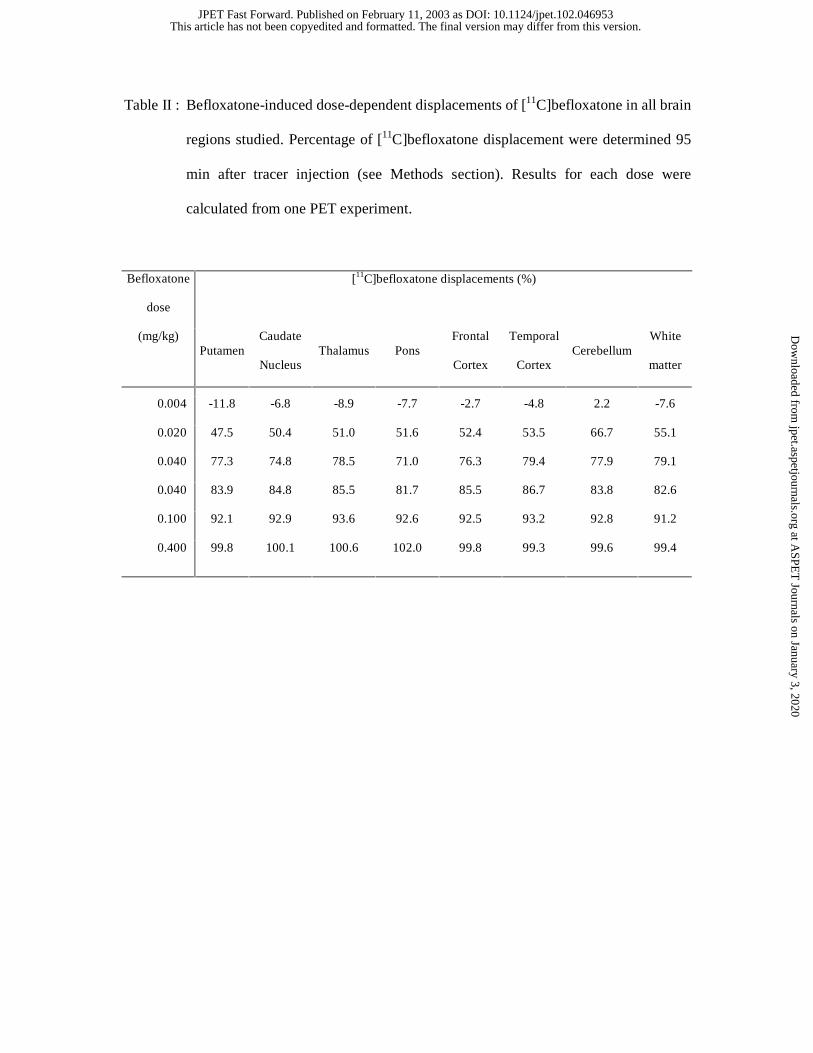
Table II : Befloxatone-induced dose-dependent displacements of [11C]befloxatone in all brain
regions studied. Percentage of [11C]befloxatone displacement were determined 95
min after tracer injection (see Methods section). Results for each dose were
calculated from one PET experiment.
Befloxatone
dose
[11C]befloxatone displacements (%)
(mg/kg) Putamen
Caudate
Nucleus Thalamus Pons
Frontal
Cortex
Temporal
Cortex Cerebellum
White
matter
0.004 -11.8 -6.8 -8.9 -7.7 -2.7 -4.8 2.2 -7.6
0.020 47.5 50.4 51.0 51.6 52.4 53.5 66.7 55.1
0.040 77.3 74.8 78.5 71.0 76.3 79.4 77.9 79.1
0.040 83.9 84.8 85.5 81.7 85.5 86.7 83.8 82.6
0.100 92.1 92.9 93.6 92.6 92.5 93.2 92.8 91.2
0.400 99.8 100.1 100.6 102.0 99.8 99.3 99.6 99.4
This article has not been copyedited and formatted. The final version may differ from this version.JPET Fast Forward. Published on February 11, 2003 as DOI: 10.1124/jpet.102.046953
at ASPE
T Journals on January 3, 2020
jpet.aspetjournals.orgD
ownloaded from