manual for sugar fortification with vitamin a part 3a2zproject.org/~a2zorg/pdf/3final.pdf · manual...
TRANSCRIPT

Manual for Sugar Fortificationwith Vitamin A
Part 3
Analytical Methods for the Control and Evaluation of
Sugar Fortification with Vitamin A
Omar Dary, Ph.D.Guillermo Arroyave, Ph.D.
with
Hernando Flores, Ph.D., Florisbela A. C. S. Campos, and
Maria Helena C. B. Lins
Dr. Omar Dary is a research biochemist at the Institute of Nutrition of Central America andPanama (INCAP), Guatemala.
Dr. Guillermo Arroyave is an international consultant in micronutrients residing in San Diego,California.
Dr. Hernando Flores, Ms. Campos, and Ms. Lins are biochemists at the Universidad dePernambuco, Brazil.
i
MANUAL FOR SUGAR FORTIFICATION
PART 3
TABLE OF CONTENTS
ACKNOWLEDGMENTS . . . . . . . . . . . . . . . . . . . . . . . . . . . . . . . . . . . . . . . . . . . . . . . . . . . . . . . . . . . v
FOREWORD . . . . . . . . . . . . . . . . . . . . . . . . . . . . . . . . . . . . . . . . . . . . . . . . . . . . . . . . . . . . . . . . . . . vii
I. INTRODUCTION . . . . . . . . . . . . . . . . . . . . . . . . . . . . . . . . . . . . . . . . . . . . . . . . . . . . . . . . . . 1
II. PROPERTIES OF RETINOL AND RETINOL COMPOUNDS USED IN SUGARFORTIFICATION . . . . . . . . . . . . . . . . . . . . . . . . . . . . . . . . . . . . . . . . . . . . . . . . . . . . . . . . . . 3
III. PRINCIPLES FOR DETERMINING RETINOL IN VITAMIN A PREMIX AND FORTIFIEDSUGAR . . . . . . . . . . . . . . . . . . . . . . . . . . . . . . . . . . . . . . . . . . . . . . . . . . . . . . . . . . . . . . . . . . 5A. Spectrophotometric method . . . . . . . . . . . . . . . . . . . . . . . . . . . . . . . . . . . . . . . . . . . . . 5B. Colorimetric method . . . . . . . . . . . . . . . . . . . . . . . . . . . . . . . . . . . . . . . . . . . . . . . . . . 6
IV. SPECTROPHOTOMETRIC DETERMINATION OF RETINOL IN PREMIX . . . . . . . . . . . 7A. References . . . . . . . . . . . . . . . . . . . . . . . . . . . . . . . . . . . . . . . . . . . . . . . . . . . . . . . . . . 7B. Principle . . . . . . . . . . . . . . . . . . . . . . . . . . . . . . . . . . . . . . . . . . . . . . . . . . . . . . . . . . . . 7C. Critical points and cautions . . . . . . . . . . . . . . . . . . . . . . . . . . . . . . . . . . . . . . . . . . . . . 7D. Equipment and materials . . . . . . . . . . . . . . . . . . . . . . . . . . . . . . . . . . . . . . . . . . . . . . . 8E. Reagents . . . . . . . . . . . . . . . . . . . . . . . . . . . . . . . . . . . . . . . . . . . . . . . . . . . . . . . . . . . 8F. Procedure . . . . . . . . . . . . . . . . . . . . . . . . . . . . . . . . . . . . . . . . . . . . . . . . . . . . . . . . . . . 8G. Calculations . . . . . . . . . . . . . . . . . . . . . . . . . . . . . . . . . . . . . . . . . . . . . . . . . . . . . . . . . 9
V. SPECTROPHOTOMETRIC DETERMINATION OF RETINOL IN FORTIFIED SUGAR . 11A. References . . . . . . . . . . . . . . . . . . . . . . . . . . . . . . . . . . . . . . . . . . . . . . . . . . . . . . . . . 11B. Principle . . . . . . . . . . . . . . . . . . . . . . . . . . . . . . . . . . . . . . . . . . . . . . . . . . . . . . . . . . . 11C. Critical points and cautions . . . . . . . . . . . . . . . . . . . . . . . . . . . . . . . . . . . . . . . . . . . . 11D. Equipment and materials . . . . . . . . . . . . . . . . . . . . . . . . . . . . . . . . . . . . . . . . . . . . . . 12E. Reagents . . . . . . . . . . . . . . . . . . . . . . . . . . . . . . . . . . . . . . . . . . . . . . . . . . . . . . . . . . 12F. Procedure . . . . . . . . . . . . . . . . . . . . . . . . . . . . . . . . . . . . . . . . . . . . . . . . . . . . . . . . . . 13G. Calculations . . . . . . . . . . . . . . . . . . . . . . . . . . . . . . . . . . . . . . . . . . . . . . . . . . . . . . . . 14H. Verification of the efficiency of the extraction . . . . . . . . . . . . . . . . . . . . . . . . . . . . . 15
VI. SEMIQUANTITATIVE COLORIMETRIC DETERMINATION OF RETINOL INFORTIFIED SUGAR . . . . . . . . . . . . . . . . . . . . . . . . . . . . . . . . . . . . . . . . . . . . . . . . . . . . . . . 17A. References . . . . . . . . . . . . . . . . . . . . . . . . . . . . . . . . . . . . . . . . . . . . . . . . . . . . . . . . . 17B. Principle . . . . . . . . . . . . . . . . . . . . . . . . . . . . . . . . . . . . . . . . . . . . . . . . . . . . . . . . . . . 17C. Critical points and cautions . . . . . . . . . . . . . . . . . . . . . . . . . . . . . . . . . . . . . . . . . . . . 17D. Materials . . . . . . . . . . . . . . . . . . . . . . . . . . . . . . . . . . . . . . . . . . . . . . . . . . . . . . . . . . 18E. Reagents . . . . . . . . . . . . . . . . . . . . . . . . . . . . . . . . . . . . . . . . . . . . . . . . . . . . . . . . . . 18

ii
F. Procedure . . . . . . . . . . . . . . . . . . . . . . . . . . . . . . . . . . . . . . . . . . . . . . . . . . . . . . . . . . 19
VII. VOLUMETRIC METHOD TO DETERMINE PEROXIDE LEVELS IN OILS . . . . . . . . . . 21A. References . . . . . . . . . . . . . . . . . . . . . . . . . . . . . . . . . . . . . . . . . . . . . . . . . . . . . . . . . 21B. Principle . . . . . . . . . . . . . . . . . . . . . . . . . . . . . . . . . . . . . . . . . . . . . . . . . . . . . . . . . . . 21C. Critical points and cautions . . . . . . . . . . . . . . . . . . . . . . . . . . . . . . . . . . . . . . . . . . . . 21D. Equipment and materials . . . . . . . . . . . . . . . . . . . . . . . . . . . . . . . . . . . . . . . . . . . . . . 21E. Reagents . . . . . . . . . . . . . . . . . . . . . . . . . . . . . . . . . . . . . . . . . . . . . . . . . . . . . . . . . . 22F. Procedure . . . . . . . . . . . . . . . . . . . . . . . . . . . . . . . . . . . . . . . . . . . . . . . . . . . . . . . . . . 23G. Calculations . . . . . . . . . . . . . . . . . . . . . . . . . . . . . . . . . . . . . . . . . . . . . . . . . . . . . . . . 24
VIII. PRINCIPALS OF METHODS TO DETERMINE RETINOL IN BLOOD . . . . . . . . . . . . . . 25A. Introduction . . . . . . . . . . . . . . . . . . . . . . . . . . . . . . . . . . . . . . . . . . . . . . . . . . . . . . . . 25B. Collection and management of blood samples . . . . . . . . . . . . . . . . . . . . . . . . . . . . . . 26
IX. SPECTROPHOTOMETRIC DETERMINATION OF BLOOD RETINOL BYULTRAVIOLET DESTRUCTION OF RETINOL . . . . . . . . . . . . . . . . . . . . . . . . . . . . . . . . 29A. References . . . . . . . . . . . . . . . . . . . . . . . . . . . . . . . . . . . . . . . . . . . . . . . . . . . . . . . . . 29B. Principle . . . . . . . . . . . . . . . . . . . . . . . . . . . . . . . . . . . . . . . . . . . . . . . . . . . . . . . . . . . 29C. Critical points and cautions . . . . . . . . . . . . . . . . . . . . . . . . . . . . . . . . . . . . . . . . . . . . 29D. Equipment and materials . . . . . . . . . . . . . . . . . . . . . . . . . . . . . . . . . . . . . . . . . . . . . . 30E. Reagents . . . . . . . . . . . . . . . . . . . . . . . . . . . . . . . . . . . . . . . . . . . . . . . . . . . . . . . . . . 31F. Procedure . . . . . . . . . . . . . . . . . . . . . . . . . . . . . . . . . . . . . . . . . . . . . . . . . . . . . . . . . . 31G. Calculations . . . . . . . . . . . . . . . . . . . . . . . . . . . . . . . . . . . . . . . . . . . . . . . . . . . . . . . . 33H. Verification of the method’s reproducibility . . . . . . . . . . . . . . . . . . . . . . . . . . . . . . . 34I. Verification of the method’s recovery . . . . . . . . . . . . . . . . . . . . . . . . . . . . . . . . . . . . 34J. Variations in assay . . . . . . . . . . . . . . . . . . . . . . . . . . . . . . . . . . . . . . . . . . . . . . . . . . . 35
X. DETERMINATION OF BLOOD RETINOL BY HIGH PERFORMANCE LIQUIDCHROMATOGRAPHY (HPLC) . . . . . . . . . . . . . . . . . . . . . . . . . . . . . . . . . . . . . . . . . . . . . . 37A. References . . . . . . . . . . . . . . . . . . . . . . . . . . . . . . . . . . . . . . . . . . . . . . . . . . . . . . . . . 37B. Principle . . . . . . . . . . . . . . . . . . . . . . . . . . . . . . . . . . . . . . . . . . . . . . . . . . . . . . . . . . . 37C. Critical points and cautions . . . . . . . . . . . . . . . . . . . . . . . . . . . . . . . . . . . . . . . . . . . . 38D. Equipment and materials . . . . . . . . . . . . . . . . . . . . . . . . . . . . . . . . . . . . . . . . . . . . . . 38E. Reagents . . . . . . . . . . . . . . . . . . . . . . . . . . . . . . . . . . . . . . . . . . . . . . . . . . . . . . . . . . 39F. Procedure . . . . . . . . . . . . . . . . . . . . . . . . . . . . . . . . . . . . . . . . . . . . . . . . . . . . . . . . . . 42G. Calculations . . . . . . . . . . . . . . . . . . . . . . . . . . . . . . . . . . . . . . . . . . . . . . . . . . . . . . . . 44H. Verification of the recovery of the method . . . . . . . . . . . . . . . . . . . . . . . . . . . . . . . . 45
XI. PRINCIPALS OF METHODS TO DETERMINE RETINOL IN BREAST MILK . . . . . . . . 47A. Introduction . . . . . . . . . . . . . . . . . . . . . . . . . . . . . . . . . . . . . . . . . . . . . . . . . . . . . . . . 47B. Collection and management of breast milk samples . . . . . . . . . . . . . . . . . . . . . . . . . 48
XII. APPLICATION OF THE SPECTROPHOTOMETRIC METHOD BY ULTRAVIOLETDESTRUCTION FOR THE DETERMINATION OF TOTAL RETINOL INBREAST MILK . . . . . . . . . . . . . . . . . . . . . . . . . . . . . . . . . . . . . . . . . . . . . . . . . . . . . . . . . . . 49

iii
XIII. DETERMINATION OF BREAST MILK RETINOL BY HIGH PERFORMANCE LIQUIDCHROMATOGRAPHY (HPLC) . . . . . . . . . . . . . . . . . . . . . . . . . . . . . . . . . . . . . . . . . . . . . . 51A. References . . . . . . . . . . . . . . . . . . . . . . . . . . . . . . . . . . . . . . . . . . . . . . . . . . . . . . . . . 51B. Principle . . . . . . . . . . . . . . . . . . . . . . . . . . . . . . . . . . . . . . . . . . . . . . . . . . . . . . . . . . . 51C. Critical points and cautions . . . . . . . . . . . . . . . . . . . . . . . . . . . . . . . . . . . . . . . . . . . . 51D. Equipment and materials . . . . . . . . . . . . . . . . . . . . . . . . . . . . . . . . . . . . . . . . . . . . . . 52E. Reagents . . . . . . . . . . . . . . . . . . . . . . . . . . . . . . . . . . . . . . . . . . . . . . . . . . . . . . . . . . 52F. Procedure . . . . . . . . . . . . . . . . . . . . . . . . . . . . . . . . . . . . . . . . . . . . . . . . . . . . . . . . . . 55G. Calculations . . . . . . . . . . . . . . . . . . . . . . . . . . . . . . . . . . . . . . . . . . . . . . . . . . . . . . . . 57H. Calculation of the recovery proportion . . . . . . . . . . . . . . . . . . . . . . . . . . . . . . . . . . . . 58I. Variations . . . . . . . . . . . . . . . . . . . . . . . . . . . . . . . . . . . . . . . . . . . . . . . . . . . . . . . . . 59
XIV. LABORATORY QUALITY CONTROL PROCEDURES . . . . . . . . . . . . . . . . . . . . . . . . . . 61A. Criteria of quality control . . . . . . . . . . . . . . . . . . . . . . . . . . . . . . . . . . . . . . . . . . . . . . 61B. Routine quality control . . . . . . . . . . . . . . . . . . . . . . . . . . . . . . . . . . . . . . . . . . . . . . . 63C. Equipment calibration . . . . . . . . . . . . . . . . . . . . . . . . . . . . . . . . . . . . . . . . . . . . . . . . 65
XVI. SUGGESTED READING . . . . . . . . . . . . . . . . . . . . . . . . . . . . . . . . . . . . . . . . . . . . . . . . . . . 69
APPENDIX 3.1: CONSTRUCTION OF AN ULTRAVIOLET IRRADIATION CHAMBER 71
APPENDIX 3.2: SUGGESTIONS FOR CLEANING GLASSWARE AND SPECTROPHOTOMETRIC CUVETTES . . . . . . . . . . . . . . . . . . . . . . . . . . . . . . . . . . . . . . . 75
APPENDIX 3.3: SUPPLIERS FOR LABORATORY EQUIPMENT AND REAGENTS . . . . 77
TABLES
Table 3.1: Correction Factors for Retinol Absorbance at Different Wavelengths Using a Source of Visible Light . . . . . . . . . . . . . . . . . . . . . . . . . . . . . . . . . . . . . . . . . . . . . . 6
FIGURES
Figure 3.1: Chemical Structure of Retinol and Derivatives . . . . . . . . . . . . . . . . . . . . . . . . . . . . . . 3Figure 3.2: Fortified Sugar Quality Control Graph . . . . . . . . . . . . . . . . . . . . . . . . . . . . . . . . . . . . 64Figure 3.3: Retinol Absorbance by Irradiation Time . . . . . . . . . . . . . . . . . . . . . . . . . . . . . . . . . . 68Appendix Figure 3.1a: Irradiation Chamber . . . . . . . . . . . . . . . . . . . . . . . . . . . . . . . . . . . . . . . . . . . 72Appendix Figure 3.1b: Dimensions of an Irradiation Chamber . . . . . . . . . . . . . . . . . . . . . . . . . . . . . 73

v
ACKNOWLEDGMENTS
This manual is based on experiences in sugar fortification at the Institute of Nutrition of CentralAmerica and Panama (INCAP). Many people have been involved in this program over the last 25years. This publication represents the work of all those involved, who are too numerous to mention.
The presentation and content of parts 1, 2, and 3 of the manual benefited greatly from thecomments and suggestions of the reviewers, whom the authors would like to thank. These include Drs.Frances R. Davidson and Timothy Quick, United States Agency for International Development(USAID); Dr. Juan R. Aguilar, United Nations Children’s Fund (UNICEF), Peru; Dr. Jose O. Mora,International Sciences and Technology Institute (ISTI); Dr. Luis Mejia, Kellogg Latin America; Mr.Alberto Nilson, Hoffman-La Roche, Brazil; Dr. Kenneth Brown, University of California at Davis; Mr.Leonardo de Leon, INCAP; Drs. Jean Humphrey and Esse Yamini, Johns Hopkins University; andespecially Dr. Penelope Nestel, Opportunities for Micronutrient Interventions (OMNI) Project.
Part 2 is based on the collective experience of the following individuals who have beeninstrumental in improving the sugar fortification process in Central America over the last 10 years: Mr.Leonel Anleu, Sugar Producers’ Association, Guatemala; Dr. Oscar Pineda, formerly of INCAP,Guatemala; Dr. Vilma Estrada, Dr. Doris Chinchilla, Mr. Plutarco Murillo, and Ms. Victoria SaraiBejarano, Honduras; and Dr. Manuel de Gracia and Dr. Enrique Murillo, Panama.
Part 3 presents the methods used at the Chemistry and Biochemistry Laboratory, INCAP, andis based on work carried out by Carolina Martinez, Dora Ines Mazariegos, Maria Elena Estrada deSandoval, Esmeralda Morales, Monica Guamuch, and Gerardo Pirir.
The manual was translated into English by Maritza Mendez de Oliva and Carlos Cipriani.Odette Sanabria de Osorio drew the illustrations. Ingrid Cabrera, Hazel de Orellana, and Ana Ruth deBurckard typed the Spanish edition, and Lisa Sherburne was instrumental in compiling and formattingand Pamela Cubberly in copyediting and formatting the English edition; their assistance is greatlyappreciated.
Support activities carried out from 1992 to 1994, which have served in part as the basis for thispublication, were financed by the Regional Office for Central America and Panama (ROCAP)/USAIDProject 596-0169.

vii
FOREWORD
In many countries, vitamin A deficiency is a widespread problem that is not necessarily limitedto specific groups of people or isolated communities. Among the interventions available, foodfortification is an accepted method of delivering micronutrients to the population at large and is widelypracticed in developed countries. In these countries, foods such as milk powder, butter and margarine,complementary infant foods, and breakfast cereals are routinely fortified with micronutrients, includingvitamin A. The above foods, however, are not regularly consumed by the vast majority of thepopulation in developing countries, especially among those at greatest risk of vitamin A deficiency. Onefood that is consumed by nearly the entire population in developing countries is sugar, which can befortified with vitamin A. Sugar fortification is practical because target populations do not need to alteror adapt a new or costly distribution system. Indeed, sugar fortification only requires the existence of awell-established sugar production and marketing system. This allows for the uniform addition of vitaminA as well as the monitoring of its content. Fortification of sugar with vitamin A is one of the safest, mostefficacious, and most cost-effective interventions to prevent and control vitamin A deficiency.
This manual in which technical guidelines are presented to systematize and facilitate theestablishment and execution of a vitamin A sugar fortification program is divided into three parts. Part 1,Guidelines for the Development, Implementation, Monitoring, and Evaluation for Vitamin ASugar Fortification Program, describes why it is important to prevent and reduce vitamin Adeficiency and how to go about establishing such a program. Existing strategies are discussed and thebasic elements to be considered in establishing an appropriate program for vitamin A sugar fortificationare described in detail. In addition, part 1 offers an overview of the entire program so that public andprivate sector officials who manage and coordinate sugar-processing activities have information on theessential components to ensure an adequate operation. Technical areas presented in this document willalso be useful to specialists involved in specific components of the fortification process. These includethe operations involved in sugar fortification, determinates for both the efficiency and efficacy ofintervention, and guidelines for determining program costs.
Part 2, Technical and Operational Guidelines for Preparing Vitamin A Premix andFortified Sugar, is written specifically for technical personnel responsible for implementing sugarfortification. Chapter I covers general aspects if the fortification process, Chapter II describes how tomanufacture the premix, and Chapter III describes procedures for adding the vitamin A premix tosugar. It also contains a detailed description of quality control procedures.
Part 3, Analytical Methods for the Control and Evaluation for Sugar Fortification withVitamin A, presents field and laboratory methods to estimate the content of vitamin A in the premix andin fortified sugar. It also gives details on how to determine retinol levels in biological samples critical inevaluating the impact of the fortification program. Part 3 is written primarily for laboratory personnelwho will be responsible for laboratory analyses.

viii
Each part of the manual is relatively self-sufficient in the essential areas of program design andimplementation. Ideally, however, it is recommended that the three parts be considered as theoreticaland practical units to be used together.
Research on sugar fortification with vitamin A first began at the Institute of Nutrition of CentralAmerica and Panama (INCAP) in the 1960s under the leadership of Guillermo Arroyave with thesupport of Dr. M. Forman of USAID. The technology was developed over a 10-year period. Startingin 1974 Costa Rica, Guatemala, Honduras, and Panama legislated that all sugar must be fortified withvitamin A. With USAID support, the INCAP team was able to show conclusively that sugarfortification with vitamin A is both efficacious and cost-effective. USAID support, which has continuedover the years, most recently through the OMNI project, has been important in ensuring that sugarfortification programs have continually improved in Central America countries. It has also stimulated thesuccessful development of sugar fortification programs in other Latin American countries.
A sustainable sugar fortification program reflects the collaborative efforts between sugarproducers, the public sector, researchers, and donors. The purpose of this document is to share theexperiences of those involved in sugar fortification in Central America, so that other countries can planand implement this important intervention to eliminate and prevent vitamin A deficiency.
Frances Davidson Hernán DelgadoOffice of Health & Nutrition, USAID INCAP/PAHOWashington, D.C. Guatemala
1. Unless stated, the methods described in this manual are currently those being used in the Chemistry andBiochemistry Laboratory at the Institute of Nutrition of Central America and Panama (INCAP).
1
I. INTRODUCTION
Access to a laboratory is essential to having an effective vitamin A sugar fortification program.This is because chemical analyses are required to verify the retinol levels in both premix and fortifiedsugar. Furthermore, the biological impact of sugar fortification is determined from the retinol levels inserum and breast milk.
It is important for the reader of this manual to note that any laboratory working on retinoldeterminations should have two basic facilities: first, to ensure the quality of results, an area away fromdirect sunlight with indirect incandescent yellow light and, second, to ensure the safety of laboratorypersonnel, a laboratory hood to reduce the risks associated with using volatile flammable solvents. If theappropriate lighting is not available, all test tubes and flasks with retinol-containing solutions can becovered with black fabric to reduce the risk of retinol losses.
This document starts by describing the general properties of retinol and the retinol compoundused in sugar fortification as the basis for understanding the analytical methods presented. It is followedby sections that describe common principles behind and details of laboratory methods to determine the1
retinol content in (a) premix and fortified sugar (including measuring peroxide levels in vegetable oil,which is used in the premix), (b) blood, and (c) breast milk. Included for each substance are guidelinesfor collecting samples and a comparison of the advantages and limitations of principles and procedures.A concluding section then describes procedures for quality control of the analytical methods.Appendixes include information on how to construct an ultraviolet irradiation chamber and properlyclean glassware and spectrophotometric cuvettes and a list of addresses for suppliers of the equipmentand reagents needed for the assays described here.


2. Nevertheless, about 600 compounds in plant tissues whose structures are related to retinol are known ascarotenoids. Only fifty of these natural compounds can be converted to retinol in animals and are known asactive carotenoids or precursors of vitamin A. The most active compound is beta-carotene, making it the mostimportant carotenoid from a nutritional perspective.
3
II. PROPERTIES OF RETINOL AND RETINOL COMPOUNDSUSED IN SUGAR FORTIFICATION
Retinol is the active form of vitamin A and is found in animal tissues and fluids but not plants. It2
is most commonly found as a fatty acid ester, predominantly as retinyl palmitate. A number of retinolderivatives with specific biochemical functions also exist. These include retinal and retinoic acid in whichthe -CHO and -COOH radicals, respectively, replace the -OH radical. The chemical structures of theretinol and derived compounds are shown below in figure 3.1.
Figure 3.1:CHEMICAL STRUCTURE OF RETINOL AND DERIVATIVES

Manual for Sugar Fortificaiton with Vitamin A - Part 3
3. Produced by Hoffman-La Roche and BASF, Germany (see appendix 3.3).
4
Retinol in blood is bound to its carrier protein, known as retinol binding protein, but, in breastmilk, it is esterified with several fatty acids. To determine blood retinol levels, the plasma or serumsamples require treatment with alkaline ethanol to destroy the protein complex, which precipitates theprotein and liberates the retinol. In breast milk, retinol should be separated from its esters bysaponification.
All the retinol compounds commercially available for industrial or pharmaceutical use aresynthetic. Most are in the form of esters, often palmitate or acetate, which are more stable and easier tohandle. Retinol and its esters are highly soluble in organic solvents and fats but not water. For thisreason, retinol is not suitable for fortifying water-based foods; however, the advent of water-miscible,dry, vitamin A compounds, including retinyl palmitate beadlets, has facilitated the fortification of water-based foods, including sugar.
The prototype retinyl palmitate used in sugar fortification is 250-CWS, in which retinyl3
palmitate is embedded in a gelatin matrix containing antioxidants as stabilizers, as well as othercompounds that make it water-miscible. The 250-CWS beadlets are spherical in shape, light yellow incolor, and have a slight but distinct odor. Its stability and other characteristics makes 250-CWS asuitable fortificant for white sugar. Because retinol is fat soluble, it is easily extracted with organicsolvents; however, the water-miscible beadlets must first be dispersed in water to release the retinylpalmitate from the other beadlet components.
Once retinol and its esters have been extracted, their distinct characteristics become evident.These characteristics, often the basis for determining retinol levels or important considerations in assays,include the following:
Ë Destruction by ultraviolet (UV) light. Retinol is destroyed when irradiated with UV light;thus, specific determinations can be made using retinol absorbance before and after itsselective destruction with UV light. At the same time, this property requires that samples and specific solutions containing retinol be protected from direct light duringpreparation and handling.
Ë Absorption of electromagnetic energy in the UV and shortest wavelengths of thevisible spectrum. Due to the conjugated double bond in retinol, this molecule absorbsradiant energy around 325 nm. This property is generally used to determine quantitativelythe level of retinol and is done by measuring its absorbance at maximum absorptionwavelength, which could vary slightly depending on the retinol derivative and solvent.
Ë Oxidation. Retinol readily takes up oxygen to form peroxide bonds, a reaction directlyrelated to temperature; thus, solutions containing retinol should be protected fromprolonged exposure to air and kept at a maximum temperature of 10°C.

5
III. PRINCIPLES FOR DETERMINING RETINOL IN VITAMIN APREMIX AND FORTIFIED SUGAR
The retinol concentration in premix or fortified sugar can be determined quantitatively using aspectrophotometric method or semi-quantitatively using a colorimetric method that is based on achromogenic reaction.
A. Spectrophotometric method
1. Collection and handling of samples. A minimum of 5 g of premix and 50 g of fortifiedsugar is needed. Samples should be stored in opaque glass or plastic bottles that aretightly capped and protected from direct sunlight, heat, and humidity. Once at thelaboratory, samples should be stored in a dry, cool place. Before weighing the amountto be analyzed, the premix or fortified sugar should be thoroughly mixed.
2. Dispersion of the retinyl palmitate beadlets. To ensure complete dispersion of the250-CWS in water, the beadlets should be dissolved and preferably incubated in waterat 50°–60°C for 15 minutes.
3. Extraction. Retinyl palmitate is extracted from the aqueous layer using an organicsolvent. The most appropriate is hexane. Because of the high concentration of retinylpalmitate in the premix, extraction is unnecessary. Instead, dilution of the aqueous phasewith 2-propanol is sufficient.
4. Determining retinol concentration. Retinol determination is based on its absorbanceat 325 nm using a spectrophotometer. The high retinyl content in premix and fortifiedsugar allows for a higher wavelength to be used if necessary but always below 350 nm;however, the accuracy and sensitivity decrease when wavelengths over 325 nm areused, which means that a correction factor must be used. This can be easily obtained bydividing the absorbance of a retinol solution at 325 nm in a referencespectrophotometer by the absorbance of the same solution at the other wavelengthused. Table 3.1 on the next page shows the correction factors for retinol at differentwavelengths as determined at INCAP.

Manual for Sugar Fortificaiton with Vitamin A - Part 3
4. Reference value for ultraviolet radiation source at 325 nm = 1.000.5. This method is not sufficiently accurate to be used as a quality control parameter for premix.
6
Table 3.1: Correction Factors for Retinol Absorbance at DifferentWavelengths Using a Source of Visible Light4
Wavelength (nm) Correction factor325 1.007330 1.066335 1.117340 1.360345 1.622350 2.030
5. Increasing the analytical specificity. This is necessary when the samples have asignificant amount of interfering substances and can be achieved in two ways: first, byseparating the retinol from other substances that absorb radiant energy at equal orsimilar wavelengths to retinol using high performance liquid chromatography (HPLC)or, second, by measuring the absorbance of the extracts before and after selectivelydestroying the retinol through exposure to UV light. The difference in absorbancebetween the unirradiated and irradiated extracts is specifically due to retinol and is usedto calculate the concentration of retinol in the sample. This procedure is used to assaypremix samples. Fortified sugar essentially contains no interfering substances; thus, theretinol can be specifically determined simply by subtracting the absorbance of thereagent’s blank from the absorbance of the sample.
B. Colorimetric method
The other method that can be used to determine retinol levels in fortified sugar is colorimetric,5
which is faster and does not require a spectrophotometer, making it adaptable to field conditions. Themethod is based on the Carr-Price reaction, in which retinol is converted to anhydroretinol whencombined with a chromogenic reagent containing trichloracetic acid and dichloromethane. A blue colordevelops from the transient protonation of anhydroretinol, the intensity of which is proportional to theconcentration of retinol in the sample. This method is less accurate and precise than thespectrophotometric one; thus, the validity of the results should be verified against those using thespectrophotometric method.

6. Based on an unpublished procedure described by O. Pineda in Fortificación de Azúcar con Vitamina A, Manualde Operaciones, Guatemala City: INCAP.
7. Section XIV.C.1 includes a description of a simple procedure to calibrate spectrophotometers routinely used inclinical laboratories.
7
IV. SPECTROPHOTOMETRIC DETERMINATION OF RETINOLIN PREMIX
A. References6
None are available.
B. Principle
This method entails solubilizing water-miscible retinyl palmitate beadlets in hot water, followedby dilution in 2-propanol. The concentration of retinol in retinyl palmitate is determined by itsspectrophotometric absorbance at 325 nm.
C. Critical points and cautions
A spectrophotometer capable of reading 325 nm is essential. This is because the concentrationof the retinol standards has to be verified by spectrophotometric analysis. Given the importance of thespectrophotometer for ensuring the accuracy and reliability of the retinol determinations, it should becalibrated frequently following the instructions provided by the manufacturer, especially to confirm thecalibration of the monochromator. This confirmation should be carried out frequently and not only7
when a new lamp is installed.
A UV light irradiation system, in which the retinol is destroyed, is also required. This system canbe as simple as a UV lamp and curtain to protect technicians from exposure to this light. The criticalfactor is to establish exactly both the optimal irradiation time and appropriate distance between the UVlight and the solutions. The efficiency of the irradiation system should be checked periodically (every 3months) and each time a new lamp is installed. Appendix 3.1 gives details of an irradiation system.
Once the sample has been solubilized in 2-propanol, the analysis should not be interrupted.
Manual for Sugar Fortificaiton with Vitamin A - Part 3
8
Based on experience at the INCAP laboratory, if the variability between duplicates of the samesolution is greater than 3 percent, the results should be rejected and the readings repeated. In addition,the results of two independently weighed subsamples of the same sample should not differ on averageby more than 8 percent. If the variation is greater than 8 percent, the assay should be repeated.
D. Equipment and materials
Vortex mixer
Water bath (50°–60°C)
Irradiation chamber with ultraviolet light (described in appendix 3.1)
UV/VIS spectrophotometer
100 mL volumetric flasks
Spectrophotometer cuvettes (preferably quartz)
Graduated serological pipettes
Volumetric pipettes
20 mL test tubes
10 mm x 75 mm glass tubes transparent to UV light
Glass rods
200–250 mL beaker
Spatulas
Aluminum foil
E. Reagents
1. Analytical grade 2-propanol ((CH CH(OH)CH ), purity = 99.7%, MW = 60.10, 3 3
d = 0.78 g/mL).
F. Procedure
1. Mix the premix sample thoroughly.
2. Weigh duplicate 1 g samples, recording the exact weights to three decimal places, anddissolve each sample in 80 mL of distilled water at 50°–60°C in a beaker. Use a glassrod to completely dissolve the sample.

Retinyl palmitate (mg/g) '(Abso & Absirrad)
å× Vl
p× FD × FCspe
Analytical Methods for the Control and Evaluation of Sugar Fortification with Vitamin A
8. If a UV-light spectrophotometer is not available, a visible light spectrophotometer may be used but at awavelength below 350 nm (see table 3.1).
9
3. Incubate in a water bath at 50°–60°C for 15 minutes. Cool at room temperature.Transfer to a 100-mL volumetric flask. Rinse the beaker with small amounts of distilledwater, transfer the washings to the volumetric flask, and make up to volume withdistilled water, and mix. This solution is cloudy.
4. Measure 2 mL of the solution in step 3 into a 20 mL test tube and add 8 mL of 2-propanol (to give a 2:10 dilution). Mix vigorously in a Vortex mixer.
5. Measure 1 mL of solution from step 4 into a 20-mL test tube and add 9 mL of 2-propanol (to give a 1:10 dilution). Mix using a Vortex mixer.
6. Separate off 1 mL of the solution from step 5 and place in a 10 mm x 75 mm glass testtube transparent to UV light. Irradiate this solution in the irradiation chamber for 35minutes (or the time required according to the performance of the irradiation chamber).
7. Adjust the zero of the spectrophotometer with 2-propanol. Read the absorbance of theirradiated and unirradiated solutions at 325 nm in 1 cm light path quartz cuvettes. 8
G. Calculations
The concentration of retinyl palmitate in the premix samples is calculated using the followingequation:
where:
Abs = reading at time zeroo
Abs = reading after irradiation.irrad

Retinol (mg/g) ' (Abso & Absirrad) × 29.04p
× FCspe
Manual for Sugar Fortificaiton with Vitamin A - Part 3
9. See section XIV.C.1.
10
The parameters for the equation are:
Parameter Explanation Value
å Retinyl palmitate absorption coefficient (mg cm mL) 94.0
Vl Volume of the initial solution of the sample (mL) 100.0
p Weight of the sample (g) weight at step IV.F.2
FD Dilution factor 50.0
Fc Correction factor of the spectrophotometer, ideally 1.000spe
!1 !1
9
To express the results as unesterified retinol, the ratio of the molecular weights of retinol/retinylpalmitate (286.46/524.84 = 0.546) must be taken into consideration. A simplified equation to estimatethe unesterified retinol is the following:

10. Section XIV.C.1 includes a description of a simple procedure to calibrate spectrophotometers routinely used inclinical laboratories.
11
V. SPECTROPHOTOMETRIC DETERMINATION OF RETINOLIN FORTIFIED SUGAR
A. References
Arroyave, G. and C. de Funes. 1974. Enriquecimiento de azúcar con vitamina A. Método para ladeterminación cuantitativa de retinol en azúcar blanca de mesa.” Arch. Latinoamer. Nutr. 24:147–53.
B. Principle
This method is an adaptation of the method developed by Arroyave and Funes (1974). Theprocedure uses five to ten times less reagents than the original method, and its accuracy is similar. Theprecision, however, is somewhat lower, although highly satisfactory. The method requires the extractionof retinyl palmitate in hexane. Retinol concentration is determined by its absorbance at 325 nm. Thismethod does not usually require irradiation with UV light, because the absorbance of the extract at 325nm is essentially only due to the retinol in sugar.
C. Critical points and cautions
A spectrophotometer capable of reading 325 nm is essential. This is because the concentrationof the retinol standards has to be verified by spectrophotometric analysis. Given the importance of thespectrophotometer for ensuring the accuracy and reliability of the retinol determinations, it should becalibrated frequently following the instructions provided by the manufacturer, especially to confirm thecalibration of the monochromator. This confirmation should be carried out frequently and not only10
when a new lamp is installed.
A UV-light irradiation system in which the retinol is destroyed is also required. This system canbe as simple as a UV lamp and curtain to protect technicians from exposure to this light. The criticalfactor is to establish exactly both the optimal irradiation time and appropriate distance between the UVlight and the solutions. The efficiency of the irradiation system should be checked periodically (every 3months) and each time a new lamp is installed. Appendix 3.1 gives details of an irradiation system.

Manual for Sugar Fortificaiton with Vitamin A - Part 3
12
Once the sugar has been extracted in hexane, the analysis should not be interrupted.
Based on experience at the INCAP laboratory, if the variability between replicates of the samesolution is greater than 5 percent, the results should be rejected and the extractions repeated. Therecovery of the method is at least 91 percent.
D. Equipment and materials
Vortex mixer
Water bath (50°–60°C)
UV/VIS spectrophotometer
100 mL volumetric flasks
Spectrophotometer cuvettes (preferably quartz)
Graduated serological pipettes
Volumetric pipettes
Pasteur pipettes
20 mL test tubes with screw caps
Glass rods
200–250 mL beaker
Aspiration bulbs for Pasteur and serological pipettes
Spatulas
E. Reagents
1. Analytical grade absolute ethanol ((C H OH), purity = 99.8%, MW = 46.07, d = 0.792 5
g/mL)
2. Analytical grade hexane ((C H ), purity = 99%, MW = 86.18, d = 0.66 g/mL)6 14
3. 0.1N sodium hydroxide ((NaOH), purity = 97%, MW = 40.00). Dissolve 4 g NaOHin 1 liter of distilled water. Store in polyethylene or polypropylene bottle.

Analytical Methods for the Control and Evaluation of Sugar Fortification with Vitamin A
11. If a UV-light spectrophotometer is not available, a visible light spectrophotometer may be used but at awavelength below 350 nm.
13
F. Procedure
1. Mix the sugar sample thoroughly.
2. Weigh approximately 20 g of sugar, recording the exact weights to three decimal placesand dissolve in 60–80 mL 0.1N NaOH in a 200–250 mL beaker. Use a glass rod tocompletely dissolve the sample.
3. Incubate in water bath at 50°C for 15 minutes. Cool at room temperature. Transfer to a100 mL volumetric flask. Rinse the beaker with small amounts of 0.1N NaOH, transferthe washings to the volumetric flask, and make up to 100 mL with 0.1N NaOH andmix.
4. Measure 4 mL of the solution in step 3 into three 20 mL test tubes. Prepare in triplicatea reagent blank with 0.1N NaOH following the same procedure as for the samples.
5. Add 4 mL of absolute ethanol to each tube. Mix in the Vortex mixer for 5 seconds.
6. Measure 5 mL of hexane and add it to each tube from step 5. Immediately stoppereach tube and mix vigorously with the Vortex mixer for 30 seconds to ensure completeextraction of the retinyl palmitate. Open the tubes briefly to release the vapor pressure.
7. Allow separation of the top organic solvent phase.
8. As soon as possible, transfer the organic phase, using a Pasteur pipette to a 1 cm lightpath spectrophotometer cuvette and read the absorbance at 325 nm. Adjust the zero11
of the spectrophotometer with hexane before each reading.

Retinyl palmitate (µg/g) 'Abscorrected
å× Vh
Vaz× Vi
p× FCspe
Retinol (µg/g) ' Abscorrected × 741.85p
× FCspe
Manual for Sugar Fortificaiton with Vitamin A - Part 3
12. See section XIV.C.1.
14
G. Calculations
The retinyl palmitate concentration of the sugar sample is calculated using the followingequation:
where:
Abs = Abs !Abs corrected sample blank
and Abs is the average for the three readings, which should be less than 0.050.blank
The parameters for the equation are:
Parameter Explanation Valueå Retinyl palmitate absorption coefficient in hexane (µg cm mL) 0.092
Vh Volume of the organic phase (mL) 5.0
Vaz Volume of the aliquot analyzed from the sugar solution (mL) 4.0
VI Volume of the initial solution of the sample (mL) 100.0
p Weight of the sample (g) Weight at step
FC Correction factor of the spectrophotometer, ideallyspe
!1 !1
12
V.F.2
1.000
To express the results as unesterified retinol, the ratio of the molecular weights of retinol/retinylpalmitate (286.46/524.84 = 0.546) must be taken into consideration. A simplified equation to estimatethe unesterified retinol is:

R 'st
× 100
Analytical Methods for the Control and Evaluation of Sugar Fortification with Vitamin A
13. This assay uses pure retinyl palmitate rather than retinyl palmitate beadlets; however, in the experience ofINCAP, the results are practically the same.
14. Prepare from a primary solution of 100 µg/mL of retinyl palmitate in ethanol; keep at !20°C in a nitrogenatmosphere and a dark container. The 20 µg/mL solution should have an absorbance near 2.0 at 325 nm againstabsolute ethanol.
15
H. Verification of the efficiency of the extraction
To verify the efficiency of the extraction, a recovery assay should be done using the followingsuggested procedures:13
1. Using a sample of unfortified sugar, follow steps V.F.1–4 of the analytical procedure.At this point, add to two of three test tubes 3 mL of absolute ethanol and then 1 mL ofan ethanol solution of retinyl palmitate of a known concentration (approximately 20µg/mL ), that is, the control. To the third tube, that is, the blank, add 4 mL of absolute14
ethanol. Continue the analytical procedure from step V.F.6. Read the absorbance of theretinyl palmitate controls and the blank. Subtract the absorbance of the blank from themean absorbance of the controls. This is the absorbance due to the retinyl palmitateadded (s).
2. Independently prepare a retinyl palmitate solution as follows. Measure 1 mL of thesame retinyl palmitate solution that was used above into a 5 mL volumetric flask. Makeup to volume with ethanol. Read the absorbance of this solution and multiply by 0.98 tocompensate for the higher absorbance of retinyl palmitate in ethanol than in hexane. Thisfigure (t) is the theoretical absorbance that would have been found if recovery efficiencywas 100 percent.
3. Calculate recovery (R) as follows:


17
VI. SEMIQUANTITATIVE COLORIMETRIC DETERMINATIONOF RETINOL IN FORTIFIED SUGAR
A. References
Arroyave, G., O. Pineda, and C. de Funes, 1974. “Enriquecimiento de azúcar con vitamina A. Métodorápido para la fácil inspección del proceso.” Arch. Latinoamer. Nutr. 24: 155–59.
Bayfield, R. F. and E. R. Cole. 1980. “Colorimetric Determination of Vitamin A with TrichloroaceticAcid.” In D. B. McCormick and L. D. Wright, eds. Methods in Enzymology; Part F,Vitamins and Coenzymes. 67: 189–95. New York: Academic Press.
B. Principle
The method described here is a modification of that proposed by Arroyave, Pineda, and Funes(1974). This method is based on the formation of anhydroretinol when retinol is mixed with achromogenic reagent containing trichloroacetic acid and dichloromethane. A blue complex is formedand the intensity of the color can be measured semiquantitatively by visual comparison against areference scale of copper sulfate solutions. The blue color is transient, so the comparison should bedone within 10 seconds of adding the reagent.
C. Critical points and cautions
The chromogenic reagent has to be prepared frequently because it is unstable. The reagentshould be used within 5 days if stored at room temperature (25°C) and within 14 days if refrigerated. Ifrefrigerated, it should be removed from the refrigerator 2 to 3 hours prior to use. If crystals develop,they can be dissolved by manual agitation of the container. To verify the quality of the reagent, a controlwith a known concentration of retinol in sugar should be analyzed and the intensity of the blue colorshould match the expected intensity according to the reference scale.
The chromogenic reagent is highly corrosive and should be handled with care by trainedpersonnel. Immediately before use, the volume required should be transferred to a beaker, from whichit can be drawn into a syringe before being added to the sugar. A syringe rather than a pipette is usedbecause the addition of the reagent should be vigorous and rapid. The reagent goes turbid in a humidenvironment, so it must be kept capped until needed. In addition, the beaker into which it is pouredmust be dry and at room temperature. Any reagent in the beaker that is not used should be discarded
Manual for Sugar Fortificaiton with Vitamin A - Part 3
18
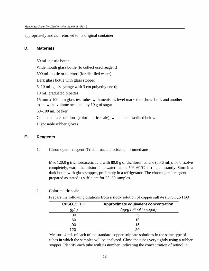
appropriately and not returned to its original container.
D. Materials
50 mL plastic bottle
Wide mouth glass bottle (to collect used reagent)
500 mL bottle or thermos (for distilled water)
Dark glass bottle with glass stopper
5–10 mL glass syringe with 3 cm polyethylene tip
10 mL graduated pipettes
15 mm x 100 mm glass test tubes with meniscus level marked to show 1 mL and another to show the volume occupied by 10 g of sugar
50–100 mL beaker
Copper sulfate solutions (colorimetric scale), which are described below
Disposable rubber gloves
E. Reagents
1. Chromogenic reagent: Trichloroacetic acid/dichloromethane
Mix 120.0 g trichloroacetic acid with 80.0 g of dichloromethane (60.6 mL). To dissolvecompletely, warm the mixture in a water bath at 50°–60°C stirring constantly. Store in adark bottle with glass stopper, preferably in a refrigerator. The chromogenic reagentprepared as stated is sufficient for 25–30 samples.
2. Colorimetric scale
Prepare the following dilutions from a stock solution of copper sulfate (CuSO .5 H O).4 2
CuSO .5 H O Approximate equivalent concentration4 2
(g/L) (µg/g retinol in sugar)
30 560 10 90 15
120 20 Measure 4 mL of each of the standard copper sulphate solutions in the same type oftubes in which the samples will be analyzed. Close the tubes very tightly using a rubberstopper. Identify each tube with its number, indicating the concentration of retinol in

Analytical Methods for the Control and Evaluation of Sugar Fortification with Vitamin A
19
µg/g that the color represents. These solutions are stable and can be kept indefinitely atroom temperature.
F. Procedure
1. Mix the sugar sample thoroughly.
2. In a 50 mL wide mouth flask, weigh 10 g of sugar (or use the test tube marked to showthe equivalent volume).
3. Add 10 mL of water at 50°C, preferably distilled. Dissolve the sugar, heating thesolution, if necessary.
4. Cool solution at room temperature.
5. Transfer the sugar solution to a test tube up to the previously marked 1 mL level.
6. Pour enough chromogenic reagent for all the samples into a clean glass beaker
7. Wearing disposable gloves, add 3 mL of chromogenic reagent to each test tube using asyringe. Mix immediately and vigorously.
8. Compare the intensity of the blue color of the samples with the copper sulfate standardswithin 10 seconds of adding the reagent, because the color change is transient.
9. Estimate the approximate concentration of retinol in the sugar sample (µg/g) bymatching the color developed to the closest tube in the reference scale. In mostinstances, the intensity of the blue color of the sample will fall between two of thereference tubes. The level of retinol in the sugar should be reported as falling within therange corresponding to the reference tubes. For example, if the intensity of the blue liessomewhere between the levels of 30 g/L and 60 g/L copper sulfate, the retinol level isbetween 5 µg/g and 10 µg/g sugar.
10. Discard residual chromogenic reagent, including the sugar-reagent mixture, in a glassbottle and take to the laboratory for proper disposal.


21
VII. VOLUMETRIC METHOD TO DETERMINE PEROXIDELEVELS IN OILS
A. References
Association of Official Analytical Chemists. 1984. Official Methods of Analysis of the Association ofOfficial Analytical Chemists. 14th. ed., Arlington, Virginia.
B. Principle
This method is recommended for determining the peroxide level in the vegetable oil used inmanufacturing the vitamin A sugar premix. Peroxides and similar substances arise from fat and oiloxidation. These substances oxidize iodide to iodate, which can be quantitatively measured by theirredox reaction with sodium thiosulfate. The reaction takes place in a slightly acid medium and in thepresence of an excess of iodide ions. Due to the hydrophobic nature of the oil, titration is carried out ina mixture of acetic acid and chloroform.
C. Critical points and cautions
Reagents should be kept free of oxygen using an inert gas (CO or N ).2 2
D. Equipment and materials
Magnetic mixer
500 mL and 1,000 mL volumetric flasks
Burette
250 mL Erlenmeyer flask preferably with glass stoppers
Dropper bottle
1,000 mL graduated cylinder
Glass rods
100 mL and 400 mL beaker
Spatula

Manual for Sugar Fortificaiton with Vitamin A - Part 3
22
E. Reagents
1. Acetic acid/chloroform
Mix 3 volumes glacial acetic acid ((CH COOH), purity = 99.7%, MW = 60.05, 3
d = 1.05 g/mL) with 2 volumes of chloroform USP (CH Cl )). Prepare only the3 3
quantity needed and place in a glass bottle.
2. 1 percent indicator starch solution
Put 0.5 g of analytical grade starch in a 100 mL beaker and add 5 mL of hot, distilledwater. Mix by constantly stirring in 45 mL of distilled water. Boil for a few minutes, letcool, and filter. Store in a dropper bottle for no more than 1 week.
3. 0.1N sodium thiosulfate
Dissolve approximately 25 g of analytical grade sodium thiosulfate ((Na S O .5H O),2 2 3 2
purity = 99.5%, MW = 248.18) in 1 liter of distilled water. Bring to a boil and gentlyboil for 5 minutes. Allow to cool. Transfer to a dark bottle, previously washed with achromic cleaning mixture and rinsed with boiled water, and store. The solution remainsstable for about 6 months. Discard the solution if it becomes turbid. The solution shouldbe standardized before use (described below). Diluted solutions are prepared usingboiled water just before use.
4. Potassium iodide free of iodate
5. 1N hydrochloric acid
Slowly add 82.8 mL of concentrated hydrochloric acid ((HCl), 37%, MW = 36.46, d= 1.2g/mL) to 300 mL distilled water in a 1,000 mL volumetric flask. Cool at roomtemperature and make up to 1,000 mL with distilled water. Store in a tightly cappedglass bottle to avoid contact with vapors and alkaline solutions.
6. Potassium dichromate ((K Cr O ), purity = 99.0, MW = 294.19)2 2 7

Na2S2O3(eq/mL) 'gK2Cr2O7
mLNa2S2O3
×1eqCr2O7
294.20gCr2O7
×6eqS2O3
1eqCr2O7
Normality Na2S2O3(eq/L) 'gK2Cr2O7
mLNa2S2O3
× 1,000(mL/L)49.032
Analytical Methods for the Control and Evaluation of Sugar Fortification with Vitamin A
23
F. Procedure
1. Standardizing the sodium thiosulfate solution
Every time a batch of samples is processed, the following procedure should be carried out intriplicate:
a. Weigh between 0.2000 g and 0.2300 g of potassium dichromate, previouslydried for 2 hours at 105°C and stored in a desiccator, and transfer to a 250 mLErlenmeyer flask.
b. Add about 80 mL of distilled water, 2 g of potassium iodide, and 20 mL of 1NHCl, and mix with a magnetic stirrer. Let stand for 10 minutes in the dark.
c. With continuous stirring, titrate the potassium dichromate solution with 0.1Nsodium thiosulfate, until the yellow color almost disappears. At this point, addten drops of the 1 percent starch solution. Continue titrating drop by drop. Thetitration is complete when the blue color disappears. Record the final volume ofthiosulfate solution required to complete the titration.
The normality of the thiosulfate solution is calculated as follows:
which can be simplified to:

Perox. (meq/kg) 'mL S2O3
g sample× S2O3 (eq/L) × 1,000(meq/eq)
Manual for Sugar Fortificaiton with Vitamin A - Part 3
24
2. Titration of samples
Each sample should be run in duplicate.
a. Weigh 5 (±0.05) g of each sample in a 250 mL Erlenmeyer flask with glassstopper.
b. Add 30 mL of acetic acid/chloroform solution and gently mix until dissolved.
c. Add approximately 1 g of potassium iodide and mix. Let stand for 1 minute.
d. Add 30 mL of distilled water and ten drops of the 1 percent starch solution.Stirring continuously, slowly titrate with 0.1N sodium thiosulfate solution untilthe blue color suddenly disappears. If the titration uses less than 0.5 mL 0.1Nsodium thiosulfate, reduce the normality of the solution to 0.01N (1:10 dilution)with boiled distilled water and repeat the titration.
The intermediate iodine formed is soluble in the organic phase; therefore, thedisappearance of the color (first yellow and then blue once the starch has beenadded) should be observed in this phase.
3. Blank titration
In each run, repeat the procedure for a blank, in which no oil is added (less than 0.1 mLof sodium thiosulfate solution should be needed). Subtract the titration volume for theblank from the titration volume for each of the samples.
G. Calculations
The milliequivalent of peroxides per kilogram of sample are calculated as follows:
The maximum permitted level for peroxides in vegetable oil is 5 meq/kg.

25
VIII. PRINCIPALS OF METHODS TO DETERMINE RETINOL INBLOOD
A. Introduction
Evaluating the nutritional impact of a sugar fortification program on vitamin A status can be doneby measuring retinol levels in blood both before and after the intervention.
Two methods for determining blood retinol levels are presented here. The first is an adaptationof the classic spectrophotometric method of Bessey et al. (1946) (see section IX). The specificity ofthis method is based on the selective destruction of free or esterified retinol present in blood byirradiation with UV light. The second method requires an apparatus that does high performance liquidchromatography (HPLC) to separate the free retinol from other substances (see section X). Thismethod is more specific and sensitive than the spectrophotometric method; however, thespectrophotometric method has the advantage of being lower in cost and allowing for two or threetimes as many samples to be analyzed during the same period of time. A limitation of thespectrophotometric method is that it does not differentiate between free retinol and its esters. Thiswould be important only if used in populations with very high intakes of retinol in postprandialconditions. This limitation does not apply in countries in which vitamin A deficiency is a public healthproblem.
The choice of the method to use will depend on the purpose of the study and the resourcesavailable. While the HPLC is undoubtably more accurate and precise, both methods can provide thenecessary information for evaluation purposes.
The determination of blood retinol levels involves the following laboratory steps:
1. Treatment with alkaly. Theoretically, this treatment hydrolyzes the retinyl esters in thesample. For the spectrophotometric method, this hydrolysis is incomplete, but this doesnot affect the usefulness of the method. In serum or plasma, this treatment is necessaryto destroy the retinol-retinol binding protein complex and precipitate proteins and otherinterfering substances.
2. Extraction. Retinol and nonhydrolyzed esters are extracted with an organic solvent. Axylene-kerosene mixture is preferred in the spectrophotometric determination, whilehexane or another low-boiling-point solvent is used in the HPLC determination.
3. Determining absorbance. In both the spectrophotometric and HPLC methods, theabsorbance of retinol at 325 nm is used for its quantitative determination.
Manual for Sugar Fortificaiton with Vitamin A - Part 3
26
4. Selective destruction of retinol. To ensure that the absorbance is from retinol only,absorbance is determined in the sample extracts both before and after selectivedestruction of the retinol by irradiation with UV light. The HPLC method does notrequire this step because, before measuring the absorbance of retinol, it ischromatographically separated from other interfering substances including its esters.
B. Collection and management of blood samples
Blood retinol can be measured in both serum or plasma. Serum is preferable when blood isprocessed within 6 hours at the site of sample collection. If these conditions cannot be met, as is oftenthe situation in many surveys, plasma is recommended because there is less risk of hemolysis.Centrifugation of plasma can be delayed for up to 24 hours, provided the samples are refrigerated.
Blood can be obtained from venipuncture or, if the laboratory uses microadaptations of theanalytical methods, from finger pricks. When venipuncture is used, between 3 and 5 mL should bedrawn from an antecubital vein using standard safety procedures. A 21- or 22-gauge needle can beused for preschool children, while a 20-gauge needle can be used for adults. When a tourniquet isapplied around the forearm, it should be removed as soon as blood begins to flow into the tube so as tominimize the risk of hemolysis. It is preferable to draw blood directly into a vacutainer tube, coveredwith aluminum foil. The tubes and their aluminum foil should be labeled with indelible ink. If collectingblood directly into a vacutainer is not possible or is difficult, it can be drawn into a syringe and injectedinto the vacutainer tube. To avoid this double step, an alternative is to use a Sarstedt Monovette testtube syringe, but they are more expensive than syringes and vacutainer tubes. Vacutainer tubescontaining serum or plasma gel separators will help to prevent hemolysis, but only if the blood iscentrifuged before it is transported.
Blood samples should not be exposed to air or direct sunlight, nor should they come into directcontact with ice. The blood samples should be packed and transported carefully to ensure that they donot become hemolyzed from too much shaking while in transit. If the blood is exposed to air, it issuggested that the atmosphere above the sample be filled with nitrogen by blowing the gas into the tubefor 30 to 60 seconds without disturbing the sample, after which the tube should be tightly capped.
Serum is separated from coagulated blood by centrifugation at 2,500 to 3,000 rpm for 15minutes. Unclear serum should be transferred with a Pasteur pipette to another centrifuge tube andrecentrifuged. The serum should be stored at !20°C in adequately sized vials so that the dead spacebetween the surface of the sample and cap is minimal, leaving enough space for expansion duringfreezing.
Plasma is separated from the blood cells using the same process as for serum, but thecentrifugation need only be done for 10 minutes. Plasma samples should be handled exactly the same as
Analytical Methods for the Control and Evaluation of Sugar Fortification with Vitamin A
15. Microvacutainers with a lip will facilitate directing the flow of blood into it.
27
serum samples and should also remain frozen until analyzed. Under this condition, both kinds ofsamples will remain viable for up to one year.
When centrifugation is done in the field, serum and plasma can be temporarily stored andtransported frozen, for which liquid nitrogen or dry ice can be used. It is essential that, once frozen,samples remain frozen at all times even when being transferred between laboratories; thus, sufficientamounts of liquid nitrogen or dry ice must be available to ensure this.
Where finger-prick blood samples are collected, an automatic lancet that pricks to a givendepth and minimizes bruising is preferable to an ordinary lancet, although they cost slightly more. It isimportant when collecting finger-prick blood samples that blood is not squeezed out of the finger. Bloodflow can be made easier if the subjects are asked to shake their hand vigorously up and down beforetheir finger is pricked. The blood can be easily collected in a serum or plasma microvacutainer, which15
can be centrifuged. After centrifugation, the serum or plasma should be transferred to a vial and storedas for venipuncture blood.
To verify that the samples have not deteriorated during storage, aliquots of a serum orplasma control whose retinol value is known should be stored under the same conditions andanalyzed at the same time.

16. Maximum absorbance wavelength of retinol in a kerosene-xylene mixture.17. Section XIV.C.1 includes a description of a simple procedure to calibrate spectrophotometers routinely used in
clinical laboratories.
29
IX. SPECTROPHOTOMETRIC DETERMINATION OF BLOODRETINOL BY ULTRAVIOLET DESTRUCTION OF RETINOL
A. References
Bessey, O. A., O. H. Lowry, M. J. Brock, and J. A. López . 1946. The determination of vitamin A andcarotene in small quantities of blood serum. J. Biol. Chem. 166: 177–88.
Araujo, C. R. C. and H. Flores. 1978. Improved spectrophotometric vitamin A assay. Clin. Chem. 24:386.
B. Principle
The procedure described below is an adaptation of the method proposed by Bessey et al.(1946). This method consists of precipitating the proteins with an alcoholic potassium hydroxidesolution and subsequent extraction of retinol (and any esters) in a kerosene-xylene mixture.Saponification, although incomplete, facilitates retinol extraction and eliminates interfering substances.The retinol in the extract is selectively destroyed by irradiation of ultraviolet light. The differencebetween absorbance at 328 nm before and after irradiation is attributable and proportional to total16
retinol.
C. Critical points and cautions
A spectrophotometer capable of reading 325 nm is essential. This is because the concentrationof the retinol standards have to be verified by spectrophotometric analysis. Given the importance of thespectrophotometer for ensuring the accuracy and reliability of the retinol determinations, it should becalibrated frequently following the instructions provided by the manufacturer, especially to confirm thecalibration of the monochromator. This confirmation should be carried out frequently and not only17
when a new lamp is installed.
A UV-light irradiation system in which the retinol is destroyed is also required. This system canbe as simple as a UV lamp and curtain to protect technicians from exposure to this light. The criticalfactor is to establish exactly both the optimal irradiation time and appropriate distance between the UVlight and the solutions. The efficiency of the irradiation system should be checked periodically (every 3months) and each time a new lamp is installed. Appendix 3.1 gives details of an irradiation system.

Manual for Sugar Fortificaiton with Vitamin A - Part 3
30
Once the retinol has been extracted into the organic solvent, the analysis should not beinterrupted.
Based on the experience at the INCAP laboratory, two independently analyzed subsamples ofthe same sample should not differ on average by more than 10 percent if the concentration of retinol isequal to or greater than 30 µg/dL or by more than 15 percent if the concentration of retinol fallsbetween 15 and 30 µg/dL. If these criteria are not met, the assay should be repeated. Samples having aretinol concentration below 15 µg/dL should be reported as 15 µg/dL. The recovery of retinol using thismethod is 93 percent or higher.
D. Equipment and materials
Vortex mixer
Water bath (56°C)
UV-light irradiation chamber with ultraviolet light (example in appendix 3.1)
Refrigerated centrifuge
Stopwatch
UV/VIS spectrophotometer
Black walled quartz cuvettes (maximum 1 mL capacity)
Dark glass bottle
Graduated serological pipettes
Automatic pipettes (Eppendorf type)
Pasteur pipettes
Graduated cylinder
10 mm x 75 mm glass test tubes transparent to UV light
12 mm x 75 mm glass test tubes
Glass rods
Aspiration bulbs for Pasteur and serological pipettes

tcorr ' tref &(273 % tref) × (760 & mmHg)
10,000
Analytical Methods for the Control and Evaluation of Sugar Fortification with Vitamin A
18. These temperatures must be adjusted to the specific barometric pressure of each place. Thus:
19. See footnote 18.
31
E. Reagents
1. Alcoholic potassium hydroxide
Combine 10 volumes of analytical grade absolute ethanol ((C H OH), purity = 99.8%,2 5
MW = 46.07, d = 0.79 g/mL) with 1 volume of 11.0N potassium hydroxide (KOH,MW = 56.11). Mix fifty times using a stirring rod. Centrifuge at 3,000 rpm for 15minutes. After centrifugation, a small white precipitate or a separate phase may beobserved at the bottom of the tube; this is caused by potassium carbonate that ispresent in some KOH preparations. The clear supernatant solution should be usedwithin 60 minutes of preparation.
2. Kerosene/xylene 1:1 volume
Mix, just before using, the necessary amounts of kerosene (fraction distilled in glass at206°–216°C ) and xylene (fraction distilled in glass at 138°–143°C ) until the18 19
solution turns clear. Both the individual solvents and the mixture should be stored indark bottles.
F. Procedure
1. If the samples have been in the freezer, thaw at room temperature (20°–25°C) and mixwell in a Vortex mixer. Centrifuge at 2,500 rpm for 10 minutes preferably in arefrigerated centrifuge to eliminate any interference due to fibrin formation.
2. Measure in duplicate 0.5 mL serum or plasma into 12 mm x 75 mm glass tubes.
3. Prepare in duplicate a reagent blank with 0.5 mL of distilled water and a control serumwith 0.5 mL of serum of known retinol concentration.

Manual for Sugar Fortificaiton with Vitamin A - Part 3
20. The volume ratio for serum:alcoholic KOH:xylene/kerosene described in the procedure is 1:1:2. An increase insensitivity may be achieved by either (a) increasing proportionally the serum (or plasma) volume and alcoholicKOH but keeping the 1:1 ratio without changing the kerosene/xylene volume. In this case, the test tube sizeshould be increased to hold the larger volumes or (b) decreasing the kerosene/xylene volume while keeping theserum or plasma and alcoholic KOH volumes unchanged. In these cases, the irradiation chamber may have tobe slightly modified to ensure complete irradiation of the samples by UV light. Keep in mind that any variationin serum or plasma and kerosene/xylene volumes implies a different dilution factor in the equation for thecalculations.
32
4. Add 0.5 mL of alcoholic KOH to all tubes. Mix in a Vortex mixer for 10 to 20seconds.
5. Close the tubes with rubber stoppers and incubate in a water bath at 56°C for 20minutes.
6. Cool at room temperature. Add 1.0 mL of 1:1 kerosene/xylene mixture. Mix in theVortex mixer for 30 seconds measured with a stopwatch. Agitation should be vigorousto ensure a complete extraction. 20
7. Centrifuge at 3,000 rpm for 10 minutes preferably in a refrigerated centrifuge.
8. Remove the organic phase using a Pasteur pipette and transfer to a spectrophotometriccuvette. Be careful not to transpose any of the aqueous phase. Read the absorbance ofthe organic phase at 328 nm against xylene/kerosene.
9. Using a Pasteur pipette, transfer the organic extract from the spectrophotometriccuvette to a 10 mm x 75 mm tube transparent to ultraviolet light. Irradiate the tubes withultraviolet light for 35 minutes or the time determined necessary to destroy retinol (seesection XIV.C.2).
10. Between samples, aspirate the residue in the spectrophotometric cuvette with a Pasteurpipette connected to a vacuum system. Wash the cuvettes with the kerosene/xylenemixture every 15 readings.
11. Read the absorbance of every irradiated extract against the mixture of xylene/keroseneat 328 nm.

Retinol (µg/dL) 'AbsRetinol × Vs × 106
å × Vm
Retinol (µg/dL) ' Absretinol × 1,273.9
Analytical Methods for the Control and Evaluation of Sugar Fortification with Vitamin A
33
G. Calculations
1. Correct the absorbance of each sample before irradiation by subtracting theabsorbance of the unirradiated blank. Subtract the absorbance of the irradiated blankfrom the absorbance of the irradiated sample. Sometimes the absorbance of the reagentblank is negative, therefore, the value should be added instead of subtracted.
2. Calculate the specific absorbance due to retinol by subtracting the corrected readings ofthe irradiated tubes from the readings of the unirradiated ones and correct by the“efficiency factor” of the irradiation chamber (see section XIV.C.2)
3. Calculate the retinol concentration using the following equation:
where:
Abs is the absorbance of each sample obtained in step IX.G.2.retinol
The parameters for the equation are:
Parameter Explanation Value
å Retinol absorption coefficient (g cm dL) in 1,570!1 !1
kerosene:xylene at 328 nm
Vs Volume of organic phase (mL) 1.0
Vm Volume of the sample (mL) 0.5
The equation is simplified to:

Absretinol ' (Abscont & Absblank & Abscontirr % Absblankirr)
Manual for Sugar Fortificaiton with Vitamin A - Part 3
21. A dilution of this solution should have an absorbance near to 0.9 when read at 325 nm against ethanol.
34
H. Verification of the method’s reproducibility
To verify the method’s reproducibility through independent runs, a pool of serum should beprepared and its exact retinol concentration determined. To do this, prepare a series of vials with 1.5mL of control serum, leaving as little space as possible between the surface of the sample and the vialcap. Blow inert gas (nitrogen) in the tubes and close tightly. Store the control vials in the freezer at!20°C or lower. Include a duplicate serum control in every analytical run. After ten or more runs of thecontrol, calculate the method’s between-run coefficient of variation.
I. Verification of the method’s recovery
The recovery of the retinol extraction should be verified periodically. The following procedure issuggested:
1. Irradiate a plasma or serum sample with UV light for 1 hour. Then, divide the sampleinto two portions. To one portion, add 10 µL of a 50 mg/mL retinol solution in absoluteethanol for each milliliter of the serum or plasma (control). To the second portion (the21
blank), add 10 µL of ethanol for every milliliter of serum or plasma. Treat both portionsas independent samples following the analytical procedure from step IX.F.2.
2. Calculate the absorbance due to the added retinol as follows:
where Abs and Abs are the absorbances before and after irradiating the retinol-cont contirr
containing controls and Abs and Abs are the absorbances for the blanksblank blankirr
before and after irradiation.
The value from this calculation (s) is the absorbance due to the added retinol.
3. Independently, place 50 mL of the same 50 µg/mL retinol solution in a 10-mLvolumetric flask and make up to volume with the kerosene/xylene mixture. Read theabsorbance of this solution at 328 nm against the kerosene/xylene mixture and multiplyby 0.99, which is the ratio between this dilution and the dilution occurring during therecovery assay (200/202). This value (t) is the theoretical absorbance that would havebeen found if recovery of retinol was 100 percent efficient.

R 'st
× 100
Analytical Methods for the Control and Evaluation of Sugar Fortification with Vitamin A
35
4. Calculate recovery (R) as follows:
J. Variations in assay
If a spectrophotometer capable of reading absorption accurately using smaller cuvettes isavailable, samples and reagent volumes can be proportionally reduced; however, the size of the test andirradiation tubes, as well as the irradiation chamber will also have to be adjusted.
When a micromethod is used, the spectrophotometer’s capability of reading small cuvettesshould be verified. To do this, prepare dilutions of a 50 µg/mL retinol in ethanol solution at 1:100,2:100, 3:100, 4:100, and 5:100. Read the absorbance of these solutions at 325 nm using normal quartzcuvettes (3 mL) and compare the absorbances with those from semimicro (1 mL) or micro (less than 1mL) quartz cuvettes. The reading for the latter two cuvettes should coincide with those for the normalcuvettes. The above retinol in ethanol solutions should have absorbances ranging from 0.100 to 0.500.


37
X. DETERMINATION OF BLOOD RETINOL BY HIGHPERFORMANCE LIQUID CHROMATOGRAPHY (HPLC)
A. References
Bankson, D. D., R. M. Russell, and J. A. Sadowski. 1986. Determination of retinyl esters and retinol inserum or plasma by normal-phase liquid chromatography: method and applications. Clin.Chem. 32: 35–40.
Bieri, J. G., T. J. Tolliver, and G. L. Catignani. 1979. Simultaneous determination of á-tocopherol andretinol in plasma or red cells by high pressure liquid chromatography. Am. J. Clin. Nutr. 32:2143.
DeRuyter, M. G. M. and A. P. Leenheer. 1976. Determination of Serum retinol (vitamin A) by high-speed liquid chromatography. Clin. Chem. 22: 1593–95.
Packer L. 1990. Methods in Enzymology, Retinoids. Part A, Molecular and Metabolic Aspects.189: 75–76, 155–67, 170–75. New York: Academic Press.
B. Principle
This is a highly selective and accurate method that requires neither blanks nor irradiation of theretinol-containing extracts. The sample does not require saponification, because the method is designedto measure specifically the nonesterified retinol in blood. The method can be modified if retinyl esterdeterminations are needed.
Serum or plasma is diluted 1 to 2 with a retinyl acetate solution in ethanol. The retinyl acetateacts as the internal standard, and the ethanol precipitates the proteins, which releases the retinol that isthen extracted with hexane. The extract is evaporated in a nitrogen atmosphere and the residueresuspended in methanol. Retinol is separated by high performance liquid chromatography using a fixedapolar phase (C ) and 95 percent methanol as the mobile phase. Retinol is detected with an ultraviolet18
detector at 325 nm, and its concentration is determined from the ratio of its peak area to that of theretinyl acetate.
Manual for Sugar Fortificaiton with Vitamin A - Part 3
22. Section XIV.C.1 includes a description of a simple procedure to calibrate spectrophotometers routinely used inclinical laboratories.
38
C. Critical points and cautions
A spectrophotometer capable of reading 325 nm, even when high pressure liquidchromatography (HPLC) is used, is essential. This is because the concentration of the retinol standardshas to be verified by spectrophotometric analysis. Given the importance of the spectrophotometer forensuring the accuracy and reliability of the retinol determinations, it should be calibrated frequentlyfollowing the instructions provided by the manufacturer, especially to confirm the calibration of themonochromator. This confirmation should be carried out frequently and not only when a new lamp is22
installed.
Once retinol has been extracted in the organic phase, the analysis should not be interrupted.
Because the retinol standard and retinol acetate internal control are not completely pure, theirpurity must be estimated. This can be done by calculating the percent of the peak area for each analyteover the total area of all the peaks detected in the chromatogram.
In situations in which no HPLC-grade water is commercially available, it can be prepared in thelaboratory by demineralizing, filtering, and boiling water that has been doubly distilled.
Based on the experience at the INCAP laboratory, two independently analyzed subsamples ofthe same sample should not differ on average by more than 10 percent. If the variation is greater than10 percent, the assay should be repeated. The recovery of retinol using this method is 95 percent ormore.
D. Equipment and materials
Vortex mixer
Refrigerated centrifuge
Liquid chromatograph with UV detector
Micropak column SP-18-5 of 150 x 4 (DI) mm
Stopwatch
UV/VIS spectrophotometer
Spectrophotometer quartz cuvettes
10 mL and 100 mL volumetric flasks
Dark glass bottles

Analytical Methods for the Control and Evaluation of Sugar Fortification with Vitamin A
39
Graduated serological pipettes
Automatic pipettes (Eppendorf type)
Pasteur pipettes
Conical centrifuge tubes (9 mm x 76 mm)
500 µL amber micro centrifuge tubes
10 mL beaker
Aspiration bulbs for Pasteur and serological pipettes
45 mm millipore membrane
E. Reagents
1. Analytical grade absolute ethanol ((C H OH), purity = 99.8%, MW = 46.07,2 5
d = 0.79 g/mL)
2. HPLC grade hexane ((C H ), purity = 95%, MW = 86.18, d = 0.66 g/mL)6 14
3. HPLC grade methanol ((CH OH), purity = 100 %, MW = 32.04, d = 0.79 g/mL)3
4. Nitrogen gas (99.5 percent purity)
5. Internal standard (retinyl acetate)
a. Stock solution (about 500 mg/mL)
Weigh 0.056 g of retinyl acetate of the highest purity (MW = 328.5, 90%purity) in a 10 mL beaker and dissolve with absolute ethanol. Transfer to a 100mL volumetric flask. Rinse the beaker several times with absolute ethanol andtransfer the washings to the volumetric flask. Add approximately 0.002 g ofBHT (3,5-bis-(t-butyl)-4-hydroxy-toluene). Make up to 100 mL with absoluteethanol. Store the solution in the freezer (!20°C) in a dark glass bottle withglass stopper. The concentration of this solution is calculated from theconcentration of the secondary solution.
b. Primary solution (about 50 µg/mL)
Dilute 1 mL of stock solution to 10 mL with absolute ethanol in a 10 mLvolumetric flask. Store as indicated for the stock solution and calculate its

Manual for Sugar Fortificaiton with Vitamin A - Part 3
23. This value is the absorption coefficient (mg cm mL) for retinyl acetate in ethanol at 325 nm.!1 !1
40
retinol concentration from the concentration of the secondary solution.
c. Secondary solution (about 0.5 µg/mL)
Dilute 0.5 mL of primary solution to 50 mL with absolute ethanol in a 50 mLvolumetric flask. Store the solution in a refrigerator. Discard the remainder afterusing it.
The concentration of retinyl acetate in the secondary solution is determined in aspectrophotometer as follows:
i. Zero the spectrophotometer using absolute ethanol. Read in triplicatethe absorbance of the secondary solution of retinyl acetate at 325 nm.
ii. Divide the average value of the absorbance by 0.156 and multiply by23
the degree of purity of the reagent as determined in HPLC (peak areaof retinyl acetate/total area of all peaks.) The result is the actualconcentration of retinyl acetate in the secondary solution in µg/mL.
The µg/mL retinyl acetate concentration in the primary and stock solutions is obtainedby multiplying this value by 100 and 1,000, respectively.
6. Standard retinol solutions
a. Stock solution (about 500 µg/mL)
Weigh 0.071 g retinol of the highest purity (MW = 286.5, 70% purity) anddissolve with absolute ethanol in a 10 mL beaker. Transfer to a 100 mLvolumetric flask. Rinse the beaker several times with absolute ethanol, andtransfer the washings to the volumetric flask. Add approximately 0.002 g ofBHT. Make up to 100 mL with absolute ethanol. Store the solution in thefreezer (!20°C) in a dark bottle with glass stopper. The concentration of thissolution is calculated from that of the secondary solution.

Analytical Methods for the Control and Evaluation of Sugar Fortification with Vitamin A
41

Manual for Sugar Fortificaiton with Vitamin A - Part 3
24. This value is the absorption coefficient (mg cm mL) for retinol in ethanol at 325 nm.!1 !1
42
b. Primary solution (about 50 µg/mL)
Dilute 1 mL stock solution to 10 mL absolute ethanol in a 10 mL volumetricflask. Store as indicated for the stock solution and calculate its concentrationfrom the concentration of the secondary solution.
c. Secondary solutions (about 1–10 µg/mL)
Dilute 5 mL primary solution to 25 mL of absolute ethanol in a 25 mLvolumetric flask. Dilute the secondary retinol solution with absolute ethanol toobtain at least 10 mL of five solutions with different concentrations of retinolranging from 1 to 10 µg/mL. Store the secondary solutions in the refrigerator.Discard the solutions after using them.
The concentration of retinol in the most concentrated secondary solutions isdetermined in a spectrophotometer as follows:
i. Zero the spectrophotometer using absolute ethanol. Read in triplicatethe absorbance of the 10 µg/mL secondary retinol solution at 325 nm.
ii. Divide the average value of the absorbance by 0.1845 and multiply by24
the degreee of purity of the reagent as determined in HPLC (peak areaof retinol/total area of all peaks.) The result is the actual concentrationof retinol in the secondary solution in µg/mL. The concentration ofretinol in the other solutions is calculated by dividing each one’s averagevalue for absorbance by its dilution factor.
The retinol concentration in the primary and stock solutions is obtained by multiplyingthe concentration of the 10 µg/mL and secondary solutions by 5 and 50, respectively.

Y ' mX % b
Analytical Methods for the Control and Evaluation of Sugar Fortification with Vitamin A
43
F. Procedure
1. Calibration curve
a. Measure 0.1 mL of primary retinyl acetate solution (50 µg/mL) and 1.0 mL ofeach secondary retinol solution into individual 10 mL volumetric flasks. Makeup to volume with HPLC-grade absolute methanol.
b. Filter each solution through the 45 µ millipore membrane. Run the solutions inthe HPLC.
c. Regress the ratio of the area of the peaks of retinol to the area of the peak ofretinyl acetate multiplied by the concentration of the internal standard of retinylacetate (y-axis) on the retinol concentration of the secondary retinol solutions inµg/mL (x-axis). The regression equation used to calculate the retinol levels ofthe samples is:
where:
Y = retinyl acetate (µg/mL) x (area peak retinol/area peak retinylacetate)
X = retinol (µg/mL)
m = slope (Y/X)
b = intercept on y-axis
The regression equation should be calculated every fifteen days to confirm theconsistency of the analytical conditions. However, for each run, at least oneretinol standard must be analyzed to confirm the validity of the curve.
Manual for Sugar Fortificaiton with Vitamin A - Part 3
44
2. Extraction
One extraction is sufficient for plasma or serum and is recommended because more samplescan be processed and few sources of error will be introduced.
a. If samples are frozen, thaw at room temperature (20°–25°C) and mixthoroughly using a Vortex mixer. Centrifuge at 2,500 rpm for 10 minutes toeliminate interference due to fibrin formation if plasma is being used.
b. Measure 0.25 mL of serum or plasma into duplicate 9 mm x 76 mm conicalcentrifuge tubes.
c. Add 0.25 mL of secondary solution (0.5 µg/mL) of the internal standard (retinylacetate). Mix in the Vortex mixer for 1 minute.
d. Add 0.5 mL hexane and mix vigorously in the Vortex mixer for 1 minute toensure complete extraction.
e. Centrifuge at 2,500 rpm for 2 minutes. A refrigerated centrifuge should be usedwhere one is available.
f. Using a Pasteur pipette, transfer the organic phase to an amber micro centrifugetube.
g. Evaporate the organic phase by blowing nitrogen into the micro centrifuge tubeinside a laboratory hood for organic solvents.
h. Resuspend the extract with 0.25 mL HPLC-grade methanol. Mix in the Vortexmixer for 1 minute.
i. Centrifuge at 2,000 rpm for 5 minutes.

Analytical Methods for the Control and Evaluation of Sugar Fortification with Vitamin A
45
3. Chromatography
a. Using the HPLC syringe, take an appropriate amount of sample solution andinject into the machine following the optimal conditions for the apparatus, asdetermined by the laboratory. For example:
Mobile phase 95% methanol/5% waterFlow 1.6 mL/minDetector UV at 325 nm with 0.002 UA/mV sensitivityIntegrator attenuation 4Paper speed 1 cm/minInjection volume 100 µLElution time Retinol: 2.5 min
Retinyl acetate: 4.5 min
b. At the end of every day, wash the HPLC system for at least 30 minutes at aflow of 0.5 mL/min with a mixture of 95 percent methanol/5 percent water,followed by 100 percent methanol for 15 minutes.
G. Calculations
1. Calculate the ratio of the peak of retinol over the peak of retinyl acetate for each sampleand multiply these ratios by the concentration of the secondary retinyl acetate internalstandard.
2. The retinol concentration in the samples is calculated directly using the calibration curve.
3. To report retinol levels in the samples in µg/dL, multiply the values calculated in thepreceding step by 100.

R 'st
× 100
Manual for Sugar Fortificaiton with Vitamin A - Part 3
25. A 1:10 dilution of the primary retinol solution should have an absorbance around 0.9 at 325 nm against ethanol.
46
H. Verification of the recovery of the method
Even though it is not essential for the calculations, it is recommended that the recovery of themethod be verified every 3 months. For this purpose the following procedure is suggested:
1. Irradiate a plasma or serum sample with ultraviolet light for 1 hour and separate intotwo portions. To one portion, add 10 µL of the primary 50 µg/mL retinol solution foreach milliliter of the sample (control). To the second portion, add 10 µL of ethanol for25
every milliliter of sample (blank). Treat both portions as independent samples andfollow the chromatographic process.
2. Using the calibration curve, calculate the retinol level for both portions. Subtract theretinol value of the blank from that of the control. The result of this calculation is theamount of recovered retinol (s).
3. Independently, place 0.1 mL of primary retinol solution (50 mg/mL), 0.1 mL of primaryacetate solution (50 µg/mL) and make to volume with HPLC grade methanol in a 10mL volumetric flask. Run this solution in the HPLC system.
4. Determine the retinol level using the calibration curve. Divide this value by 1.01 toadjust for the dilution of the primary retinol solution in the control sample. This value (t)is the theoretical amount of retinol that would have been found if the recovery was 100percent efficient.
5. Calculate recovery (R) as follows:

XI. PRINCIPALS OF METHODS TO DETERMINE RETINOL INBREAST MILK
A. Introduction
Evaluating the nutritional impact of a sugar fortification program on vitamin A status can bedone by measuring retinol levels in breast milk both before and after the intervention.
Two methods for determining breast milk retinol levels are presented here. The first is anadaptation of the classic spectrophotometric method of Bessey et al. (1946) (see section XII). Thespecificity of this method is based on the selective destruction of free or esterified retinol present inbreast milk by irradiation with UV light. The second method requires an apparatus that does highperformance liquid chromatography (HPLC) after saponification and extraction of the sample (seesection XIII). This method is more specific than the spectrophotometric method; however, thespectrophotometric method has the advantage of being lower in cost and allowing for two or threetimes as many samples to be analyzed during the same period of time.
The choice of the method to use will depend on the purpose of the study and the resourcesavailable. While the HPLC is undoubtably more accurate and precise, both methods can provide thenecessary information for evaluation purposes.
The determination of breast milk retinol levels involves the following laboratory steps:
1. Saponification. Samples are treated with alcoholic potassium hydroxide to hydrolyzethe retinyl esters in the sample. Most of the vitamin A in breast milk is in the esterifiedform and therefore saponification is necessary to hydrolyze the esters and obtain a totalretinol value. In the spectrophotometric method, saponification also improves theaccuracy of the determination by avoiding errors due to differences in extraction ratesand thus spectrophotometric characteristics between free retinol and its esters.
2. Extraction. Retinol and nonhydrolyzed esters are extracted with an organic solvent. Axylene-kerosene mixture is preferred in the spectrophotometric determination, whilehexane or another low-boiling-point solvent is used in the HPLC determination.
3. Determining absorbance. In both the spectrophotometric and HPLC methods, theabsorbance of retinol at 325 nm is used for its quantitative determination.

Manual for Sugar Fortificaiton with Vitamin A - Part 3
48
4. Selective destruction of retinol. To ensure that the absorbance is from retinol only,absorbance is determined in the sample extracts both before and after selectivedestruction of the retinol by irradiation with UV light. The HPLC method does notrequire this step because, before measuring the absorbance of retinol, it ischromatographically separated from other interfering substances including its esters.
B. Collection and management of breast milk samples
Ideally, the milk sample should be the total milk from one breast that has not been emptied forat least 2 hours, because the concentration of certain milk components, in particular fats and fat-solublesubstances such as vitamin A, increase over time during suckling. This is impractical in field work,however, and it is recommended that the first 10 mL from a breast that has not been suckled for at least1 hour be taken. It is essential that the procedure followed in collecting breast milk samples be clearlydocumented and consistently followed.
Breast milk can be expressed manually or with a hand pump into a 20 to 30 mL wide mouth,screw top tube/bottle made of glass or polypropylene (for example, Nalgene). Samples should be keptcool and away from air, sunlight, bright light, and high temperatures. Ideally, precise alliquots to be usedin the analysis should be removed and placed in separate vials for long-term storage as soon aftercollection as possible. If the samples are likely to remain frozen for a long period of time, theatmosphere above the sample surface should be filled with nitrogen by blowing the gas withoutdisturbing the sample for 30 seconds, after which they should be tightly closed. Samples should befrozen as soon as possible after collection and not thawed until ready for analysis. Prior to analysis,breast milk samples should be completely homogenized, by sonification wherever possible.

49
XII. APPLICATION OF THE SPECTROPHOTOMETRIC METHODBY ULTRAVIOLET DESTRUCTION FOR THEDETERMINATION OF TOTAL RETINOL IN BREAST MILK
Even though the method described in section IX was originally proposed for thespectrophotometric determination of retinol in serum or plasma, it can be easily applied to breast milk.The principle of the method, as well as the equipment, materials, and reagents are the same for bothblood and breast milk.
Milk samples should be carefully homogenized, treated, and analyzed in exactly the same wayas for serum or plasma. The size of the aliquot remains the same, because the retinol concentrationranges are similar for both fluids. The results are also calculated using the same equations.
It should be noted that this method measures total preformed vitamin A and does notdifferentiate between free and esterified retinol. Because breast milk contains a sizable proportion ofretinyl esters, the accuracy of the results, in terms of free retinol, is inferior to that of the method forblood. This is because saponification is incomplete and residues of nonhydrolyzed retinyl esters maybehave slightly different during extraction. This limitation is amply compensated for by the practicabilityand low cost of this method, which are characteristics that make it suitable for surveys to evaluate theimpact of vitamin A interventions. In these situations, the total content of retinol in breast milk (free andesters), that is, its activity as preformed vitamin A, is the main concern, because evidence exists that thisparameter provides a good picture of the vitamin A status of the lactating women.


26. This section was written by Sherry Tanumihardjo, Department of Biochemistry and Biophysics, Iowa StateUniversity.
27. This method is based on the procedure developed by S. Tanumihardjo and J.A. Olson from the Departmentof Biochemistry and Biophysics, Iowa State University.
28. Section XIV.C.1 includes a description of a simple procedure to calibrate spectrophotometers routinely usedin clinical laboratories.
51
XIII. DETERMINATION OF BREAST MILK RETINOL BY HIGHPERFORMANCE LIQUID CHROMATOGRAPHY (HPLC)26
A. References27
B. Principle
Retinol in breast milk is found mostly esterified to fatty acids. The ester linkage must besaponified to release the retinol. 3,4-Didehydroretinyl acetate, added to the milk before saponification,is used as an internal standard to determine extraction efficiencies. After saponification, the retinol andthe didehydroretinol are extracted and analyzed by HPLC using a C reversed-phase column and a18
methanol/water mobile phase.
C. Critical points and cautions
A spectrophometer capable of reading 325 nm, even when high pressure liquid chromatography(HPLC) is used, is essential. This is because the concentration of the retinol standards has to be verifiedby spectrophotometric analysis. Given the importance of the spectrophotometer for ensuring theaccuracy and reliability of the retinol determinations, it should be calibrated frequently following theinstructions provided by the manufacturer, especially to confirm the calibration of the monochromator .28
This confirmation should be carried out frequently and not only when a new lamp is installed.
Once retinol has been extracted in the organic phase, the analysis should not be interrupted.
In situations in which no HPLC-grade water is commercially available, it can be prepared in thelaboratory by demineralizing, filtering, and boiling water that has been doubly distilled.
Based on the experience of the Department of Biochemistry and Biophysics, Iowa StateUniversity, two independently analyzed subsamples of the same sample should not differ on average bymore than 10 percent. If the variation is greater than 10 percent, the assay should be repeated.
Manual for Sugar Fortificaiton with Vitamin A - Part 3
52
D. Equipment and materials
Vortex mixer
Clinical centrifuge
Liquid chromatograph with UV detector
Water “Resolve” 5 µm C reverse-phase column18
Stopwatch
UV/VIS spectrophotometer
Spectrophotometer quartz cuvettes
25 mL volumetric flask
Graduated serological pipettes
Automatic pipettes (Eppendorf type)
Pasteur pipettes
Aspiration bulbs for Pasteur and serological pipettes
10 mL screw top centrifuge tubes
7 mL test tubes with caps
E. Reagents
1. Analytical grade absolute ethanol ((C H OH), purity=99.8%, MW=46.07, 2 5
d=0.79 g/mL)
2. Analytical grade 2-propanol ((CH CH(OH)CH ), purity=99.7%, MW=60.10, d=0.783 3
g/mL)
3. 50:50 w/v potassium hydroxide in water (KOH/H O)2
4. Purified water
5. HPLC-grade hexane ((C H ), purity=95%, MW=86.18, d=0.66 g/mL)6 14
6. HPLC-grade methanol ((CH OH), purity=100%, MW=32.04, d=0.79 g/mL)3
7. HPLC-grade ethylene dichloride ((ClCH CH Cl), purity=99.8%, MW=98.96, d=1.252 2
g/mL) or HPLC-grade methylene dichloride ((CH Cl ), purity=99.9%, MW=84.94,2 2
d=1.32 g/mL).

Analytical Methods for the Control and Evaluation of Sugar Fortification with Vitamin A
29. Small quantities of this reagent dissolved in corn oil for use as an internal standard for breast milk analysiscan be obtained from Iowa State University for a shipping and handling charge.
53
8. Nitrogen or argon gas (99.5 percent purity)
9. Internal standard solution of 3,4-didehydroretinyl acetate (DRA) (C H O ,2922 30 2
MW=326)
DRA is an excellent choice for an internal standard because:
a. DRA can be added to the breast milk before the analysis is started and can becarried through until the HPLC determination.
b. DRA is in the esterified form and will therefore undergo hydrolysis as do theretinyl esters in the breast milk.
c. By monitoring a few HPLC runs longer than usual, the completeness of thesaponification step can be determined in each laboratory.
d. Didehydroretinol is easily separated from retinol during the HPLC analysis.
DRA is prepared as follows:
From the DRA stock solution (35 mM in corn oil), take 10 µL and mix with 25 mLisopropanol in a volumetric flask. This is the primary DRA solution.

Manual for Sugar Fortificaiton with Vitamin A - Part 3
30. If retinol of high purity is not available in the market, it can be prepared by the following methods: (a)reduction of retinaldehyde (which is chemically more stable) with sodium borohydride (NaBH ); (b)4
saponification of retinyl acetate with alcoholic potassium hydroxide (KOH); or (c) HPLC purification ofcommercial retinol by injecting a concentrated solution onto the HPLC and collecting the retinol peak as itelutes from the column.
54
10. Standard retinol solutions
a. Stock solution (about 500 µg/mL)
Weigh 0.050 g retinol of the highest purity (MW=286.5 70% purity) anddissolve with absolute ethanol in a 10 mL beaker . Transfer to a 100 mL30
volumetric flask. Rinse the beaker several times with absolute ethanol, andtransfer the washing to the volumetric flask. Add approximately 0.002 g ofBHT (3,5-bis-(t-butyl)-4-hydroxytoluene). Make up to 100 mL with absoluteethanol. Store the solution in the freezer (-20°C) in a dark bottle with glassstopper. The concentration of this solution is calculated from that of thesecondary solution.
b. Primary solution (about 50 µg/mL)
Dilute 1 mL stock solution to 10 mL absolute ethanol in a 10 mL volumetricflask. Store as indicated for the stock solution and calculate its concentrationfrom the concentration of the secondary solution.
c. Secondary solutions (about 1-10 µg/mL)
Dilute 5 mL primary solution to 25 mL of absolute ethanol in a 25 mLvolumetric flask. Dilute this secondary retinol solution with absolute ethanol toobtain at least 10 mL of five solutions with different concentrations of retinolranging from 1 to 10 µg/mL. Store the secondary solutions in the refrigerator.Discard the solutions after using them.
The concentration of retinol in the most concentrated secondary solutions isdetermined in a spectrophotometer as follows:
i. Zero the spectrophotometer using absolute ethanol. Read in triplicatethe absorbance of the 10 µg/mL secondary retinol solution at 325 nm.
Y ' mX % b
x '(y&b)
m
Analytical Methods for the Control and Evaluation of Sugar Fortification with Vitamin A
31. This value is the absorption coefficient (mg cm mL) for retinol in ethanol at 325 nm.-1 -1
55
ii. Divide the average value of the absorbance by 0.1845 , and multiply31
by the degree of purity of the reagent as determined in HPLC (peakarea of retinol/total area of all peaks).
The retinol concentration in the primary and stock solutions is obtained by multiplyingthe concentration of the 10µ/mL secondary solution by 5 and 50, respectively.
F. Procedure
1. Calibration curve
a. Measure 1.0 mL of each secondary retinol solution into individual 10 mLvolumetric flasks. Make up to volume with isopropanol.
b. Filter each solution through the 45 µm millipore membrane. Run the solutions inthe HPLC.
c. Construct a standard curve for retinol by plotting integrator peak areas versusthe amount of retinol injected (ng). Determine the best fit straight line:
Where:
Y = peak area of retinol
X = mass of retinol injected (ng)
m = slope (Y/X)
b = intercept on y-axis
Determine the mass of retinol in the samples from their peak area either directlyfrom the graph or from the relationship:

Manual for Sugar Fortificaiton with Vitamin A - Part 3
56
The regression equation should be calculated every fifteen days to confirm theconsistency of the analytical conditions. However, for each run, at least oneretinol standard must be analyzed to confirm the validity of the curve.
2. Saponification
a. Pipet an aliquot of breast milk (0.5 mL) into a 10 mL screw top centrifuge tube.
b. Add 40 µL of the DRA primary solution (internal standard).
c. Suspend in 0.75 mL ethanol by mixing in a Vortex mixer for 15 seconds.
d. Add 0.4 mL 50:50 (w/v) KOH/H O. Screw on top. Mix in a Vortex mixer for2
15 seconds.
e. Place in a water bath at 45°C for 1 hour. Mix in the Vortex mixer every 15minutes for about 15 seconds.
3. Extraction
a. After saponification extract by adding 1 mL hexane. Mix in the Vortex mixer for30 seconds and centrifuge at 2,000 rpm for 30 seconds. Using a Pasteurpipette, transfer the top organic layer to a clean test tube. Repeat the extractionwith hexane two more times, that is, mixing in the Vortex mixer and centrifuging.
b. Pool the organic layers and evaporate by blowing argon or nitrogen into the testtube, inside a laboratory hood for organic solvents.
c. Resuspend the extract with 100 µL of the appropriate solvent (i.e., 50:50methanol:ethylene [or methylene] dichloride). Mix in the Vortex mixer for 15seconds, centrifuge, and keep on ice packs until injection into the HPLCsystem.

Analytical Methods for the Control and Evaluation of Sugar Fortification with Vitamin A
32. This is an intermediate point between the wavelengths of maximum absorbance for retinol, 325 nm, and fordidehydroretinol, 350 nm.
57
4. Chromatography
a. Using the HPLC syringe, take 25 µL of sample solution and inject into themachine following the optimal conditions for the apparatus, as determined bythe laboratory. For example:
Mobile phase 85% methanol/15% water OR
FlowDetectorIntegrator attenuationPaper speedElution time
90% methanol/10% water1 mL/minUV at 335 nm 32
60.5 cm/minRetinol: 5.6 minDidehydroretinol: 4.6 min
b. At the end of every day, wash the HPLC system at least for 15 minutes at aflow of 1.0 mL/min with a mixture of 50% methanol/50% methylene dichloride,followed by 100% methanol for 15 minutes.
G. Calculations
1. Determine the amount of retinol in ng in the injected aliquot from the calibration curve.
2. Divide by the equivalent amount of breast milk volume used (i.e. 125 µL).
3. Correct the value of each sample by dividing it by the recovery proportion (see sectionXIII.H below).
4. To report retinol levels in the samples in µg/dL, multiply the values calculated in thepreceding step by 100.

R 'st
Manual for Sugar Fortificaiton with Vitamin A - Part 3
58
H. Calculation of the recovery proportion
The following procedure is suggested:
1. Dilute 0.4 mL of the primary DRA solution to 1 mL with isopropanol, and inject 25 µLin the HPLC system.
2. Wait for the DRA to elute (about 10 minutes on a 15 cm C column). Depending on18
the column, the cis-trans isomers may be separated. If so, add the integrator areastogether; this is the area if 100 percent recovery of the didehydroretinol has beenacheived after saponification and extraction (t). On reverse-phase columns, thedehydroretinol isomers most likely will not be separated. Each laboratory may want toverify this on their HPLC system.
3. Independently, add 40 µL of the primary DRA solution into 0.5 mL water. Then,follow the procedure from step F.1.c. to F.4.a. The integrator area of didehydroretinolis the actual amount extracted in the system(s).
4. Calculate the recovery proportion of didehydroretinol (R) as follows:
Each sample will have a unique value for (s) and should be corrected by dividing by (R).

Analytical Methods for the Control and Evaluation of Sugar Fortification with Vitamin A
33. Various chemical methods can be found in: Ferris, A.M. and R.G. Jensen. 1984. Lipids in human milk: areview. 1:Sampling, determination, and content. J. Pediatr. Gastroenterol. Nutr. 3: 108-122.
59
I. Variations
When expressing the vitamin A content by volume, the collection period for breast milk samplesneeds to be randomly distributed throughout the day to account for the wide variation in the fat contentof the milk. If this is not possible, the fat content of the fresh milk should be accounted for by using a“creamatocrit” method. If fresh samples cannot be analyzed, the fat content must be determined using achemical method .33
The creamatocrit method is as follows:
1. Draw about 75 µL of well-mixed fresh breast milk into capillary tubes. Seal at one endand spin in a hematocrit centrifuge.
2. Measure the length of the cream layer and determine the amount of fat in reference to astandard curve of fat concentrations established by a chemical method.
3. Express the retinol amount on a per gram milk fat basis.


34. This section was prepared with the assistance of D. I. Mazariegos, INCAP.
61
XIV. LABORATORY QUALITY CONTROL PROCEDURES
A. Criteria of quality control34
All analytical methods have their own intrinsic characteristics, which determine theirperformance under optimal working conditions. This section focuses on the systematic quality control oflaboratory operations rather than the intrinsic characteristics of each particular method. Analyticalprocedures are subject to errors, depending on the conditions under which they are carried out. Theseinclude the “human factor”; environmental factors such as temperature, humidity, and exposure to light;and the characteristics of the equipment used.
The two qualities of an analytical method that are subject to variation are accuracy, which is thedegree to which the results represent the true or real value, and precision, which is the reproducibilityof the results obtained under normal day-to-day conditions.
1. Determining accuracy
When setting up an analytical method, every laboratory should confirm its accuracy. Ideally, theaccuracy of the method should be close to that of the original method, run under optimal conditions, asdescribed in the literature. The approach to determining the accuracy of a method under routine normalconditions may vary from method to method. The most common approach involves the recovery of aknown amount of the compound being analyzed. In this case, known amounts of retinol or retinylpalmitate are added to a portion of the sample. The samples with and without the added analyte aretreated and analyzed in exactly the same way. The quantity of the analyte recovered, expressed as thepercent of the quantity added, is the mathematical expression for the accuracy of the method. Thevalidity of this assessment is improved if three concentrations of the analyte are used. Theseconcentrations should correspond to levels in the “insufficient,” “adequate,” and “high” ranges of retinolas found under actual conditions in the samples. For example, a high level of retinol in fortified sugarcould be 25 µg/g, an adequate level, 15 µg/g, and an insufficient level, 5 µg/g.
Another method for determining accuracy is to analyze certified standard controls, preparedby centers that specialize in producing controls such as the National Institute of Standards andTechnology (NIST) (see appendix 3.3). The values obtained should be the same as those indicated bythe issuing laboratory.

CV (%) 'standard deviation
mean× 100
Ea '12
CV 2within run % CV 2
betwn. run
Manual for Sugar Fortificaiton with Vitamin A - Part 3
62
An alternative way to determine accuracy is to compare the results obtained in the laboratorydoing the testing with those obtained from the same samples in a reference laboratory. This is known asexternal validation. It is suggested that a minimum of twenty samples containing different levels of retinolbe analyzed in both laboratories. The results should be plotted on a graph with the results of thereference laboratory plotted on the x-axis and those of the laboratory being tested on the y-axis. Thecorrelation coefficient should also be calculated. Ideally, the slope of the regression line should be oneand its intercept zero.
2. Determining precision
Precision is the expression of the method’s reproducibility and has two components: the within-run variation, which is the random variation between replicates in the same run, and the between-runvariation, which is the variation between independent runs. The way to determine the within- andbetween-run variations is described below.
Within-run variation. Select three retinol-containing samples with different concentrationscorresponding to the “insufficient,” “adequate,” and “high” ranges that would be expected for thesamples to be analyzed. Analyze each of the three samples at least ten times. The precision of themethod is expressed as the coefficient of variation (CV), which is calculated as follows:
Between-run variation. Take the same three samples used in the within-run variation andanalyze them in duplicate in ten different independent runs. The precision of the method is expressed asthe coefficient of variation, using the same equation shown above. Because this test is done over time, itis important that the samples are preserved so that their concentration of retinol does not vary duringthe period of time required to complete the assays.
Total analytical error. The total analytical error (E ) is the variation that results from the addeda
effect of the within- and between-run variations, and is calculated as follows:
As a general rule, a method that has an E of 5 percent is considered a good method. A methoda
having an E of 10 percent may be considered acceptable when the analytical method is very complex.a
Each laboratory will have to decide on the lower limit of accuracy and precision acceptable for their

Analytical Methods for the Control and Evaluation of Sugar Fortification with Vitamin A
63
purposes.
B. Routine quality control
Once the method has been initially validated, a routine systematic approach must be put in placeto ensure that the accuracy and precision of the method is maintained over time. All analytical methodsshould be run, at least in duplicate, as a standard procedure in any laboratory. Results should only beaccepted if the value of each duplicate does not deviate from the average by more than twice thewithin-run precision of the method. When it is not possible to analyze all samples in duplicate, thereproducibility of the assay should be confirmed by analyzing duplicates of some of the samples (forexample, 10 percent) selected randomly. The above situation should only be used when the number ofsamples surpasses the capacity of the laboratory, when the amount of each sample is limiting, or whenthe assay is too expensive.
In addition to the initial determinations of accuracy and precision, possible changes in thesecharacteristics with time should be checked using reference control samples at three concentrations ofretinol (“insufficient,” “adequate,” and “high”) every time the assay is done. The complete run should berepeated if the results for two of the three reference controls is outside the limits of two standarddeviations around the previously estimated mean for those reference controls. If only one referencecontrol can be included in each run, the “insufficient” level should be used, because retinol assays aregenerally concerned with identifying samples below the low critical level.
The results of the accuracy and precision assays through time should be plotted on a graph ofquality control, when the x-axis is the ordinal number or date of each run and the y-axis is the retinolvalue found for the reference control sample (see figure 3.2 on the next page). In this graph, linesparallel to the x-axis correspond to the values of the average plus or minus one and two standarddeviations and are based on calculations reflecting between-run variations in the method.
If the method uses a calibration curve calculated with standards, this curve should be obtainedideally for each run by analyzing at least five different concentrations of retinol covering the range ofretinol that the laboratory is interested in. The correlation coefficient (r ) for each calibration curve2
should be equal or greater than 0.98. Samples falling outside the upper limit on the calibration curvemust be diluted and reanalyzed. Those that have results that fall below the standard having the lowestconcentration should be reported as having a “concentration lower than” the value of the loweststandard. It is incorrect to extrapolate the calibration curve beyond the experimentally determinedrange. Finally, it is important to stress that the approval of a method by the laboratory manager dependson whether the expected quality of the method is maintained through time; therefore, when new factorsare introduced (for example, modification of the technique or changes in procedures, new personnel, ornew equipment), the method should be repeated until acceptable values for accuracy and precision areagain achieved.
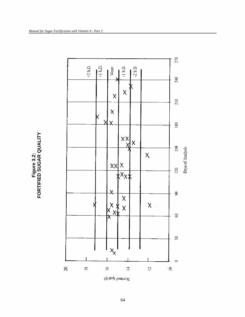
Manual for Sugar Fortificaiton with Vitamin A - Part 3
64
Fig
ure
3.2
:F
OR
TIF
IED
SU
GA
R Q
UA
LIT
Y
CF 'Absorbancereference
Absorbancecalibrated
Analytical Methods for the Control and Evaluation of Sugar Fortification with Vitamin A
65
C. Equipment calibration
1. Spectrophotometer calibration
A simple spectrophotometer can be used in retinol assays if previously calibrated against areference spectrophotometer. To do this, the absorbance range of a retinol solution in ethanol orhexane should be obtained. The spectrum obtained for both spectrophotometers must match. After this,the absorbance at the peak of maximum absorption should be obtained in both spectrophotometersusing a standard retinol solution. The correction factor (CF) for the calibrating spectrophotometer is:
The range of linearity of the spectrophotometer should also be tested using retinol solutions inethanol with concentrations ranging from 0.1 to 5.0 µg/mL (absorbances should be between 0.020 and1.000), which can be obtained by appropriate dilution of a 50 µg/mL retinol solution, for example,0.2:100, 0.5:100, 1:100, 2:100, 5:100, and 10:100. Readings should be made using standard-sizedquartz cuvettes (3 mL).
2. Verification of the efficiency of the irradiation chamber
The efficiency of the irradiation unit should be verified periodically. If it is used daily or often,verification should be done at least once a month and every time a new UV lamp is installed.
A standard solution of retinol or retinyl palmitate is prepared (using the same solvent used forthe sample) with a retinol concentration of 5 µg/mL. The absorbance of this standard solution should beclose to 0.9 at 325 nm. The standard should be prepared freshly every time and protected from directlight at all times.
The unit is calibrated by determining the time required to destruct the retinol in the standardsolution. To do this, two procedures are needed. First, confirm that all the positions in the irradiationchamber’s test tube rack receive the same intensity of irradiation. Second, determine the optimal time ofirradiation.
Manual for Sugar Fortificaiton with Vitamin A - Part 3
66
To check that irradiation is uniform along the test tube rack, all the test tube positions must beoccupied by a tube containing the same retinol solution. After a fixed period of irradiation, for example,15 minutes, the absorbance of each solution is determined. The test tube positions that destroy retinol ata lower magnitude than the norm should be labeled and not used; these are usually the positions ateither end of the rack.
To determine the optimal time of irradiation, the same standard solution of retinol is irradiated intriplicate tubes for specific periods of time, for example, 0, 5, 10, 15, 20, 25, 30, 35, 40, 45, 50, and60 minutes.
The procedures for calibrating the unit are as follows:
a. Turn on the UV lamps at least 30 minutes before use.
b. Place 1 mL of standard solution in all the tubes labeled to indicate the irradiationtime. It is essential that the label is positioned so that it does not obstruct thepath of the UV light.
c. Close the tubes using a stopper made of Teflon or high quality rubber, forexample, vacutainer type. Mark the meniscus level in each tube before placingthe tube in the rack.
d. Place the three controls, that is, those not irradiated (0-time) in the dark untilread.
e. At the end of each irradiation time period, remove the tubes corresponding tothat time period and place them in the dark together with the 0-time controls.
f. Continue the process until all the time periods are completed.
g. Check the level of liquid in each tube to ensure that there was no loss of solventby evaporation. If it is lower than the mark (see step c), add solvent up to thelevel of the mark and mix the contents. (If the tubes are properly closed, therewill be no evaporation.)
h. Adjust the zero of the spectrophotometer, using the solvent used to prepare thestandards. Measure the absorbance of all solutions, including the controls, at325 nm, using the same spectrophotometric cuvettes used for the sampleextracts and under the same conditions.

Analytical Methods for the Control and Evaluation of Sugar Fortification with Vitamin A
35. Irradiation is not prolonged until retinol is completely destroyed for three reasons. First, destruction of retinolfollows an asymptotic logarithmic curve; thus, destruction of the last traces of retinol would take an indefiniteperiod of time that would be impossible to define exactly. Second, the sample extracts may contain substancesthat are not retinol but that absorb at 325 nm and are destroyed by irradiation after a prolonged period of time.Third, interfering new complexes that absorb at 325 nm may be formed during prolonged irradiation.
67
i. Plot a graph of irradiation time (x-axis) against the mean absorbance for eachpoint (y-axis) (see figure 3.3 on the next page).
The optimal irradiation time is the period of time in which at least 95 percent of the retinol isdestroyed. Once determined, this time should be the one used to irradiate the sample extracts. The35
results have to be corrected by the “efficiency factor,” that is, the percent destruction of thepredetermined irradiation time. For example, if it is found that 95 percent of the retinol is destroyed in35 minutes and this amount of time is used to irradiate the sample extracts, the results are multiplied by1.053 (100/95).

Manual for Sugar Fortificaiton with Vitamin A - Part 3
68
Fig
ure
3.3
:R
ET
INO
L A
BS
OR
BA
NC
E B
Y IR
RA
DIA
TIO
N T
IME

69
XVI. SUGGESTED READING
Arroyave, G., C. O. Chichester, H. Flores, J. Glover, L. Mejía, J. A. Olson, K. L. Simpson, and B. A.Underwood. 1982. Biochemical Methodology for the Assessment of Vitamin A Status.Washington, D.C.: IVACG.
Carey, N. and C. Garber. 1989. “Evaluation of Methods.” In L. Kaplan and A. Pesce, eds. ClinicalChemistry: Theory, Correlation, and Analysis. 2nd edition. Chapter 19. pp. 290–310. SaintLouis: Mosby Company.
Conacher, H. 1990. Validation of methods used in crisis situations: task force report. J. Assoc. Off.Anal. Chem. 73(2): 332–34.
Cornbleet, J. and N. Gochman. 1979. Incorrect least squares regression coefficients in method-comparison analysis. Clin. Chem. 25(3): 432–38.
Association of Official Analytical Chemists. 1988. Guidelines for collaborative study procedure tovalidate characteristics of a method of analysis. J. Assoc. Off. Anal. Chem. 71(1): 161–73.
Ruig, W., R. Stephany, and J. Dijkstra. 1989. Criteria for the detection of analytes in test samples. J.Assoc. Off. Anal. Chem. 72(3): 487–90.


36. General Electric F20 T 12/B<13 20-watt lamps have proved good.
71
Appendix 3.1
CONSTRUCTION OF AN ULTRAVIOLETIRRADIATION CHAMBER
The irradiation unit (see appendix figure 3.1a on the next page) is a simple, low-cost unit madeof wood on which two ultraviolet (UV) lamps (350–390 nm emission) are mounted. 36
The base has two parallel lateral walls (point B, appendix figure 3.1a). The lower part of thelateral walls has openings that extend along the length of the walls and expose the test tubes containingthe retinol solutions to the UV light (point C, appendix figure 3.1a). The test tube rack slots into the gapbetween the lateral walls (point D, appendix figure 3.1a), when the base contains small depressions tohold the test tubes in place. The holes on one side of the test tube rack alternate with those on the otherside. The spacing of the holes on the test tube rack is such that the distance between the center of anytwo adjacent tubes is enough to allow the light from both UV lamps to fully reach and expose theextracts to the same amount of UV light. In this model, the distance between the holes is twocentimeters and the diameter of the test tubes is one centimeter.
The UV lamps and their transformers and starters (point E, appendix figure 3.1a) are attachedto the base of the unit. Both lamps are operated simultaneously by a switch (point F, appendix figure3.1a) that is outside of the unit. The system is covered by a wooden top (point A, appendix figure3.1a), with a small hole at the side for the electric cord.
Appendix figure 3.1b gives details of the construction plan.

72
Appendix Figure 3.1a
IRRADIATION CHAMBER

73
Appendix Figure 3.1b
DIMENSIONS OF AN IRRADIATION CHAMBER


75
Appendix 3.2
SUGGESTIONS FOR CLEANING GLASSWARE ANDSPECTROPHOTOMETRIC CUVETTES
The cleanliness of glassware and spectrophotometric cuvettes is essential for obtaining reliableresults. The spectrophotometric cuvettes should be carefully washed and handled to avoid scratchingand staining. The following steps should be followed to ensure that the condition of the glassware andspectrophotometric cuvettes do not affect the results.
I. Glassware
A. Discard any sample residues and reagents in appropriate containers. Organic solventsshould be incinerated or, in some instances, recovered by distillation. Water-soluble orinnocuous substances can be poured down the drain followed by abundant water.Before discarding biological materials, they should be decontaminated by treatment witha sodium hypochlorite solution for 24 hours or autoclaved for 30 minutes at 121°C.
B. Submerge the glassware in tap water. Large pieces of glassware should be brushedwith a 2–3 percent solution of an alkaline detergent (for example, Extran or Teepol).Small glassware, vials, and pipettes should be boiled for 15 minutes in a 5 percentalkaline detergent solution.
C. Rinse with tap water.
D. Soak overnight in a 10 percent nitric acid solution. Stained glassware can be treatedwith a 50 percent solution of nitric acid.
E. Rinse twenty times with tap water and then ten times with distilled water.
F. Dry in an oven at 60°C.
G. Store in a closed cabinet.

76
II. Spectrophotometric cuvettes
A. After each use, rinse the cuvettes with the solvent used to dilute the samples. Then,rinse in a 1:2:1 solution of water/95 percent ethanol/concentrated hydrochloric acid.
B. If the cuvettes are frequently used, store submerged in the above 1:2:1 solution.
C. If the cuvettes are infrequently used, remove from the solution, rinse five times withacetone, and blow the inside dry with air filtered through a dry cotton filter. Never drythe cuvettes using heat or in an oven.
D. Store cuvettes in a closed container.
E. Prior to using the cuvettes, rinse the inside with the solvent used to dissolve the samplesand suck out the remaining solvent using a vacuum.
As a routine practice, especially to detect any problems with a cuvette (dirt, stains, andscratches), it is important to verify that the absorbance reading of all the cuvettes in anyposition, including that for the reference cuvette, is essentially the same when filled with thesame solution. Sometimes a small, but constant, difference may be found among cuvettes. Inthis case, this difference should be corrected for.

77
Appendix 3.3
SUPPLIERS FOR LABORATORY EQUIPMENTAND REAGENTS
I. Suppliers
A. ALDRICH
P.O. Box 355
Milwaukee, WI 53201-9358
Tel: (414) 273-3850
Fax: (414) 273-4979
B. BASF
6700 Ludwigfhafen-Rhein, Ludwingfhafen, Germany
Tel: (049) 621-600
Fax: (049) 622-525
C. Beckman Instruments Inc.
2500 Harbor Boulevard, Fullerton, CA 92634
Tel: (714) 521-3700
Fax: (714) (213) 691 -0841
D. Fisher Scientific
Headquarters
711 Forbes Avenue
Pittsburgh, PA 15219-4785
USA
Tel: (412) 562-8300
International Headquarters, Latin America, Mexico, and Caribbean
50 Fadem Road
Springfield, NJ 07081-3193
USA
Tel: (201) 467-6400

78
Fax: (201) 379-7415
Europe-Mid East/Africa
New Enterprise House
St. Helen's Street
Derby, DE 1 3GY
United Kingdom
Tel: (44) 332-200755
Fax: (44) 332-296546
Asia and Pacific
No. 1 101 Thomson Road
16-05 United Square
Singapore 1130
Tel: (65) 250-9766
Fax: (65) 253-2286
E. Hoffman-La Roche
CH-4002, Basel, Switzerland
Tel: (061) 688-1111
Fax: (061) 691-9600
F. J. T. Baker
222 Red School Lane
Phillipsburg, NJ 08865
USA
Tel (908) 859-2151
Fax: (908) 857-4318
G. Millipore Intertech
P.O. Box 10
Marlborough, MA 01752
USA
Tel: (508) 624-8400
Fax: (508) 624-8630

79
H. National Institute of Standards and Technology (NIST)
Bldg. 202, Room 204
Gaithersburg, MD 20899
Tel: (301) 975-6776
Fax: (301) 948-3730
I. Perkin Elmer Corporation
761 Main Ave., Norwalk, CT 06859-0012
USA
Tel: (203) 762-1000
Fax: (203) 762-6000
Bodenseewerk Perkin-Elmer GmbH
Postfach 10 11 64, D-7770 Uberlingen, Germany
Tel: (075) 51-81-0
Fax: (075) 51 -1612
Perkin-Elmer Ltd.
Post Office Lane, Beaconsfield, Bucks HP9 1 QA
England
Tel: (0494) 676161
Fax: (0494) 679333
J. Sarstedt
P.O. Box 468, Newton, NC 28658-0468
USA
Tel: (704) 465-4000
Fax: (704) 465-4003
K. Sigma Chemical Company
P.O. Box 14508
St. Louis, MO 63178-9916
USA
Tel: (314) 771-5750
Fax: (314) 771-5757

80
L. VARIAN
Varian Associates
Parts and supplies center
220 Humboldt Court
Sunnyvale, CA 94089
USA
Tel: (408) 734-5370
Fax: (408) 744-0261
II. Equipment (examples)
A. Spectrophotometer UV/VIS, VARIAN DMS- 100S or Perkin Elmer Lamda 3BHPLC
B. Varian Chromatography 5500 with UV detector. Includes Autosampler Vista 9090,interphase Varian IIM-A, DS-650 computer, and HP ThinkJet printer
C. Micropak Column Sp-18-5 of 150 x 4 (Dl) mm (Varian 03-912042.42)
D. Test tubes 10 x 75 mm. Kimble KG-33
E. Masked semi-microcell spectrophotometer cuvettes. Beckman, Product 533043
III. Reagents (examples of supliers with catalogue number)
A. Spectrophotometric determination of retinol in premix
2-propanol. Merck Art. 9634
B. Spectrophotometric determination of retinol in fortified sugar
Absolute ethanol. Merck Art. 983.25
Hexane. Sigma Art. H-9379
Sodium hydroxide. Sigma S-0899
C. Colorimetric determination of retinol in fortified sugar
Trichloroacetic acid. Merck No. 1839
Dichloromethane. Merck No. 1593
Cupric sulfate. Fisher Scientific Company Art. No. 493

81
D. Peroxides in oil
Glacial acetic acid. Sigma Art. A-0808
Chloroform, USP. Aldrick 31,998-8
Starch. Merck Art. 1252
Sodium thiosulfate. Merck Art. 6516
Potassium iodide. Merck Art. 5043 Kl
Concentrated hydrochloric acid. Sigma Art. H-7020
Potassium dichromate. Sigma Art. P-6435
E. Spectrophotometric determination of blood and milk retinol by UV destruction
Absolute ethanol. Merck Art. 983.25
Potassium hydroxide. Aldrick Art. 30,656-8
Deorodized kerosene. J.T. Baker Art. P339-00
Xylene (analytical grade). J.T. Baker Art 9490-03
F. HPLC determination of blood and milk retinol
Absolute ethanol. Merck Art. 983.25
2-propanol. Merck Art. 9634
Hexane (HPLC grade). J.T. Baker Art. 9303-3
Methanol (HPLC grade). J.T. Baker Art. 9093
Ethylene dichloride (HPLC grade). Sigma-Aldrich Art. 27,056-3
Methylene dichloride (HPLC grade). Sigma-Aldrich Art. 27,057-1
Retinyl acetate. Sigma Art. R-3513
BHT. Sigma Art. B-1378
Retinol. Sigma Art. R-7632
G. Irradiation chamber
Irradiation lamps. General Electric F20 T 12 BLB