management of class iii malocclusion ...ijchmr.com/uploadfiles/109-112.20190812021552.pdfmanagement...
TRANSCRIPT

Verma M et al. Class III malocclusion management.
109 HECS International Journal of Community Health and Medical Research |Vol. 5|Issue 3| July – September 2019
Harsukh Educational Charitable Society International Journal of Community Health and Medical Research
Journal home page:www.ijchmr.com doi: 10.21276/ijchmr
ISSN E: 2457-0117 ISSN P: 2581-5040 Index Copernicus ICV 2018=62.61
CASE REPORT MANAGEMENT OF CLASS III MALOCCLUSION: TREATMENT DILEMMA- A CASE REPORT Munish Verma1, Sampann Chowdhry2, Bhupinder Singh Bedi3, Sunil Kumar Bhatti4
1Graded specialist orthodontist, 2Graded specialist Endodontist, 3Graded specialist oral and maxilla facial surgery, 4Graded specialist orthodontist, Command military dental center, Western command Chandimandir Cantt, Panchkula, Haryana. ABSTRACT Background: Management of skeletal class III cases has always been a confusing and problematic issue in everyday practice. For this purpose various appliances have been developed for the ease of correction and management of maxillary deficiency. Aim: To minimize the dentoskeletal effects of facemask by class III traction using miniplates in growing patients. Method: This case report comprises of treatment in a deficient maxilla of an 12-year-old boy by using face mask, a bonded maxillary acrylic splint expander and miniplates. Acrylic expander was placed in maxilla for surural disruption. Two miniplates were inserted in the premolar area of the maxilla under local anaesthesia. Facemask with class III traction resulted in favorable correction of malocclusion. Results: The SNA and ANB angles increased by 2� and 4 � respectively. Conclusion: class III traction and miniplates proved to be a successful method for correction of class III malocclusion in this case. Key words: Dental, Malocclusion, Treatment Corresponding author: Dr. Lt Col Munish Verma, Graded specialist orthodontist, Command military dental center, Western command Chandimandir Cantt, Panchkula, Haryana This article may be cited as: Verma M, Chowdhry S, Bedi BS, Bhatti SK. Management of class III malocclusion: treatment dilemma- A case report. HECS Int J Comm Health Med Res 2019; 5(3):109-112.
NTRODUCTION Class III malocclusion can be of dental or skeletal type. Dental class III malocclusion is generally treated with camouflage treatment whereas skeletal class III cases require a more constructed treatment plan. Skeletal Class III malocclusions
are relatively rare and are associated with genetic factors. The etiology may involve a retrognathic maxilla or a prognathic mandible, or both1, 2. It has been found that 65% to 67% of all Class III malocclusions were characterized by maxillary deficiency3. In patients having a deficient maxilla in which the mandible is not markedly affected, treatment proceeds with stimulation and guidance of maxillary growth by orthopaedic forces. Various extraoral appliances, such as facemasks and reverse pull headgears have been used to correct maxillary deficiency 4–6; however, because the force is applied to the teeth, the inevitable mesial migration of the dentition can result in severe anterior crowding7. Furthermore, skeletal effects are often difficult to achieve with this approach. To overcome these disadvantages, many clinicians are using dental implants, mini-plates, and modified fixation screws which provide bone anchorage in orthodontic treatment 8– 10. Mini-screws (mini-implants) are a
more popular choice because of their ease of insertion and removal11, 12. In this case report, two mini-plates were inserted in the anterior part of the maxilla in the canine area and connected to a facemask appliance by use of elastics for correction of maxillary deficiency. DIAGNOSIS AND ETIOLOGY Patient was a 12-year-old boy who came to the hospital with a chief complaint of backwardly placed upper front teeth. He had no medical problems and there were no signs of temporomandibular joint dysfunction. The patient had a skeletal Class III malocclusion with maxillary deficiency whereas the parents had no such features. The facial photographs (figure 1) showed a Class III appearance with a concave profile because of maxillary deficiency. The pretreatment intraoral photographs (figure 2) and dental casts showed Class III molar relationship bilaterally and anterior cross-bite with reverse overjet of 2 mm. Cephalometric analysis (figure 3) (table 1) confirmed the Class III skeletal pattern.
I

Verma M et al. Class III malocclusion management.
110 HECS International Journal of Community Health and Medical Research |Vol. 5|Issue 3| July – September 2019
Figure 1- Pretreatment extraoral photographs
Figure 2- Pretreatment intraoral photographs
Figure 3- Pretreatment cephalometric radiograph
Figure 4- Miniplate insertion and bonded maxillary expander
Table 1- Pretreatment cephalometric values
Figure 5- Facemask therapy
Figure 6: Post-treatment extraoral photographs TREATMENT OBJECTIVES The objectives we want to achieve for this patient were:
1. Improvement of profile 2. Improvement of smile esthetics 3. Achieving Class I molar and canine relationship
bilaterally 4. Correction of skeletal relationship
Ø SNA 770
Ø SNB 790
Ø ANB -20
Ø UI-NA 290
(07)
Ø LI-NB 300
(10)
Ø GoGn –SN 370
Ø FMA 360
Ø IMPA 920
Ø A-N perpendicular -9 Ø Co-Gn 121mm Ø Co-A (94) 89mm
Ø Nasolabial angle 890
Ø ANS:PNS-Go:Pog 1:1.6 Ø SN:ANS-PNS 1:0.6 Ø SN:Go-Pog 1:1.6
Verma M et al. Class III malocclusion management.
111 HECS International Journal of Community Health and Medical Research |Vol. 5|Issue 3| July – September 2019
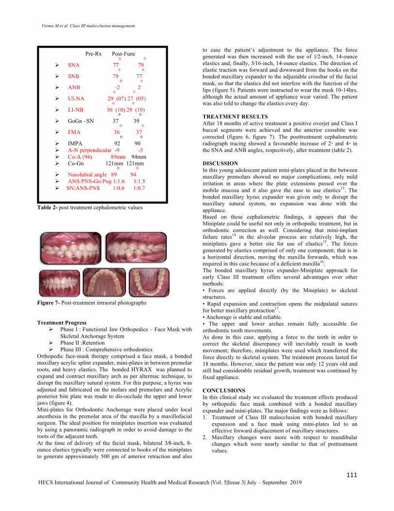
Table 2- post treatment cephalometric values
Figure 7- Post-treatment intraoral photographs Treatment Progress
Ø Phase I : Functional Jaw Orthopedics – Face Mask with Skeletal Anchorage System
Ø Phase II :Retention Ø Phase III : Comprehensive orthodontics
Orthopedic face-mask therapy comprised a face mask, a bonded maxillary acrylic splint expander, mini-plates in between premolar roots, and heavy elastics. The bonded HYRAX was planned to expand and contract maxillary arch as per altermac technique, to disrupt the maxillary sutural system. For this purpose, a hyrax was adjusted and fabricated on the molars and premolars and Acrylic posterior bite plate was made to dis-occlude the upper and lower jaws (figure 4). Mini-plates for Orthodontic Anchorage were placed under local anesthesia in the premolar area of the maxilla by a maxillofacial surgeon. The ideal position for miniplates insertion was evaluated by using a panoramic radiograph in order to avoid damage to the roots of the adjacent teeth. At the time of delivery of the facial mask, bilateral 3⁄8-inch, 8-ounce elastics typically were connected to hooks of the miniplates to generate approximately 500 gm of anterior retraction and also
to ease the patient’s adjustment to the appliance. The force generated was then increased with the use of 1⁄2-inch, 14-ounce elastics and, finally, 5⁄16-inch, 14-ounce elastics. The direction of elastic traction was forward and downward from the hooks on the bonded maxillary expander to the adjustable crossbar of the facial mask, so that the elastics did not interfere with the function of the lips (figure 5). Patients were instructed to wear the mask 10-14hrs, although the actual amount of appliance wear varied. The patient was also told to change the elastics every day. TREATMENT RESULTS After 18 months of active treatment a positive overjet and Class I buccal segments were achieved and the anterior crossbite was corrected (figure 6, figure 7). The posttreatment cephalometric radiograph tracing showed a favourable increase of 2◦ and 4◦ in the SNA and ANB angles, respectively, after treatment (table 2). DISCUSSION In this young adolescent patient mini-plates placed in the between maxillary premolars showed no major complications; only mild irritation in areas where the plate extensions passed over the mobile mucosa and it also gave the ease to use elastics13. The bonded maxillary hyrax expander was given only to disrupt the maxillary sutural system, no expansion was done with the appliance. Based on these cephalometric findings, it appears that the Miniplate could be useful not only in orthopedic treatment, but in orthodontic correction as well. Considering that mini-implant failure rates14 in the alveolar process are relatively high, the miniplates gave a better site for use of elastics15. The forces generated by elastics comprised of only one component; that is in a horizontal direction, moving the maxilla forwards, which was required in this case because of a deficient maxilla16. The bonded maxillary hyrax expander-Miniplate approach for early Class III treatment offers several advantages over other methods: • Forces are applied directly (by the Miniplate) to skeletal structures. • Rapid expansion and contraction opens the midpalatal sutures for better maxillary protraction17. • Anchorage is stable and reliable. • The upper and lower arches remain fully accessible for orthodontic tooth movements. As done in this case, applying a force to the teeth in order to correct the skeletal discrepancy will inevitably result in tooth movement; therefore, miniplates were used which transferred the force directly to skeletal system. The treatment process lasted for 18 months. However, since the patient was only 12 years old and still had considerable residual growth, treatment was continued by fixed appliance. CONCLUSIONS In this clinical study we evaluated the treatment effects produced by orthopedic face mask combined with a bonded maxillary expander and mini-plates. The major findings were as follows: 1. Treatment of Class III malocclusion with bonded maxillary
expansion and a face mask using mini-plates led to an effective forward displacement of maxillary structures.
2. Maxillary changes were more with respect to mandibular changes which were nearly similar to that of pretreatment values.
Pre-Rx Post-Func
Ø SNA 770
790
Ø SNB 790
770
Ø ANB -20
20
Ø UI-NA 290
(07) 270
(05)
Ø LI-NB 300
(10) 290
(10)
Ø GoGn –SN 370
390
Ø FMA 360
370
Ø IMPA 920
900
Ø A-N perpendicular -9 -3 Ø Co-A (94) 89mm 94mm Ø Co-Gn 121mm 121mm
Ø Nasolabial angle 890
940
Ø ANS:PNS-Go:Pog 1:1.6 1:1.5 Ø SN:ANS-PNS 1:0.6 1:0.7
Verma M et al. Class III malocclusion management.
112 HECS International Journal of Community Health and Medical Research |Vol. 5|Issue 3| July – September 2019
References: 1. Litton, S.F.; Ackermann, L.V.; Isaacson, R.J.; and Shapiro, B.L.: A
genetic study of Class 3 malocclusion, Am. J. Orthod. 58:565-577, 1970.
2. 2. Proffit, W.R.; Fields, H.W. Jr.; and Moray, L.J.: Prevalence of malocclusion and orthodontic treatment need in the United States: Estimates from the NHANES III survey, Int. J. Adult Orthod. Orthog. Surg. 13:97-106, 1998.
3. E. E. Ellis and J. A. McNamara Jr., “Components of adults Class III open bit malocclusion,” American Journal of Orthodontics and Dentofacial Orthopedics, vol. 85, pp. 277– 290, 1984.
4. M. Hegmann and A. K. R¨uther, “The Grummons face mask as an early treatment modality within a Class III therapy concept,” Journal of Orofacial Orthopedics, vol. 64, no. 6, pp. 450–456, 2003.
5. Z. Altug and A. D. Arslan, “Skeletal and dental effects of a mini maxillary protraction appliance,” Angle Orthodontist, vol. 76, no. 3, pp. 360–368, 2006.
6. H.W. Fields andW.R. Proffit, “Treatment of skeletal problems in children,” in Contemporary Orthodontics, W. R. Proffit, H. W. Fields, and D. M. Sarver, Eds., pp. 495–511, Mosby, St. Louis,Mo, USA, 4th edition, 2007.
7. Williams, M.D.; Sarver, D.M.; Sadowsky, P.L.; and Bradley, E.: Combined rapid maxillary expansion and protraction facemask in the treatment of Class III malocclusions in growing children: A prospective long-term study, Semin. Orthod. 3:265-274, 1997.
8. W. E. Roberts, C. L. Nelson, and C. J. Goodacre, “Rigid implant anchorage to close a mandibular first molar extraction site,” Journal of Clinical Orthodontics, vol. 28, no. 12, pp. 693– 704, 1994.
9. S. Miyawaki, I. Koyama, M. Inoue, K. Mishima, T. Sugahara, and T. Takano-Yamamoto, “Factors associated with the stability of titanium screws placed in the posterior region for orthodontic anchorage,” American Journal of Orthodontics and Dentofacial Orthopedics, vol. 124, no. 4, pp. 373–378, 2003.
10. S. Kuroda, Y. Sugawara, T. Deguchi, H. M. Kyung, and T. Takano-Yamamoto, “Clinical use of miniscrew implants as orthodontic anchorage: success rates and postoperative discomfort,” American Journal of Orthodontics and Dentofacial Orthopedics, vol. 131, no. 1, pp. 9–15, 2007.
11. A. Costa, M. Raffainl, and B. Melsen, “Miniscrews asorthodontic anchorage: a preliminary report,” The International Journal of Adult Orthodontics and Orthognathic Surgery,vol. 13, no. 3, pp. 201–209, 1998.
12. S. Kuroda, A. Katayama, and T. Takano-Yamamoto, “Severe anterior open-bite case treated using titanium screw anchorage,” Angle Orthodontist, vol. 74, no. 4, pp. 558–567, 2004.
13. Wilmes, B. and Drescher, D.: A miniscrew system with inter-changeable abutments, J. Clin. Orthod. 42:574-580, 2008.
14. A. Jamilian and R. Showkatbakhsh, “Treatment of maxillary deficiency by miniscrew implants—a case report,” Journal of orthodontics, vol. 37, no. 1, pp. 56–61, 2010.
15. H. J. De Clerck, M. A. Cornelis, L. H. Cevidanes, G. C.Heymann, and C. J. F. Tulloch, “Orthopedic traction of the maxilla with miniplates: a new perspective for treatment of midface deficiency,” Journal of Oral andMaxillofacial Surgery, vol. 67, no. 10, pp. 2123–2129, 2009.
16. Baccetti T, McGill, Lorenzo Franchi, James A. McNamara Jr., and Isabella Tollaro. Skeletal effects of early treatment of Class III malocclusion with maxillary expansion and face-mask therapy. Am J Orthod Dentofacial Orthop 1998;113:333-43
17. Wilmes, Nienkemper, Ludwig, Kau, and Drescher. Early Class III Treatment with a Hybrid Hyrax-Mentoplate Combination. JCO 2011 Volume XLV Number 1; 1-7