mammalian n-glycan branching protects against innateimmune ...lwang/bcmb8020/n-glycan-paper3.pdf ·...
TRANSCRIPT
Immunity
Article
Mammalian N-Glycan Branching Protects againstInnate Immune Self-Recognition and Inflammationin Autoimmune Disease PathogenesisRyan S. Green,1,2 Erica L. Stone,1,2,3 Mari Tenno,1,2,3 Eero Lehtonen,1 Marilyn G. Farquhar,1
and Jamey D. Marth1,2,*1Department of Cellular and Molecular Medicine2Howard Hughes Medical InstituteUniversity of California, San Diego, La Jolla, CA 92093, USA3 These authors contributed equally to this work.
*Correspondence: [email protected]
DOI 10.1016/j.immuni.2007.06.008
SUMMARY
Autoimmune diseases are prevalent and oftenlife-threatening syndromes, yet the pathogenictriggers and mechanisms involved remainmostly unresolved. Protein asparagine linked-(N-) glycosylation produces glycan structuresthat substantially differ among the extracellu-lar compartments of evolutionarily divergentorganisms. Alpha-mannosidase-II (aM-II) defi-ciency diminishes complex-type N-glycanbranching in vertebrates and induces an auto-immune disease in mice similar to humansystemic lupus erythematosus. We found thatdisease pathogenesis provoking glomerulone-phritis and kidney failure was nonhematopoieticin origin, independent of complement C3 andthe adaptive immune system, mitigated by intra-venous administration of immunoglobulin-G,and linked to chronic activation of the innateimmune system. N-glycans produced in aM-IIdeficiency bear immune-stimulatory mannose-dependent ligands for innate immune lectin re-ceptors, disrupting the phylogenic basis of thisglycomic recognition mechanism. Thus, mam-malian N-glycan branching safeguards againstthe formation of an endogenous immunologicsignal of nonself that can provoke a sterileinflammatory response in the pathogenesis ofautoimmune disease.
INTRODUCTION
Autoimmune diseases arise when immune stimuli override
mechanisms of self-tolerance and are often diagnosed by
elevations in autoantibody titers. Pathogenesis is gener-
ally attributed to the effector functions of the adaptive im-
mune system. Systemic lupus erythematosus (SLE) is an
autoimmune syndrome in which autoantibodies to nuclear
antigens and immune complex formation are induced with
308 Immunity 27, 308–320, August 2007 ª2007 Elsevier Inc.
a high prevalence of kidney disease (Jorgensen et al.,
2004; Lauwerys and Wakeland, 2005). The etiology of
the SLE-like syndrome that develops from the absence
of alpha-mannosidase-II (aM-II) is puzzling (Chui et al.,
2001). aM-II is conserved among mammals, and its
deficiency in mice alters protein N-glycosylation in some
cell types by blocking the formation of complex-type
N-glycans that normally constitute the predominant N-
glycan branching structure on the vertebrate cell surface
(Chui et al., 1997).
Incomplete N-glycan branching in the Golgi apparatus
results from aM-II deficiency, leading to the appearance
of hybrid-type N-glycan structures at the cell surface.
As mice age without aM-II function, signs of SLE invari-
ably appear with an increase in anti-nuclear antibody
(ANA) titers, a hematologic abnormality characterized as
dyserythropoietic anemia, and glomerular deposition of
immunoglobulins and complement component C3. Glo-
merulonephritis is the major pathologic feature in these
animals leading to sclerosis, renal dysfunction, and kid-
ney failure in a syndrome indicative of lupus nephritis
(Chui et al., 2001). Remarkably, diminished N-glycan
branching is observed among only a subset of cell line-
ages in aM-II deficiency resulting from the presence of an-
other mannosidase, termed aM-IIx, that can compensate
to promote complex-type N-glycan branching (Chui et al.,
1997; Akama et al., 2006). Although this compensation
occurs among lymphoid and myeloid cells, it is absent
from the erythroid lineage and variable in efficacy among
multiple cell types (Chui et al., 1997, 2001; Akama et al.,
2006). For example, T and B lymphocytes that lack aM-
II retain high amounts of complex-type N-glycans. Not
surprisingly, lymphoid cells develop normally in aM-II de-
ficiency and respond to immunologic stimulation without
alterations (Chui et al., 2001). These findings have sug-
gested that aM-II-deficient autoimmune disease may re-
flect a distinct etiology encompassing a pathogenic
mechanism that involves endogenous epitope modifica-
tion (Wakeland et al., 2001).
We have used molecular and cellular approaches that
discriminate among the roles of cell lineages to determine
the origin and mechanism of the SLE-like autoimmune dis-
ease resulting from aM-II deficiency. Our findings show

Immunity
N-Glycosylation in Autoimmune Disease
a pathogenic trigger among nonhematopoietic cell types
and glycoproteins that undergo altered protein N-glycan
branching, indicating a sterile signal of infection and in-
flammation composed of extracellular mannose-depen-
dent N-glycan ligands of innate immune lectin receptors.
The adaptive immune system does not appear to play
a pathogenic role and instead moderates the disease
course, attenuating the inflammatory route that encom-
passes macrophage recruitment, glomerulonephritis,
and kidney failure. Pathogenesis in aM-II deficiency arises
by disrupting a mechanism of vertebrate nonself discrim-
ination by which the innate immune system normally dis-
tinguishes the glycomes of lower eukaryotic and prokary-
otic organisms.
RESULTS
Normal Kidney Morphogenesis in aM-II DeficiencyTissue and organ damage in SLE is often focused and
most severe in the kidney. To investigate the pathogenic
basis of this autoimmune disease syndrome in aM-II defi-
ciency, we first studied kidney development and early
postnatal function. Among kidneys of animals analyzed
at birth, we observed unaltered glomerular organization
at the electron microscope level with normal glomerular
basement membrane, epithelial foot processes, and filtra-
tion slits (Figure S1 in the Supplemental Data available on-
line). In addition, kidney function in mice lacking aM-II was
unimpaired during the first 6 months of life as determined
by urinalysis measurements of hematuria and proteinuria.
These findings indicate that the development of glomeru-
lonephritis between 6 and 9 months of age is not associ-
ated with an ontogenic or functional disruption of the kid-
ney per se.
Nonhematopoietic Origin of Autoimmune Diseasein aM-II DeficiencyAutoimmune diseases may be of hematopoietic origin
and resolved to abnormalities in lymphoid cell function,
as evidenced in numerous bone-marrow transplantation
studies (Ikehara, 1998). We subjected 8-week-old wild-
type and aM-II null littermates to irradiation levels that
were invariably lethal without hematopoietic reconstitu-
tion by bone-marrow grafts. At this age, disease pathol-
ogy is not yet present and all mice are healthy. Syngeneic
bone-marrow recipients that were fully reconstituted by
donor-derived hematopoietic cells by 8–10 weeks and
which attained normal peripheral leukocyte numbers in
circulation were further studied up to 9 months after
transplantation. Recipients analyzed remained donor de-
rived throughout the course of the study as determined
by hematopoietic cell-surface N-glycan markers and ge-
nomic DNA analyses (Figure 1A and data not shown). No
complications reflecting graft-versus-host disease were
noted in the experimental populations. Lymphoid lineage
development, abundance, and activation responses
were unaffected, as in systemic aM-II-deficient mice,
because of the presence of the aM-IIx isozyme (Chui
et al., 2001). Remarkably, elevated autoantibody titers
to cellular and nuclear antigens, glomerular deposition
of immunoglobulin-G and complement C3, as well as
kidney inflammation and dysfunction were linked to
aM-II-deficient recipients (Figures 1B–1F). Autoimmune
disease pathogenesis was neither provoked by aM-
II-deficient bone-marrow-derived cells nor attenuated
by wild-type marrow grafts. Instead, the pathogenic trig-
ger that leads to the induction of autoantibodies and
kidney disease in aM-II deficiency resides among nonhe-
matopoietic cell types.
Among disease markers, only the dyserythropoietic
anemia was linked to aM-II-deficient bone-marrow-
derived and hematopoietic lineages (Figure 1G). This is
consistent with the erythroid dependence on aM-II, a cell
lineage wherein no compensation by the aM-IIx isozyme
occurs, resulting in hybrid-type N-glycans at the cell sur-
face and increased turnover of the anisotropic population
of circulating erythrocytes (Chui et al., 1997). It is evident
from these studies that this erythroid defect does not con-
tribute to the elevation of autoantibody titers or to kidney
disease.
Attenuation of Disease by the AdaptiveImmune SystemThe adaptive immune system fails to develop without
recombinase-activating gene-1 (RAG-1); mature lympho-
cytes, antibodies, and immune complexes do not exist in
such animals (Mombaerts et al., 1992). In the context of
aM-II deficiency, it was possible to determine the contribu-
tion of the adaptive immune system to disease pathogen-
esis with a focus on the nephritic component. Unexpect-
edly, mice lacking both aM-II and RAG-1 were more
severely affected with loss of weight, alopecia, and exac-
erbation of kidney disease, as compared with littermates
lacking aM-II or RAG-1 alone (Figure S2). Glomerulone-
phritis was intensified and a high degree of nephron loss
occurred concurrent with hematuria and proteinuria
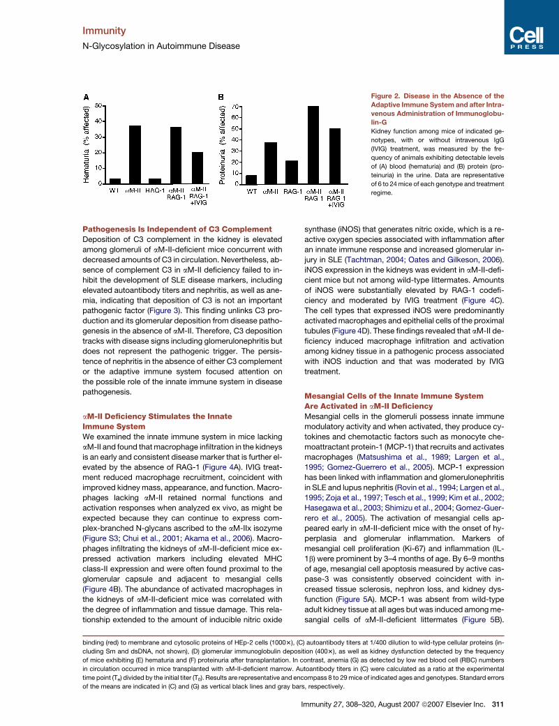
(Figures 2A and 2B).
Molecules produced by lymphocytes that can modulate
autoimmune disease include immunoglobulin-G (IgG),
which is a ligand for Fc receptors expressed on various
cell types (Nimmerjahn and Ravetch, 2006). Fc receptor
function can decrease immune responses. For example,
FcgRIIb attenuates immune activation and mice lacking
this Fc receptor spontaneously develop autoimmune dis-
ease (Takai et al., 1996). We suspected that the absence
of inhibitory signaling through Fc receptors among mice
lacking RAG-1 might accelerate inflammation and nephri-
tis in the aM-II null background. Cohorts of animals were
given multiple intravenous injections of normal mouse
IgG (IVIG) for a period of 4–6 months while disease signs
were followed. In the absence of both aM-II and RAG-1,
IVIG therapy diminished disease signs coincident with im-
proved kidney function (Figure 2). The adaptive immune
system therefore was not the source of the pathogenic
trigger, nor was it required for disease progression, but
instead appeared to moderate inflammation and
glomerulonephritis.
Immunity 27, 308–320, August 2007 ª2007 Elsevier Inc. 309

Immunity
N-Glycosylation in Autoimmune Disease
Figure 1. Nonhematopoietic Origin of Autoimmune Disease in aM-II Deficiency
(A) Bone-marrow transplantation to reconstitute the hematopoietic system was accomplished among 2-month-old recipients. At indicated times after
transplantation, donor and recipient genotypes were detected by erythrocyte cell-surface binding of the erythroagglutinin (E-PHA) that binds complex
N-glycan structures dependent upon aM-II function. Results were similar to hematopoietic cell DNA genotyping studies (not shown).
(B–G) Autoimmune markers were analyzed routinely for 6–9 months after transplantation, and disease occurred only in recipients that lacked aM-II
function. These findings encompassed (B) anti-nuclear antibody (ANA) titers in sera diluted 1/200 with IgG binding (blue) to nuclear epitopes and IgM
310 Immunity 27, 308–320, August 2007 ª2007 Elsevier Inc.
Immunity
N-Glycosylation in Autoimmune Disease
Figure 2. Disease in the Absence of the
Adaptive Immune System and after Intra-
venous Administration of Immunoglobu-
lin-G
Kidney function among mice of indicated ge-
notypes, with or without intravenous IgG
(IVIG) treatment, was measured by the fre-
quency of animals exhibiting detectable levels
of (A) blood (hematuria) and (B) protein (pro-
teinuria) in the urine. Data are representative
of 6 to 24 mice of each genotype and treatment
regime.
Pathogenesis Is Independent of C3 ComplementDeposition of C3 complement in the kidney is elevated
among glomeruli of aM-II-deficient mice concurrent with
decreased amounts of C3 in circulation. Nevertheless, ab-
sence of complement C3 in aM-II deficiency failed to in-
hibit the development of SLE disease markers, including
elevated autoantibody titers and nephritis, as well as ane-
mia, indicating that deposition of C3 is not an important
pathogenic factor (Figure 3). This finding unlinks C3 pro-
duction and its glomerular deposition from disease patho-
genesis in the absence of aM-II. Therefore, C3 deposition
tracks with disease signs including glomerulonephritis but
does not represent the pathogenic trigger. The persis-
tence of nephritis in the absence of either C3 complement
or the adaptive immune system focused attention on
the possible role of the innate immune system in disease
pathogenesis.
aM-II Deficiency Stimulates the InnateImmune SystemWe examined the innate immune system in mice lacking
aM-II and found that macrophage infiltration in the kidneys
is an early and consistent disease marker that is further el-
evated by the absence of RAG-1 (Figure 4A). IVIG treat-
ment reduced macrophage recruitment, coincident with
improved kidney mass, appearance, and function. Macro-
phages lacking aM-II retained normal functions and
activation responses when analyzed ex vivo, as might be
expected because they can continue to express com-
plex-branched N-glycans ascribed to the aM-IIx isozyme
(Figure S3; Chui et al., 2001; Akama et al., 2006). Macro-
phages infiltrating the kidneys of aM-II-deficient mice ex-
pressed activation markers including elevated MHC
class-II expression and were often found proximal to the
glomerular capsule and adjacent to mesangial cells
(Figure 4B). The abundance of activated macrophages in
the kidneys of aM-II-deficient mice was correlated with
the degree of inflammation and tissue damage. This rela-
tionship extended to the amount of inducible nitric oxide
synthase (iNOS) that generates nitric oxide, which is a re-
active oxygen species associated with inflammation after
an innate immune response and increased glomerular in-
jury in SLE (Tachtman, 2004; Oates and Gilkeson, 2006).
iNOS expression in the kidneys was evident in aM-II-defi-
cient mice but not among wild-type littermates. Amounts
of iNOS were substantially elevated by RAG-1 codefi-
ciency and moderated by IVIG treatment (Figure 4C).
The cell types that expressed iNOS were predominantly
activated macrophages and epithelial cells of the proximal
tubules (Figure 4D). These findings revealed that aM-II de-
ficiency induced macrophage infiltration and activation
among kidney tissue in a pathogenic process associated
with iNOS induction and that was moderated by IVIG
treatment.
Mesangial Cells of the Innate Immune SystemAre Activated in aM-II DeficiencyMesangial cells in the glomeruli possess innate immune
modulatory activity and when activated, they produce cy-
tokines and chemotactic factors such as monocyte che-
moattractant protein-1 (MCP-1) that recruits and activates
macrophages (Matsushima et al., 1989; Largen et al.,
1995; Gomez-Guerrero et al., 2005). MCP-1 expression
has been linked with inflammation and glomerulonephritis
in SLE and lupus nephritis (Rovin et al., 1994; Largen et al.,
1995; Zoja et al., 1997; Tesch et al., 1999; Kim et al., 2002;
Hasegawa et al., 2003; Shimizu et al., 2004; Gomez-Guer-
rero et al., 2005). The activation of mesangial cells ap-
peared early in aM-II-deficient mice with the onset of hy-
perplasia and glomerular inflammation. Markers of
mesangial cell proliferation (Ki-67) and inflammation (IL-
1b) were prominent by 3–4 months of age. By 6–9 months
of age, mesangial cell apoptosis measured by active cas-
pase-3 was consistently observed coincident with in-
creased tissue sclerosis, nephron loss, and kidney dys-
function (Figure 5A). MCP-1 was absent from wild-type
adult kidney tissue at all ages but was induced among me-
sangial cells of aM-II-deficient littermates (Figure 5B).
binding (red) to membrane and cytosolic proteins of HEp-2 cells (10003), (C) autoantibody titers at 1/400 dilution to wild-type cellular proteins (in-
cluding Sm and dsDNA, not shown), (D) glomerular immunoglobulin deposition (4003), as well as kidney dysfunction detected by the frequency
of mice exhibiting (E) hematuria and (F) proteinuria after transplantation. In contrast, anemia (G) as detected by low red blood cell (RBC) numbers
in circulation occurred in mice transplanted with aM-II-deficient marrow. Autoantibody titers in (C) were calculated as a ratio at the experimental
time point (Te) divided by the initial titer (T0). Results are representative and encompass 8 to 29 mice of indicated ages and genotypes. Standard errors
of the means are indicated in (C) and (G) as vertical black lines and gray bars, respectively.
Immunity 27, 308–320, August 2007 ª2007 Elsevier Inc. 311

Immunity
N-Glycosylation in Autoimmune Disease
Figure 3. Pathogenesis in the Absence of Complement C3
Among mice lacking aM-II, complement C3, both aM-II and C3, as well as wild-type littermates, disease signs were measured at 6–9 months of age
including glomerular immunoglobulin deposition (4003), the presence of anti-nuclear antibodies (ANA) (4003), reduced RBC numbers (anemia), and
kidney dysfunction assessed by frequencies of hematuria and proteinuria. Analyses depicted are representative of results with six or more littermate
pairs of indicated genotypes. Standard errors of the means are indicated as gray bars in the RBC analysis.
Glomerular expression of MCP-1 was moderated by IVIG
treatment, as was the abundance of activated macro-
phages found adjacent to mesangial cells and the glomer-
ular capsule (Figure 5C). These studies revealed that the
activation of mesangial cells in the absence of aM-II led
to the production of proinflammatory proteins that are
known pathogenic factors in the etiology of SLE and kid-
ney disease.
Innate Immune Lectin Receptor Modulationand Proinflammatory Ligand FormationCells of the innate immune system, including mesangial
cells and macrophages, express pattern-recognition re-
ceptors including lectins that bind to mannose-enriched
glycan structures typical of microbial and pathogen cell
surfaces (Barton and Medzhitov, 2003; McGreal et al.,
2004). For example, the macrophage mannose receptor
(MMR) and the mannose-binding lectins (MBL-A and
MBL-C in the mouse) bind to glycan structures bearing
312 Immunity 27, 308–320, August 2007 ª2007 Elsevier Inc.
mannose linkages typical of lower eukaryotic and micro-
bial glycans (Sharon, 1987; Ezekowitz et al., 1990; Sastry
et al., 1995; Hansen et al., 2000; Gordon, 2002; McGreal
et al., 2004). Mammalian lectins of the innate immune sys-
tem therefore appear to distinguish evolutionarily dispa-
rate organisms based in part upon unique differences in
mannose linkages expressed among cellular glycomes.
Mesangial cells are exposed to a variety of stimuli that
exist in vascular circulation and which enter the kidney
glomerulus during normal filtration processes. Although
macrophages are recruited and activated by MCP-1,
macrophages were not directly stimulated by hybrid N-
glycans produced in the absence of aM-II (Figure S3).
The source of the mesangial cell activation stimulus was
investigated among N-glycans in circulation. Serum from
wild-type mice induced some MCP-1 production in iso-
lated cultures of glomeruli, whereas the addition of the
yeast cell wall constituent mannan induced a significantly
greater response. MCP-1 production was also markedly

Immunity
N-Glycosylation in Autoimmune Disease
Figure 4. Innate Immune Activation and Kidney Inflammation
(A) Macrophage recruitment was detected by means of anti-CD68 antibody, and fluorescent signals were quantified among multiple kidney sections.
Results were plotted from five 8-month-old mice of indicated genotypes.
(B) Expression of MHC class II among CD68+ macrophages in the kidneys of aM-II null mice (top, fluorescent colocalization in yellow, effective mag-
nification 20003). RCA-1 (ricinis communis agglutinin-1) lectin binding in the glomerulus marks mesangial cells adjacent to CD68+ macrophages (bot-
tom, magnification 2003).
(C) iNOS protein expression was measured by fluorescence in kidney sections from mice of the indicated genotypes.
(D) iNOS expression was analyzed among various cell types including kidney proximal tubular epithelial cells (nonglomerular RCA-1+ binding, top),
activated macrophages associated with the glomerular capsule (middle), and endothelial cells via the MECA-32 marker (bottom). Data are represen-
tative of three results with more littermate pairs; means and standard deviations are indicated in (A) and (C).
increased in cultures of isolated glomeruli treated with se-
rum from aM-II-deficient mice. Moreover, this response,
like that of mannan, was inhibited in the presence of the
mannose analog and mannose lectin binding inhibitor al-
pha-methylmannoside (aMM) (Figure 6A). These findings
indicated that hybrid N-glycans present in the sera of
aM-II-deficient mice induced mesangial cell activation
and proinflammatory cytokine production by a man-
nose-dependent binding mechanism.
Among innate immune lectins that exhibit mannose-
binding activity, the expression of the MMR, MBL-A, and
MBL-C was analyzed. In aM-II-deficient mice, we ob-
served that MMR protein expression was induced on me-
sangial cells in the kidney and was present among a pro-
portion of intercalating macrophages (Figure 6B and data
not shown). In studies of MBL protein abundance, we
noted reduced serum concentrations of both MBL-A and
MBL-C that correlated with their increased deposition in
the kidney of aM-II-deficient mice (Figures 6C and 6D).
The modulation of mannose-binding lectin expression
observed in aM-II deficiency implicated the presence of
endogenous glycan ligands.
By using recombinant mammalian MMR and MBL
chimeras, mannose-dependent ligands of these lectins
were detected in the sera of aM-II-deficient mice
(Figure 6E). Multiple glycoproteins exhibited ligands that
were recognized by both lectins, and this binding was
competed by aMM. The presence of endogenous ligands
among various cell and tissue types was further investi-
gated and revealed accumulation of mannose-dependent
MMR ligands primarily among kidney tissue and mesangial
cells within the glomerular capsule, but not among various
other organs and cell types (Figure 6F). Ligands of MBL-A
and MBL-C lectins were also present among glycoproteins
Immunity 27, 308–320, August 2007 ª2007 Elsevier Inc. 313

Immunity
N-Glycosylation in Autoimmune Disease
Figure 5. Mesangial Cell Activation and Apoptosis
(A) Detection of Ki-67, IL-1b, and activated caspase-3 proteins among mesangial cells in the glomeruli of kidney sections from 3- to 6-month-old mice
of indicated genotypes.
(B) Expression of monocyte chemoattractant protein-1 (MCP-1) in mesangial cells at 3–6 months of age.
(C) MCP-1 expression (rhodamine, red) and macrophage recruitment (FITC, green) in the absence or presence of previous IVIG treatment. Magnifi-
cations are 4003 unless indicated. Data are representative of results obtained with three or more littermates of the indicated ages and genotypes.
in the kidneys of aM-II-deficient mice as well as among
glycoproteins expressed on the erythrocyte cell surface
(not shown). In further studies, inherited deficiency of the
MBL-A lectin with the coabsence of aM-II continued to
induce autoimmune disease (Figure S4; and data not
shown). The absence of disease modulation by a single
lectin deficiency may represent cell-type-specific func-
tions and compensation involving a growing list of man-
nose-binding lectins expressed among cells of the innate
immune system including Dec-205, Endo180, DC-SIGN,
Langerin, Dectin-2, and BDCA-2. Our findings have shown
that altered N-glycosylation because of loss of aM-II activ-
ity led to the production of proinflammatory mannose-
dependent glycan ligands for multiple innate immune lectin
receptors. These endogenous ligands have structural
similarity with glycan linkages on the cell surface of lower
eukaryotes and microbes (Figure S5).
DISCUSSION
Protein N-glycosylation is altered in aM-II deficiency re-
sulting in the formation of hybrid-type N-glycans bearing
mannose-dependent ligands of innate immune lectin re-
ceptors that induce inflammation. Endogenous ligands
of innate immune lectins do not normally accumulate
among extracellular compartments of vertebrates, and
those produced in the absence of aM-II appear to act as
a pathogenic trigger of nonhematopoietic origin consis-
tent with the etiologic features of the resulting SLE-like
autoimmune disease syndrome. We found that disease
progression tracked with chronic stimulation of the innate
314 Immunity 27, 308–320, August 2007 ª2007 Elsevier Inc.
immune system involving mesangial cell activation and
macrophage recruitment in a sterile milieu lacking an in-
fectious and causative pathogen. Inflammation and cell
death in the kidney was accompanied by markers of nitric
oxide production and increased antigen presentation, yet
neither elevated autoantibody titers nor complement C3
were factors in disease progression, and neither appeared
to play a substantial pathogenic role.
The increased severity of kidney disease among ani-
mals lacking both aM-II and RAG-1 indicated an etiology
independent of peptide antigen-presentation mecha-
nisms and the adaptive immune system. On balance, the
adaptive immune system provides a moderating effect in
disease progression. This moderation does not appear
to reflect alterations in lymphoid cell subpopulations in-
cluding regulatory T cells, which were similar between mu-
tant and wild-type genotypes (data not shown). Among
genetic lesions that alter the immune system and induce
kidney disease, a lymphocyte-independent mechanism
of pathogenesis has been similarly detected by loss of
RAG-1 function among mice lacking the Fyn tyrosine ki-
nase (Yu et al., 2001). Fyn deficiency accelerated autoim-
mune disease symptoms induced by the absence of the
Lyn tyrosine kinase, appearing to reflect a kidney-intrinsic
role of Fyn. SLE-like disease signs induced by aM-II defi-
ciency followed normal kidney development and early
postnatal function and may be augmented by immuno-
logic disease processes similarly engendered by other ge-
netic lesions.
Administration of IVIG in aM-II deficiency reduced
macrophage recruitment and disease severity evoked by
RAG-1 codeficiency, providing the moderating effect

Immunity
N-Glycosylation in Autoimmune Disease
ascribed to the adaptive immune system. Autoantibodies
detected in aM-II deficiency do not exhibit N-glycan bind-
ing (Chui et al., 2001), implying that conserved regions
within the IgG molecule participate in disease attenuation.
IVIG is a therapeutic treatment in human diseases of
inflammation and autoimmunity including myasthenia
gravis, Guillain-Barre syndrome, graft-versus-host dis-
ease, Kawasaki’s syndrome, as well as SLE, in which it
may operate by promoting inhibitory signaling of Fc
receptors (Ott et al., 2001; Kazatchkine and Kaveri, 2001;
Sherer and Shoenfeld, 2006; Park-Min et al., 2007). It is
possible that loss of FcgRIIb in the aM-II-deficient back-
ground may also elevate innate immune inflammatory
responses and increase disease severity. Our findings
support the view that the therapeutic efficacy of IVIG treat-
ment reflects at least in part a reduction in innate immune
activation, and we suggest that genetic or spontaneous
variation in human protein N-glycan branching frequency
may also alter IVIG efficacy in disease intervention.
The nonhematopoietic origin of innate immune activa-
tion and autoimmune disease in aM-II deficiency can be
further resolved into a molecular and pathogenic trigger.
Alterations in the functions of N-glycoproteins that nor-
mally regulate the innate immune system remain possible
but unlikely, because inflammation did not appear sys-
temically and immune cell response parameters were un-
affected by the absence of aM-II. Moreover, a pathogenic
trigger was detected among the hybrid N-glycans bearing
exposed mannose linkages resulting from the loss of aM-II
function. These proinflammatory N-glycans reside on cir-
culating glycoproteins and perhaps immune complexes
that accumulate among cells of the kidney filtration appa-
ratus where they can induce MCP-1 production and an or-
gan-selective disease process. The nonhematopoietic
and cellular origin of these ligands may include various
cell types that secrete N-glycoproteins into circulation
and may also involve the kidney should some cells within
this organ lack sufficient compensation afforded by ex-
pression of the aM-IIx isozyme.
Innate immune mannose-binding lectins, of the C-type
family, include endocytotic and phagocytic receptors
that promote foreign antigen uptake, intracellular process-
ing, and antigen presentation (McGreal et al., 2004; Rob-
inson et al., 2006). At least some stimulate intracellular
signaling pathways involving tyrosine kinases and phos-
phatases (Lopez-Herrera et al., 2005; Robinson et al.,
2006; Sheng et al., 2006). The stimulation of MCP-1
production among isolated glomeruli by aM-II-deficient
sera was blocked by aMM, implying that a mannose-
dependent binding event induces inflammatory signaling
by mesangial cells. Elevated expression of MCP-1 is com-
mon in glomerulonephritis among humans and mice
wherein this cytokine recruits and activates macrophages
(Matsushima et al., 1989; Largen et al., 1995; Gomez-
Guerrero et al., 2005). Moreover, MCP-1 is linked to path-
ogenesis and nephritis in some autoimmune syndromes
(Rovin et al., 1994; Largen et al., 1995; Zoja et al., 1997;
Tesch et al., 1999; Kim et al., 2002; Hasegawa et al.,
2003; Shimizu et al., 2004; Gomez-Guerrero et al., 2005).
Ligands of C-type lectins with mannose-binding activity
have been proposed to consist of repetitive arrays of ter-
minal mannose residues that normally reside among path-
ogen-derived oligomannose structures. A significant but
limited degree of molecular spacing among mannose link-
ages is believed to be important in pathogen recognition
by facilitating lectin domain interactions (McGreal et al.,
2004). Nevertheless, low levels of endogenous ligands
exist among few mammalian glycoproteins including the
lysosomal hydrolases that are secreted in inflammation
(Stahl et al., 1978; Sheperd et al., 1985). Mammalian N-
glycoproteins that are trafficked to the lysosome by the
mannose-6-phosphate signal are also unprocessed by
aM-II (Varki and Kornfeld, 1980), and therefore such N-
glycoproteins display mannose linkages also present in
the absence of aM-II. Nevertheless, substantial binding
specificity is evident among mannose-binding lectins of
the mammalian innate immune system that normally pre-
cludes extensive self-recognition.
Although erythrocytes bear hybrid N-glycans at their
cell surface in aM-II deficiency that are also endogenous
MBL and MMR ligands, they do not contribute to kidney
disease or to the elevation of autoantibody titers. This
may reflect a lesser degree of coclustering involving ex-
posed mannose linkages among N-glycoproteins when
tethered to the cell surface or perhaps the sequestering
of erythrocyte accumulation and turnover in the spleen. In-
terestingly, aM-II-deficient mice bear an increased num-
ber of splenic macrophages concurrent with an elevation
in circulating monocyte numbers in the blood. Although
humans deficient in aM-II activity may exist among
some cases of congenital dyserythropoietic anemia type
II (also known as HEMPAS), genetic linkage studies of
this syndrome have excluded the aM-II gene (Iolascon
et al., 1997; Lanzara et al., 2003). Clinical diagnosis is in-
exact at present, and whether N-glycosylation defects as-
sociated with human HEMPAS are identical to those in-
duced in the absence of aM-II, extend to all cell types,
and provoke autoimmune disease as these patients age
remains to be established.
The phylogenic and structural contexts of mannose
linkages are important. Although a portion of total cellular
N-glycans in the Golgi of wild-type mammalian cells are
hybrid N-glycans, these are predominantly synthetic inter-
mediates and do not normally reach the cell surface in
substantial amounts. In phylogeny, vertebrates are unique
as compared with invertebrates and lower eukaryotes in
the mannose linkages exposed among extracellular gly-
cans. Those high-mannose N-glycans found on mature
glycoproteins in vertebrates, as well as those on budding
viruses, typically bear terminal a2 mannose linkages ema-
nating from and masking the underlying a3 and a6 man-
nose linkages of the core N-glycan structure. The fraction
of N-glycans with a2-linked terminal mannose is unaltered
by aM-II deficiency (Chui et al., 2001). Instead, extracellu-
lar hybrid-type N-glycans bearing terminal a3 and a6 man-
nose linkages are produced at the expense of complex-
type N-glycan branching, mimicking structural features
among the exposed terminal mannose linkages typical
Immunity 27, 308–320, August 2007 ª2007 Elsevier Inc. 315

Immunity
N-Glycosylation in Autoimmune Disease
Figure 6. Mannose-Binding Lectin Expression and Endogenous Ligand Formation
(A) MCP-1 production among isolated glomeruli after incubation with sera from 6- to 9-month-old mice of indicated genotypes. Differences observed
between aM-II null and wild-type sera as well as between aM-II null sera with or without pretreatment with 1 mg/ml of alpha-methylmannoside (aMM)
were significant (p = 0.038 and p = 0.016; respectively). MCP-1 levels in culture upon addition of mouse sera and FCS at t = 0 were 2 pg/ml or less.
Data shown are the mean and standard error from six or more independent experiments with each condition.
(B) Macrophage mannose receptor (MMR) expression among mesangial cells in kidney sections (RCA-1+) (colocalization in yellow, 4003). Large
panels at left are magnified 2003.
(C) Abundance of mannose-binding lectins MBL-A and MBL-C in the sera of mice of indicated genoptypes. Six or more mice of indicated genotypes
were evaluated.
(D) Glomerular MBL deposition in kidney glomeruli of 6-month-old mice of the indicated genotypes (4003).
316 Immunity 27, 308–320, August 2007 ª2007 Elsevier Inc.

Immunity
N-Glycosylation in Autoimmune Disease
of invertebrates and many lower organisms. Among yeast
mannan mutants comprised of differing proportions of
mannose linkages, the a3 and especially the a6 linkages
are the most potent in assays of mannose receptor ligand
binding and glycoprotein clearance (Stahl et al., 1978).
The SLE-like syndrome that develops in aM-II deficiency
originates from an endogenous nonhematopoietic and
organ-selective pathogenic stimulus composed of hybrid-
type N-glycans that promote chronic inflammation, lead-
ing to the elevation of systemic autoantibody titers, cell
activation and apoptotic death, and kidney failure. Autoin-
flammatory diseases such as Muckle-Wells and familial
cold urticaria bear some etiologic similarities, because
they also arise from the dysfunction of endogenous cellular
components (Sutterwala et al., 2006). Although the dis-
ease trigger in aM-II deficiency appears to mimic a foreign
entity, our findings are consistent with a pathogenic mech-
anism of autoimmune disease in which endogenous cellu-
lar signals of danger and infection may contribute in the
absence of an exogenous pathogenic stimulus (Matzinger,
2002). Other glycans from various sources that include
chitin, N-acetylglucosamine residues, and fragments of
hyaluronan also activate the innate immune system and
may in some cases contribute to disease pathogenesis
(Malhotra et al., 1995; Jiang et al., 2006; Reese et al.,
2007). We infer that the development of complex N-glycan
branching during vertebrate evolution in part enabled the
immune system to acquire and retain lectin receptors to
detect and signal the presence of nonself upon encounter-
ing the extracellular glycomes of potential pathogens. This
interaction may be exploited in immunization protocols
should terminal a3 and a6 mannose linkages provide an
adjuvant function that explains the heightened immune
responses to some N-glycoprotein antigens synthesized
in yeast and insect cells. The transfer of mammalian N-gly-
cosylation pathways into microbial organisms may usurp
this mechanism of nonself discrimination and perhaps alter
the outcome of infection. Transcriptional networks and
mutagenic events that depress the formation of complex
N-glycan branching among mammals can induce chronic
inflammation and may serve as the pathogenic bases of
some autoimmune and autoinflammatory diseases.
EXPERIMENTAL PROCEDURES
Mice
Strains included those deficient in aM-II (Chui et al., 1997), RAG-1
(C57BL/6J-Rag-1tm1Mom; Mombaerts et al., 1992), MBL-A
(B.6129Mb19; Hansen et al., 2000; Liu et al., 2001; kindly provided
by T. Kawasaki, Kyoto University, Japan), and C3 (B6.129S4-C3tm1Crr;
Wessels et al., 1995). Mutations were analyzed in the C57BL/6J back-
ground after 10 or more generations of breeding prior to experimenta-
tion. All animals were housed and maintained in compliance with eth-
ical and institutional guidelines.
Flow Cytometry
Flow cytometric experiments were carried out as previously described
(Chui et al., 2001) with the following modifications: whole blood was
collected and 5 3 105 erythrocytes were incubated with fluorescein
(FITC)-conjugated E-PHA (1 mg/ml) (Vector Laboratories, Burlingame,
CA) in a final volume of 100 ml. All incubations and washes were
performed on ice in FACS buffer (2% fetal calf serum in phosphate-
buffered saline). Cells were analyzed with a FACSCalibur flow cytom-
eter and CellQuest software (Becton Dickinson, Mountain View, CA).
Sera Collection and Hematology
Mice were anesthetized with methoxyfluorane (Medical Develop-
ments, Smithvale, Australia) and blood was collected from the tail
vein into EDTA- or heparin-coated polypropylene tubes (Becton Dick-
inson). A Hemavet 850FS (Drew Scientific, Oxford, CT) programmed
for normal mouse parameters was used to acquire hematology data.
For sera preparation, blood was collected from the tail vein into BD
Microtainer tubes with serum separator (Becton Dickinson) and incu-
bated at 22�C for 45 min. Samples were then centrifuged on a
bench-top centrifuge at 14,000 rpm. Serum was transferred to
eppendorf tubes prior to analysis and storage at �80�C.
Bone-Marrow Transplantation
Recipient mice of each relevant genotype were administered 10–11
Grays of gamma-radiation from a Cs137 source at 6–8 weeks of age
for optimal transplant engraftment by donor cells (Cui et al., 2002). Do-
nor cells were isolated from femurs of 8- to 10-week-old sex-matched
nonirradiated donors. 24 hr after irradiation, recipient mice were in-
jected via the tail vein with 107 bone-marrow cells resuspended in
200 ml phosphate-buffered saline (PBS). In addition to RBC analyses
of donor engraftment (Roy et al., 1990; Down et al., 1991), upon termi-
nation of the study, genomic DNA from bone-marrow cells of recipi-
ents (Purgene, Gentra Systems) were analyzed by polymerase chain
reaction.
Autoantibody Analyses
For membrane protein isolation, tissues were homogenized in ice-cold
buffer (10 mM HEPES [pH 7.4], 1 mM EDTA, protease inhibitor cock-
tail). Homogenates were centrifuged at 600 3 g for 10 min at 4�C. Pel-
lets were discarded, and supernatant was centrifuged at 32,000 rpm
for 1 hr at 4�C. Supernatants were discarded and pellets were resus-
pended in lysis buffer on ice for 30 min. After a final centrifugation at
14,000 rpm for 10 min at 4�C, supernatant was transferred to new
tubes and stored at �80�C until further analysis.
For detection of auto-reactivity toward cellular proteins, 96-well mi-
croplates (Nunc, Rochester, NY) were incubated with isolated mem-
brane protein fractions at 10 mg/ml in PBS for 2 hr at 37�C. Plates
were washed three times with 200 ml PBS + 0.05% NP-40, and wells
were then blocked with PBS + 2% BSA for 2 hr at 22�C. Plates were
washed and serial dilutions (1:400–1:1600) of wild-type or mutant
sera (in PBS with 1% BSA) were added at 100 ml/well for 2 hr at
22�C. Plates were washed and wells were incubated 45 min at 22�C
with 100 ml of an alkaline phosphatase-conjugated or HRP-conjugated
anti-mouse Ig kappa light-chain monoclonal antibody (PharMingen,
San Diego, CA) diluted 1:5000 in PBS with 1% BSA. Plates were
washed again and then developed with 100 ml of p-nitrophenyl
(E) Detection of endogenous ligands of innate immune mannose-binding lectins in the sera of mice via MMR and MBL-C chimeras. MBL-C lectin in the
sera is also detected by this approach (�30 kDa). Total sera protein is shown stained by Ponceau S. Binding of MBL-C and MMR chimeras to gly-
coprotein ligands was inhibited by 1 mg/ml of alpha-methyl-mannoside (aMM).
(F) Endogenous MMR ligands, detected by binding of the MMR-his chimera, among various tissues of mice of indicated genotypes (8003). MMR-his
binding is also prominent among proximal tubule cells and their brush borders in aM-II-deficient mice (not shown). Binding was competed with 150
mM aMM, as well as either 1 mg/ml of mannan or 10 mM EDTA (not shown). Results are representative of findings with three or more littermate pairs of
indicated genotypes unless otherwise stated.
Immunity 27, 308–320, August 2007 ª2007 Elsevier Inc. 317

Immunity
N-Glycosylation in Autoimmune Disease
phosphate (Sigma) for 15 min at 22�C, and reactions were stopped
with 50 ml of 0.1 M EDTA. Signals were measured with a VersaMax mi-
croplate reader at 405 nm (Molecular Devices Corporation, Sunnyvale,
CA). For HRP-conjugated antibody, plates were developed by adding
100 ml Tetramethylbenzidine (TMB) liquid substrate solution (Sigma)
and signals measured at 650 nm.
HEp-2 anti-nuclear antibody (ANA) assays were performed as de-
scribed (Chui et al., 2001) with minor modification. For anti-nuclear an-
tibody detection, sera from mice were diluted (1:40–1:320) in PBS and
incubated with HEp-2 cell substrate slides (Immuno Concepts, Sacra-
mento, CA) in a covered, humidified chamber for 30 min at 22�C. Slides
were then rinsed in PBS for 20 min, and antibody binding was detected
with the use of AMCA-conjugated or FITC-conjugated anti-mouse IgG
and a Rhodamine-conjugated anti-mouse IgM (Jackson ImmunoRe-
search, West Grove, PA) at 1:400 for 30 min at 22�C. Slides were
next washed in PBS and mounted with coverslips for fluorescent
microscopy with a Zeiss Axioplan fluorescent microscope (Zeiss,
Gottingen, Germany).
Kidney Histology and Function
Tissues were frozen in O.C.T. medium (Sakura Finetech USA Inc., Tor-
rence, CA) sectioned to 5 mm (Leica Microsystems CM3050 S, Wetzlar,
Germany) and dried 2 hr at 22�C. Sections were fixed in �20�C ace-
tone for 2 min, allowed to dry, hydrated in PBS, and rinsed 33 with
PBS, prior to incubation in PBS with 2% BSA with or without Fc recep-
tor-blocking antibody for 1 hr before further incubation with primary
antibodies or lectins used to detect tissue proteins. Slides were
washed 43 between each step (PBS with 1% BSA, with or without
0.05% Tween-20). In studies of C-type lectin binding, 2 mM CaCl2was included in all buffers. Incubation with primary antibodies was car-
ried out overnight in a dark, humidified chamber at 4�C. Secondary an-
tibody incubation proceeded for 1 hr in a dark humidified chamber at
22�C. After a final wash, slides were mounted in Prolong Gold (Invitro-
gen, Carlsbad, CA) and observed with a Zeiss Axioplan fluorescent
microscope (Zeiss). Images were obtained with a Photometrics Cool-
SNAP ES CCD camera (Roper Scientific/Photometrics, Tucson, AZ)
and analyzed by AxioVision (Carl Zeiss) or MetaMorph (Molecular De-
vices Corporation, Downingtown, PA) software. All images were
acquired and processed identically in comparative studies among
tissues of different genotypes.
Antibodies and lectins (with dilutions or concentrations) included:
CD68 (1:200), CD68-FITC (1:200), CD206 (1:100), MHC Class II I-Ab
(1:25) (Serotec, Raleigh, NC), iNOS (1:100) (Upstate, Lake Placid,
NY), IL-1b (1:200), Ki-67 (1/100) (Abcam), Caspase 3 active form
(1/100) (PharMingen), MCP-1 (1/100) (Serotec), anti-IgA-FITC (1:200),
CD16/32 (0.5 mg/ml) (PharMingen, San Diego, CA), MBL-A (2 mg/ml),
MBL-C (2 mg/ml) (gift from S. Thiel, University of Aarhus, Denmark),
anti-mouse C3 (1:200), goat anti-hamster-FITC (1:200) (ICN/Cappel,
Aurora, OH), anti-IgG-FITC (1:400), anti-IgM-Rhodamine (1:400),
mouse F(ab’)2 anti-Rat IgG-FITC (1:400), mouse F(ab’)2 anti-rat IgG-
Rhodamine (1:400), mouse anti-Rat IgG-CY5 (1:200), goat anti-rabbit
IgG-rhodamine (1:200), mouse anti-goat IgG-FITC (1:400) (Jackson).
Ricinis communis agglutinin-1 (RCA)-1 lectin FITC conjugate (1/
2000) (Vector Laboratories) was used to visualize mesangial cells
and proximal tubules (Laitinen et al., 1987).
Kidney function assessed by hematuria and proteinuria was accom-
plished after isolation of urine with Multistix 9SG reagent strips (Bayer,
Elkhart, IN). Hematuria (trace-large) and proteinuria (trace-2000 mg/dl)
were quantified by color. For proteinuria, a positive result was deter-
mined as a minimum value of 100 mg/dl.
IVIG Treatment
Total murine IgG (Equitec, Kerrville TX) was resuspended in sterile PBS
and administered to 4- to 8-week-old mice via tail-vein injection. IgG
concentrations in circulation were maintained at approximately 200
mg/ml for the duration of treatment. Sham treatments were carried
out in parallel with PBS or PBS + BSA.
318 Immunity 27, 308–320, August 2007 ª2007 Elsevier Inc.
Lectin and Ligand Analyses
For analysis of MBL protein levels in serum, sera were diluted 1:200 in
Tris-buffered saline (TBS [pH 7.5], 1% BSA, 5 mM Ca2+) and incubated
in wells of microplates (Nunc). Biotinylated anti-MBL antibody (gift
from S. Thiel) was detected with a Streptavidin-HRP secondary (Phar-
Mingen) and TMB liquid substrate solution (Sigma). Plates were read at
650 nm with a VersaMax microplate reader (Molecular Devices).
For analysis of circulating lectin ligands, serum protein (20 mg) was
separated by SDS-PAGE via a 4%–15% Tris-HCl gradient gel (Bio-
Rad) and transferred to a PVDF membrane (Bio-Rad). Blots were incu-
bated overnight at 4�C with 2% IgG-free BSA (Jackson ImmunoRe-
search) in TBST with 2 mM CaCl2. Blots were then incubated in either
50 pg/ml of the MBL-C chimera or the MMR-his chimera overnight at
4�C. For binding competition analyses, 150 mM aMM was used. Re-
combinant MMR and MBL-C were detected with either biotinylated
anti-MMR or biotinylated anti-MBL-C (R&D Systems) (250 ng/ml), re-
spectively, followed by incubation with Avidin-HRP (BD PharMingen)
(1:2000). All washes and reactions were done with 2% BSA in TBS
plus 2 mM CaCl2. Peroxidase was detected with ECL plus (GE Health-
care) and visualized with EpiChemi3 Darkroom (UVP BioImaging
Systems).
To detect MMR ligands in the kidney, the polyhistidine-tagged re-
combinant mouse MMR chimera (R&D Systems, Minneapolis, MN)
was incubated (2.5 mg/ml) on 5 mm tissue sections and detected with
rabbit polyclonal antibody to 6-Histidine (2.5 mg/ml) (Novus Biologi-
cals, Littleton, CO) followed by goat anti-rabbit IgG-rhodamine
(1:100) (Jackson). Mannan or aMM (Sigma) was used at indicated
concentrations to confirm mannose-dependent binding.
Macrophage Isolation and Activation Assays
Isolation and activation of peritoneal macrophages was performed as
described (Fortier, 1994; Fortier and Falk, 1994).
Isolation of Glomeruli and MCP Production
Isolation of glomeruli was performed as previously described (Take-
moto et al., 2002). Glomeruli from wild-type mice were cultured in
200 ml of DMEM plus L-glutamine, penicillin, and streptomycin. Either
5% heat-inactivated fetal calf serum, 20% wild-type mouse sera,
20% aM-II null mouse sera, or 10 mg/ml of mannan was added.
aMM (1 mg/ml) was added 2 hr prior to the above stimuli where indi-
cated. MCP-1 concentration in cell-culture media was measured by
ELISA (eBiosciences).
C3 Measurements
Microplates (Nunc Maxisorp) were coated with 100 ml/well of rat anti-
mouse C3 mAb (at 2 mg/ml) (HyCmlt Biotechnology, Uden, Nether-
lands) overnight at 4�C. Plates were washed 43 with PBS + .05%
Tween-20 (PBST). Plate wells were then incubated with 200 ml PBST
with 2% BSA (0.45 mm filtered) for 2 hr at 22�C, and then washed 43
with 250 ml PBST. Sera was diluted (1:40,000) in PBS, with 1% BSA
then added to plates at 100 ml/well and incubated 2 hr at 22�C. Plates
were washed 43 and goat anti-mouse C3 (ICN/Cappel) was added at
1:2000 in PBS with 1% BSA (100 ml/well) for 1 hr at 22�C. Plates were
washed and HRP-conjugated anti-goat was diluted (1:5000) in PBS
with 1% BSA, prior to incubation at 100 ml/plate well for 1 hr at 22�C.
Binding was detected with TMB substrate solution (Sigma) and mea-
sured at 650 nm with a VersaMax microplate reader (Molecular
Devices).
Statistical Methods
Unless otherwise indicated, the Student’s t test was performed to de-
termine statistical significance.
Supplemental Data
Five figures and Experimental Procedures are available at http://www.
immunity.com/cgi/content/full/27/2/308/DC1/.

Immunity
N-Glycosylation in Autoimmune Disease
ACKNOWLEDGMENTS
We thank S. Thiel for providing MBL antibodies, J. Tuler and T. Kalka
for technical assistance, and M. Aebi for helpful discussions. This re-
search was funded in part by the NIH NIDDK grants DK48247 to
J.D.M. and DK17724 to M.G.F. J.D.M. is also supported by the Howard
Hughes Medical Institute. The authors declare that they have no com-
peting financial interests.
Received: April 30, 2007
Revised: June 7, 2007
Accepted: June 12, 2007
Published online: August 2, 2007
REFERENCES
Akama, T.O., Nakagawa, H., Wong, N.K., Sutton-Smith, M., Dell, A.,
Morris, H.R., Nakayama, J., Nishimura, S., Pai, A., Moremen, K.W.,
et al. (2006). Essential and mutually compensatory roles of {alpha-
mannosidase II and {alpha-mannosidase IIx in N-glycan processing
in vivo in mice. Proc. Natl. Acad. Sci. USA 103, 8983–8988.
Barton, G.M., and Medzhitov, R. (2003). Toll-like receptor signaling
pathways. Science 300, 1524–1525.
Chui, D., Oh-Eda, M., Liao, Y.F., Panneerselvam, K., Lal, A., Marek,
K.W., Freeze, H.H., Moremen, K.W., Fukuda, M.N., and Marth, J.D.
(1997). Alpha-mannosidase-II deficiency results in dyserythropoiesis
and unveils an alternate pathway in oligosaccharide biosynthesis.
Cell 90, 157–167.
Chui, D., Sellakumar, G., Green, R., Sutton-Smith, M., McQuistan, T.,
Marek, K., Morris, H., Dell, A., and Marth, J.D. (2001). Genetic remod-
eling of protein glycosylation in vivo induces autoimmune disease.
Proc. Natl. Acad. Sci. USA 98, 1142–1147.
Cui, Y.-Z., Hisha, H., Yang, G.-X., Fan, T.-X., Jin, T., Li, Q., Lian, Z., and
Ikehara, S. (2002). Optimal protocol for total body irradiation for alloge-
neic bone marrow transplantation in mice. Bone Marrow Transplant.
30, 843–849.
Down, J.D., Tarbell, N.J., Thames, H.D., and Mauch, P.M. (1991).
Syngeneic and allogeneic bone marrow engraftment after total body
irradiation: dependence on dose, dose rate, and fractionation. Blood
77, 661–669.
Ezekowitz, R.A., Sastry, K., Bailly, P., and Warner, A. (1990). Molecular
characterization of the human macrophage mannose receptor: dem-
onstration of multiple carbohydrate recognition-like domains and
phagocytosis of yeasts in Cos-1 cells. J. Exp. Med. 172, 1785–1794.
Fortier, A.H. (1994). Activation of murine macrophages. In Current Pro-
tocols in Immunology, J.E. Coligan, A.M. Kruisbeek, D.H. Margulies,
E.M. Shevach, and W. Strober, eds. (New York: Wiley), pp. 14.4.1–
14.4.5.
Fortier, A.H., and Falk, L.A. (1994). Isolation of murine macrophages. In
Current Protocols in Immunology, J.E. Coligan, A.M. Kruisbeek, D.H.
Margulies, E.M. Shevach, and W. Strober, eds. (New York: Wiley),
pp. 14.1.1–14.1.9.
Gomez-Guerrero, C., Hernandez-Vargas, P., Lopez-Franco, O.,
Ortiz-Munoz, G., and Egido, J. (2005). Mesangial cells and glomerular
inflammation: from the pathogenesis to novel therapeutic approaches.
Curr. Drug Targets Inflamm. Allergy 4, 341–351.
Gordon, S. (2002). Pattern recognition receptors: doubling up for the
innate immune response. Cell 111, 927–930.
Hansen, S., Thiel, S., Willis, A., Holmskov, U., and Jensenius, J.C.
(2000). Purification and characterization of two mannan-binding lectins
from mouse serum. J. Immunol. 164, 2610–2618.
Hasegawa, H., Kohno, M., Sasaki, M., Inoue, A., Ito, M.R., Terada, M.,
Hieshima, K., Maruyama, H., Miyazaki, J., Yoshie, O., et al. (2003).
Antagonist of monocyte chemoattractant protein 1 ameliorates the
initation and progression of lupus nephritis and renal vasculitis in
MRL/lpr mice. Arthritis Rheum. 48, 2555–2566.
Ikehara, S. (1998). Bone marrow transplantation for autoimmune
diseases. Acta Haematol. 99, 116–132.
Iolascon, A., Miraglia del Giudice, E., Perrotta, S., Granatiero, M.,
Zelante, L., and Gasparini, P. (1997). Exclusion of three candidate
genes as determinants of congenital dyserythropoietic anemia type II
(CDA-II). Blood 90, 4197–4200.
Jiang, D., Liang, J., and Noble, P.W. (2006). Hyaluronan in tissue injury
and repair. Annu. Rev. Cell Dev. Biol., in press. Published online
September 8, 2006. 10.1146/annurev.cellbio.23.090506.123337.
Jorgensen, T.N., Gubbels, M.R., and Kotzin, B.L. (2004). New insights
into disease pathogenesis from mouse lupus genetics. Curr. Opin. Im-
munol. 16, 787–793.
Kazatchkine, M.C., and Kaveri, S.V. (2001). Immunomodulation of
autoimmune and inflammatory diseases with intravenous immune
globulin. N. Engl. J. Med. 345, 747–755.
Kim, H.L., Lee, D.S., Yang, S.H., Lim, C.S., Chung, J.H., Kim, S., Lee,
J.S., and Kim, Y.S. (2002). The polymorphism of monocyte chemoat-
tractant protein-1 is associated with the renal disease of SLE. Am. J.
Kidney Dis. 40, 1146–1152.
Laitinen, L., Virtanen, I., and Saxen, L. (1987). Changes in the glycosyl-
ation pattern during embryonic development of mouse kidney as
revealed with lectin conjugates. J. Histochem. Cytochem. 35, 55–65.
Lanzara, C., Ficarella, R., Totaro, A., Chen, X., Roberto, R., Perrotta, S.,
Lasalandra, C., Gasparaini, P., Iolascon, A., and Carella, M. (2003).
Congenital dyserythropoietic anemia type II: exclusion of seven candi-
date genes. Blood Cells Mol. Dis. 30, 22–29.
Largen, P.J., Tam, F.W., Rees, A.J., and Cattell, V. (1995). Rat mesan-
gial cells have selective role in macrophage recruitment and activation.
Exp. Nephrol. 3, 34–39.
Lauwerys, B.R., and Wakeland, E.K. (2005). Genetics of lupus nephri-
tis. Lupus 14, 2–12.
Liu, H., Jensen, L., Hansen, S., Petersen, S.V., Takahashi, K., Ezeko-
witz, A.B., Hansen, F., and Jensen, T.G. (2001). Characterization and
quantification of mouse mannan-binding lectins (MBL-A and MBL-C)
and study of acute phase responses. Scand. J. Immunol. 53, 489–497.
Lopez-Herrera, A., Liu, Y., Rugeles, M.T., and He, J.J. (2005). HIV-1 in-
teraction with human mannose receptor (hMR) induces production of
matrix metalloproteinase 2 (MMP-2) through hMR-mediated intracellu-
lar signaling in astrocytes. Biochim. Biophys. Acta 1741, 55–64.
Malhotra, R., Wormald, M.R., Rudd, P.M., Dwek, R.A., and Sim, R.B.
(1995). Glycosylation changes of IgG associated with rheumatoid ar-
thritis can activate complement via the mannose-binding protein.
Nat. Med. 1, 237–243.
Matsushima, K., Larsen, C.G., DuBois, G.C., and Oppenheim, J.J.
(1989). Purification and characterization of a novel monocyte chemo-
tactic and activating factor produced by a human myelomonocytic
cell line. J. Exp. Med. 169, 1485–1490.
Matzinger, P. (2002). The danger model: a renewed sense of self.
Science 296, 301–305.
McGreal, E.P., Martinez-Pomares, L., and Gordon, S. (2004). Diver-
gent roles for C-type lectins expressed by cells of the innate immune
system. Mol. Immunol. 41, 1109–1121.
Mombaerts, P., Iacomini, J., Johnson, R.S., Herrup, K., Tonegawa, S.,
and Papaioannou, V.E. (1992). RAG-1-deficient mice have no mature B
and T lymphocytes. Cell 68, 869–877.
Nimmerjahn, F., and Ravetch, J.V. (2006). Fc gamma receptors: old
friends and new family members. Immunity 24, 19–28.
Oates, J.C., and Gilkeson, G.S. (2006). The biology of nitric oxide and
other reactive intermediates in systemic lupus erythematosus. Clin.
Immunol. 121, 243–250.
Immunity 27, 308–320, August 2007 ª2007 Elsevier Inc. 319

Immunity
N-Glycosylation in Autoimmune Disease
Ott, V.L., Fong, D.C., and Cambier, J.C. (2001). Fc gamma RIIB as a
potential molecular target for intravenous gamma globulin therapy.
J. Allergy Clin. Immunol. 108, 95–98.
Robinson, M.J., Sancho, D., Slack, E.C., Leibundgut-Landmann, S.,
and Reis e Sousa, C. (2006). Myeloid C-type lectins in innate immunity.
Nat. Immunol. 12, 1258–1265.
Park-Min, K.H., Serbina, N.V., Yang, W., Ma, X., Krystal, G., Neel, B.G.,
Nutt, S.L., Hu, X., and Ivashkiv, L.B. (2007). FcgammaRIII-dependent
inhibition of interferon-gamma responses mediates the suppressive
effects of intravenous immune globulin. Immunity 26, 67–78.
Reese, T.A., Liang, H.E., Tager, A.M., Luster, A.D., Van Rooijen, N.,
Voehringer, D., and Locksley, R.M. (2007). Chitin induces accumula-
tion in tissue of innate immune cells associated with allergy. Nature
447, 92–96.
Rovin, B.H., Rumancik, M., Tan, L., and Dickerson, J. (1994). Glomer-
ular expression of monocyte chemoattractant protein-1 in experimen-
tal and human glomerulonephritis. Lab. Invest. 71, 536–542.
Roy, D.C., Tantravahi, R., Murray, C., Dear, K., Gorgone, B., Anderson,
K.C., Freedman, A.S., Nadler, L.M., and Ritz, J. (1990). Natural history
of mixed chimerism after bone marrow transplantation with CD6-
depleted allogeneic marrow: a stable equilibrium. Blood 75, 296–304.
Sastry, R., Wang, J.S., Brown, D.C., Ezekowitz, R.A., Tauber, A.I., and
Sastry, K.N. (1995). Characterization of murine mannose-binding pro-
tein genes Mbl1 and Mbl2 reveals features common to other collectin
genes. Mamm. Genome 6, 103–110.
Sharon, N. (1987). Bacterial lectins, cell-cell recognition and infectious
disease. FEBS Lett. 217, 145–157.
Sheng, K.C., Pouniotis, D.S., Wright, M.D., Tang, C.K., Lazoura, E.,
Pietersz, G.A., and Apostolopoulos, V. (2006). Mannan derivatives in-
duce phenotypic and functional maturation of mouse dendritic cells.
Immunology 118, 372–383.
Sheperd, V.L., Konish, M.G., and Stahl, P. (1985). Dexamethasone in-
creases expression of mannose receptors and decreases extracellular
lysosomal enzyme accumulation in macrophages. J. Biol. Chem. 260,
160–164.
Sherer, Y., and Shoenfeld, Y. (2006). Intravenous immunoglobulin for
immunomodulation of systemic lupus erythematosus. Autoimmun.
Rev. 5, 153–155.
Shimizu, S., Nakashima, H., Matsutani, K., Inoue, Y., Miyake, K.,
Akahoshi, M., Tanaka, Y., Egashira, K., Hirakata, H., Otsuka, T., and
Harada, M. (2004). Anti-monocyte chemoattractant protein-1 gene
320 Immunity 27, 308–320, August 2007 ª2007 Elsevier Inc.
therapy attenuates nephritis in MRL/lpr mice. Rheumatology (Oxford)
43, 1121–1128.
Stahl, P.D., Rodman, J.S., Miller, M.J., and Schlesinger, P.H. (1978).
Evidence for receptor-mediated binding of glycoproteins, glycoconju-
gates, and lysosomal glycosidases by alveolar macrophages. Proc.
Natl. Acad. Sci. USA 75, 1399–1403.
Sutterwala, F.S., Ogura, Y., Szczepanik, M., Lara-Tejero, M., Lichten-
berger, G.S., Grant, E.P., Bertin, J., Coyle, A.J., Galan, J.E., Askenase,
P.W., and Flavell, R.A. (2006). Critical role for NALP3/CIAS1/Cryopyrin
in innate and adaptive immunity through its regulation of caspase-1.
Immunity 24, 317–327.
Tachtman, H. (2004). Nitric oxide glomerulonephritis. Semin. Nephrol.
24, 324–332.
Takai, T., Ono, M., Hikida, M., Ohmori, H., and Ravetch, J.V. (1996).
Augmented humoral and anaphylactic responses in Fc gamma RII-
deficient mice. Nature 379, 346–349.
Takemoto, M., Asker, N., Gerhardt, H., Lyndkvist, A., Johansson, B.R.,
Saito, Y., and Betsholtz, C. (2002). A new method for large scale isola-
tion of kidney glomeruli from mice. Am. J. Pathol. 161, 799–805.
Tesch, G.H., Maifert, S., Schwarting, A., Rollins, B.J., and Kelley, V.R.
(1999). Monocyte chemoattractant protein 1-dependent leukocyte in-
filtrates are responsible for autoimmune disease in MRL-Fas(lpr) mice.
J. Exp. Med. 190, 1813–1824.
Varki, A., and Kornfeld, S. (1980). Structural studies of phosphorylated
high mannose-type oligosaccharides. J. Biol. Chem. 225, 10847–
10858.
Wakeland, E.K., Liu, K., Graham, R.R., and Behrens, T.W. (2001). De-
lineating the genetic basis of systemic lupus erythematosus. Immunity
15, 397–408.
Wessels, M.R., Butko, P., Ma, M., Warren, H.B., Lage, A.L., and Car-
roll, M.C. (1995). Studies of group B streptococcal infection in mice de-
ficient in complement component C3 or C4 demonstrate an essential
role for complement in both innate and acquired immunity. Proc.
Natl. Acad. Sci. USA 92, 11490–11494.
Yu, C.C.K., Benedict Yen, T.S., Lowell, C.A., and DeFranco, A.L.
(2001). Lupus-like kidney disease in mice deficient in the Src family
tyrosine kinases Lyn and Fyn. Curr. Biol. 11, 34–38.
Zoja, C., Liu, X.-H., Donadelli, R., Abbate, M., Testa, D., Corna, D.,
Taraboletti, G., Vecchi, A., Dong, Q.G., Rollins, B.J., et al. (1997). Renal
expression of monocyte chemoattractant protein-1 in lupus autoim-
mune mice. J. Am. Soc. Nephrol. 8, 720–729.