Mala Chakraborty - Home | AASLD of the Adaptive Immune Response in Drug-Induced Liver Injury Mala Chakraborty March 23, 2016 1 Drug-Induced Liver Injury (DILI) Conference XVI Wednesday
28
Role of the Adaptive Immune Response in Drug-Induced Liver Injury Mala Chakraborty March 23, 2016 1 Drug-Induced Liver Injury (DILI) Conference XVI Wednesday 23 March 2016
Role of the Adaptive Immune Response in Drug-Induced Liver Injury
Mala Chakraborty March 23, 2016
1
Drug-Induced Liver Injury (DILI) Conference XVI Wednesday 23 March 2016
Presenter
Presentation Notes
Hi. I would like to thank Dr. Senior, first, for giving me the opportunity to talk in this conference. And I'll be discussing about the role of adaptive immune response in DILI. Especially the role of myeloid-derived suppressor cells in the model.
• It is nearly impossible to predict which new drugs will cause hepatotoxicity and who will be susceptible to DILI
• This is due in large part to the idiosyncratic nature of DILI and the lack of animal models for most drugs where mechanisms of liver injury and susceptibility factors can be uncovered
• Our understanding of DILI is improving based on animal model studies of acetaminophen/halothane and clinical findings
The Dilemma
2
Presenter
Presentation Notes
The problem in DILI is that it's nearly impossible to predict what new drug will cause hepatotoxicity and which individual will be susceptible to DILI. This is mainly due to the part of idiosyncratic nature of DILI and the lack of animal models for most of the drugs where mechanisms can be started and the susceptibility factors can be uncovered. However, our understanding of DILI is improving based on animal model of acetaminophen, halothane and some other drugs, and clinical findings are also helping us to understand the mechanism of DILI.
• Fever, skin rash, and/or hepatic eosinophilia are often seen
• Hepatic lesions contain mononuclear cells, neutrophils, eosinophils and lymphocytes
• Toxicity occurs often after more than one exposure of the drug
• Susceptible patients have circulating antibodies against protein adducts of drugs or unlabeled carrier proteins
• Susceptible patients have drug metabolite or adducts of drug specific circulating T cells
3
Presenter
Presentation Notes
Clinical evidence and studies show that DILI may be caused by allergic reactions against the liver mediated by adaptive immune responses. Common clinical features are fever, skin rash, hepatic eosinophilia. And the hepatic lesions always contains, mainly contains mononuclear cells, neutrophils and eosinophils and sometimes lymphocytes. Toxicity usually occurs after more than one exposure to the drug. And susceptible patients have humoral and T cell response against the protein adducts or unlabeled carrier proteins. �
A.K. Daly and C.P. Day, Drug Metab. Rev., 2012
HLA Associations Suggest Roles For Adaptive Immune Reactions in DILI
Genome-Wide Association Studies in Drug-Induced Liver Injury: Step Change in Understanding the Pathogenesis
Guruprasad P. Aithal and Jane I. Grove, Semin Liver Dis 2015
Human leukocyte antigen (HLA)-B*57:01-restricted activation of drug-specific T cells provides the immunological basis for flucloxacillin-induced liver injury
HEPATOLOGY 2013 Naisbitt et al
Genetic association studies in drug-induced liver injury
4
Presenter
Presentation Notes
HLA association also suggests the role of adaptive immune response in DILI. The first two studies, the publications showed large number of case-control association studies, including candidate gene, as well as genome-wide association studies. And the strongest association was found in HLA I and II and NAT2 also. The underlying mechanism of HLA association is most likely the involvement of T cells as antigens are presented in the context of HLA molecules. The last publications have shown that B*57:01 allele is a susceptibility factor for flucoxacillin-induced hepatotoxicity. They cloned the T cells from the liver injury patient and also they showed that nine T cells from the B*57 volunteer can be activated by the drug if they are presented in the context of dendritic cells.
Proctor WR et al, Hepatology. 2013 57(5);Cheng et al. Biochem Pharmacol 80:255-261 (2010); Dugan et al. JPET, 333:364-372 (2011); You et al. Hepatology 44:1421-1431 (2006); Feng et al. Biochem Pharmacol
77:277-284 (2009); Kobayashi et al. Toxicol Sci 111:302-311 (2009)
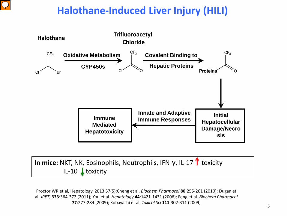
Innate and Adaptive Immune Responses
Halothane Trifluoroacetyl Chloride
Initial Hepatocellular Damage/Necro
sis
Immune Mediated
Hepatotoxicity
Oxidative Metabolism
CYP450s
Covalent Binding to
Hepatic Proteins
5
Presenter
Presentation Notes
Though clinical evidence and HLA association suppose that adaptive immune response play a major role in DILI, we need to develop an animal model for the definitive experimental proof. The drug we used in our animal model is halothane. Halothane is an inhalation anesthetic, was widely used, as Dr. Senior mentioned, until 1980s. And it was withdrawn from the U.S. market due to the liver toxicity, but still is being used in Middle East, Sub-Saharan Africa and third-world countries due to its low cost and effectiveness. Halothane is metabolized in the liver by cytochrome P450 to intermediate trifluoroacetyl chloride, which covalently binds to mainly liver proteins and it can cause toxicity to the hepatocytes directly, or it can cause enough stress to hepatocytes that secrete many proinflammatory cytokines, which activates the innate immune cells and subsequently the adaptive immune system. We reported that eosinophils in the initial injury of halothane-mediated liver injury, whereas IL-10 decreases the toxicity.
Mouse model of Halothane-Induced Liver Injury (HILI)
6
24h VEH
24h HAL
Presenter
Presentation Notes
LIVER NECROSIS IS SIMILAR TO WHAT IS SEEN IN HUMANS AFTER HALOTHANE EXPOSURE. THERE IS CENTRILOBULAR NECROSIS AROUND THE CENTRAL VEIN AREA (ZONE 3) WHICH IS RICH IN P450 ENZYMES. NECROSIS INVOLVES CELL SWELLING, MEMBRANE BLEB FORMATION, AND EVENTUALLY THE RUPTURE OF PLASMA MEMBRANE. THE RELEASE OF CELLULAR COMPONENTS FROM NECROTIC CELLS ELICITS AN INFLAMMATORY RESPONSE. THIS TYPE OF CELL DEATH CHARACTERISES ACUTE DAMAGE THAT OCCURS AT INITIAL EXPOSURE TO HALOTHANE.
Evidence for Liver Tolerance in a Guinea Pig Model of Halothane-Induced Liver Injury
M. Chen and J. Gandolfi, Drug Metab. Rev., 29, 103 (1997)
7
Presenter
Presentation Notes
Folks have been trying to develop an animal model of DILI for a long time. In 1997, Gandolfi, et al., published that they give three exposure of halothane in a guinea pig model, but they couldn't recognize any secondary immune response, which showed -- give a hint that tolerance might play a role in halothane-induced liver injury.
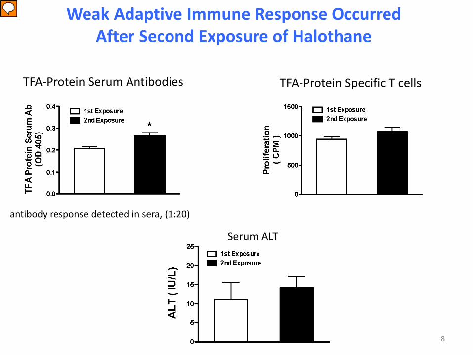
Weak Adaptive Immune Response Occurred After Second Exposure of Halothane
TFA-Protein Serum Antibodies TFA-Protein Specific T cells response
antibody response detected in sera, (1:20)
★
Serum ALT
8
Presenter
Presentation Notes
In our model, also, we expose the mice to halothane. We couldn't see any different trifluoroacetyl protein-specific T cells or increased similarity.
The idiosyncratic nature of DILI is due at least in part to immune tolerance in the liver
• Broken immune tolerance in the liver may lead to the
development of animal model of DILI mediated by adaptive immune response
• Patients who develop DILI may be deficient in liver tolerance
Hypothesis
9
Presenter
Presentation Notes
We could detect some antibody directed against trifluoroacetyl protein, but the trifluoroacetyl was very low. After one to 20 dilution, we couldn't detect any antibody directed against trifluoroacetyl protein. So, we and others hypothesize that the idiosyncratic nature of DILI is due at least in part to the immune tolerance in the liver. If we can break the immune tolerance in the liver, we may develop an animal model of DILI mediated by the adaptive immune response. And similarly, patients who develop DILI may be deficient in some liver tolerance.
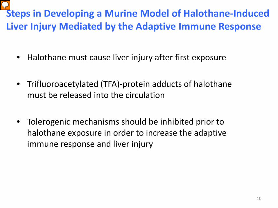
Steps in Developing a Murine Model of Halothane-Induced Liver Injury Mediated by the Adaptive Immune Response
• Halothane must cause liver injury after first exposure
• Trifluoroacetylated (TFA)-protein adducts of halothane must be released into the circulation
• Tolerogenic mechanisms should be inhibited prior to halothane exposure in order to increase the adaptive immune response and liver injury
10
Presenter
Presentation Notes
These are the steps we have chosen, because they are representative of the human halothane hepatitis. First, we wanted to detect if we can detect the trifluoroacetyl protein in the mouse liver, as well as in the serum.
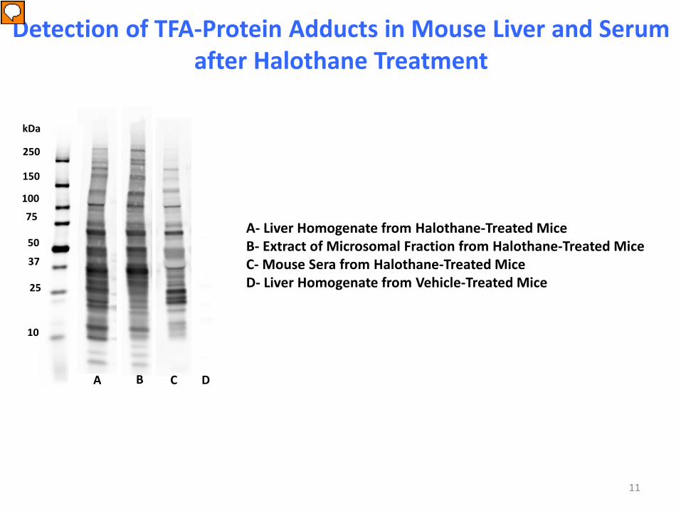
A B D C
100
37
75
kDa
250
150
50
25
10
A- Liver Homogenate from Halothane-Treated Mice B- Extract of Microsomal Fraction from Halothane-Treated Mice C- Mouse Sera from Halothane-Treated Mice D- Liver Homogenate from Vehicle-Treated Mice
Detection of TFA-Protein Adducts in Mouse Liver and Serum after Halothane Treatment
11
Presenter
Presentation Notes
WESTERN BLOT SHOWS THE DETECTION OF TFA-PROTEIN ADDUCTS IN MOUSE LIVER AND SERUM 10 HOURS AFTER HALOTHANE TREATMENT. TFA-PROTEIN ADDUCTS LEAK OUT OF THE CYTOPLASM AFTER ABOUT 10-12 HOURS, HENCE WE ANALYZE OUR TISSUES BEFORE THAT TIME POINT. EXTRACT OF MICROSOMAL FRACTION FROM HALOTHANE TREATED MICE WILL BE USED TO IDENTIFY ANTI-TFA ANTIBODIES IN SUBSEQUENT WESTERN BLOTS AND ALSO AS TFA PROTEINS IN PROLIFERATION ASSAYS.
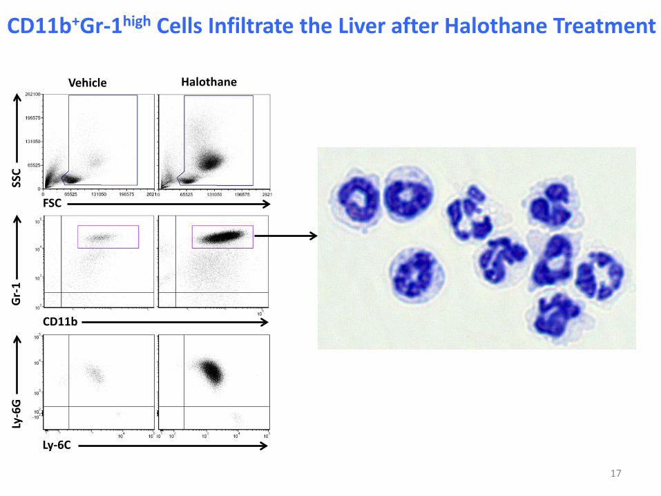
Ly-6C
Ly-6
G
CD11b
Gr-
1
FSC
SSC
Vehicle Halothane
CD11b+Gr-1high Cells Infiltrate the Liver after Halothane Treatment
12
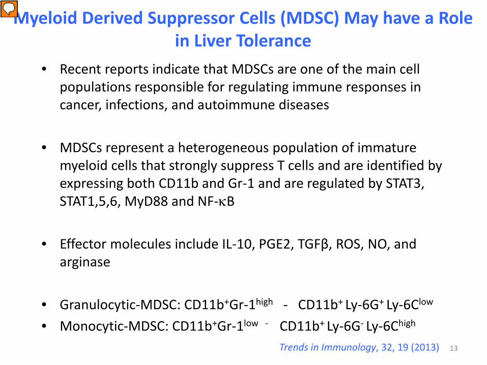
• Recent reports indicate that MDSCs are one of the main cell populations responsible for regulating immune responses in cancer, infections, and autoimmune diseases
• MDSCs represent a heterogeneous population of immature myeloid cells that strongly suppress T cells and are identified by expressing both CD11b and Gr-1 and are regulated by STAT3, STAT1,5,6, MyD88 and NF-κB
• Effector molecules include IL-10, PGE2, TGFβ, ROS, NO, and arginase
Myeloid Derived Suppressor Cells (MDSC) May have a Role in Liver Tolerance
13
Presenter
Presentation Notes
And recent reports indicate that myeloid derived suppressor cells are one of the main cell populations responsible for regulating immune responses in cancer, in infectious diseases and also in autoimmune diseases. MDSCs represent a heterogeneous population of immature myeloid cells that strongly suppress T cells and identified by the expression of CD11b and Gr-1 on their surface. And MDSCs are regulated by the transcription factors STAT1, 3, 5, 6 and NF-kappaB. The effector molecules which access the suppression of the T cells are IL-10, prostaglandin E2, TGFbeta, reactive oxygen species, nitric oxide and arginase. The other two subsets has been cited in the literature. One is the granulocytic kind, which express CD11b and Gr-1 high. And the monocytic kind actually CD11b positive Gr-1 low, but they are Ly-6G negative and mainly Ly-6C high.
CD11b+Gr-1high Cells Produce High Levels of ROS after Halothane Treatment
14
Presenter
Presentation Notes
So, then we also in our animal model, we assess the kinetics of the CD11b Gr-1 cells. The CD11b Gr-1 cells reach its peak at 24 hours, but it drops by 72 hours. As in mouse, in the model neutrophils also express CD11b and Gr-1 on their surface. It's very difficult to differentiate between myeloid derived suppressor cells and neutrophil other than their functional activity. So, one of the functional activity is the expression of reactive oxygen species. So, we measured reactive oxygen species in the CD11b Gr-1 cells by a fluorogenic dye, DCFD. You can see that though the total number of cells is higher at 24 hours, but the per cell basis reactive oxygen species is more at 72 hours, which indicated that after neutrophil leaves the liver, that those are the cells are present in the liver, they are mainly myeloid derived suppressor cells.
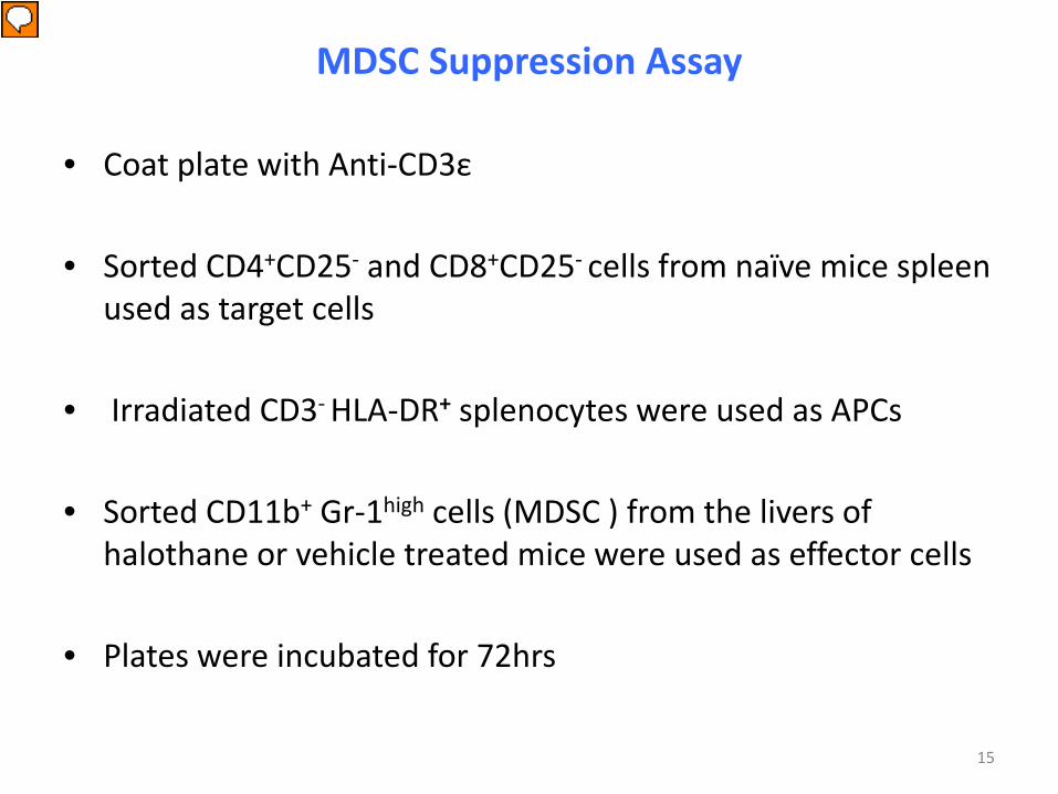
MDSC Suppression Assay
• Coat plate with Anti-CD3ε
• Sorted CD4+CD25- and CD8+CD25- cells from naïve mice spleen used as target cells
• Irradiated CD3- HLA-DR+ splenocytes were used as APCs
• Sorted CD11b+ Gr-1high cells (MDSC ) from the livers of halothane or vehicle treated mice were used as effector cells
• Plates were incubated for 72hrs
15
Presenter
Presentation Notes
To assess the functional activity, we measured the suppression assay of this myeloid derived suppressor cells by coating the plate with anti-CD3e and we used sorted CD4+CD25 negative cells or CD8+CD25 negative cells as a -- from the mice spleen as a target cells.
Hepatic MDSC From Halothane Treated Mice Suppressed T Cell Proliferation by Producing Nitric Oxide
CD4+ T cells CD4+ T cells
CD8+ T cells CD8+ T cells
16
Presenter
Presentation Notes
And we used CD3 negative HLA-DR positive splenocytes as antigen-presenting cells and sorted CD11b+Gr-1 high cells, the MDSCs from the livers of halothane or vehicle-treated mice were used as effector cells and we cultured the cells for 72 hours. And we measured the T cell proliferation by tritiated thymidine uptake. You can see when we didn't use any myeloid derived suppressor cells in the culture, the T cells were proliferated. And when we added the myeloid derived suppressor cells, the suppression of the T cell proliferation went down. And several factors have been implicated for the function of -- for the suppressive activity of myeloid derived suppressor cells.
Ly-6C
Ly-6
G
CD11b
Gr-
1
FSC
SSC
Vehicle Halothane
CD11b+Gr-1high Cells Infiltrate the Liver after Halothane Treatment
17
Isotype Treated
Anti-Gr-1 Treated
18
Liver Injury after depletion of MDSCs Prior To Halothane Treatment
Presenter
Presentation Notes
Histology shows microcalcification, but no inflammation or cytopathic injury. Histology shows mild perivenular lymphocytic inflammation and hepatocyte cytoplasmic vacuolation and dropout in zone 3
19
Liver Injury after depletion of MDSCs Prior To Halothane Treatment
Anti-Gr-1 Treated Anti-Gr-1 Treated
Presenter
Presentation Notes
Perivenular region showing multiple small foci of inflammation including a small cluster of plasma cells and hepatocyte dropout. Hepatocyte dropout and apoptotic hepatocyte is surrounded by small lymphocytes and macrophages. THE PRESENCE OF PLASMA CELLS IS A CLASSICAL INDICATION OF AUTO-IMMUNE HEPATITIS. AGAIN, THE PRESENCE OF APOPTOTIC HEPATOCYTES AND LYMPHOCYTES IS INDICATIVE OF AN ADAPTIVE IMMUNE REACTION.
20
Histology of Severely Injured Liver Treated With Anti-Gr-1
ALT - 1513
Presenter
Presentation Notes
Severely injured liver showed inflammatory infiltration of cells as well as necrosis and apoptosis in Zone 3. Necrotic hepatocytes were found mainly near the veins and apoptotic hepatocytes scattered around the edges of the injury.
21
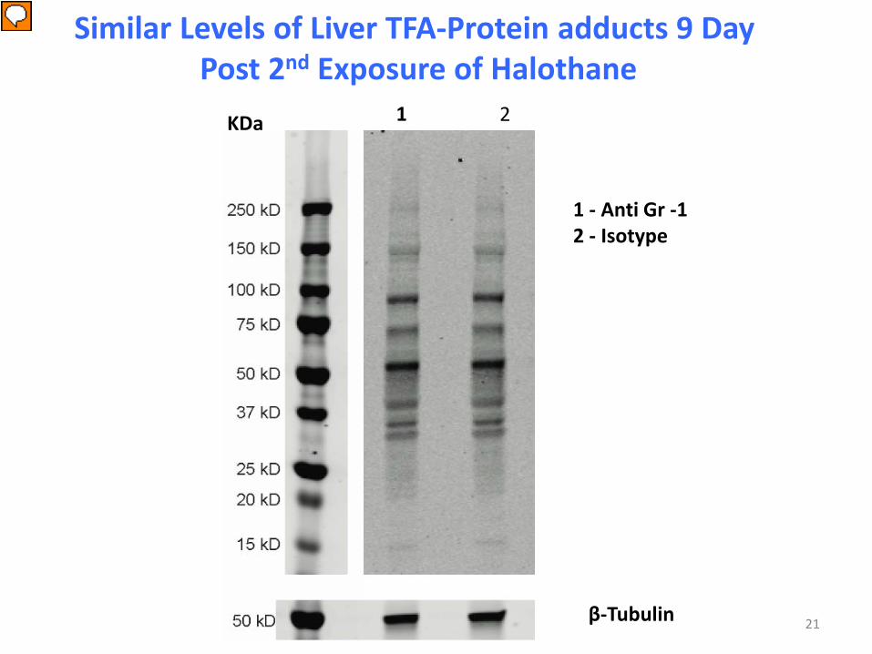
Similar Levels of Liver TFA-Protein adducts 9 Day Post 2nd Exposure of Halothane
KDa
1 - Anti Gr -1 2 - Isotype
β-Tubulin
1 2
Presenter
Presentation Notes
As it is known that halothane hepatitis is initiated by TFAPA, we verified that anti Gr-1 treatment did not alter halothane metabolism by immunoblotting .
22
Depletion of MDSCs Prior To Halothane Treatment Increased The Titer of Anti-TFA Antibodies
IL-4
Presenter
Presentation Notes
Next slide also shows that anti-Gr-1-treated mouse showed some small foci of plasma cells. Plasma cells is a classical indication of allergic hepatitis. Note: Slide #23 has been deleted from this list because it was too large (128.5 Mb) to be sent by electronic means. It can be seen in the published paper by Chakraborty et al, Hepatology 2015 Aug; 62(2):546 -57. in Figure 4AB.
Anti Gr-1
Depletion of MDSCs Prior to Halothane Exposure Increased Eosinophil Infiltration
Isotype
23
Presenter
Presentation Notes
We recently reported the the pathogenic role of eosinophils in the initial halothane exposure and eosinophilia is often reported as clinical symptom for patients with DILI.
Depletion of Hepatic MDSC Prior To Halothane Treatment Resulted In Increased TFA-Protein Specific CD4+ T Cells In The Liver
24
Presenter
Presentation Notes
And as we know that trifluoroacetyl protein adduct is important in halothane-mediated liver injury, we first decided to know that anti-Gr-1 antibody has any effect on this -- on the metabolism. And we found that after nine days of the second exposure, there is no difference in the TFA-protein adduct in the two groups. And then we wanted to know whether this humoral antibody, humoral response against this TFA-protein adduct is playing a role in this model. We found out that anti-Gr-1-treated mice, there is a significant increase of the total IgG after nine days of the second exposure and whether we couldn't detect any difference in the antibody after seven days.
25
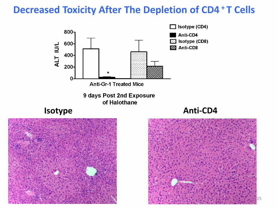
Decreased Toxicity After The Depletion of CD4 + T Cells
Isotype Anti-CD4
Cytokines after Halothane Rechallenge
IL-10 TGF-β
26
Presenter
Presentation Notes
And next we depleted the CD4 cells and both CD8 cells in those mice treated after second exposure of the halothane. Anti-CD4 depletion lowered the toxicity of those anti-Gr-1-treated mice. We saw a trend in the decrease in the ALT level, but that was not just statistically significant. Though we developed a model mediated by adaptive immune response, we didn't see any fulminant liver failure, which made us think that other tolerogenic molecules are playing compensatory mechanism in this model for CTLA4, pG1 or regulatory T cells. But in this serum when we measured IL-10 and TGF beta there is an increased IL-10 in the anti-Gr-1-treated mice. So, maybe this Kupffer cells are secreting this IL-10 where MDSCs are not present there.
Summary
• Protein adducts of halothane produced in the liver and released in the blood were able to induce both specific humoral and T cell responses against protein adducts when tolerogenic MDSC were depleted from the liver prior to halothane treatment
• This approach also led to a significant inflammatory liver injury that appeared to be mediated at least in part by adaptive immune system
• We provide the evidence for the development of an animal model of drug induced liver injury mediated by adaptive immune system
27
Presenter
Presentation Notes
So, in summary, we can say that we have developed a model by depleting these myeloid derived suppressor cells where halothane produce the trifluoroacetyl protein adducts are produced and released in the blood and were able to induce both specific humoral and T cell response against the protein adducts. And this approach also led to a significant inflammatory liver injury that appeared to be mediated at least in part by adaptive immune response.
• Lance Pohl • Kenrick Semple • Julia Berkson • Aaron Fullerton • William Proctor • Mohammed Bourdi • NHLBI Flow Cytometry Core Lab • Dr. Kleiner (Pathologist, NCI)
28
Acknowledgements
Presenter
Presentation Notes
I would like to thank Dr. Pohl for giving me the opportunity to work in his lab, and our lab colleagues. And I would like to thank Dr. Kleiner for carefully looking at my slides and giving me his valuable opinion. Thanks. (Applause.)