macrophage binding to receptor vcam-1 transmits survival signals in breast cancer cells that invade...
TRANSCRIPT

Cancer Cell
Article
Macrophage Binding to Receptor VCAM-1Transmits Survival Signals in Breast Cancer Cellsthat Invade the LungsQing Chen,1 Xiang H.-F. Zhang,1 and Joan Massague1,2,*1Cancer Biology and Genetics Program2Howard Hughes Medical Institute
Memorial Sloan-Kettering Cancer Center, New York, NY 10065, USA*Correspondence: [email protected]
DOI 10.1016/j.ccr.2011.08.025
SUMMARY
Aberrant expression of vascular cell adhesion molecule-1 (VCAM-1) in breast cancer cells is associated withlung relapse, but the role of VCAM-1 as a mediator of metastasis has remained unknown. We report thatVCAM-1 provides a survival advantage to breast cancer cells that infiltrate leukocyte-richmicroenvironmentssuch as the lungs. VCAM-1 tethers metastasis-associatedmacrophages to cancer cells via counter-receptora4-integrins. Clustering of cell surface VCAM-1, acting through Ezrin, triggers Akt activation and protectscancer cells from proapoptotic cytokines such as TRAIL. This prosurvival function of VCAM-1 can be blockedby antibodies against a4-integrins. Thus, newly disseminated cancer cells expressing VCAM-1 can thrive inleukocyte-rich microenvironments through juxtacrine activation of a VCAM-1–Ezrin-PI3K/Akt survivalpathway.
INTRODUCTION
Primary tumors can release a large number of cells into the circu-
lation long before the tumor is diagnosed and removed. Although
distant relapse may eventually occur, the limited number of
metastatic lesions that emerge suggests that only a small
proportion of the cells that leave a primary tumor succeed at infil-
trating, surviving, and ultimately overtaking a distant organ (Fi-
dler, 2003; MacDonald et al., 2002). Recent progress in metas-
tasis research has led to the identification of genes and
mechanisms that mediate cancer cell extravasation (Bos et al.,
2009; Gupta et al., 2007; Padua et al., 2008; Ricono et al.,
2009). Other recently identified metastasis genes directly partic-
ipate in the ultimate colonization of the invaded organs, an event
that may take place after a latency period lasting months or
decades depending on the type of cancer (Jones et al., 2006;
Kang et al., 2003; Muller et al., 2001; Paez-Ribes et al., 2009;
Yin et al., 1999). However, less is known about the mechanisms
Significance
Interactions with the stroma are key for the survival of disseminThe identification of thesemechanisms is essential for the deveVCAM-1 is aberrantly expressed in lung metastatic breast canwhen engaged by macrophages. As a result, VCAM-1 primesrich lung parenchymamicroenvironment. The interaction betwea validated target in diseases of rampant leukocyte recruitmenrenders these cells vulnerable to proapoptotic signals.
538 Cancer Cell 20, 538–549, October 18, 2011 ª2011 Elsevier Inc.
that allow the survival of cancer cells immediately upon entering
a distant organ and being exposed to an often lethal micro-
environment. Cell death upon infiltration of a distant organ is
regarded as the single-most important bottleneck for the estab-
lishment of distant metastases (Cameron et al., 2000; Luzzi et al.,
1998; Wong et al., 2001). To cope with the newly invaded tissue,
cancer cells that leave the circulation must interact with the
newfound stroma and obtain crucial survival and viability signals.
A better understanding of these survival mechanisms is needed
for the development of therapeutic strategies to target dissemi-
nated cancer cells (DTCs) and thereby eliminate residual disease
after the removal of a primary tumor.
The mechanisms that mediate metastasis depend, in part, on
organ-specific determinants (Fidler, 2003; Nguyen et al., 2009).
For example breast cancer metastasis may affect the lungs,
bones, liver, and brain (Anan et al., 2010), organs that present
distinct barriers to the entry and survival of circulating cancer
cells (CTCs). To have a certain probability of entering these
ated cancer cells (DTCs) and the development ofmetastasis.lopment of strategies to target DTCs. The leukocyte receptorcer cells and is shown here to transduce prosurvival signalsmetastatic cells for survival and outgrowth in the leukocyte-en leukocyte a4-integrins and VCAM-1 in endothelial cells ist. Targeting this interaction in VCAM-1+ breast cancer cells
F
E D
Ctrl sh2 sh1 Lun
g ph
oton
flux
(104
)0
2.5
5.0
7.5
10.0
p=
0.037
p=
0.035
Ctrl
sh2
sh1
H I
MDA-LM2
mVCAM-1
-actin
C sh2 sh14T1
Ctrl
sh1
0
4
8
12
16
20p=
0.011
p=
0.018
4T1
Ctrl sh2 sh1
Pho
ton
flux
(105
)
Week 3
Tail vein
Cancercells
C
Weeks after injection
Ctrlsh1sh2
4 2 3 1
Tum
or v
olum
e (m
m2 )
0
100
200
300
400
0
BC sh2 sh1
hVCAM-1 -tubulin
MDA-LM2
G
0
20
40
60
80
p=
0.015
p=
0.001
Pho
ton
flux
(103
)
0.2
0.4
0.6
0.8
1.0
0
MDA-LM2 J
sh2
sh1
Ctrl
Ctrl sh2 sh1Week 1
Ctrl sh2 sh1Week 6
A
Gland #4
Mastectomy
Cancercells
1 wk
4 wks
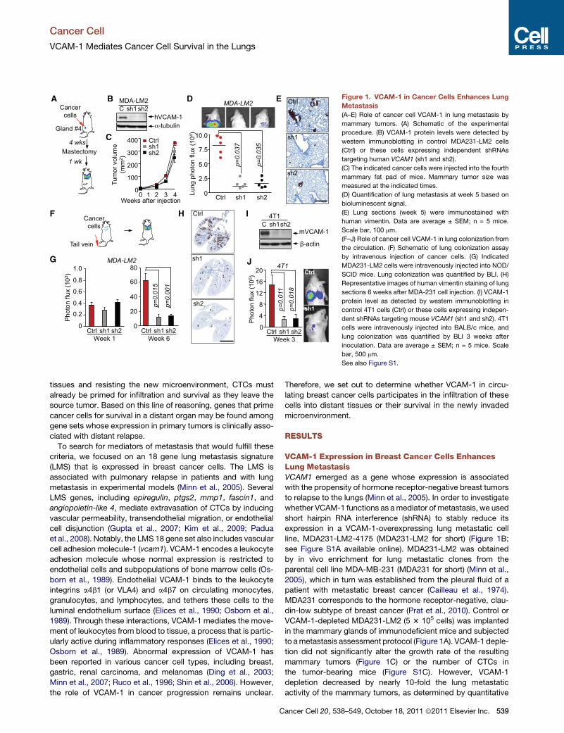
Figure 1. VCAM-1 in Cancer Cells Enhances Lung
Metastasis
(A–E) Role of cancer cell VCAM-1 in lung metastasis by
mammary tumors. (A) Schematic of the experimental
procedure. (B) VCAM-1 protein levels were detected by
western immunoblotting in control MDA231-LM2 cells
(Ctrl) or these cells expressing independent shRNAs
targeting human VCAM1 (sh1 and sh2).
(C) The indicated cancer cells were injected into the fourth
mammary fat pad of mice. Mammary tumor size was
measured at the indicated times.
(D) Quantification of lung metastasis at week 5 based on
bioluminescent signal.
(E) Lung sections (week 5) were immunostained with
human vimentin. Data are average ± SEM; n = 5 mice.
Scale bar, 100 mm.
(F–J) Role of cancer cell VCAM-1 in lung colonization from
the circulation. (F) Schematic of lung colonization assay
by intravenous injection of cancer cells. (G) Indicated
MDA231-LM2 cells were intravenously injected into NOD/
SCID mice. Lung colonization was quantified by BLI. (H)
Representative images of human vimentin staining of lung
sections 6 weeks after MDA-231 cell injection. (I) VCAM-1
protein level as detected by western immunoblotting in
control 4T1 cells (Ctrl) or these cells expressing indepen-
dent shRNAs targeting mouse VCAM1 (sh1 and sh2). 4T1
cells were intravenously injected into BALB/c mice, and
lung colonization was quantified by BLI 3 weeks after
inoculation. Data are average ± SEM; n = 5 mice. Scale
bar, 500 mm.
See also Figure S1.
Cancer Cell
VCAM-1 Mediates Cancer Cell Survival in the Lungs
tissues and resisting the new microenvironment, CTCs must
already be primed for infiltration and survival as they leave the
source tumor. Based on this line of reasoning, genes that prime
cancer cells for survival in a distant organ may be found among
gene sets whose expression in primary tumors is clinically asso-
ciated with distant relapse.
To search for mediators of metastasis that would fulfill these
criteria, we focused on an 18 gene lung metastasis signature
(LMS) that is expressed in breast cancer cells. The LMS is
associated with pulmonary relapse in patients and with lung
metastasis in experimental models (Minn et al., 2005). Several
LMS genes, including epiregulin, ptgs2, mmp1, fascin1, and
angiopoietin-like 4, mediate extravasation of CTCs by inducing
vascular permeability, transendothelial migration, or endothelial
cell disjunction (Gupta et al., 2007; Kim et al., 2009; Padua
et al., 2008). Notably, the LMS 18 gene set also includes vascular
cell adhesion molecule-1 (vcam1). VCAM-1 encodes a leukocyte
adhesion molecule whose normal expression is restricted to
endothelial cells and subpopulations of bone marrow cells (Os-
born et al., 1989). Endothelial VCAM-1 binds to the leukocyte
integrins a4b1 (or VLA4) and a4b7 on circulating monocytes,
granulocytes, and lymphocytes, and tethers these cells to the
luminal endothelium surface (Elices et al., 1990; Osborn et al.,
1989). Through these interactions, VCAM-1 mediates the move-
ment of leukocytes from blood to tissue, a process that is partic-
ularly active during inflammatory responses (Elices et al., 1990;
Osborn et al., 1989). Abnormal expression of VCAM-1 has
been reported in various cancer cell types, including breast,
gastric, renal carcinoma, and melanomas (Ding et al., 2003;
Minn et al., 2007; Ruco et al., 1996; Shin et al., 2006). However,
the role of VCAM-1 in cancer progression remains unclear.
C
Therefore, we set out to determine whether VCAM-1 in circu-
lating breast cancer cells participates in the infiltration of these
cells into distant tissues or their survival in the newly invaded
microenvironment.
RESULTS
VCAM-1 Expression in Breast Cancer Cells EnhancesLung MetastasisVCAM1 emerged as a gene whose expression is associated
with the propensity of hormone receptor-negative breast tumors
to relapse to the lungs (Minn et al., 2005). In order to investigate
whether VCAM-1 functions as amediator of metastasis, we used
short hairpin RNA interference (shRNA) to stably reduce its
expression in a VCAM-1-overexpressing lung metastatic cell
line, MDA231-LM2-4175 (MDA231-LM2 for short) (Figure 1B;
see Figure S1A available online). MDA231-LM2 was obtained
by in vivo enrichment for lung metastatic clones from the
parental cell line MDA-MB-231 (MDA231 for short) (Minn et al.,
2005), which in turn was established from the pleural fluid of a
patient with metastatic breast cancer (Cailleau et al., 1974).
MDA231 corresponds to the hormone receptor-negative, clau-
din-low subtype of breast cancer (Prat et al., 2010). Control or
VCAM-1-depleted MDA231-LM2 (5 3 105 cells) was implanted
in the mammary glands of immunodeficient mice and subjected
to ametastasis assessment protocol (Figure 1A). VCAM-1 deple-
tion did not significantly alter the growth rate of the resulting
mammary tumors (Figure 1C) or the number of CTCs in
the tumor-bearing mice (Figure S1C). However, VCAM-1
depletion decreased by nearly 10-fold the lung metastatic
activity of the mammary tumors, as determined by quantitative
ancer Cell 20, 538–549, October 18, 2011 ª2011 Elsevier Inc. 539

BA
Leukocyte
VCAM-1
4 1/7Integrin
D
0.2
0.4
0.6
1.2
Rat
io (U
937:
Tum
or)
anti- 4 - + - + - +
0.8
Ctrl sh1 sh2
p<
0.0001
p<
0.0001
p<
0.0001
1.0
Rat
io (U
937:
Tum
or)
0.5
1.5
2.5
3.5
4.5
p<
0.0001
U937 DAPI
MD
A23
1M
DA
231-
LM2
F E
GFP+ cancer cells Gr1+ Neutro
F4/80+ Macro CD31+ Endoth
rVCAM-1
% M
ax
10
15
p=
0.0006
p=
0.0017
0
5
F4/8
0+/G
FP+
cells
(10-
2 )
shVCAM1
Ctrl 21
C Figure 2. VCAM-1 on Cancer Cells Binds Tumor-
Associated Macrophages
(A) Schematic illustration of VCAM-1 interactions with
leukocyte a4-integrins.
(B and C) Adhesion assays of U937 cells on monolayers of
parental MDA231 or MDA231-LM2 cells. (B) Representa-
tive photomicrographs of adherent TRITC-labeled U937
cells on themonolayer of indicated cancer cells. Scale bar,
100 mm. (C) Quantification of the ratio of adherent U937
cells and cancer cells. Data are average ± SEM; n = 10 unit
areas.
(D) U937 cells treated with anti-a4-integrin antibody
(anti-a4) or Ig control (�) were tested for binding to
monolayers of control or VCAM1-depleted MDA231-LM2.
VCAM-1 knockdown was done using two independent
shRNAs (sh1, sh2). Data are average ± SEM; n = 10 unit
areas.
(E) rVCAM-1 binging assay with different stromal cell
populations from lung metastatic nodules, including
F4/80+ macrophages (Macro), Gr-1+ neutrophils (Neutro),
and CD31+ endothelial cells (Endoth) and GFP+ cancer
cells. Cell surface binding of bovine serum albumin control
(gray closed histogram) or rVCAM-1 (black open histo-
gram) was determined by flow cytometry.
(F) Adhesion assays of stromal cells from lung metastatic nodules onmonolayers of control or VCAM1-depleted (sh1, sh2) MDA231-LM2 cells. The ratio between
adherent F4/80+ macrophages to GFP+ cancer cells was quantified by flow cytometry.
See also Figure S2.
Cancer Cell
VCAM-1 Mediates Cancer Cell Survival in the Lungs
bioluminescence imaging (BLI) of an integrated luciferase gene
(Figure 1D) or by antihuman vimentin immunohistochemistry
(Figure 1E). In a tail vein inoculation protocol to assess lung colo-
nization by a synchronous influx of CTCs (Figure 1F), VCAM-1
depletion did not affect the accumulation of MDA231-LM2 in
the lungs during the first week after inoculation (Figure 1G).
However, VCAM-1 depletion significantly inhibited the subse-
quent development of metastatic colonies (Figures 1G and 1H).
To probe the role and significance of VCAM-1 in breast cancer
metastasis in a syngeneic, immunocompetent model, we inves-
tigated 4T1 breast cancer cells. 4T1 is a lung metastatic cell line
derived from a mammary tumor that spontaneously emerged in
BALB/c mice (Aslakson and Miller, 1992). Western immunoblot-
ting and qRT-PCR analysis demonstrated that 4T1 cells express
VCAM-1 (Figure 1I; Figure S1B). These data show that lung
metastatic cells from a spontaneous mouse mammary tumor
shared with the human breast cancer counterparts the remark-
able feature of expressing this leukocyte-binding endothelial
cell receptor. Furthermore, shRNA-mediated depletion of
VCAM-1 in 4T1 cells (Figure 1I; Figure S1B) severely inhibited
the lung metastatic activity of these cells in immunocompetent
BALB/c mice (Figure 1J).
VCAM-1 Mediates Binding of Tumor-AssociatedMacrophages to Cancer CellsThe extracellular region of VCAM-1 consists of seven immuno-
globulin (Ig) domains, one of which (domain 4) is missing in the
VCAM-1-6D splice variant (Cybulsky et al., 1991). a4-Integrins
bind to separate sites on Ig domains 1 and 4 (Figure 2A), and
therefore, the VCAM-1-6D variant is of low affinity (Vonderheide
et al., 1994). By means of isoform-specific RT-PCR, we deter-
mined that MDA231 and another human metastatic breast
cancer cell model, CN34 (see below), predominantly express
the long, high-affinity VCAM-1 variant (Figure S2A). To determine
540 Cancer Cell 20, 538–549, October 18, 2011 ª2011 Elsevier Inc.
whether VCAM-1 on breast cancer cells can bind leukocytes, we
incubated MDA231monolayers with a suspension of U937 cells,
a human monocyte cell line that expresses a4b1-integrin (Kalo-
geris et al., 1999). The MDA231-LM2 cells bound over 3-fold
as many U937 cells as did the parental MDA231 cells (Figures
2B and 2C). shRNA-mediated depletion of VCAM-1 in
MDA231-LM2 cells or addition of a blocking antibody against
a4-integrin inhibited the binding of U937 cells (Figure 2D). We
concluded that VCAM-1 on cancer cells can bind monocytes.
Monocytic cells including macrophages enhance tumor
progression (Joyce and Pollard, 2009; Qian and Pollard, 2010).
Blockage of macrophage recruitment significantly decreases
lung colonization by MDA231-LM2 cells in mice (Qian et al.,
2009, 2011). However, the specific contribution of macrophages
to metastatic colonization is not well understood. To identify
metastasis stromal cell types that bind VCAM-1, we generated
lung metastatic nodules by tail vein inoculation of MDA231-
LM2 cells expressing green fluorescent protein (GFP). Single-
cell suspensions prepared from the resulting nodules were
incubated with fluorochrome-conjugated recombinant VCAM-1
ectodomain (rVCAM-1) or bovine serum albumin as a control.
Specific cell surface markers were used to identify and quantify
different types of stromal cells (Figure S2B). VCAM-1 bound
to metastasis-associated CD45+F4/80+ macrophages and
CD45�CD31+ vascular endothelial cells, but not to Gr1+ neutro-
phils or GFP+ cancer cells (Figure 2E). Next, we prepared cell
suspensions from lung metastasis nodules, depleted these
suspensions of human EpCAM+ cancer cells, and panned the
remaining cells over control or VCAM-1-depleted MDA231-
LM2 monolayers. Analysis of the monolayer-bound fraction
demonstrated VCAM-1-dependent binding of F4/80+ cells to
MDA231-LM2 cells (Figure 2F).
In 4T1 lung metastatic nodules, CD45+CD4+ and CD45+CD8+
lymphocytes expressed low levels of cell surface a4-integrin

F
Gland #2
Unlabeled cells
7 days
GFP/Luc+MDA231-LM2
1-4 weeks
G
Ctrl
sh1
Pho
ton
flux
(106
)
Weeks after I.C. injection p
<0
.0
5
0.5
1.0
1.5
2.0
1 2 3 4
Ctrlsh1sh2
H
CD
68
4T1-control
CMFDA DAPI
CD
8C
D4
C
4T1-VCAM1 sh-1CMFDA labeled cancer cells
Tail vein
24h
Lung
CD68
CD8
CD4
staining
B
E
D
MDA-LM2
020406080
100
% o
f int
erac
ting
tum
or c
ells
CD68 CD8 CD4 CD684T1
Ctrlsh1
Num
ber o
f C
D68
ce
lls/tu
mor
cel
l
Ctrl0
0.4
0.8
1.2
1.6
00.5
1.5
2.5
3.5
4T1 MDA-LM2sh1 Ctrl sh1
— U937
A
Ctrlsh1sh2
0
0.5
1.0
1.5
2.0
2.5
Tran
s-H
PM
EC
mig
ratio
n 6 h EC
U937(+PMA)
Cancer cells
Control sh1
GFP CD68 DAPI
Figure 3. Cancer Cell VCAM-1 Is Dispensable for Transendothelial Migration or Leukocyte Recruitment but Enhances Tumor Seeding
(A) MDA231-LM2 cell migration across monolayers of HPMECs, with or without activated monocytes in the lower chamber of a transwell. Migrated cancer cells
were quantified. Data are average ± SEM; n = 10 unit areas.
(B–E) Association of cancer cells with leukocytes in the lungs. (B) Schematic of the experimental procedure. (C) CMFDA-labeled 4T1 cells were intravenously
inoculated into BALB/c mice, and the association between cancer cells and CD4+ and CD8+ T cells and CD68+ macrophages was analyzed 24 hr later. Shown
are two representative micrographs per experimental group. Scale bar, 10 mm. (D) Percentage of cancer cells interacting with the indicated leukocyte cells.
(E) Quantification of CD68+ cells surrounding cancer cells. Data are average ± SEM; n > 30 cancer cells.
(F–H) Tumor self-seeding assay. (F) Schematic of experimental procedure. GFP/luciferase expressing cancer cells of inoculation of into the arterial circulation
of mice bearing primary tumors formed by unlabeled cancer cells. (G) Representative images of mammary tumors seeded with indicated GFP+ MDA-231 LM2
cells. GFP+ cancer cells and tumor-associated macrophages were stained with anti-GFP and anti-CD68 antibodies in primary tumors (week 4). Scale bar, 10 mm.
(H) Seeding of control (Ctrl) or VCAM-1 depleted (sh1 and sh2) MDA231-LM2 cells in mammary tumors after intracardiac inoculation was determined by BLI
quantification. Data are average ± SEM; n = 10 mice.
See also Figure S3.
Cancer Cell
VCAM-1 Mediates Cancer Cell Survival in the Lungs
compared to CD45+F4/80+ macrophages (Figure S2C). Macro-
phages comprised approximately 7% of the total cell population
in the metastatic nodules (Figure S2C), whereas CD4+ and CD8+
lymphocytes collectively comprised 4% of the tumor cell
population. Moreover, endothelial cells, which also bind to
rVCAM-1 (Figure 2E), comprised <1% of the cell population
from MDA231-LM2 lung nodules (Figure S2B). Overall, macro-
phages were the most abundant source of potential a4-integrin
contacts in these lung nodules.
Cancer Cell VCAM-1 Is Dispensable for Extravasationand Macrophage RecruitmentContacts with macrophages enhance cancer cell extravasation
(Qian et al., 2009). Although U937 monocytes enhanced the
C
ability of MDA231-LM2 cells to migrate through human pulmo-
nary microvascular endothelial cell (HPMEC) monolayers,
VCAM-1 depletion did not influence this process. This was
observed when the monocytes were activated with PMA to
induce adherence (Yamamoto et al., 2009) and were placed on
the lower side of the transwell membrane (Figure 3A). Similar
results were obtained when monocytes stimulated with cyclic-
AMP were placed with cancer cells in the upper transwell
chamber, with complement factor C5a as a monocyte chemoat-
tractant (Rubin et al., 1991) in the bottom chamber (Figure S3A).
VCAM-1 depletion also had no effect when U937 cells were
placed together with cancer cells, with CXCL12 as a cancer
cell chemoattractant (Zhang et al., 2009) in the bottom chamber
(Figure S3B). These results are consistent with our in vivo
ancer Cell 20, 538–549, October 18, 2011 ª2011 Elsevier Inc. 541
Cancer Cell
VCAM-1 Mediates Cancer Cell Survival in the Lungs
evidence that VCAM-1 is not required for entry of breast cancer
cells into the lung parenchyma.
To determine if VCAM-1 on cancer cells influenced the recruit-
ment of macrophages into the tumor, we labeled 4T1 or
MDA231-LM2 cells with CellTracker Green CMFDA, and inocu-
lated these cells into mice via the tail vein. After 24 hr, we exam-
ined the association of CD4+, CD8+ T lymphocytes (for 4T1
model) and CD68+ macrophages (for both 4T1 and MDA231-
LM2 models) with CMFDA+ cancer cells in the lung parenchyma
(Figures 3B and 3C; Figure S3C). Over 95% of cancer cells were
surrounded by macrophages in close juxtaposition, whereas
fewer than 30% of cancer cells were associated with CD4+ or
CD8+ T lymphocytes (Figure 3D). VCAM-1 depletion in cancer
cells changed neither the percentage of cancer cells surrounded
by immune cells nor the average number of interacting macro-
phages per cancer cell (Figures 3D and 3E). Furthermore, the
lung nodules formed by the VCAM-1-depleted cells showed
the same content of F4/80+, CD31+, and Gr1+ cells as did size-
matched control nodules (Figure S2B). Thus, VCAM-1 expres-
sion in cancer cells is not required for their recruitment of, or
exposure to, macrophages.
VCAM-1 inCancer Cells Enhances the Seeding of BreastTumorsWe further tested the role of VCAM-1 under conditions that
challenged cancer cells to seed new or established tumors.
VCAM-1 depletion significantly diminished the rate of tumor
outgrowth when only 5 3 104 cells were implanted in mammary
fat pad tumor assays (Figures S3D–S3F, compare to tumor
formation by 5 3 105 implanted cells in Figure 1C). Recently,
we showed in experimental models that established breast
tumors are powerful magnets for CTCs (Kim et al., 2009). In
this process, called ‘‘self-seeding,’’ CTCs that pass through a
breast tumor can effectively reinvade the tumor by virtue of their
metastatic abilities, the leaky tumor vasculature, and the attrac-
tion of inflammatory cytokines IL6 and IL8 in the tumor microen-
vironment. Once in that environment, aggressive seeder clones
can attract myeloid cells through the release of CXCL1 and
expand in that myeloid-rich microenvironment (Kim et al.,
2009). In a tumor self-seeding assay (Figure 3F), MDA231-LM2
seeder cells were indeed surrounded by CD68+ macrophages
(Figure 3G). MDA231 seeder cells that were depleted of
VCAM-1 showed a marked decrease in the ability to populate
the recipient tumors (Figure 3H). Collectively, these results
suggest that VCAM-1 supports the outgrowth of cancer cells
that infiltrate the lungs and other restrictive sites. Moreover,
these results raised the possibility that VCAM-1-leukocyte inter-
actions are a source of this advantage.
VCAM-1 Protects Breast Cancer Cells from Apoptosisin the Lung MicroenvironmentTo determine how VCAM-1 expression in breast cancer cells
contributes to tumor outgrowth in the lungs, we transduced
a doxycycline-driven VCAM1 shRNA expression system in
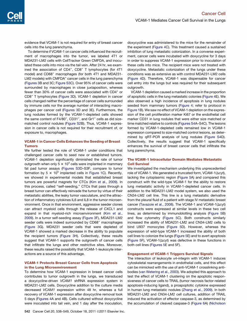
MDA231-LM2 cells. Doxycycline addition to the culture media
decreased VCAM1 expression within 48 hr, whereas a full
recovery of VCAM-1 expression after doxycycline removal took
5 days (Figures 4A and 4B). Cells cultured without doxycycline
were inoculated into tail vein, and 1 day after the inoculation,
542 Cancer Cell 20, 538–549, October 18, 2011 ª2011 Elsevier Inc.
doxycycline was administered to the mice for the remainder of
the experiment (Figure 4C). This treatment caused a sustained
inhibition of lung metastatic colonization. In a converse experi-
ment, cancer cells were incubated with doxycycline for 3 days
in order to suppress VCAM-1 expression prior to inoculation of
these cells into mice. The recipient mice were not treated with
doxycycline. Metastatic colonization of the lungs under these
conditions was as extensive as with control MDA231-LM2 cells
(Figure 4D). Therefore, VCAM-1 was dispensable for cancer
cell entry into the lungs but was required for their subsequent
outgrowth.
VCAM-1 depletion caused amarked increase in the proportion
of apoptotic cells in the lung metastatic colonies (Figure 4E). We
also observed a high incidence of apoptosis in lung nodules
seeded from mammary tumors (Figure 4; refer to protocol in
Figure 1B).We saw no effect of VCAM-1 depletion on the expres-
sion of the cell proliferation marker Ki67 or the endothelial cell
marker CD31 in lung nodules that were either size matched or
timematched relative to controls (Figures S4A–S4C). The lesions
formed by VCAM-1-depleted cells remained low in VCAM-1
expression compared to size-matched control lesions, as deter-
mined by qRT-PCR analysis of lung nodules (Figure S4D).
Collectively, the results suggest that VCAM-1 specifically
enhances the survival of breast cancer cells that infiltrate the
lung parenchyma.
The VCAM-1 Intracellular Domain Mediates MetastaticCell SurvivalWe investigated the mechanism underlying this unprecedented
role of VCAM-1. We generated a truncated form, VCAM-1(Dcyt),
lacking the cytoplasmic region (Figure 5A) and compared this
construct with the wild-type VCAM-1 for the ability to restore
lung metastatic activity in VCAM-1-depleted cancer cells. In
addition to the MDA231-LM2 model system, we also used the
CN34-LM2 cell line. This line is a lung metastatic derivative
from the pleural fluid of a patient with stage IV metastatic breast
cancer (Tavazoie et al., 2008). The VCAM-1 and VCAM-1(Dcyt)
constructs were expressed at similar levels in these two cell
lines, as determined by immunoblotting analysis (Figure 5B)
and flow cytometry (Figure 5C). Both constructs similarly
increased the ability of MDA231-LM2 and CN34-LM2 cells to
bind U937 monocytes (Figure 5D). However, whereas the
expression of wild-type VCAM-1 increased the ability of both
cell lines to colonize the lungs (Figure 5E) and to avert apoptosis
(Figure 5F), VCAM-1(Dcyt) was defective in these functions in
both cell lines (Figures 5E and 5F).
Engagement of VCAM-1 Triggers Survival SignalsThe interaction of leukocyte a4-integrin with VCAM-1 induces
cytoskeletal rearrangements in endothelial cells, and this effect
can be mimicked with the use of anti-VCAM-1 crosslinking anti-
bodies (van Wetering et al., 2003). We adopted this approach to
test the effect of VCAM-1 clustering on the apoptotic respon-
siveness of cancer cells to TRAIL (tumor necrosis factor-related
apoptosis-inducing ligand), a proapoptotic cytokine expressed
in human lung metastatic nodules (Zhang et al., 2009). In both
MDA231-LM2 and CN34-LM2 cell cultures, addition of TRAIL
induced the activation of effector caspase-3, as determined by
the accumulation of cleaved caspase-3 (Figure 6A) (Nicholson
FE
Con
trol
VC
AM
1sh
1
hVimentin Cleaved Casp-3
TUN
EL+
cel
ls/u
nit a
rea
Ctrl 210
0.5
1.0
1.5
2.5
2.0
p<0.001
shVCAM1
BA
DC
00.20.40.60.81.01.2
VC
AM
1 m
RN
A
0 1 2 3Days with Dox
Ctrlsh1
Cancer cells
+Dox
1-3 days
RT-PCR Days after Dox removal
0 1 2 3 4 5
VC
AM
1 m
RN
A
00.20.40.60.81.01.2
Ctrlsh1
Cancer cells Dox
3 days
Dox removal
1-5 days
RT-PCR
sh2
Ctrl
sh1
Ctrl
TUNEL
Cancercells
1 day Dox in water
BLI
Cancer cells, 3 days + Dox
BLI Lung
pho
ton
flux
(103
)0
10
20
30
40
1 7 14 21 28Days post injection
Ctrlsh1
Lung
pho
ton
flux
(103
)
Days post injection
0
10
20
30
40
1 7 14 21 28
Ctrlsh1
p=
0.0
00
1
Figure 4. VCAM-1 Protects Cancer Cells from
Apoptosis in the Lung Microenvironment
(A and B) MDA231-LM2 cells with a doxycycline (Dox)-
inducible shVCAM-1 vector were cultured in the presence
of doxycycline (A) or were placed in doxycycline-free
medium after culturing with doxycycline (B), in order to
determine the kinetics of VCAM1 depletion and recovery,
respectively.
(C) Control or doxycycline-driven VCAM1 shRNA cells
were inoculated into mice, and all mice were treated with
doxycycline. Lung colonization was assessed by quanti-
tative BLI.
(D) BLI quantification of lung colonization after the indi-
cated cancer cells pretreated with doxycycline were
injected into mice that were not given doxycycline. The
mice were not administrated with drug. Data are average ±
SEM; n = 8 mice.
(E) TUNEL staining and quantification in the lungs har-
vested 6 weeks after intravenous injection of cancer cells
as indicated in Figure 1E. Representative photomicro-
graphs, and quantification of TUNEL+ cells. Data are
average ± SEM; n = 10 unit area/mouse, 3 mice/group.
Scale bar, 50 mm.
(F) The indicated cancer cells were injected into the
fourth mammary fat pad of mice, and the lungs were
harvested as indicated in Figure 1A. Immunostaining for
human vimentin and cleaved caspase-3was performed on
consecutive lung tissue sections. Scale bar, 100 mm.
See also Figure S4.
Cancer Cell
VCAM-1 Mediates Cancer Cell Survival in the Lungs
et al., 1995). Incubation with anti-VCAM-1 antibody and
secondary F(ab0)2 fragment markedly blunted this effect in the
cells expressing full-length VCAM-1, but importantly, not in cells
expressing VCAM-1(Dcyt) (Figure 6A).
To determine the effect of engaging VCAM-1 with its natural
a4-integrin counter-receptors, we incubated cancer cells with
U937 monocytes. Cancer cells expressing VCAM-1, VCAM-1
(Dcyt), or no exogenous VCAM-1 were prelabeled with CMFDA,
and apoptosis was assessed by TUNEL staining (Figure 6B).
Expression of VCAM-1 reduced to half the incidence of apo-
ptosis in cancer cells that were exposed to TRAIL, whereas
VCAM-1(Dcyt) provided no protection (Figure 6C). Furthermore,
addition of anti-a4-integrin-blocking antibody prevented
VCAM-1 from exerting this antiapoptotic effect (Figure 6C).
Cancer cell counts corroborated these findings (Figure 6D).
Thus, upon engagement by a4-integrin on leukocytes, VCAM-1
in breast cancer cells delivers antiapoptotic signals via its cyto-
plasmic tail.
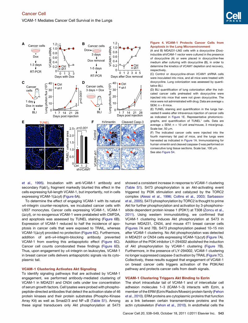
VCAM-1 Clustering Activates Akt SignalingTo identify signaling pathways that are activated by VCAM-1
engagement, we performed antibody-mediated clustering of
VCAM-1 in MDA231 and CN34 cells under low concentration
of serum growth factors. Cell lysates were probedwith phospho-
peptide-directed antibodies that detect the activation state of 46
protein kinases and their protein substrates (Phospho-Kinase
Array Kit) as well as Smad2/3 and NF-kB (Table S1). Among
these signal transducers only Akt phosphorylation at S473
C
showed a consistent increase in response to VCAM-1 clustering
(Table S1). S473 phosphorylation is an Akt-activating event
triggered by PI3K stimulation and catalyzed by the TORC2
complex (Alessi et al., 1996; Collins et al., 2003; Sarbassov
et al., 2005). S473 phosphorylation by TORC2 is thought to prime
Akt for further phosphorylation and activation by 3-phosphoino-
sitide dependent protein kinase 1 (PDK1) at T308 (Zoncu et al.,
2011). Using western immunoblotting, we confirmed that
VCAM-1 clustering induces Akt phosphorylation at S473 in
human MDA231, CN34, and mouse 4T1 breast cancer cells
(Figures 7A and 7B). S473 phosphorylation peaked 10–15 min
after VCAM-1 clustering. No Akt phosphorylation was detected
in MDA231 or CN34 cells expressing VCAM-1(Dcyt) (Figure 7A).
Addition of the PI3K inhibitor LY-294002 abolished the induction
of Akt phosphorylation by VCAM-1 clustering (Figure 7B).
Furthermore, in the presence of LY-294002, VCAM-1 clustering
no longer suppressed caspase-3 activation by TRAIL (Figure 7C).
Collectively, these results suggest that engagement of VCAM-1
on breast cancer cells triggers activation of the PI3K/Akt
pathway and protects cancer cells from death signals.
VCAM-1 Clustering Triggers Akt Binding to EzrinThe short intracellular tail of VCAM-1 and of intercellular cell
adhesion molecules 1–3 (ICAM-1–3) interacts with Ezrin, a
member of the ERM (Ezrin/Radixin/Moesin) protein family (Fehon
et al., 2010). ERM proteins are cytoplasmic proteins that function
as a link between certain transmembrane proteins and the
actin cytoskeleton (Fehon et al., 2010). In endothelial cells the
ancer Cell 20, 538–549, October 18, 2011 ª2011 Elsevier Inc. 543

E
BA
F
VCAM-1Ctrl WT cytMDA-LM2-sh1
Tubulin p
<0
.0
05
1.0
2.0
3.0
1.0
2.0
3.0
4.0p
<0
.0
02
Rat
io (U
937:
Tum
or)
MDA-LM2-sh1 CN34-LM2
Ctrl WT cyt Ctrl WT cyt
Lung
pho
ton
flux
(103
)
10
30
50
70
2
6
10
14
18
p=
0.0
16
p=
0.0
07
p=
0.0
28
p=
0.0
34
MDA-LM2-sh1 CN34-LM2
Ctrl WT cyt Ctrl WT cyt
TUN
EL+
cel
ls/u
nit a
rea
0.4
0.8
1.2
1.6
p=
0.0
01
p<
0.0
00
1
0.4
0.8
1.2
1.6
p=
0.0
05
p<
0.0
00
1
MDA-LM2-sh1 CN34-LM2
Ctrl WT cyt Ctrl WT cyt
19aa
VCAM-1WT cyt
TMIC
EC
C CN34-LM2MDA231-LM2-sh1
cytWT Ctrl
VCAM1
% M
ax
D
Figure 5. The VCAM-1 Intracellular Domain Mediates Survival of Cancer Cells in the Lungs
(A) Domain structure of the wild-type (WT) VCAM-1 with extracellular (EC), transmembrane (TM), and intracellular (IC) domains and VCAM-1 (DCyt) construct
lacking the IC. WT or DCyt VCAM-1 was overexpressed in VCAM1-depleted (by shRNA 1) MDA231-LM2 cells (MDA-LM2-sh1) and CN34-LM2 cells.
(B and C) Expression of the wild-type VCAM-1, VCAM-1(DCyt), or empty vector (Ctrl) in MDA231-LM2 cells that were stably depleted of endogenous VCAM-1 by
the expression of shRNA1 and CN34-LM2 cells. (B) VCAM-1 was detected by western immunoblotting in MDA231-LM2 cells. (C) The expression of VCAM-1 on
the cell surface was detected by flow cytometry.
(D) U937 cell adhesion assays on monolayers of the indicated MDA231 or CN34 cell lines. Data are average ± SEM; n = 10 unit areas.
(E and F) The indicated cell lines were intravenously injected into mice. Six weeks after tumor cell injection, lung BLI was measured (E), and TUNEL+ cells were
quantified in lung tissue sections (F). Data are average ± SEM; n = 5 mice.
Cancer Cell
VCAM-1 Mediates Cancer Cell Survival in the Lungs
interaction of VCAM-1 with Ezrin triggers cytoskeletal rearrange-
ments for the translocation of VCAM-1-bound leukocytes from
blood into tissue (Barreiro et al., 2002; van Wetering et al.,
2003). In addition to this role, Ezrin can signal cell survival
through the PI3K pathway (Gautreau et al., 1999). Antibody-
mediated crosslinking of ICAM-2 triggers phosphorylation of
Ezrin at Y353, which results in binding and activation of PI3K
(Perez et al., 2002). Y353 is located within the a-helical domain
of Ezrin, and Y353 phosphorylation is thought to indirectly
involve Src kinases (Chuan et al., 2006; Krieg and Hunter,
1992). Based on these clues, we hypothesized that VCAM-1
A
CleavedCasp3
X-linkTRAIL - + +
- - +
VCAM-1- + + - - +
VCAM-1( cyt)
Casp3
Tubulin
- + + - - +
Control- + + - - +
VCAM-1- -
VCAM- + + - - +
Control 2ML-43NC 1hs-2ML-ADM
Vector:
B
% o
f TU
NE
L+ c
ells
0.5
1.0
1.5
2.0
2.5
anti- 4 - + - + - +Ctrl WT cyt
p=
0.002
p=0.003p=0.003
DCMFDA
TUNEL
DAPI
Merge
C
12
16
4
8
1
2
3
4
5
6
anti- 4 - + - + - +Ctrl WT cyt
Can
cer c
ells
/wel
l (10
3 )
p<
0.001
p<
0.001
p<
0.001
C
MDA-LM2-sh1 MDA-LM2-sh1
544 Cancer Cell 20, 538–549, October 18, 2011 ª2011 Elsevier Inc.
engagement may trigger Akt activation though Ezrin. Indeed,
antibody-mediated clustering of VCAM-1 in MDA231-LM2 or
CN34 cells in low-serum conditions led to a rapid phosphoryla-
tion of Ezrin at Y353 (Figure 7D). VCAM-1(Dcyt) failed to mediate
this effect (Figure 7D).Moreover, this effect was accompanied by
the formation of a complex between Ezrin and Akt (Figure 7D)
and an enhanced recruitment of Akt to themembrane (Figure 7E;
Figure S5). The results suggest that VCAM-1 signaling activates
Ezrin Y353 phosphorylation to help PI3K recruit Akt to the
membrane, leading to TORC2 activation of Akt signaling
(summarized in Figure 7F).
+ + - +
-1( cyt)
- + - + - +trl WT cyt
p<
0.001
p<
0.001
p<
0.001
CN34-LM2
Figure 6. VCAM-1 Engagement by a4-Integrins
Triggers Antiapoptotic Signals via the Cytoplasmic
Domain
(A) VCAM-1 clustering antibodies (Ab X-linking) were
added to the indicated cell lines in the presence or
absence of 10 ng/ml TRAIL for 3 hr. Caspase-3 activation
was detected by western immunoblotting.
(B–D) The indicated cancer cells were cocultured with
U937 cells in the presence of TRAIL and a4-integrin anti-
body (anti-a4) or comparable Ig control (�). (B) After 24 hr,
TUNEL+ (red) and Green CMFDA-labeled cancer cells
were scored under a fluorescent microscope. Arrowheads
indicate a TUNEL+/CMFDA+ cancer cell, and asterisks
indicate a (CMFDA-negative) U937 cell. (C) Percentage of
TUNEL+/CMFDA+ cancer cells. Data are average ± SEM;
n = 10 unit areas; p value calculated using t test. Scale bar,
20 mm. (D) Cancer cells were quantified after 48 hr of
coculture. Data are average ± SEM; n = 4 samples.
- + + - - +
++
- - - +
CN34-LM2/VCAM-1+-+
TRAIL - + + - - +
++
- - - +
+-+
CleavedCasp3
X-linkLY
Casp3
Tubulin
MDA-LM2-sh1/VCAM-1C
MDA231+ + X-link
LY - +
Akt
p-A
kt/A
kt
CN34+
- + +
BA
X-link - 5’ 15’ VCAM-1
60’ p-Akt
(S473)Akt
30’ - 5’ 15’ VCAM-1( cyt)
60’ 30’
p-A
kt/A
kt
MDA-LM2-sh1
- 10’ 30’ VCAM-1 VCAM-1( cyt)
60’ - 10’ 30’ 60’
CN34-LM2
- 10’ 60’ 30’ 4T1
D
E
Akt
MDA-LM2-sh1/VCAM-1
X-link - 5’ 15’ 60’ 30’ - 5’ 15’ 60’ 30’
Cytosol Membrane
EGFR
-actin
MDA-LM2-sh1/VCAM-1( cyt)
Cytosol Membrane- 5’ 15’ 60’ 30’ - 5’ 15’ 60’ 30’
p-EzrinX-link - 5’ 15’
VCAM-160’ 30’ - 5’ 15’
VCAM-1( cyt)60’ 30’
MDA-LM2-sh1
AktEzrin
- 10’ 30’ VCAM-1 VCAM-1( cyt)
60’ - 10’ 30’ 60’
CN34-LM2
IP: E
zrin
-actinEzrin
Inpu
t
CleavedCasp3
X-linkTRAIL
LY Casp3
Tubulin
p-Akt(S473)
F
AKT EzrinP
P
Survival signal
PI3K
TORC2
VCAM-1
Ezrin Y353
S473
Figure 7. VCAM-1 Clustering Activates PI3K/Akt Signaling with the Involvement of Ezrin
(A) Time course of Akt phosphorylation at the TORC2 site S473 after addition of VCAM-1 clustering antibodies. The indicated human MDA-231 and CN34 and
mouse 4T1 breast cancer cells were used in low-serum media. The ratio of phospho-Akt to total Akt is shown below each lane.
(B) Cancer cells overexpressing VCAM-1 were pretreated with PI3K inhibitor LY294002 (LY) or vehicle DMSO (�) for 30 min. VCAM-1 antibody clustering was
applied for 10 min, and activated Akt (S473) was detected by western blotting. The ratio of phospho-Akt to Akt is shown below each lane.
(C) Cancer cells overexpressing VCAM-1 were pretreated with LY294002 (LY) or vehicle (�) for 30 min, and VCAM-1 clustering antibodies and TRAIL (10 ng/ml)
were added for 3 hr as indicated. Caspase-3 activation was detected by western immunoblotting.
(D) Time course of Ezrin phosphorylation at Y353 and Ezrin-Akt interaction after addition of VCAM-1 clustering antibodies to the indicated cells. The indicated
proteins were analyzed by western immunoblotting of whole-cell lysates (Input) or anti-Ezrin immunoprecipitates (IP: Ezrin).
(E) Time course of Akt association with the cell membrane fraction after addition of VCAM-1 clustering antibodies to the indicated cells. Immunoblotting with
antibodies against EGFR and b-actin served as markers of membrane and cytosolic fractions, respectively.
(F) Model of VCAM-1-mediated Akt activation. VCAM-1 clustering triggers Ezrin phosphorylation at Y353, which is a known docking site for PI3K. This event is
accompanied by Akt binding to Ezrin, Akt association with the cell membrane, and Akt phosphorylation at the TORC2 site S473 for propagation of survival signals.
See also Figure S5 and Table S1.
Cancer Cell
VCAM-1 Mediates Cancer Cell Survival in the Lungs
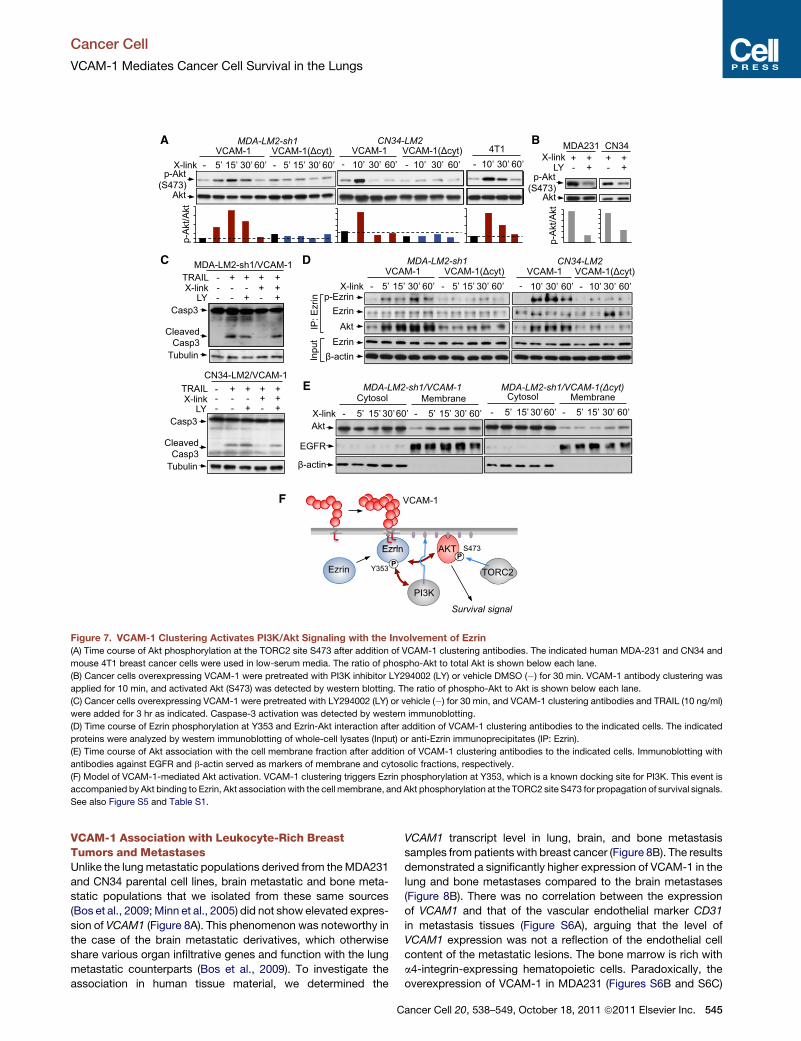
VCAM-1 Association with Leukocyte-Rich BreastTumors and MetastasesUnlike the lungmetastatic populations derived from theMDA231
and CN34 parental cell lines, brain metastatic and bone meta-
static populations that we isolated from these same sources
(Bos et al., 2009;Minn et al., 2005) did not show elevated expres-
sion of VCAM1 (Figure 8A). This phenomenon was noteworthy in
the case of the brain metastatic derivatives, which otherwise
share various organ infiltrative genes and function with the lung
metastatic counterparts (Bos et al., 2009). To investigate the
association in human tissue material, we determined the
C
VCAM1 transcript level in lung, brain, and bone metastasis
samples from patients with breast cancer (Figure 8B). The results
demonstrated a significantly higher expression of VCAM-1 in the
lung and bone metastases compared to the brain metastases
(Figure 8B). There was no correlation between the expression
of VCAM1 and that of the vascular endothelial marker CD31
in metastasis tissues (Figure S6A), arguing that the level of
VCAM1 expression was not a reflection of the endothelial cell
content of the metastatic lesions. The bone marrow is rich with
a4-integrin-expressing hematopoietic cells. Paradoxically, the
overexpression of VCAM-1 in MDA231 (Figures S6B and S6C)
ancer Cell 20, 538–549, October 18, 2011 ª2011 Elsevier Inc. 545

A 4
0
2
3
1
CN3410
VC
AM
1 m
RN
A
4
0
2
6
8
MDA231
Metastaticderivatives
Metastaticderivatives
D C
LungBone
BrainOther sites
-4 40-8Row Z-score
VC
AM
1 e
xpre
ssio
n (lo
g2)
0
2
4
-1
1
3
Leukocyte signature
highlow-2
p=0.0001
EB
p<
0.0001
VC
AM
1 e
xpre
ssio
n (lo
g2)
Lung Brain -2
0
2
4
-1
1
3
Bone
p<
0.0001
Leukocyte
4 integrin
P
VCAM-1– cancer cell VCAM-1+ cancer cell Leukocyte
Figure 8. VCAM1 Expression Is Associated with
Leukocyte Content in Breast Tumors
(A) Relative VCAM1 mRNA level in lung metastatic, brain
metastatic, and bone metastatic cells derived from
parental MDA231 or CN34 cell lines. Data are average ±
SEM; n = 3.
(B) VCAM1 expression in metastatic tissues from patients
with cancer including samples from lung (n = 18), brain (n =
19), and bone metastasis (n = 17).
(C and D) VCAM1 expression in metastatic lesions with
high or low leukocyte content. (C) Clustering of 67 meta-
static samples of patients with breast cancer by LeukoS.
(D) VCAM1 expression in leukocyte signature high and low
clusters of metastatic lesions.
(E) Model of the survival advantage obtained by breast
cancer cells that express VCAM-1 in leukocyte-rich tissues
such as the lung. On exposure to macrophages and other
leukocytes, the engagement of VCAM-1 by a4-integrin
counter-receptors triggers Ezrin phosphorylation and
activation of PI3K/Akt signaling. PI3K/Akt signaling
opposes the effects of death cytokines in the pulmonary
microenvironment.
See also Figure S6.
Cancer Cell
VCAM-1 Mediates Cancer Cell Survival in the Lungs
did not enhance the ability of these cells to initiate bone marrow
colonization (Figures S6D–S6F).
The selective association of VCAM1 expression with lung
metastasis was intriguing in light of our functional evidence link-
ing stromal leukocytes and VCAM-1-mediated cancer cell
survival. Therefore, we sought evidence for a possible associa-
tion of VCAM1 expression with the leukocyte content of primary
tumors. From a gene expression signature of CD45+CD10�
leukocytes isolated from human breast tumors (Allinen et al.,
2004), we generated a bioinformatic classifier. Using this tool,
we classified 67 metastasis tissue samples from patients with
breast cancer into leukocyte gene expression signature (Leu-
koS)-high and -low groups (Figure 8C). The LeukoS-high group
was highly enriched for lung metastasis samples and excluded
most of the bone and brain metastasis samples (Figure 8C).
This finding is consistent with the natural capacity of the lung
parenchyma to be infiltrated by lymphoid and myeloid cells
(Lipscomb et al., 1995). Furthermore, a comparison of the
VCAM1 transcript levels with the leukocyte signature score of
these tumors revealed that VCAM1 expression was enriched in
the LeukoS-high metastases (Figure 8D). These results with clin-
ical samples are consistent with the implication from our func-
tional studies that VCAM-1-expressing cancer cells have
a survival advantage to initiate metastasis at sites that are rich
in leukocytes, such as the lungs (Figure 8E).
DISCUSSION
The adaptation of newly arrived cancer cells to themicroenviron-
ment of distal organs is a stringent rate-limiting step in metas-
tasis, and the probability of completing this step varies widely
depending on the tumor type and the target organ. The specific
stromal interactions that dictate the compatibility of DTCs with
a particular organ site have remained largely amystery. Focusing
on VCAM-1 as an overexpressed gene in breast cancer cells that
preferentially colonize the lungs, we show that this cell adhesion
molecule is engaged by counter-receptor a4-integrins on leuko-
546 Cancer Cell 20, 538–549, October 18, 2011 ª2011 Elsevier Inc.
cytes to trigger PI3K/Akt activation and cancer cell survival in the
pulmonary parenchyma (Figure 8E). Our findings provide a bio-
chemical explanation for the clinical association of VCAM-1
expression in breast tumors with cancer relapse to the lungs
(Minn et al., 2005). Our findings also show that VCAM-1 expres-
sion is high in leukocyte-rich lung metastasis samples from
patients with breast cancer compared to brain metastasis
samples. We propose that these associations are based on a
prosurvival advantage provided by VCAM-1 in cancer cells that
invade leukocyte-rich sites such as the lungs.
Tumor-associated macrophages and other stromal leuko-
cytes play crucial roles in various aspects of tumorigenesis
through the secretion of specific cytokines and proteases
(Cheng et al., 2007; Giraudo et al., 2004; Gocheva et al., 2010;
Lin et al., 2006; Sangaletti et al., 2008; Wyckoff et al., 2004).
The present results reveal an unprecedented role for juxtacrine
stimulation of cancer cells by macrophages during metastatic
colonization. VCAM-1 on the surface of cancer cells mediates
a4-integrin-dependent binding of macrophages, and its oligo-
merization triggers PI3K/Akt signaling in the cancer cells. High-
resolution imaging techniques recently demonstrated physical
contacts between macrophages and breast cancer cells during
macrophage-facilitated CTC extravasation in mice (Qian et al.,
2009). However, we found no contribution of VCAM-1 to cancer
cell extravasation or macrophage recruitment into tumors.
Rather, our evidence suggests that the a4-integrin-VCAM-1
interaction promotes cancer cell survival through PI3K/Akt
pathway activation by the VCAM-1 cytoplasmic domain. During
leukocyte transmigration, VCAM-1 on endothelial cells can
communicate with the cytoplasmic proteins Ezrin and Moesin,
and trigger Rac1 activation (Barreiro et al., 2002; van Wetering
et al., 2003). Our data indicate that VCAM-1 clustering in breast
cancer cells mediates Ezrin phosphorylation, Akt binding to
Ezrin, and Akt phosphorylation at the TORC2 site S473 to acti-
vate prosurvival signaling. The protein kinase(s) responsible
for Ezrin phosphorylation at Y353 remains unknown, although
Src has been implicated as an indirect mediator (Chuan et al.,
Cancer Cell
VCAM-1 Mediates Cancer Cell Survival in the Lungs
2006; Krieg and Hunter, 1992). Thus, leukocyte engagement of
VCAM-1 in breast cancer cells may serve to amplify the activa-
tion of the PI3K/Akt pathway in a mitogen-poor microenviron-
ment during the initial stages of seeding and adaptation of
DTCs to new sites.
We propose that VCAM-1 expression confers breast cancer
cells with a selective survival advantage in leukocyte-rich
tissues. As organs exposed to the outside environment, the
lungs are rich in resident immune cells (Lipscomb et al., 1995).
In tumor-bearing mice the lungs accumulate bone marrow-
derived mononuclear phagocytes and endothelial progenitors,
cell types that express a4-integrins (Gao et al., 2008; Kaplan
et al., 2005). Our data suggest that a4-integrin-expressing leuko-
cytes and endothelial cells in lungs mediate Akt prosurvival
signaling by directly interacting with VCAM-1 in metastatic
breast cancer cells. The pulmonary parenchyma thereby
provides a matching receptive soil for CTCs that express
VCAM-1, explaining the observed link between VCAM-1 expres-
sion in primary tumors and selective relapse to the lungs (Minn
et al., 2005).
Cancer cells departing from a breast tumor with high expres-
sion of VCAM-1 would be primed for survival in the leukocyte-
rich microenvironment of the lungs. In comparison the brain
has a limited presence of microglial macrophages, which is in
line with the lack of an association of VCAM-1 with brain metas-
tasis that we observed. In the case of bonemetastasis, we found
no association between VCAM-1 expression in primary tumors
and bone relapse, and no effect of VCAM-1 overexpression on
the ability of breast cancer cells to initiate bonemarrow coloniza-
tion in mice. VCAM-1 may not provide an early advantage in the
bone marrow because Akt activation in cancer cells that enter
this site can be provided by other mechanisms, such as Src-
dependent CXCL12 signaling (Zhang et al., 2009). We do not
rule out a role of VCAM-1 later in the osteolytic phase of bone
metastasis.
What drives the selection for VCAM-1-expressing cancer cells
in breast tumors is unknown, but it is possible that VCAM-1-rich
clones are positively selected in breast tumor areas of active
infiltration or inflammation. Macrophage-rich areas of a primary
tumor would nurture the expansion of VCAM-1-expressing
cancer cell clones. Furthermore, breast cancer cells in lung
metastatic nodules can reenter the circulation, seed back a
mammary tumor, and abundantly proliferate in leukocyte-rich
tumor areas (Kim et al., 2009), and we provide evidence that
VCAM-1 favors the outgrowth of CTCs that infiltrate a self-seed-
ing mammary tumor. Accordingly, lung metastatic clones ex-
pressing VCAM-1 might undergo recurrent cycles of amplifica-
tion by shuttling between lung nodules and a leukocyte-rich
primary tumor microenvironment.
By disrupting the a4-integrin-VCAM-1 interaction between
macrophages and cancer cells with anti-a4-integrin-blocking
antibodies, we were able to cancel the prosurvival action of
VCAM-1 in the cancer cells. The interaction between endothelial
VCAM-1 and leukocyte a4-integrins is a validated target in
multiple sclerosis and other diseases that involve rampant
recruitment of circulating leukocytes into tissue (Comi, 2009;
Schmidt et al., 2009). Our results point at a potential application
of such targeted drugs to suppress VCAM-1-mediated survival
of DTCs after the removal of a primary tumor.
C
EXPERIMENTAL PROCEDURES
Animal Studies
All experiments using animals were done in accordance to a protocol
approved by MSKCC Institutional Animal Care and Use Committee (IACUC).
Female NOD/SCID or BALB/c mice (NCI; Charles River Lab and Taconic
Farm) between 5 and 7 weeks old were used. Lung metastasis assays from
orthotopic inoculation followed previously described procedures (Padua
et al., 2008). A total of 5 3 105 cancer cells in 50 ml of 1:1 mix of PBS/growth
factor-reduced Matrigel (BD Biosciences) was injected into the fourth right
mammary fat pad of mice. The primary tumors were surgically removed
when they each reached approximately 300 mm3. After 7 days, lung metas-
tases were quantified using BLI. For lung colonization assays, 2 3 105 cells
in 100 ml PBSwere intravenously injected, and lung colonization was quantified
using BLI. For inducible knockdown experiments, doxycycline hyclate (Sigma-
Aldrich) was added into the drinking water (2 mg/ml). For the primary tumor
implantation with a low number of cells, 5 3 104 cells were injected. Tumor-
seeding assays followed previously described procedures (Kim et al., 2009).
A total of 5 3 105 unlabeled MDA231 parental cells were injected into the
second right mammary fat pad of mice. After 7 days, 100,000 GFP/lucif-
erase-integrated LM2 cells were intracardially injected into tumor-bearing
mice. To detect tumor-immune cell interactions in the lungs, cancer cells
were labeled with 5 mM CellTracker Green CMFDA (Molecular Probes), and
106 labeled cells were intravenously injected. Lung tissues were harvested
24 hr after injection, and CD4, CD8, and CD68 staining was performed. To
detect CTCs, the whole-blood perfusate from tumor-bearing mice was
collected, and total RNA was extracted. The relative expression of human
b2-microglobulin was assessed by qRT-PCR and normalized to murine
b2-microglobulin.
Cell-Cell Binding Assays
A total of 2 3 106 prelabeled U937 cells were allowed to adhere to the mono-
layer of cancer cells. Adherent cells (red) and the nucleus of total cells (DAPI
staining in blue) were scored by fluorescence microscopy. The ratio of
adherent U937 cells to cancer cells was calculated as red counts/(blue
counts � red counts). In the indicated experiments, U937 cells were preincu-
bated with functional blocking antibody of a4-integrin (10 mg/ml; clone PS/2
fromAbD Serotec) or Ig control for 30min. To identify the cell types that adhere
to cancer cells, human EpCAM-depleted stromal cell suspensions from lung
metastatic nodules were allowed to adhere to GFP+ cancer cell monolayers.
Single-cell suspensions of cancer cells and adherent cells were incubated
with fluorochrome-conjugated F4/80 antibody. F4/80+ macrophages and
GFP+ cancer cells were quantified using flow cytometry.
VCAM-1 Clustering Assays
VCAM-1 in human cancer cells was crosslinked with 10 mg/ml of mouse
anti-human VCAM-1 monoclonal antibody (clone 1G11; Beckman Coulter)
and 50 mg/ml goat anti-mouse IgG1 F(ab0)2 (Jackson ImmunoResearch) for
indicated time frames, whereas 10 mg/ml of mouse anti-human VCAM-1
monoclonal antibody (Clone MVCAM.A429; AbD Serotec) and 50 mg/ml goat
anti-rat IgG1 F(ab0)2 (Jackson ImmunoResearch) were used in 4T1 mouse
breast cancer cells. Mouse or rat IgG was used as control. For apoptosis
analysis, cancer cells were incubated with 100 ng/ml TRAIL (PeproTech) for
3 hr. Caspase-3 was detected by western immunoblotting. To identify
signaling mediators, VCAM-1 clustering was performed in serum-starved
human breast cancer cells for the indicated time periods. Where indicated,
50 mM LY294002 or DMSO control was added during the incubation with
anti-VCAM-1 and F(ab0)2. Cell lysates were analyzed using Human Phospho-
Kinase Array Kit (R&D Systems). In the indicated experiments, western blot
was performed using antibodies against phospho-Akt (S473), Akt, phospho-
NF-kB p65 (S468), or phospho-Smad2 (S465/467) and phospho-Smad3
(S423/425) (all from Cell Signaling). The ratio of phospho-Akt to total Akt was
quantified by ImageJ software (Abramoff et al., 2004). For coimmunoprecipita-
tion assays, cell pellets were lysed with IP buffer (Pierce). Equal amounts of
protein were incubated with anti-Ezrin (clone 3C12; Abcam) and protein G
(GE Healthcare). Ezrin, phospho-Ezrin (Y353; BD PharMingen), and Akt were
detected in the immunoprecipitates. For cell fractionation, membrane and
cytosolic fractions were harvested using Subcellular Protein Fractionation kit
ancer Cell 20, 538–549, October 18, 2011 ª2011 Elsevier Inc. 547

Cancer Cell
VCAM-1 Mediates Cancer Cell Survival in the Lungs
(Pierce). Akt was detected in each fraction, whereas EGFR or b-actin (Cell
Signaling) was used as the loading control for membrane or cytosolic fraction,
respectively.
To clustering VCAM-1 in tumor cells by physiologically interacting with
a4-integrin in macrophages, confluent cancer cells were cocultured with
U937 cells (2.5 3 105 U937 cells/cm2) in the presence of 20 ng/ml TRAIL. To
detect the percentage of apoptotic cancer cells, cancer cells were prelabeled
with Green CMFDA before the coculture. TUNEL staining was performed 24 hr
after incubation and observed under the fluorescent microscope. To quantify
survived tumor cells, cells were lysed by Passive Lysis Buffer (Promega), and
the luciferase activity was detected by Luciferase Assay System (Promega)
using GloMax 96 Microplate Luminometer (Promega). Standard curve was
generated by detecting the luciferase activity of a series number of cancer
cells, and the absolute number of survived cancer cells was calculated. In
the indicated experiments, a4-integrin antibody was applied in the coculture
system.
Analysis of Human Metastatic Tissue Samples
VCAM1 transcript level was analyzed in 18 lung, 19 brain, and 17 bone meta-
static samples from patients with breast cancer (GSE14020) (Zhang et al.,
2009). A correlation coefficient was calculated between VCAM1 and CD31
expression in each data set. A LeukoS, including 182 genes, was based on
the SAGE data from CD45+CD10� leukocytes isolated from breast tumors
(Allinen et al., 2004). After mapping these genes on the Affymetrix U133A plat-
form, the probe sets of these genes were queried in a data set from 67 meta-
static samples from patients with breast cancer (GSE14020) (Zhang et al.,
2009). We clustered all these samples using the heatmap.2 function in the R
statistical package. The most robust subcluster was defined as the leukocyte
signature high (LeukoS-high) group. VCAM1 expressions were compared
between these two groups.
Statistical Analysis
Pairwise comparisons were performed by two-tailed Student’s t test. Signifi-
cance was set at p values less than 0.05.
SUPPLEMENTAL INFORMATION
Supplemental Information includes six figures, one table, and Supplemental
Experimental Procedures and can be found with this article online at
doi:10.1016/j.ccr.2011.08.025.
ACKNOWLEDGMENTS
The authors thank J. Joyce and members of the J.M. lab for insightful discus-
sion and technical suggestions, and the MSKCC Molecular Cytology Core
Facility for histological sample staining and analysis. This work was supported
by National Institutes of Health Grants CA126518 and CA94060, and the Alan
and Sandra Gerry Metastasis Research Initiative. Q.C. is supported by Life
Sciences Research Foundation Fellowship. J.M. is an investigator of the
Howard Hughes Medical Institute.
Received: January 30, 2011
Revised: July 7, 2011
Accepted: August 17, 2011
Published: October 17, 2011
REFERENCES
Abramoff, M.D., Magelhaes, P.J., and Ram, S.J. (2004). Image processing with
ImageJ. Biophotonics International 11, 36–42.
Alessi, D.R., Andjelkovic, M., Caudwell, B., Cron, P., Morrice, N., Cohen, P.,
and Hemmings, B.A. (1996). Mechanism of activation of protein kinase B by
insulin and IGF-1. EMBO J. 15, 6541–6551.
Allinen, M., Beroukhim, R., Cai, L., Brennan, C., Lahti-Domenici, J., Huang, H.,
Porter, D., Hu, M., Chin, L., Richardson, A., et al. (2004). Molecular character-
ization of the tumor microenvironment in breast cancer. Cancer Cell 6, 17–32.
548 Cancer Cell 20, 538–549, October 18, 2011 ª2011 Elsevier Inc.
Anan, K., Mitsuyama, S., Koga, K., Tanabe, R., Saimura, M., Tanabe, Y.,
Watanabe, M., Suehara, N., Matsunaga, H., Nishihara, K., et al. (2010).
Disparities in the survival improvement of recurrent breast cancer. Breast
Cancer 17, 48–55.
Aslakson, C.J., and Miller, F.R. (1992). Selective events in the metastatic
process defined by analysis of the sequential dissemination of subpopulations
of a mouse mammary tumor. Cancer Res. 52, 1399–1405.
Barreiro,O.,Yanez-Mo,M.,Serrador, J.M.,Montoya,M.C.,Vicente-Manzanares,
M., Tejedor, R., Furthmayr, H., and Sanchez-Madrid, F. (2002). Dynamic in-
teraction of VCAM-1 and ICAM-1 with moesin and ezrin in a novel endothelial
docking structure for adherent leukocytes. J. Cell Biol. 157, 1233–1245.
Bos, P.D., Zhang, X.H., Nadal, C., Shu, W., Gomis, R.R., Nguyen, D.X., Minn,
A.J., van de Vijver, M.J., Gerald, W.L., Foekens, J.A., andMassague, J. (2009).
Genes that mediate breast cancer metastasis to the brain. Nature 459, 1005–
1009.
Cailleau, R., Young, R., Olive, M., and Reeves, W.J., Jr. (1974). Breast tumor
cell lines from pleural effusions. J. Natl. Cancer Inst. 53, 661–674.
Cameron, M.D., Schmidt, E.E., Kerkvliet, N., Nadkarni, K.V., Morris, V.L.,
Groom, A.C., Chambers, A.F., and MacDonald, I.C. (2000). Temporal progres-
sion of metastasis in lung: cell survival, dormancy, and location dependence of
metastatic inefficiency. Cancer Res. 60, 2541–2546.
Cheng, J., Huo, D.H., Kuang, D.M., Yang, J., Zheng, L., and Zhuang, S.M.
(2007). Human macrophages promote the motility and invasiveness of osteo-
pontin-knockdown tumor cells. Cancer Res. 67, 5141–5147.
Chuan, Y.C., Pang, S.T., Cedazo-Minguez, A., Norstedt, G., Pousette, A., and
Flores-Morales, A. (2006). Androgen induction of prostate cancer cell invasion
is mediated by ezrin. J. Biol. Chem. 281, 29938–29948.
Collins, B.J., Deak, M., Arthur, J.S., Armit, L.J., and Alessi, D.R. (2003). In vivo
role of the PIF-binding docking site of PDK1 defined by knock-in mutation.
EMBO J. 22, 4202–4211.
Comi, G. (2009). Treatment of multiple sclerosis: role of natalizumab. Neurol.
Sci. 30 (Suppl 2 ), S155–S158.
Cybulsky, M.I., Fries, J.W., Williams, A.J., Sultan, P., Eddy, R., Byers, M.,
Shows, T., Gimbrone,M.A., Jr., and Collins, T. (1991). Gene structure, chromo-
somal location, and basis for alternative mRNA splicing of the human VCAM1
gene. Proc. Natl. Acad. Sci. USA 88, 7859–7863.
Ding, Y.B., Chen, G.Y., Xia, J.G., Zang, X.W., Yang, H.Y., and Yang, L. (2003).
Association of VCAM-1 overexpressionwith oncogenesis, tumor angiogenesis
and metastasis of gastric carcinoma. World J. Gastroenterol. 9, 1409–1414.
Elices, M.J., Osborn, L., Takada, Y., Crouse, C., Luhowskyj, S., Hemler, M.E.,
and Lobb, R.R. (1990). VCAM-1 on activated endothelium interacts with the
leukocyte integrin VLA-4 at a site distinct from the VLA-4/fibronectin binding
site. Cell 60, 577–584.
Fehon, R.G., McClatchey, A.I., and Bretscher, A. (2010). Organizing the cell
cortex: the role of ERM proteins. Nat. Rev. Mol. Cell Biol. 11, 276–287.
Fidler, I.J. (2003). The pathogenesis of cancer metastasis: the ‘seed and soil’
hypothesis revisited. Nat. Rev. Cancer 3, 453–458.
Gao, D., Nolan, D.J., Mellick, A.S., Bambino, K., McDonnell, K., and Mittal, V.
(2008). Endothelial progenitor cells control the angiogenic switch in mouse
lung metastasis. Science 319, 195–198.
Gautreau, A., Poullet, P., Louvard, D., and Arpin, M. (1999). Ezrin, a plasma
membrane-microfilament linker, signals cell survival through the phosphatidy-
linositol 3-kinase/Akt pathway. Proc. Natl. Acad. Sci. USA 96, 7300–7305.
Giraudo, E., Inoue, M., and Hanahan, D. (2004). An amino-bisphosphonate
targets MMP-9-expressing macrophages and angiogenesis to impair cervical
carcinogenesis. J. Clin. Invest. 114, 623–633.
Gocheva, V., Wang, H.W., Gadea, B.B., Shree, T., Hunter, K.E., Garfall, A.L.,
Berman, T., and Joyce, J.A. (2010). IL-4 induces cathepsin protease activity
in tumor-associated macrophages to promote cancer growth and invasion.
Genes Dev. 24, 241–255.
Gupta, G.P., Nguyen, D.X., Chiang, A.C., Bos, P.D., Kim, J.Y., Nadal, C.,
Gomis, R.R., Manova-Todorova, K., and Massague, J. (2007). Mediators of
vascular remodelling co-opted for sequential steps in lung metastasis.
Nature 446, 765–770.
Cancer Cell
VCAM-1 Mediates Cancer Cell Survival in the Lungs
Jones, D.H., Nakashima, T., Sanchez, O.H., Kozieradzki, I., Komarova, S.V.,
Sarosi, I., Morony, S., Rubin, E., Sarao, R., Hojilla, C.V., et al. (2006).
Regulation of cancer cell migration and bone metastasis by RANKL. Nature
440, 692–696.
Joyce, J.A., and Pollard, J.W. (2009). Microenvironmental regulation of
metastasis. Nat. Rev. Cancer 9, 239–252.
Kalogeris, T.J., Kevil, C.G., Laroux, F.S., Coe, L.L., Phifer, T.J., and Alexander,
J.S. (1999). Differential monocyte adhesion and adhesionmolecule expression
in venous and arterial endothelial cells. Am. J. Physiol. 276, L9–L19.
Kang, Y., Siegel, P.M., Shu, W., Drobnjak, M., Kakonen, S.M., Cordon-Cardo,
C., Guise, T.A., and Massague, J. (2003). A multigenic program mediating
breast cancer metastasis to bone. Cancer Cell 3, 537–549.
Kaplan, R.N., Riba, R.D., Zacharoulis, S., Bramley, A.H., Vincent, L., Costa, C.,
MacDonald, D.D., Jin, D.K., Shido, K., Kerns, S.A., et al. (2005). VEGFR1-posi-
tive haematopoietic bonemarrow progenitors initiate the pre-metastatic niche.
Nature 438, 820–827.
Kim, M.Y., Oskarsson, T., Acharyya, S., Nguyen, D.X., Zhang, X.H., Norton, L.,
and Massague, J. (2009). Tumor self-seeding by circulating cancer cells. Cell
139, 1315–1326.
Krieg, J., and Hunter, T. (1992). Identification of the two major epidermal
growth factor-induced tyrosine phosphorylation sites in the microvillar core
protein ezrin. J. Biol. Chem. 267, 19258–19265.
Lin, E.Y., Li, J.F., Gnatovskiy, L., Deng, Y., Zhu, L., Grzesik, D.A., Qian, H., Xue,
X.N., and Pollard, J.W. (2006). Macrophages regulate the angiogenic switch in
a mouse model of breast cancer. Cancer Res. 66, 11238–11246.
Lipscomb,M.F., Bice, D.E., Lyons, C.R., Schuyler, M.R., andWilkes, D. (1995).
The regulation of pulmonary immunity. Adv. Immunol. 59, 369–455.
Luzzi, K.J., MacDonald, I.C., Schmidt, E.E., Kerkvliet, N., Morris, V.L.,
Chambers, A.F., and Groom, A.C. (1998). Multistep nature of metastatic inef-
ficiency: dormancy of solitary cells after successful extravasation and limited
survival of early micrometastases. Am. J. Pathol. 153, 865–873.
MacDonald, I.C., Groom, A.C., and Chambers, A.F. (2002). Cancer spread and
micrometastasis development: quantitative approaches for in vivo models.
Bioessays 24, 885–893.
Minn, A.J., Gupta, G.P., Siegel, P.M., Bos, P.D., Shu, W., Giri, D.D., Viale, A.,
Olshen, A.B., Gerald, W.L., and Massague, J. (2005). Genes that mediate
breast cancer metastasis to lung. Nature 436, 518–524.
Minn, A.J., Gupta, G.P., Padua, D., Bos, P., Nguyen, D.X., Nuyten, D., Kreike,
B., Zhang, Y., Wang, Y., Ishwaran, H., et al. (2007). Lung metastasis genes
couple breast tumor size and metastatic spread. Proc. Natl. Acad. Sci. USA
104, 6740–6745.
Muller,A.,Homey,B., Soto,H.,Ge,N.,Catron,D.,Buchanan,M.E.,McClanahan,
T., Murphy, E., Yuan, W., Wagner, S.N., et al. (2001). Involvement of chemokine
receptors in breast cancer metastasis. Nature 410, 50–56.
Nguyen, D.X., Bos, P.D., and Massague, J. (2009). Metastasis: from dissemi-
nation to organ-specific colonization. Nat. Rev. Cancer 9, 274–284.
Nicholson, D.W., Ali, A., Thornberry, N.A., Vaillancourt, J.P., Ding, C.K.,
Gallant, M., Gareau, Y., Griffin, P.R., Labelle, M., Lazebnik, Y.A., et al.
(1995). Identification and inhibition of the ICE/CED-3 protease necessary for
mammalian apoptosis. Nature 376, 37–43.
Osborn, L., Hession, C., Tizard, R., Vassallo, C., Luhowskyj, S., Chi-Rosso, G.,
and Lobb, R. (1989). Direct expression cloning of vascular cell adhesion mole-
cule 1, a cytokine-induced endothelial protein that binds to lymphocytes. Cell
59, 1203–1211.
Padua, D., Zhang, X.H., Wang, Q., Nadal, C., Gerald, W.L., Gomis, R.R., and
Massague, J. (2008). TGFbeta primes breast tumors for lung metastasis seed-
ing through angiopoietin-like 4. Cell 133, 66–77.
Paez-Ribes, M., Allen, E., Hudock, J., Takeda, T., Okuyama, H., Vinals, F.,
Inoue, M., Bergers, G., Hanahan, D., and Casanovas, O. (2009).
Antiangiogenic therapy elicits malignant progression of tumors to increased
local invasion and distant metastasis. Cancer Cell 15, 220–231.
Perez, O.D., Kinoshita, S., Hitoshi, Y., Payan, D.G., Kitamura, T., Nolan, G.P.,
and Lorens, J.B. (2002). Activation of the PKB/AKT pathway by ICAM-2.
Immunity 16, 51–65.
C
Prat, A., Parker, J.S., Karginova, O., Fan, C., Livasy, C., Herschkowitz, J.I., He,
X., and Perou, C.M. (2010). Phenotypic and molecular characterization of the
claudin-low intrinsic subtype of breast cancer. Breast Cancer Res. 12, R68.
Qian, B., Deng, Y., Im, J.H., Muschel, R.J., Zou, Y., Li, J., Lang, R.A., and
Pollard, J.W. (2009). A distinct macrophage population mediates metastatic
breast cancer cell extravasation, establishment and growth. PLoS One 4,
e6562.
Qian, B.Z., and Pollard, J.W. (2010). Macrophage diversity enhances tumor
progression and metastasis. Cell 141, 39–51.
Qian, B.Z., Li, J., Zhang, H., Kitamura, T., Zhang, J., Campion, L.R., Kaiser,
E.A., Snyder, L.A., and Pollard, J.W. (2011). CCL2 recruits inflammatory
monocytes to facilitate breast-tumour metastasis. Nature 475, 222–225.
Ricono, J.M., Huang, M., Barnes, L.A., Lau, S.K., Weis, S.M., Schlaepfer, D.D.,
Hanks, S.K., and Cheresh, D.A. (2009). Specific cross-talk between epidermal
growth factor receptor and integrin alphavbeta5 promotes carcinoma cell
invasion and metastasis. Cancer Res. 69, 1383–1391.
Rubin, J., Titus, L., and Nanes, M.S. (1991). Regulation of complement 5a
receptor expression in U937 cells by phorbol ester. J. Leukoc. Biol. 50, 502–508.
Ruco, L.P., de Laat, P.A., Matteucci, C., Bernasconi, S., Sciacca, F.M., van der
Kwast, T.H., Hoogsteden, H.C., Uccini, S., Mantovani, A., and Versnel, M.A.
(1996). Expression of ICAM-1 and VCAM-1 in humanmalignant mesothelioma.
J. Pathol. 179, 266–271.
Sangaletti, S., Di Carlo, E., Gariboldi, S., Miotti, S., Cappetti, B., Parenza, M.,
Rumio, C., Brekken, R.A., Chiodoni, C., and Colombo, M.P. (2008).
Macrophage-derived SPARC bridges tumor cell-extracellular matrix interac-
tions toward metastasis. Cancer Res. 68, 9050–9059.
Sarbassov, D.D., Guertin, D.A., Ali, S.M., and Sabatini, D.M. (2005).
Phosphorylation and regulation of Akt/PKB by the rictor-mTOR complex.
Science 307, 1098–1101.
Schmidt, K.J., Buning, J., Jankowiak, C., Lehnert, H., and Fellermann, K.
(2009). Crohn’s targeted therapy: myth or real goal? Curr. Drug Discov.
Technol. 6, 290–298.
Shin, J., Kim, J., Ryu, B., Chi, S.G., and Park, H. (2006). Caveolin-1 is associ-
ated with VCAM-1 dependent adhesion of gastric cancer cells to endothelial
cells. Cell. Physiol. Biochem. 17, 211–220.
Tavazoie, S.F., Alarcon, C., Oskarsson, T., Padua, D., Wang, Q., Bos, P.D.,
Gerald, W.L., and Massague, J. (2008). Endogenous human microRNAs that
suppress breast cancer metastasis. Nature 451, 147–152.
van Wetering, S., van den Berk, N., van Buul, J.D., Mul, F.P., Lommerse, I.,
Mous, R., ten Klooster, J.P., Zwaginga, J.J., and Hordijk, P.L. (2003). VCAM-
1-mediated Rac signaling controls endothelial cell-cell contacts and leukocyte
transmigration. Am. J. Physiol. Cell Physiol. 285, C343–C352.
Vonderheide, R.H., Tedder, T.F., Springer, T.A., and Staunton, D.E. (1994).
Residues within a conserved amino acid motif of domains 1 and 4 of
VCAM-1 are required for binding to VLA-4. J. Cell Biol. 125, 215–222.
Wong, C.W., Lee, A., Shientag, L., Yu, J., Dong, Y., Kao, G., Al-Mehdi, A.B.,
Bernhard, E.J., and Muschel, R.J. (2001). Apoptosis: an early event in meta-
static inefficiency. Cancer Res. 61, 333–338.
Wyckoff, J., Wang, W., Lin, E.Y., Wang, Y., Pixley, F., Stanley, E.R., Graf, T.,
Pollard, J.W., Segall, J., and Condeelis, J. (2004). A paracrine loop between
tumor cells and macrophages is required for tumor cell migration in mammary
tumors. Cancer Res. 64, 7022–7029.
Yamamoto, T., Sakaguchi, N., Hachiya,M., Nakayama, F., Yamakawa,M., and
Akashi, M. (2009). Role of catalase inmonocytic differentiation of U937 cells by
TPA: hydrogen peroxide as a second messenger. Leukemia 23, 761–769.
Yin, J.J., Selander, K., Chirgwin, J.M., Dallas, M., Grubbs, B.G., Wieser, R.,
Massague, J., Mundy, G.R., and Guise, T.A. (1999). TGF-beta signaling
blockade inhibits PTHrP secretion by breast cancer cells and bonemetastases
development. J. Clin. Invest. 103, 197–206.
Zhang, X.H., Wang, Q., Gerald, W., Hudis, C.A., Norton, L., Smid, M., Foekens,
J.A., and Massague, J. (2009). Latent bone metastasis in breast cancer tied to
Src-dependent survival signals. Cancer Cell 16, 67–78.
Zoncu, R., Efeyan, A., and Sabatini, D.M. (2011). mTOR: from growth signal
integration to cancer, diabetes and ageing. Nat. Rev. Mol. Cell Biol. 12, 21–35.
ancer Cell 20, 538–549, October 18, 2011 ª2011 Elsevier Inc. 549