a macrophage cell line
TRANSCRIPT
Proc. Nati. Acad. Sci. USAVol. 87, pp. 914-918, February 1990Immunology
Interferon y regulates binding of two nuclear protein complexes ina macrophage cell line
(class H major histocompaibility complex genes/DNA-binding protein/transcription/lymphokine/Ia antigens)
PATRICIA W. FINN*, CATHERINE J. KARA*t, JOHN DOUHAN III*, Tu TRAN VAN*, VIRGINIA FOLSOMt,AND LAURIE H. GLIMCHER*§*Department of Cancer Biology, Harvard School of Public Health, Boston, MA 02115; §Department of Medicine, and tProgram in Immunology, HarvardMedical School, Boston, MA 02115; and tLife Technologies Inc., Gaithersburg, MD 20877
Communicated by Elkan Blout, October 30, 1989
ABSTRACT Interferon y (IFN-y) is a potent inducer ofmajor histocompatibility complex (MHC) antigens during nor-mal immune responses and in abnormal responses in autoim-mune disease. In this report we identify two nuclear factorswhose binding to the murine Ep class II MHC 3-chain gene isregulated by this cytokine. IFN-y stimulation of murine mac-rophages results in the appearance of increased binding of oneprotein complex, complex A, and decreased binding of asecond, faster migrating protein complex, complex B. Al-though the contact residues for both of these proteins lie withinthe highly conserved Y-box transcriptional element, theirbinding specificity differs. The protein in complex B is aCCAAT-box-binding protein that may be similar or identical toNF-Y or YB1, previously identified class II Y-box-bindingproteins. The DNA sequence requirements for the binding ofthe slower migrating complex, complex A, are not limited toCCAAT-box sequences but include sequences upstream of theY box. These upstream sequences are required both forIFN-y-induced gene transcription and for IFN-y-induced mod-ulation ofbinding activity. These data suggest a model in whichupstream sequences contribute to formation of a lymphokine-regulated complex downstream. The IFN-r-induced bindingprotein described as complex A in this report differs from theIFN-y, -a, or -13-induced nuclear factors previously identified.
The level of class II major histocompatibility complex (MHC)antigens on macrophages can be increased by several stimuli,the most potent of which is the cytokine interferon y (IFN-y).This increase in class II results in an increase in the intensity ofthe T-cell immune response (1). Furthermore, cultured cellsfrom virtually every organ as well as a variety oftumor cell linescan be induced to express class II in response to IFN-'y (2). Insome instances these cells then acquire the ability to act asantigen-presenting cells toT lymphocytes (3). There is evidencethat abundant de novo expression of class II in target organsamplifies autoimmune tissue destruction (4). This inductionmay well be due to IFN-y, since astrocytes in experimentalallergic encephalomyelitis and synovial cells in rheumatoidarthritis express class II, and IFN-y has been shown to induceclass II on these cells in vitro (5, 6). Understanding the mech-anism by which IFN-y induces class II gene expression in itstarget cells is therefore of importance.
MATERIALS AND METHODSCell Line. The P388D1 murine macrophage-like cell line
was grown in RPMI 1640 medium (GIBCO) supplementedwith 8% fetal bovine serum, 10 mM Hepes, 100 Ag ofpenicillin per ml, 100 1Lg of streptomycin per ml, 0.1 mM2-mercaptoethanol, and 2 mM L-glutamine.
DNA Transfection. P388D1 cells were transfected by amodification of the DEAE-dextran method (7). Chloram-phenicol acetyltransferase (CAT) assays were performed asdescribed (8) and quantitated by scintillation counting. CATactivity was assayed at 72 hr after transfection, in thepresence or absence of recombinant IFN-y (rIFN-y; gift ofGenentech and the American Cancer Society), 20 units/ml,which was added for the last 48 hr.
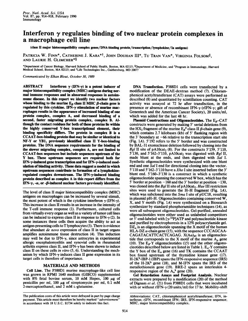
Plasmid Constructions and Oligonucleotides. The Ep-CATconstructs were generated by making 5' serial deletions fromthe HX2 fragment of the murine End class II(-chain gene (9),which contains 2.7 kilobases (kb) of 5' flanking region withthe 3' boundary at -66 relative to the transcription start site(Fig. 1A). 5'192 refers to the 5' border and was constructedby BAL-31 exonuclease deletion followed by cloning into theBgl II site of pAlOcat2 (8). For the constructs 5'159, 5'110,5'130, and 5'162-3'110, pAlOcat2 was digested with Bgl II,made blunt at the ends, and then digested with Sal I.Synthetic oligonucleotides were synthesized with one bluntend and one Sal I end for directional cloning into the vector.5'110 and 5'162-3'110 have aXho I site inserted before the 3'blunt end. 5'160-3'130 is a construct in which a syntheticoligonucleotide spanning the conservedW box, which has the5' border at position -160 and the 3' border at position -130,was cloned into the Bgl II site ofpAlOcat2. Hae III restrictionsites were used to generate the H-H fragment (Fig. lA),which was subcloned into the Sma I site of pUC18, resultingin plasmid pH-H. Oligonucleotides containing conserved W,X, and Y motifs (Fig. 1A) were synthesized on a Biosearchsynthesizer by standard phosphoramidite techniques (14), aswere all subsequent oligonucleotides. Gel-purified, annealedoligonucleotides were either used as unlabeled competitorsor 5'-end-labeled with ['y-32P]ATP and polynucleotide kinaseand purified by electrophoresis in a 12% polyacrylamide gel.DZ, is an oligonucleotide spanning the X motif of the humanHLA-DZ a-chain gene (15), with the sequence CCCAGCAA-CAGATACATTCACTCAGAG. X(Aa)45 is an oligonucleo-tide that corresponds to the X motif of the murine A,, gene(10). The E,8-Y oligonucleotides (15) and the other oligonu-cleotides described below are listed in Table 1. E,-Y containsthe Y box of the Ea gene (16) and TK contains the CCAATbox found upstream of the thymidine kinase gene (17).H-2Kb-IBP-1 (IBP) spans the IFN-responsive sequence (IRS)of the H-2Kb gene (18), and M-IFN spans the IRS of themetallothionein gene (19). BRE-2 spans an interleukin 4-responsive region of the A", gene (20).Gel Retardation Assays and Footprint Analysis. Nuclear
extracts were prepared by a modification (20) of the methodof Dignam et al. (21) from P388D1 cells that were incubatedwith or without rIFN-y (20 units/ml) for 17 hr. Mobility-shift
Abbreviations: CAT, chloramphenicol acetyltransferase; IFN, in-terferon; rIFN, recombinant IFN; IRS, IFN-responsive sequence;MHC, major histocompatibility complex.
914
The publication costs of this article were defrayed in part by page chargepayment. This article must therefore be hereby marked "advertisement"in accordance with 18 U.S.C. §1734 solely to indicate this fact.
Proc. Natl. Acad. Sci. USA 87 (1990) 915
-aY6ol go -
- W 01lg9 - H-160 -130 -90 -76 -66
.._._. a t a e 1 P 1*- tE-PtMt to oA ______A.A .___ ................I............................I.............I. rS_
X Box Y Box
I l"IC7ATIV4Opxomoee
310SV40promoie
B
C) a)CM Z 0 Z CZ c) z cr c> Lx - LL 0 L T L LO LL(n - < ~~- C~j L-L*- 7
CL + CL + C) + Ln + If) + If
4b404
0 0_: rn_
D Z O I CMi+ LO + LO +
* @@e.. , . ,_
* *0 Q+q
1 2 3 4 5 6 7i9
8 9 10 I11 12 13 14 15 16 17 18
FIG. 1. (A) Epd-CAT constructs and oligonucleotides. Part of the 5' flanking sequence of the End gene (9) is shown, including conservedW, X, and Y motifs (refs. 10-13; boxed). Hae III sites (H) are indicated. Bars above the sequence represent synthetic oligonucleotides usedin gel retardation analyses. Constructs used in transfection experiments are shown below the sequence and contained the indicated portionsofthe sequence inserted upstream ofthe simian virus 40 (SV40) promoter and CAT gene ofpAlOcat2. (B) IFN-y induction ofE'sd-CAT constructsin the P388D1 macrophage cell line. Epd-CAT constructs were assayed for CAT activity in response to rIFN-y in transient transfection assays.P388D1 cells transfected with pSV2cat (contains SV40 promoter and enhancer; ref. 8) (lanes 1 and 2), pAlOcat2 (contains SV40 promoter only)(lanes 3 and 4), and constructs as in A (lanes 5-12) were assayed for CAT activity expressed in the absence (odd-numbered lanes) or presence(even-numbered lanes) of rIFN-y. Each transfection and CAT assay was performed as five independent experiments and a representative resultis shown.
assays were performed as described (20). The H-H fragmentprobe (Fig. lA) was prepared by BamHI cleavage of pH-H(described above) and end-labeling with either [ac-32P]dNTPand Klenow fragment of DNA polymerase I or [Yy-32P]ATPand polynucleotide kinase, followed by digestion with EcoRIand isolation by gel electrophoresis. Approximately 10,000cpm ("1 ng) of end-labeled DNA was used for each 10-,ulreaction mixture containing 2.4-4.0 pug of protein from nu-clear extract. Nuclear extracts were normalized for protein(Bio-Rad protein assay) and equal amounts were addedwithin experiments. As an additional control for proteinconcentration, extracts were incubated with a 220-base-pair(bp) fragment of the Aa upstream region. This resulted inthree retarded bands, none of which was affected by IFN-ytreatment (data not shown). DNA-protein complexes wereresolved in a 4% polyacrylamide gel in 45 mM Tris/45 mMboric acid/1 mM EDTA (0.Sx TBE) run at 11 V/cm withcooling. The gels were dried and autoradiographed withintensifying screens at -70'C. For competition analysis,
nuclear extracts were preincubated with water or competitorDNA for 5 min at room temperature; all other componentswere then added and the samples were processed as formobility shift analysis. For footprint analysis, ten identical10-,tl reaction mixtures were pooled into one large lane of a4% acrylamide gel. After electrophoresis, the gel was sub-jected to copper cleavage as described (22), except thatincubation was for 11.5 min. Bands of interest were excised,eluted, purified, and run in a 6% acrylamide/7 M urea gel.Sequences were aligned by comparison to the G, G+AMaxam-Gilbert ladder (23) of the end-labeled DNA.
RESULTS AND DISCUSSIONMacrophages are sensitive targets for the actions of IFN-y.A 2- to 5-fold IFN-y-mediated increase in class II genetranscription has been reported (24-28), and we have foundthat the cell surface expression of class II MHC antigens inP388D1, a murine macrophage cell line, is induced after 24 hrof treatment with as little as 1 unit of rIFN-y per ml (data not
Table 1. Sequences of oligonucleotidesName Sequence Ref.
EwY36 CTACCTTTGATGCTGATTGGCTCCCAGCACTGGCCT 15E#-Y56 TGGAGACTCCTTTGATGCTGATTGGCTCCAGCACTGGCCTTTACCCAATCTCGAG 15Ea-Y GTCTGAAACATTTTTCTGATTGGTTAAAAGTTGAGTGCT 16TK GCGTCTTGTCATTGGCGAATTCGAACACGC 17H-2KbIBP-1 CAGGTTAGGTGCAGAAGTGAAAQ22ZGGAGATGGGGAATCC 18M-IFN T 19M-IFN I~~TCTCCACCTCGGCAGIT-TCTMIQCTCSS1BRE-2 (Aa) GATCCCGTGATTACCTTAATATGTTTGCCTAGAAGGAGGCAAA 20
Solid underlining, Y-box or CCAAT-box motif; broken underlining, IFN-responsive sequence (IRS).
A
H- 207 -192
W Box
5192
5'159
rE)
U-0
zIL+"
GAGTATCCATGTAATGAaGAGAACTGCAAG TT[TCAGAAGGGGACMA.TUATC TC TALACTAGCAACTGATGIATGCTGGACTCCTTTGATG..TCCCAGCACTGGCCTTACCkAATICCA
Immunology: Finn et al.
Proc. Natl. Acad. Sci. USA 87 (1990)
shown). To identify the upstream sequences responsible forthe transcriptional effects of IFN-'y, P388D1 cells were trans-fected with plasmid constructs containing 5' upstream se-quences of the mouse Ed gene fused to the CAT gene.Starting with 2.6 kb of upstream sequence, serial 5' deletionswere made and tested for IFN-y inducibility. Preliminaryresults identified a 127-bp region required for IFN-y induc-ibility (Fig. 1). This region extends from - 192 to -66 relativeto the start site of transcription and includes three conservedmotifs, W, X, and Y. The X and Y motifs are two conservedelements located in the same positions in all class II genessequenced to date (10-12). W is a conserved motif locatedjust upstream of the X motif in all class II P-chain genes (13).A construct containing residues -159 to -67 also was IFN-yinducible (Fig. 1B). The level of induction averaged 3-fold,which is consistent with data obtained in several otherlaboratories that define IRS elements upstream of the humanDQs and DR, and murine Ea and En class II genes (29-32) andis comparable to the 2- to 5-fold IFN-y-mediated increases intranscription noted previously (24-28). This construct (- 159to -67) contains the three conserved motifs, W, X, and Y,and initiated at the appropriate transcription start site (datanot shown). Constructs containing X and Y alone (-130 to-67) or Y alone (-110 to -67) did not confer IFN-y induc-ibility. A construct containing W alone (-160 to -130) alsowas not induced above the constitutive level by IFN-ytreatment. These results suggest that the IRS element liesbetween - 159 and - 130 but requires downstream sequences,including X and Y motifs, for full IFN-'y responsiveness. Inaddition, constructs containing W and X (Fig. 1B, lane 15) orY alone (lane 13) had greater constitutive activity than thosecontaining X and Y alone (lane 11), suggesting the presenceof a negative regulatory element between - 130 and - 110 thatalso requires downstream elements. Both negative and pos-itive regulatory elements have been demonstrated for theDQp and Ea genes (29, 32). Constructs containing X alone(-130 to -110) may further define the boundaries of theseelements in the Ed gene.The activation of transcription by specific DNA sequences
is usually accomplished by transcription factors that bind tothese sequences. An IFN (a8)-induced nuclear factor thatbinds upstream of the class I MHC gene family has beenidentified by several groups (33); however, there is noobvious homology between the class I MHC IFN consensus
NoIFN
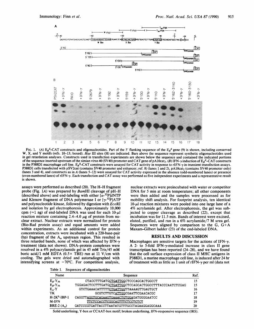
sequence and proximal upstream class II sequences (unpub-lished results) with the exception of a sequence located at-564 upstream of the DRa gene (19). When nuclear extractsfrom P388D1 cells treated for 17 hr with rIFN-y (20 units/ml)were analyzed by mobility-shift analysis with a radiolabeled130-bp Hae III (H-H) El3 DNA fragment containing all threeconserved motifs, two distinct retarded bands were observed(Fig. 2A). These two DNA-protein complexes are termed Aand B. Densitometry showed that complex A (upper band)was increased 6-fold by treatment of P388D1 cells withrIFN-y for 17 hr (lane 3), whereas complex B was diminished2-fold. Proteinase K treatment abolished complex formation(data not shown). In 10 separate experiments with differentP388D1 nuclear extracts, the increase in complex A rangedfrom 2.8-fold to 17-fold and the decrease in complex B rangedfrom 1.3-fold to 2-fold.To determine the binding site of these proteins, competi-
tion experiments were performed with unlabeled DNAprobes representing the W, X, and Y motifs (Fig. 2B).Addition of a 40-fold molar excess of E#-Y36 oligonucleotideeffectively decreased formation of complex B (lane 15) with-out affecting complex A formation. In order to prevent theformation of complex A, 400-fold molar excess Y-box com-petitor (lane 17) was required. However, the Y oligonucleo-tide did not prevent the formation of complex A or B asefficiently as the intact upstream region (H-H); a 50-foldmolar excess ofH-H inhibited the formation of complexes Aand B (lane 4). As little as 10-fold molar excess H-Heffectively decreased complex A formation (data not shown).Furthermore, when W or X oligonucleotide alone was usedas competitor, no inhibition was seen of either complex A orB (lanes 6-13). When W and X were used together, or addedwith Y, there was no difference as compared to the use of Yalone (not shown). Interestingly, an oligonucleotide from theupstream region of the Aa gene, BRE-2 (Table 1; this se-quence has been shown to be responsive to interleukin 4),behaved in a different way, inhibiting formation of complexA but not of complex B (Fig. 2C, lanes 5 and 6). This effectwas specific, as X(Aa)45, an oligonucleotide spanning the Xmotif of the A, gene (13), prevented the formation of neithercomplex (lane 7). Thus, the proteins in complexes A and Bappear to have different sequence requirements for binding,since BRE-2 competed only with complex A.
+ IFN -a
A
Extract _ 2- 99
Complex A-
Complex B ' i w
Free Probe --H-H(E) 130
12 3
Competitor H- H W X y
molcr excess 2 500 8 40D' 2C00400 8 40 200400 8a 20C40 - C
dl 66 d& 44 M i by 34T w et0A omdlC eor
Complex A-d- 1.i molor excess~:N - -T
Complex B - .iMbi
Free Probe-. hIH-H(E3)130
UL-
5,_ki.....
.4r.4 0400-4C
4.w4mieA.
12 3 4 6 C;. 7 8 9 10 1 2 3 '- 5 F..,
FIG. 2. (A) Regulation of binding of nuclear protein to the class II Ed gene by IFN-y. Mobility-shift assays were conducted with nuclearextracts from P388D1 cells untreated (lane 2) or treated with rIFN-y at 20 units/ml (lane 3). Lane 1 shows free H-H probe (no extract added).(B) Unlabeled Y box preferentially inhibits formation of labeled complex B. Competitors were H-H, the unlabeled 130-bp homologous HaeIII-Hae III fragment (lanes 3-5); W (lanes 6-9), X (lanes 10-13), and a 36-bp Y oligonucleotide (lanes 16 and 17), which are unlabeledoligonucleotides from the Ep upstream region corresponding to conserved W, X, and Y motifs (Fig. 1A); and DZa (lane 18). The amount ofcompetitor indicated above each lane represents nanograms; 10 ng equals approximately 40-fold molar excess of probe concentration for W,X, and Y. (C) BRE-2 inhibits formation of only complex A. Competitors were BRE-2 (lanes 3-6) and X(Aa)45 (lane 7).
916 Immunology: Finn et al.
Proc. Natl. Acad. Sci. USA 87 (1990) 917
c-85 B - +cCTcG y -i~Tr -
>-98 F,-107
-10
2
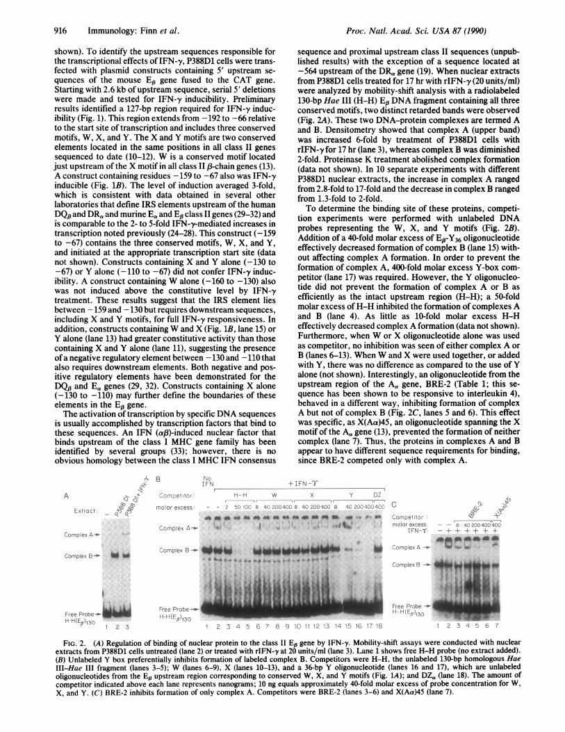
FIG. 3. (A) Footprint analysis of binding of nuclear extracts ofcells treated with IFN-y to probe H-H (noncoding strand). Lane 1,complex A; lane 2, complex B; lane 3, free probe. G and G/Arepresent Maxam-Gilbert ladders. (B) DNA-binding activity ofP388D1 nuclear proteins to Y boxes as an isolated target sequence.The probe was a radiolabeled oligonucleotide Ep-Y36 (Table 1). Lane1, unstimulated sample; lane 2, IFN-y treated sample. The mobility-shift assays were performed as in Fig. 2.
To further define the target sequence, footprinting analysisof the noncoding strand was performed on each of the twocomplexes by using the nuclease activity of 1,10-phenanthro-line-copper ion (22). In complex B, which is downregulatedby rIFN-y, the Y motif (positions -90 to -97) and part of thesequence between X and Y (bases -85 to -107) wereprotected (Fig. 3A). In complex A, which is upregulated byrIFN-y, a more restricted region was protected that laycompletely within the B footprint and included only the Ymotif (bases -85 to -98). The 3' border of both A and Bprotection extended beyond the Y motif to position -85.Identical results were obtained for the coding strand (data notshown). These results suggest the existence of two distinct
DNA-protein complexes that are regulated by rIFN-y. Com-plex B has a footprint pattern very similar to NF-Y, an E,,Y-box-binding protein described by Dorn et al. (11). Ofinterest in this regard is the isolation by Didier et al. (34) ofa cDNA, YB1, encoding a DRa Y-box-binding protein whoseexpression is negatively regulated by IFN-y at the mRNAlevel.The localization of the footprint of complex A to the Y
motif was consistent with our earlier results (Fig. 1B) sug-gesting that downstream elements are necessary for fullrIFN-y responsiveness. The results of others (32) had alsodemonstrated that the presence of all three motifs, W, X, andY, was required for rIFN-y-induced gene transcription. Onepossibility is that complex A proteins must initially interactwith sequences upstream of Y before forming a final lym-phokine-responsive complex at the Y-box site. It was ofinterest therefore to determine whether binding to the Y motifalone was increased by IFN-y treatment. When the Y-boxoligonucleotide was used as probe, two complexes, Ys(upper band) and Y,2 (lower band), were detected (Fig. 3B,lane 1) and a third complex, which migrated between thesetwo, was occasionally observed. The Y-box complexes werealso present in IFN-y-stimulated samples but were not in-creased as evidenced by densitometry (lane 2). Formation ofthe complexes binding to the Y box was sequence-specific, asdemonstrated by competition experiments using homologousand irrelevant DNA probes. A 10-fold molar excess ofhomologous Y box effectively prevented formation of bothYpl and Y,2 (data not shown). Complexes Y,31 and Y,02 did notbehave like complex A, since their formation was not mod-ulated by rIFN-y and was prevented by a much lower amountof Y-box oligonucleotide than required to inhibit complex Aformation (Fig. 2B, lanes 16 and 17). This suggests a modelin which upstream sequences contribute to formation of alymphokine-regulated complex downstream.
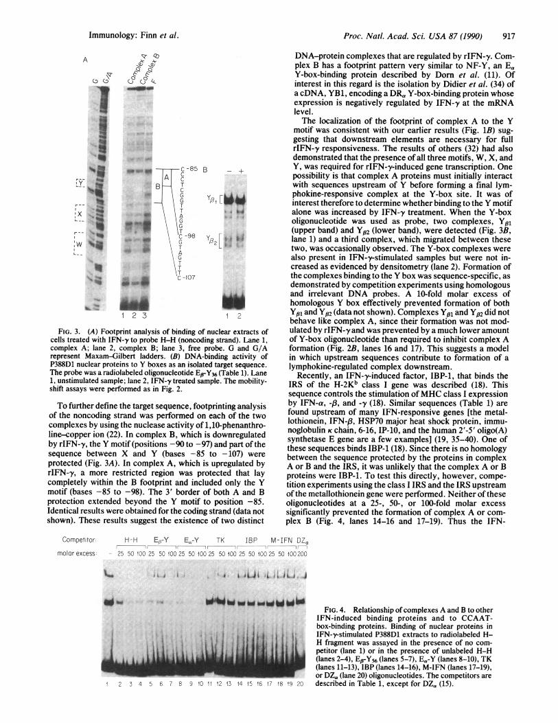
Recently, an IFN-'y-induced factor, IBP-1, that binds theIRS of the H-2Kb class I gene was described (18). Thissequence controls the stimulation ofMHC class I expressionby IFN-a, -,3, and -y (18). Similar sequences (Table 1) arefound upstream of many IFN-responsive genes [the metal-lothionein, IFN-,B, HSP70 major heat shock protein, immu-noglobulin K chain, 6-16, IP-10, and the human 2'-5' oligo(A)synthetase E gene are a few examples] (19, 35-40). One ofthese sequences binds IBP-1 (18). Since there is no homologybetween the sequence protected by the proteins in complexA or B and the IRS, it was unlikely that the complex A or Bproteins were IBP-1. To test this directly, however, compe-tition experiments using the class I IRS and the IRS upstreamof the metallothionein gene were performed. Neither of theseoligonucleotides at a 25-, 50-, or 100-fold molar excesssignificantly prevented the formation of complex A or com-plex B (Fig. 4, lanes 14-16 and 17-19). Thus the IFN-
Competitor- H-H EB-Y Ea-Y TK IBP M-IFN DZamolar excess: - 25 50 100 25 50 100 25 50 100 25 50 100 25 50 100 25 50 IOC 200
'M il 4 i I J i rf , Ji I A
O'b
L2 3 4 5 6 7 8 9 10 11 12 13 14 15 16 17 18 19 20
FIG. 4. Relationship ofcomplexes A and B to otherIFN-induced binding proteins and to CCAAT-box-binding proteins. Binding of nuclear proteins inIFN-y-stimulated P388D1 extracts to radiolabeled H-H fragment was assayed in the presence of no com-petitor (lane 1) or in the presence of unlabeled H-H(lanes 2-4), EBY56 (lanes 5-7), EaY (lanes 8-10), TK(lanes 11-13), IBP (lanes 14-16), M-IFN (lanes 17-19),or DZa (lane 20) oligonucleotides. The competitors aredescribed in Table 1, except for DZa (15).
000C) Cj)
oa--q
A
I--I
I--
r
I _..N:wY V41I_ _
% .__-Adl 2 3
Immunology: Finn et al.
Proc. Natl. Acad. Sci. USA 87 (1990)
y-induced class 11-binding proteins appear to represent aspecies distinct from IBP-1.The Y-box sequence of all class II genes is highly conserved
and contains an inverted CCAATbox. The Y-box motifs ofthehuman DR& and the murine Ea genes are identical to the Ep Ybox but differ substantially in the flanking sequences (Table 1).These DRa and Ea Y-box motifs have been shown to bindproteins YB1 (34) and NF-Y (11, 16), respectively, which areCCAAT-box-binding proteins distinct from the CCAAT-box-binding proteins previously described (e.g., CBP andCTF/NF-1). To determine the relationship between complexA and B proteins and the YB1 and NF-Y CCAAT-box-bindingproteins, competition experiments were performed. Forma-tion of complex B was prevented by a 25-fold excess ofoligonucleotide containing the Ea Y box (lanes 8-10) or the EpY box (lanes 5-7) but not as well by an oligonucleotidecontaining the CCAAT motifupstream ofthe thymidine kinasegene (lanes 11-13). The complex B protein therefore behaveslike YB1 and NF-Y and may be similar or identical to one orboth of these proteins. Final resolution of this issue awaits theisolation of cDNA clones encoding the complex B protein.Complex A formation can be most efficiently prevented by theDNA fragment containing W, X, and Y motifs (compare Fig.4, lanes 2-4, and Fig. 2B, lanes 3-5, to Fig. 4, lanes 5-7 and8-10) and therefore does not appear to be a CCAAT-box-binding protein.
This report demonstrates the binding of IFN-y-modulatedproteins to class II gene transcriptional control regions.Overall, our results are most easily explained by invokingtwo different Y-box-binding proteins with distinct but over-lapping target sequences within the Y box. Another possi-bility is that the different mobilities of the complexes and thedistinct footprints result from posttranslational modificationof a single binding protein or from formation of a dimericprotein. Indeed, complex A may be generated by the bindingof complex B protein multimers or ofB protein complexed toanother protein. The observation that the W- and X-boxregions are also required for IFN-y-induced gene transcrip-tion and sequence-specific binding suggests a cooperativeinteraction among all three motifs.
We thank Drs. J. Cairns, D. Perkins, E. Gravallese, and G.Viglianti for careful critique of the manuscript, H.-C. Liou and M.Boothby for helpful discussions, and D. Ballard for advice on copperfootprinting. We acknowledge L. Blood for expert manuscript prep-aration. This work was supported by grants from the NationalInstitutes of Health (P.W.F. and L.H.G.), the Leukemia Society(L.H.G.), the March of Dimes Foundation (L.H.G.), and PfizerPharmaceuticals (P.W.F.).
1. Matis, L. A., Glimcher, L. H., Paul, W. E. & Schwartz, R. H.(1983) Proc. Nati. Acad. Sci. USA 80, 6019-6023.
2. Skoskiewicz, M. J., Colvin, R. B., Schneeberger, E. E. &Russell, P. S. (1985) J. Exp. Med. 162, 1645-1664.
3. Pober, J. S., Collins, T., Gimbrone, M. A., Jr., Cotran, R. S.,Gitlin, J. D., Fiers, W., Clayberger, C., Krensky, A. M.,Burakoff, S. & Reiss, C. S. (1983) Nature (London) 305,726-729.
4. Adelman, N. E., Watling, D. L. & McDevitt, H. 0. (1985)Proc. Natl. Acad. Sci. USA 82, 6627-6631.
5. Matsumoto, Y., Hara, N., Tanaka, R. & Fujiwara, M. J. (1986)J. Immunol. 136, 3668-3676.
6. Teyton, L., Lotteau, V., Turmel, P., Arenzana-Seisdedos, F.,Virelizier, J.-L., Pujol, J.-P., Loyau, G., Piatier-Tonneau, D.,Auffray, C. & Charron, D. J. (1987) J. Immunol. 138, 1730-1738.
7. Dorsett, D. L., Keshet, I. & Wincour, E. (1983) J. Virol. 48,218-228.
8. Gorman, C. M., Moffat, L. F. & Howard, B. H. (1982) Mol.Cell. Biol. 2, 1044-1055.
9. Gillies, S. D., Folsom, V. & Tonegawa, S. (1984) Nature(London) 310, 594-597.
10. Boothby, M. R., Liou, H. C. & Glimcher, L. H. (1989) J.Immunol. 142, 1005-1014.
11. Dorn, A., Durand, B., Marfings, C., LeMeur, M., Benoist, C.& Mathis, D. (1987) Proc. Natl. Acad. Sci. USA 84,6249-6253.
12. Widera, G., Burkly, L. C., Pinkert, C. A., Bottger, E. C.,Cowing, C., Palmiter, R. D., Brinster, R. L. & Flavell, R. A.(1987) Cell 51, 175-187.
13. Miwa, K., Doyle, C. & Strominger, J. L. (1987) Proc. Nati.Acad. Sci. USA 84, 4939-4943.
14. Liou, H.-C., Boothby, M. R. & Glimcher, L. H. (1988) Science242, 69-71.
15. Kelly, A. & Trowsdale, J. (1985) Nucleic Acids Res. 13,1607-1621.
16. Dorn, A., Bollekens, J., Staub, A., Benoist, C. & Mathis, D.(1987) Cell 50, 863-872.
17. Kreidberg, J. A. & Kelly, T. J. (1986) Mol. Cell. Biol. 6,2903-2909.
18. Blanar, M. A., Baldwin, A. S., Jr., Flavell, R. A. & Sharp,P. A. (1989) EMBO J. 8, 1139-1144.
19. Friedman, R. L. & Stark, G. R. (1985) Nature (London) 314,637-639.
20. Boothby, M., Gravallese, E., Liou, H.-C. & Glimcher, L. H.(1988) Science 242, 1559-1562.
21. Dignam, J. D., Lebowitz, R. H. & Roeder, R. G. (1983) Nu-cleic Acids Res. 11, 1475-1488.
22. Kiwabara, M. D. & Sigman, D. S. (1987) Biochemistry 26,7234-7238.
23. Maniatis, T., Fritsch, E. F. & Sambrook, J. (1982) MolecularCloning:A Laboratory Manual (Cold Spring Harbor Lab., ColdSpring Harbor, NY).
24. Bottger, E. C., Blanar, M. A. & Flavell, R. A. (1988) Immu-nogenetics 28, 215-220.
25. Fertsch-Ruggio, D., Schoenberg, D. R. & Vogel, S. N. (1988)J. Immunol. 141, 1582-1589.
26. Blanar, M. A., Boettger, E. C. & Flavell, R. A. (1988) Proc.Natl. Acad. Sci. USA 85, 4672-4676.
27. Amaldi, I., Reith, W., Berte, C. & Mach, B. (1989) J. Immunol.142, 999-1004.
28. Woodward, J. G., Omar, K. W. & Stewart, P. M. (1989) J.Immunol. 142, 4062-4069.
29. Boss, J. M. & Strominger, J. (1986) Proc. Natl. Acad. Sci. USA83, 9139-9143.
30. Sherman, P. A., Basta, P. V. & Ting, J. P.-Y. (1987) Proc.Natl. Acad. Sci. USA 84, 4254-4258.
31. Tsang, S. T., Nakanish, M. & Peterlin, B. M. (1988) Proc.Natl. Acad. Sci. USA 85, 8598-8602.
32. Thanos, D., Mavrothalassitis, G. & Papamatheakis, J. (1988)Proc. Natl. Acad. Sci. USA 85, 3075-3079.
33. Shirayoshi, Y., Burke, P. A., Apella, E. & Ozato, K. (1988)Proc. Natl. Acad. Sci. USA 85, 5884-5888.
34. Didier, D., Schifferbauer, J., Woulfe, S. L., Zacheis, M. &Schwartz, B. J. (1988) Proc. Natl. Acad. Sci. USA 85, 7322-7326.
35. Goodbourn, S., Burnstein, H. & Maniatis, T. (1986) Cell 45,601-610.
36. Wu, B. J., Kingston, R. E. & Morimoto, R. I. (1986) Proc.Natl. Acad. Sci. USA 83, 629-633.
37. Max, E. E., Maizel, J. V., Jr., & Leder, P. (1981) J. Biol.Chem. 256, 5116-5120.
38. Porter, A. C. G., Chernajovsky, Y., Dale, T. C., Gilbert,C. S., Stark, G. R. & Kerr, I. M. (1988) EMBO J. 7, 85-92.
39. Luster, A. & Ravetch, J. (1987) Mol. Cell. Biol. 7, 3723-3731.40. Benech, P., Vigneron, M., Peretz, D., Revel, M. & Chebath, J.
(1987) Mol. Cell. Biol. 7, 4498-4504.
918 Immunology: Finn et al.