m6a-mediated stabilization of fstl5 progression through
TRANSCRIPT

Page 1/23
miR-186-5p Prevents Hepatocellular CarcinomaProgression Through Targeting METTL3 to Regulatem6A-Mediated Stabilization of FSTL5DengYong Zhang
: First A�liated Hospital of Bengbu Medical CollegeFangFang Chen
: First A�liated Hospital of Bengbu Medical CollegeShuoShuo Ma
: First A�liated Hospital of Bengbu Medical CollegeYongChun Zhou
: First A�liated Hospital of Bengbu Medical CollegeWanliang Sun
: First A�liated Hospital of Bengbu Medical CollegeDongdong Wang
: First A�liated Hospital of Bengbu Medical CollegeShiRu Chang
Bengbu Medical CollegeZheng Lu ( [email protected] )
: First A�liated Hospital of Bengbu Medical College https://orcid.org/0000-0002-9090-0875
Primary research
Keywords: microRNA, hepatocellular carcinoma, METTL3, FSTL5, mRNA stability
Posted Date: June 15th, 2021
DOI: https://doi.org/10.21203/rs.3.rs-598803/v1
License: This work is licensed under a Creative Commons Attribution 4.0 International License. Read Full License

Page 2/23
AbstractBackground: Hepatocellular carcinoma (HCC) processes in multi-steps which involves the sophisticatedinteractions of genetics, epigenetics, and transcriptional changes. According to before investigations,methyltransferase-like 3 (METTL3)-mediated m6A modi�cation regulates the development of variouscancers by regulating gene stability. However, the studies focusing on miRNA’s regulatory effect of N6-methyladenosine (m6A) modi�cation on HCC progression are still limited.
Methods: Immunochemistry (IHC) staining detected the histopathological changes in the tumor tissues.Cell Counting Kit-8 (CCK-8), clone formation, and transwell assay investigated the changes in cancer cellproliferation, invasion, and migration. The RNA m6A level was con�rmed by methylated RNAimmunoprecipitation. The RNA stability assay indicated the half-life (t1/2) of RNA in HCC cells. Theprognosis of the indicated patients’ cohort was analyzed using the cancer genome atlas (TCGA)datasets. Luciferase report analysis was used to study the potential binding between microRNA (miRNA)and mRNA. A mice tumor transplant model was further established to study the changes in tumorprogression.
Results: Follistatin-like 5 (FSTL5) was found to be signi�cantly downregulated in HCC, and it inhibited thefurther progression of HCC. The RNA stability analysis indicated that the mRNA t1/2 gene of HCC cellswas shortened. Besides, METTL3 reduced the stability of FSTL5 mRNA in a m6A-YTH domain family2(YTHDF2)-dependent manner. Functional experiments revealed that the downregulated METTL3inhibited the HCC progression by up-regulating FSTL5 in vitro and in vivo. Luciferase report analysiscon�rmed that miR-186-5p directly targeted the METTL3. Additionally, miR-186-5p inhibited theproliferation, migration, and invasion of HCC cells by downregulating METTL3. We identi�ed that miR-186-5p prevented the HCC progression by targeting METTL3 to regulate m6A-mediated FSTL5stabilization.
Conclusions: The miR-186-5p/METTL3/YTHDF2/FSTL5 axis perhaps point out a new direction for thetargeted therapy of HCC.
IntroductionHepatocellular carcinoma (HCC), a deadly cancer, ranks sixth in the common types of cancer globally [1,2]. HCC might be induced by nonalcoholic fatty liver disease, type 2 diabetes mellitus, obesity and etc.,making the incidence of HCC grow annually [3]. The clinical treatment of HCC is limited because of itsinconspicuous early symptoms and late diagnosis [4]. Thus, it is of vital importance to understand themolecular mechanism of HCC pathogenesis to provide a direction for HCC diagnosis and therapy.
The follistatin family is a kind of extracellular matrix glycoprotein. Its participation in physiologicalprocesses, in�ammatory response, tissue remodeling, and embryonic development has been veri�ed [5].As a member of the follistatin family, FSTL5 is a protein that directly binds to activin [6]. More and morestudies have shown that FSTL5 also participated in regulating tumor occurrence. Remke M et al. found

Page 3/23
that FSTL5 was a poor prognostic marker of non-WNT/non-SHH medulloblastoma [7]. FSTL5 maybecome an attractive therapeutic target and prognostic indicator [8]. However, the potential mechanism ofFSTL5 in HCC still needs further exploration.
More studies have shown that gene mutations and epigenetic regulations, including methylation,chromatin modi�cation, and remodeling, are the drivers of HCC [9]. N6-methyladenosine (m6A) RNAmethylation, a most abundant RNA modi�cations, plays a critical role in tumor progression [10, 11]. m6Amodi�cation regulates gene expressions by regulating the mRNA stability, splicing, translation, andmicroRNA maturation [12]. m6A modi�cation is mediated by m6A “writers”, “erasers”, and “readers”,which function in adding, removing, and recognizing m6A modi�cations, respectively [13]. METTL3, themain component of m6A “writers”, promotes m6A RNA modi�cation, which is recognized by m6A“readers”, including the YTHDF, heterogeneous nuclear ribonucleoprotein A2B1 (hnRNPA2B1), and insulin-like growth factor 2 mRNA-binding protein 2 (IGF2BP) [14, 15]. It has been found that m6A “writers”,“erasers”, and “readers” function vital in regulatory in the pathogenesis of cancers, including bladdercancer, gastric cancer, colorectal cancer, and HCC [16–19].
MicroRNA (miRNA) is an endogenous small non-coding RNA with 19–22 nucleotides; it can down-regulate gene expression after transcription via targeting the 3'UTR gene [20]. miRNAs participate invarious cancer development and progression [21]. Recently, miRNAs were found to be regulated byMETTL3 through m6A modi�cation to in�uence the tumor genesis and development [22]. However, thestudies on whether miRNAs could regulate the m6A process in HCC development are still limited. Here, weidenti�ed that FSTL5 was downregulated in HCC and reported that FSTL5 has anti-tumor effects on HCC.Besides, METTL3 promoted the FSTL5 mRNA degradation, and miR-186-5p negatively regulated theexpression of METTL3. More importantly, miR-186-5p, as concluded, inhibited the progression of HCC bydownregulating METTL3, leading to FSTL5 upregulation by decreasing the m6A modi�cation of FSTL5.
Materials And MethodsTissue sample collection
Twenty-eight pairs of HCC tissues and adjacent non-tumor tissues were acquired from the First A�liatedHospital of Bengbu Medical College from January 2017 to January 2018. The agreement was signedwith the approval of its Ethics Committee (No. 2017059). All patients involved in this study did not receivechemotherapy or radiotherapy before the operation. The Supplementary Table 1 shows thecharacteristics of all participants.
Cell culture and transfection
The human HCC cell lines HepG2 and Huh-7 were purchased from American Type Culture Collection(ATCC, USA) and cell bank of Chinese Academy of Sciences (Guangzhou, China), respectively. Both cellswere grown in a Dulbecco’s modi�ed Eagle’s medium (DMEM) medium (Gibco, USA) supplemented with10% fetal bovine serum (FBS, Gibco, USA) at 37℃ in a water-saturated atmosphere with 5% CO2. For in
Page 4/23
vitro transfection, Lipofectamine 2000 (Invitrogen, USA) was used to help transfer 50nm mimic control,miR-186-5p mimic, or inhibitor (Ribo bio, Shanghai, China) into the cells. For the overexpression of FSTL5,METTL3, or YTHDF2 in cells, the cDNA of human FSTL5, METTL3, and YTHDF2 was cloned into pLV-EF1a-EGFP(2A) Puro vector by Genepharma (Guangzhou, China), respectively. The short hairpin RNA(shRNA) against METTL3 and FSTL5 was from Genepharma (Guangzhou, China). The SupplementaryTable 2 shows the sequences for transfection.
RNA isolation and Reverse Transcription Polymerase Chain Reaction (RT-qPCR)
The total RNA was separated by a Trizol reagent (Invitrogen, USA). The concentration of RNA wasdetected using NanoDrop (Thermo, USA). We quanti�ed the miRNAs and mRNAs using TaqMan miRNAprobes (Applied Biosystems, USA) and Synergy BrandsSynergy Brands (SYBR) Green Dyes (Ambion,USA), respectively. We used the HiScript III 1st Strand cDNA Synthesis Kit (Vazyme, China) and the mainchamp SYBR qPCR mixture (Vazyme, China) for retroviral polymerase chain reaction and quantitativepolymerase chain reaction, respectively. The calculated equation is shown below: ▲▲CT = (CTmiRNA or
mRNA – CTU6 or GAPDH) tumor – (CTmiRNA or mRNA-CTU6 or GAPDH) control. The primers are shown inSupplemental Table 3.
Western blot
Radio-Immunoprecipitation Assay (RIPA) lysis buffer and PMSF (Beyotime, China) extracted the totalprotein in the tissue or cell of tumor. The total protein concentration was determined using a BCAdetection kit (Santa Cruz, USA). After electrophoresis, the prepared protein sample was transferred toPVDF, where it hatches along with the prepared antibody. Finally, an enhanced chemiluminescence (ECL,ThermoFisher, USA) was used to visualize the membrane. ImageJ software was used to analyze theprotein bands. The antibodies against METTL3 and GAPDH were from CST (Beverly, USA). Theantibodies against FSTL5, methyltransferase-like 14 (METTL14), AlkB homolog 5 (ALKBH5), and Fatmass and obesity-associated protein (FTO) were purchased from Abcam (Cambridge, USA).
CCK-8 assay
The cells were placed in a 96-well plate, with 1 × 10⁴ cells/well. Then, 10 μL of CCK-8 solution was addedto the cell for 1 h at 37°C. The light density of the cells was measured at 450 nm by a microchannel platereader.
Clone formation assay
The cells were digested with 0.25% trypsin, plated in a 6-cm culture dish at a density of 2 × 10³/well, andincubated at 5% CO2 and 37°C for two weeks. Next, the cells in the dish were washed with PBS, �xed with5 mL of methanol for 15 min, and subjected to Giemsa dyeing for another 15 min. The colonies wasobserved and counted using an optical microscope.

Page 5/23
Cell migration and invasion assay
Transwell assay measured cell migration and invasion. After diluting (1:4) BD glue (BD biosciences, USA)with Opti-MEM™ I (Gibco,USA) , add 50ul Matrigel into the upper chamber. HepG2 and Huh-7 cells wereharvested and inoculated in an 8-mm well transmembrane plate in the upper transmembrane chamber ata density of 50,000 cells/well (Costar, USA) pre-treated with Matrigel or not. The upper and lowerchambers each were added serum-free medium and 20% FBS medium. After 24 h incubation,the unmigrated or uninvaded cells were wiped out cotton swabs. Pictures were taken, and the number ofcells was calculated. The average value of cells number was calculated after counting the photos of 6�elds.
Luciferase reporter assay
The wild-type or mutant 3’UTR of METTL3 was ampli�ed and cloned separately into pGL3-basic vector(Promega, USA). The binding site of the 3’UTR of METTL3 was mutated fromUGUACAAUAGCUUUCUUCUUUAU to UGUACAAUAGCUUUCAAGAAAAU for miR-186-5p. Then, the livercancer cells were placed in a 24-well plate and dyed with a wild or mutant luciferase plasmid and miR-186-5p mimic, miR-186-5p inhibitor. A Dual-Luciferase Reporter Assay System (Promga, USA) measuredthe activity of the two �uorescent enzymes.
m6A RNA methylation quanti�cation
An m6A RNA methylation analysis kit (Abcam, USA) was used to measure the total methylation contentof m6A. Brie�y, after adding the detection antibody solution in each reaction, 200 ng of the total RNA wasadded in each reaction. The m6A level was quanti�ed by coloration, and the absorbance of each reactionwas measured at 450 nm. For the measurement of FSTL5 m6A enrichment, the total RNA of HepG2 cellswas extracted; then, immunoprecipitation was done using a Magna MeRIP m6A kit (Merck milliporecompany, Germany) with protein A beads combined with m6A antibody in advance. The partial RNA wasanalyzed by RT-qPCR after immunoprecipitation.
RNA immunoprecipitation-RT-PCR
The HepG2 cells were transfected with YTHDF2 overexpression lentivirus. The virus was washed twicewith PBS, and it was dissolved in a lysine buffer with a protease inhibitor mixture and ribozyme inhibitorfor 30 min at 4°C. The cytolytic enzyme was centrifuged, and the �ltrate was collected. The supernatantsolution was heated for 4 h with magnetic beads (Sigma, USA), which were pre-processed by IgG orYTHDF2 antibodies (Abcam, USA); then, it was washed �ve times with a wash buffer (50 mM Tris, 0.5mM DTT, RNase inhibitor, 200 mM sodium chloride, 0.05% NP40, 2 mM ethylamine tetraethyl). Then, thebeads were washed in a solution containing 0.1% sodium dodecyl sulfate and 10 mL of potassiumenzyme and hatched for 30 min at 55°C. The imported nucleic acids and immune precipitates wereseparated using Trizol reagents (Invitrogen, USA) and analyzed quantitatively by a chain reaction ofpolymerase and gel electrophoresis of agarose.

Page 6/23
RNA Stability
The transfection of HepG2 cells with sh-NC was done to detect the stability of RNA in cells. sh-METTL3,the control lentivirus, or the YTHDF2 overexpression lentivirus for 24 h; then, 5 mg/mL actinomycin D(Sigma, USA) treated the cells. RNA isolation was done with a Trizol reagent (Invitrogen, USA) at 0, 3, and6 h. Then, it was normalized to GAPDH by real-time polymerase chain reaction analysis. The t1/2 ofmRNA was calculated.
Immunohistochemical (IHC) analysis
The tumor tissues covered with para�n were waxed and treated with 3% H2O to disable the endogenousperoxidase. After 1 h of blocking the bond with non-speci�c antigens at 37°C with 5% cow serumalbumin, the speci�c primary antibodies (1:100 diluted, Abcam, USA) against FSTL5, METTL3, orYTHDF2 were incubated at 4°C for 12 h. About 1 h after hatching the second antibody at 37°C, the slicewas dyed with diamino-ventylamine and dyed again in a small-purpose. A representative image wastaken with an optical microscope (Olympus, Japan).
Tumor xenograft model
About 1 × 10⁷ HepG2 cells were suspended in 200 μL of PBS and injected into each side of BALB/c nu/numale mice in the age of 4-6 weeks (Charles River Lab, Beijing, China). The tumor volume in mice wasrecorded every three days (volume = (length × width 2)/2). The knockdown of METTL3 and FSTL5 wasperformed using METTL3 and FSTL5 knockdown lentivirus (GenePharma, China), respectively, byintratumoral injection of 50 μL of virus (4 × 10⁷ IU/mL) after tumor cell injection. After 15 days, the micewere killed. Western blot analysis was carried out on the tumor tissues. Euthanasia method of mice: putthe mice into the box, inject carbon dioxide into the box at the rate of 20% per minute, observe that themice have no breathing, turn off carbon dioxide, and check again to con�rm that the mice have died (noheartbeat, pupil dilation). This animal experiment was consented by the Animal Health and UseCommittee of the First A�liated Hospital of Bengbu Medical College.
Hematoxylin and eosin (HE) staining
The tumor tissues were �xed in 4% paraformaldehyde (Sigma, USA) for 48 h and embedded in para�n.Thereafter, the tissues were sliced, and the sections were dyed with hematoxylin for 2 min and eosin for 1min. The images were viewed using a light microscope (Nikon, Japan).
Statistical analysis
The Prism Graphpad 8.0 software analyzed the data, which are in the form of mean values. Two-tailedStudent's t-test compared the two groups. One-way ANOVA compared multiple groups. p < 0.05 showedstatistical signi�cance.
Results

Page 7/23
FSTL5 inhibits HCC proliferation, migration, and invasionThe FSTL5 expression level was identi�ed in tissues and cell lines of HCC for �nding out its effects. Andwe found that its expression in adjacent non-tumor tissues was vitally higher than that in HCC tissues(Fig. 1A, B). Similarly, the level of FSTL5 in HCC cell line Hh-7 was downregulated in MHCC97-H, HepG2,SMMC-7721 cells (Fig. 1C, D). Since the average expression of FSTL5 was higher in Huh-7 and HepG2cells than in other HCC cells, these two cell lines were chosen for subsequent experiments. Then, theFSTL5 overexpression plasmid was transfected into Huh-7 and HepG2 cells. RT-qPCR analysis con�rmedthe transfection e�ciency of FSTL5 (Fig. 1E). The results indicated that the overexpression of FSTL5inhibited the cell viability, clone formation, migration, and invasion in Hh-7 and HepG2 cells (Fig. 1F-H).
Mettl3 Promotes Fstl5 Mrna DegradationThen, we investigated why FSTL5 was downregulated in HCC and served as a suppressor. The RNAstability test showed that the t1/2 gene of HCC cells was shortened, suggesting the possible presence ofRNA modi�cation (Fig. 2A). m6A is a relatively common chemical formula in eukaryotes and plays animportant role in many biological processes. Besides, m6A modi�cation regulates the mRNA stability toregulate the gene expression in cancer [23, 24]. Then, we detected the m6A level in HCC tissues. We foundthat the total m6A level in adjacent non-tumor tissues was lower than that in tumor (Fig. 2B). The m6Amodi�cation was mediated by m6A methyltransferases METTL3 and METTL14, which were cleared byfat mass and FTO [25, 26]. Interestingly, only METTL3 expression was markedly increased in HCC(Fig. 2C). We assumed that the upregulated METTL3 might regulate the stability of FSTL5 by m6Amodi�cation in HCC. MeRIP qPCR analysis was used to assess the impact of METTL3 on m6Amodi�cations aimed at the FSTL5 mRNA. As a result, m6A-speci�c antibodies were signi�cantly enrichedwith the FSTL5 gene compared to the immunoglobulin group. Besides, the inhibition of METTL3signi�cantly reduced the m6A level of FSTL5 mRNA (Fig. 2D). Additionally, the knockdown of METTL3extended the mRNA t1/2 (Fig. 2E). Then, we found that METTL3 negatively regulated the FSTL5expression of HCC cells, as indicated by RT-qPCR and western blot assays (Fig. 2F, G). We constructed aluciferase reporter plasmid containing the wild type and 3'UTR mutant of FSTL5. Luciferase reporter geneanalysis showed that METTL3 inhibition signi�cantly increased the activity of wild-type luciferase but notthat of the mutant FSTL5 3'UTR, indicating that the adjustment of FSTL5 representation was affected bythe m6A trim associated with METTL3 (Fig. 2H). Moreover, RT-qPCR and IHC assays indicated thatMETTL3 expression was increased in HCC tissues (Fig. 2I, J). Pearson’s correlation assay indicated anegative correlation between METTL3 and FSTL5 expression in HCC (Fig. 2K). In summary, our �ndingsshow that the METTL3-mediated m6A formula inhibits the expression of FSTL5.
YTHDF2 promotes the FSTL5 mRNA degradation in a m6A-dependent manner

Page 8/23
Recent studies revealed that YTHDF1/2/3, a unique m6A reader family, can target thousands of genes byidentifying m6A sequencing [27]. Therefore, it is unsure whether YTHDF2 participated in the FSTL5transcription expression of HCC cells inhibited by METTL3. Western blot showed that YTHDF2overexpression decreased the FSTL5 protein expression (Fig. 3A). RIP-RT-PCR illustrated that YTHDF2directly interacted with the FSTL5 mRNA; this shows that YTHDF2 can target the FSTL5 gene (Fig. 3B).The RNA stability assay indicated that the mRNA t1/2 was shortened by YTHDF2 overexpression(Fig. 3C). We further found that only the full length or wild type of the CDS region of FSTL5 mRNA couldpull down the YTHDF2 protein, which was detected by immunoblot, indicating that YTHDF2 regulated thestability of FSTL5 mRNA by binding to the CDS region of FSTL5 (Fig. 3D). Moreover, YTHDF2 was foundupregulating in HCC tissues (Fig. 3E). TCGA dataset analysis revealed that the high expression ofMETTL3 and YTHDF2 indicated the poor prognosis of HCC (Fig. 3F). Overall, METTL3 inhibited theexpression of FSTL5 by YTHDF2-dependent FSTL5 mRNA degradation.
Downregulated METTL3 inhibits HCC cell proliferation, migration, and invasion by up-regulating FSTL5
To further assess whether METTL3 regulated the FSTL5 in HCC, METTL3 and FSTL5 were stablyknocked down in Huh-7 and HepG2 cells. We used two sequence-speci�c shRNAs to exclude the off-target effect. The transfection e�ciency of sh-METTL3-1, sh-METTL3-2, sh-FSTL5-1, and sh-FSTL5-2 inHuh-7 and HepG2 cells was con�rmed by RT-qPCR assay (Fig. 4A and S1A). Functionally, METTL3inhibition was found to signi�cantly inhibit the proliferation of Hh-7 and HepG2 cells. However, the co-transfection of sh-FSTL5 reversed the reduced cell proliferation (Fig. 4B-C and S1B-C). Consistently, sh-METTL3 had a signi�cant inhibitory effect on cell migration and invasion, which was fully demonstratedby transwell assay, while sh-FSTL5 antagonized the impact of sh-METTL3 (Fig. 4D and S1D). Namely,METTL3 served as a facilitator in HCC by negatively targeting FSTL5.
Downregulated METTL3 inhibits HCC tumor growth by up-regulating FSTL5 in vivo
To further con�rm the role of METTL3/FSTL5 on HCC tumor growth, an in vivo xenotransplantationmodel was realized. The HepG2 cells were subcutaneously inoculated in nude mice. Based on the results,we can see that knocking out METTL3 has an obvious inhibitory effect on tumor growth and xenograftsize, while co-transfection with FSTL5 silencing promoted the tumorigenicity (Fig. 5A and B).Furthermore, sh-METTL3 led to the augment of FSTL5, while FSTL5 silencing reversed the upregulatedexpression caused by sh-METTL3 (Fig. 5C). Besides, HE and IHC staining showed that the knockdown ofMETTL3 inhibited the tumor in�ltration and expression of proliferative protein Ki67, while FSTL5silencing reversed the role of sh-METTL3 (Fig. 5D). Above all, these results suggested that thedownregulated METTL3 inhibited the HCC tumor growth by up-regulating FSTL5 in vivo.
A tumor xenograft model was constructed through the subcutaneous injection of 1 × 10⁷ HepG2 cells.The knockdown of METTL3 and FSTL5 was performed using the METTL3 knockdown lentivirus andFSTL5 knockdown lentivirus (GenePharmA, Shanghai, China), respectively, by the intratumoral injectionof 50 µL of virus (4 × 10⁷ IU/mL) after the tumor cell injection. (A) Pictures of the isolated tumors of the

Page 9/23
indicated group. n = 5. (B) The tumor of mice in the indicated group is shown. n = 5. (C) The expression ofMETTL3, FSTL5, and GAPDH in the tumor tissues were detected by western blot assay. (D) HE and IHCstaining were used to assess tumor in�ltration and Ki67 expression. Bar = 200 µm. The data representthree separate experiments and are expressed as mean ± SEM. ***p < 0.001, *p < 0.05, **p < 0.01.
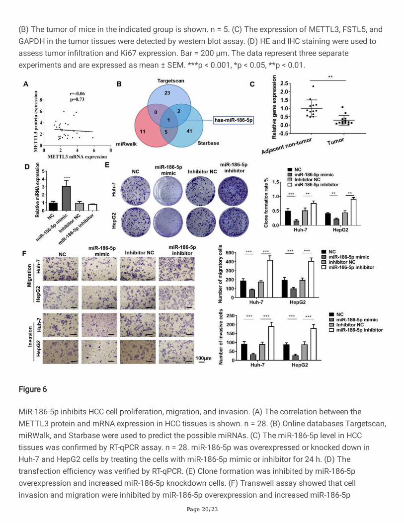
Mir-186-5p Inhibits Hcc Cell Proliferation, Migration, AndInvasionThe protein and mRNA expression of METTL3 was increased in HCC tissues. There was no link betweenthe METTL3 protein and gene expression in the raise rate of patients in each group (Fig. 6A). Themismatch between the METTL3 protein and mRNA expression levels in HCC suggests that METTL3 isinvolved in the development of HCC through post-transcriptional translation. As an important member ofpost-transcriptional regulatory factors, miRNA plays an indispensable role in HCC [28]. To further assessthe molecular mechanism of METTL3 that participated in the occurrence of HCC, online databasesTargetscan, miRWalk, and Starbase were used to predict the possible miRNAs (Fig. 6B). We found thatmiR-186-5p was the common miRNA in the three databases (Fig. 6B). Besides, the RT-qPCR analysisshowed that miR-186-5p indicates the down control in HCC tissues. This shows that miR-186-5pdownregulation can act on the mechanism of the onset of HCC disease (Fig. 6C). This data inspired us tostudy the role of miR-186-5p in HCC. We used the miR-186-5p mimic and inhibitors to raise or inhibit themiR-186-5p of cells, respectively (Fig. 6D). The clone formation assay showed that the overexpression ofmiR-186-5p inhibited the clone formation in Huh-7 and HepG2 cells, while the inhibition of miR-186-5pincreased the clone formation in Huh-7 and HepG2 cells (Fig. 6E). Transwell assay showed that theoverexpression of miR-186-5p inhibited Huh-7 and HepG2 cell migration and invasion, while the inhibitionof miR-186-5p promoted cell migration and invasion (Fig. 6F).
Mettl3 Is A Direct Target Of Mir-186-5pIn HCC cells, miR-186-5p decreased and inhibited the m6A level (Fig. 7A). As shown in the western blotassay, miR-186-5p negatively regulated the METTL3 expression in HCC cells (Fig. 7B). We also providedthe potential binding sites between the miR-186-5p and 3’UTR of METTL3 (Fig. 7C) and constructedluciferase reporter plasmids containing the wild type and synaptic variants of the 3’UTR of METTL3. The�uorescent enzyme analysis shows that miR-186-5p signi�cantly reduced the �uorescent enzyme activityof wild METTL3 3’UTR rather than that of the mutant, indicating that the speci�city of miR-186-5pcombines the 3’UTR of METTL3 (Fig. 7D). Western blot further showed that the miR-186-5p mimicdownregulated the METTL3 expression and upregulated the expression of FSTL5, which were reversed bythe overexpression of METTL3 (Fig. 7E). The MeRIP qPCR assay also revealed that the miR-186-5p mimicsigni�cantly reduced the m6A level of FSTL5 mRNA, while that co-transfected with METTL3 led to anobvious increase (Fig. 7F). Therefore, through our research, we can see that miR-186-5p affects themodi�cation of FSTL5 mRNA gene mediated by METTL3.

Page 10/23
MiR-186-5p inhibits HCC cell proliferation, migration, and invasion by downregulating METTL3
We then veri�ed whether miR-186-5p regulated the HCC cell malignancy by regulating METTL3. CCK-8and clone formation assay showed that miR-186-5p inhibited the HCC cell visibility and clone formation,while the overexpression of METTL3 reversed the role of miR-186-5p (Fig. 8A, B). Transwell assay showedthat the overexpression of METTL3-inhibited miR-186-5p inhibited the migration and invasion of HCCcells (Fig. 8C). It can be seen from these data that the malignant degree of HCC cells is regulated by miR-186-5p through the expression of METTL3.
DiscussionHCC is an interactive multi-step process, including genetic, epigenetic and transcriptional changes [29]. Inthe current study, we found that miR-186-5p prevented HCC progression by targeting METTL3 to regulatethe m6A-mediated stabilization of FSTL5 (Fig. 9).
FSTL5 is a member of the follistatin gene family that codes secretory glycogen. Our previous studieshave found that FSTL5 inhibited the HCC progression by inducing apoptosis and regulating the Wnt/YAP(Yes-associated protein) pathway [30]. YAP/TAZ (transcriptional coactivator with PDZ-binding motif) is atranscription-assisted activation factor that interacts with transcription factors combined with DNA toregulate the genes that participate in HCC proliferation and transformation. Li C et al. suggested thatFSTL5 accelerated the HCC cell apoptosis in a caspase-dependent manner [8]. However, what promptsthe downregulation of FSTL5 expression in HCC has not yet been reported.
Interestingly, our result indicated that the FSTL5 mRNA t1/2 was shortened in HCC cells, suggesting thepossible presence of RNA modi�cation. These data inspired us to hypothesize that m6A is involved inregulating the stability of FSTL5 in HCC. Emerging studies reported that epigenetic modi�cations, such asmiRNA regulation, histone modi�cation, and DNA methylation, are critically involved in the pathogenesisand development of HCC [31, 32]. m6A RNA methylation is the most common RNA modi�cation. Variousstudies have elucidated the effects of m6A modi�cation, including its effect on mRNA stability whichfunctioned in the development of HCC [33, 34]. For instance, Chen M et al. indicated that METTL3inhibited the expression of SOCS2(suppressor of cytokine signaling 2) mRNA by mediating themodi�cation of SOCS2 mRNA m6A [35]. METTL3 plays its regulatory role through m6A modi�cation-mediated mRNA stability [36]. We found that METTL3 promoted the FSTL5 mRNA degradation. YTHDF2is a functional m6A-binding protein that mainly regulates the stability of mRNA based on su�cientveri�cation and research [37]. According to reports, the METTL3/YTHDF2 m6A axis promotes the tumordevelopment by decomposing the SET domain-containing lysine methyltransferase 7 (SETD7) andKrüppel-like factor 4 (KLF4) mRNAs in bladder cancer [38].
Consistently, we found that the overexpression of YTHDF2 promoted the degradation of FSTL5 mRNA toinhibit the FSTL5 expression by recognizing the CDS region of FSTL5 mRNA. In an ongoing study, wefound that FSTL5 was lowered in HCC tissues, while METTL3 and YTHDF2 were upregulated in HCCtissues, which indicates the poor prognosis of HCC in the TCGA database. Moreover, METTL3 abolished
Page 11/23
the anti-tumor effect of FSTL5, indicating that FSTL5 was regulated by METTL3-dependent m6Amodi�cation in HCC. miRNAs participate in various disease processes by regulating the gene expressionnegatively at the post-transcriptional level [39]. The abnormal regulation of miRNA expression isassociated with tumor progression. Cai X et. al indicated that hepatitis B X-interacting protein (HBXIP)promoted the progression of breast cancer by inhibiting miRNA let-7g to regulate METTL3, and let-7gdownregulated the METTL3 expression by targeting 3'UTR [40].
However, the studies on whether miRNAs could regulate the m6A process in HCC development remainlimited. Herein, we screened the miR-186-5p by taking the intersection of online databases Targetscan,miRWalk, and Starbase. The RT-qPCR assay showed that miR-186-5p was signi�cantly downregulated inHCC tissues. An in vitro study on the effects of miR-186-5p on HCC cells further revealed that it inhibitsthe proliferation, invasion, and migration of HCC cells. We hypothesize that miR-186-5p may regulate them6A modi�cation to increase the stability or decrease the degradation of the FSTL5 level. Then, wedetected the m6A level in miR-186-5p-treated HCC cells and found that the m6A level decreased with miR-186-5p overexpression. We found that miR-186-5p negatively regulated the m6A level by directly targetingthe METTL3 in HCC cells. The overexpression of METTL3 abolished the miR-186-5p’s anti-tumor effect,indicating that miR-186-5p’s effect was dependent on the downregulation of METTL3.
In conclusion, we identi�ed the downregulated FSTL5 in HCC and the m6A-regulated FSTL5 stabilitymediated by YTHDF2. We reported the crosstalk of miRNA with m6A-mediated protein expression. ThemiR-186-5p/METTL3/YTHDF2/FSTL5 axis may provide a new idea for the targeted therapy of HCC.
AbbreviationsHCCHepatocellular carcinoma; IHC:Immunochemistry; miRNA:microRNA; m6A:N6-methyladenosine;FSTL5:Follistatin-like 5; YTHDF2:YTH domain family 2; METTL3:methyltransferase-like 3; TCGA:thecancer genome atlas; ATCC:American Type Culture Collection; DMEM:Dulbecco’s modi�ed Eagle’smedium; FBS:fetal bovine serum; shRNA:short hairpin RNA; IGF2BP:insulin-like growth factor 2 mRNA-binding protein 2; HE:Hematoxylin and eosin; ECL:Enhanced chemiluminescence; RIPA:Radio-Immunoprecipitation Assay; CCK-8:Cell Counting Kit-8; RT-qPCR:Reverse Transcription Polymerase ChainReaction; hnRNPA2B1:heterogeneous nuclear ribonucleoprotein A2B1; METTL14:Methyltransferase-like14; ALKBH5:AlkB homolog 5; FTO:Fat mass and obesity-associated protein; SOCS2:suppressor ofcytokine signaling 2; YAP:Yes-associated protein; TAZ:transcriptional coactivator with PDZ-binding motif;KLF4:Krüppel-like factor 4; SETD7:SET domain-containing lysine methyltransferase 7; HBXIP:hepatitis BX-interacting protein.
DeclarationsAcknowledgements

Page 12/23
Not applicable
Authors' contributions
DengYong Zhang completed most of the experiments in vitro; ShuoShuo Ma and FangFang Chencompleted the in vivo assays and helped to draft the manuscript; Yong Chun Zhou and WanLiang Suncollected the tissue samples and help to analysed the experimental data; DongDong Wang and ShiRuChang �nished the data collection and analysition; Zheng Lu guided the thoughts of the study andamended the manuscript. All authors read and approved the �nal manuscript.
Funding
This study was supported by Anhui Province Science Foundation for Youths (1808085QH288), BengbuMedical College Science Foundation (BYKY2019116ZD).
Availability of data and materials
The prognosis of the indicated patients’ cohort were obtained from TCGA (https://www.cancer.gov/tcga).The processed data are available from the corresponding author upon reasonable request.
Ethics approval and consent to participate
The study was approved by the Ethics Committee of Bengbu Medical College (No. 2017059).
Consent for publication
All authors agree to publish this article.
Competing interests
The authors declare that they have no competing interests.
References1. Bray F, Ferlay J, Soerjomataram I, Siegel RL, Torre LA. ,Jemal A.Global cancer statistics 2018:
GLOBOCAN estimates of incidence and mortality worldwide for 36 cancers in 185 countries.CA.Cancer J Clin. 2018;68(6):394–424.
2. Zhan L, Cao H, Wang G, Lyu Y, Sun X, An J, Wu Z, Huang Q, Liu B,Xing J.Drp1-mediated mitochondrial�ssion promotes cell proliferation through crosstalk of p53 and NF-κB pathways in hepatocellularcarcinoma.Oncotarget.2016; 7(40):65001–11.
3. Marengo A, Rosso C, Bugianesi ELiver, Cancer. Connections with Obesity, Fatty Liver, and Cirrhosis.Annu Rev Med. 2016;67:103–17.
4. Dimitroulis D, Damaskos C, Valsami S, Davakis S, Garmpis N, Spartalis E, Athanasiou A, Moris D,Sakellariou S, Kykalos S, et al. From diagnosis to treatment of hepatocellular carcinoma: An

Page 13/23
epidemic problem for both developed and developing world. World J Gastroenterol.2017;23(29):5282–94.
5. Fullerton PT Jr, Monsivais D, Kommagani R. Matzuk M M.Follistatin is critical for mouse uterinereceptivity and decidualization. Proc Natl Acad Sci U S A. 2017;114(24):E4772-e81.
�. Kingwell K. FSTL5–a new prognostic biomarker for medulloblastoma. Nat Rev Neurol.2011;7(11):598.
7. Remke M, Hielscher T, Korshunov A, Northcott PA, Bender S, Kool M, Westermann F, Benner A, Cin H,Ryzhova M, et al. FSTL5 is a marker of poor prognosis in non-WNT/non-SHH medulloblastoma. JClin Oncol. 2011;29(29):3852–61.
�. Li C, Dai L, Zhang J, Zhang Y, Lin Y, Cheng L, Tian H, Zhang X, Wang Q, Yang Q, et al.Follistatin-likeprotein 5 inhibits hepatocellular carcinoma progression by inducing caspase-dependent apoptosisand regulating Bcl-2 family proteins.2018; 22(12):6190–201.
9. Wilson CL, Mann DA, Borthwick. L A.Epigenetic reprogramming in liver �brosis and cancer. Adv DrugDeliv Rev. 2017;121:124–32.
10. Desrosiers R, Friderici K. Rottman F.Identi�cation of methylated nucleosides in messenger RNA fromNovikoff hepatoma cells. Proc Natl Acad Sci U S A. 1974;71(10):3971–5.
11. Sun T, Wu R,Ming L.The role of m6A RNA methylation in cancer.Biomed Pharmacother.2019;112:108613.
12. Chen J, Fang X, Zhong P, Song Z,Hu X.N6-methyladenosine modi�cations: interactions with novelRNA-binding proteins and roles in signal transduction.2019; 16(8):991–1000.
13. Zhao W, Qi X, Liu L, Ma S, Liu J, Wu J. Epigenetic Regulation of m(6)A Modi�cations in HumanCancer. Mol Ther Nucleic Acids. 2020;19:405–12.
14. Xu K, Yang Y, Feng GH, Sun BF, Chen JQ, Li YF, Chen YS, Zhang XX, Wang CX, Jiang LY, et al. Mettl3-mediated m(6)A regulates spermatogonial differentiation and meiosis initiation. Cell Res.2017;27(9):1100–14.
15. Zhuang M, Li X, Zhu J, Zhang J, Niu F, Liang F, Chen M, Li D, Han P. Ji S J.The m6A reader YTHDF1regulates axon guidance through translational control of Robo3.1 expression. Nucleic Acids Res.2019;47(9):4765–77.
1�. Han J, Wang JZ, Yang X, Yu H, Zhou R, Lu HC, Yuan WB, Lu JC, Zhou ZJ, Lu Q, et al.METTL3promote tumor proliferation of bladder cancer by accelerating pri-miR221/222 maturation in m6A-dependent manner.2019; 18(1):110.
17. Xu D, Shao W, Jiang Y, Wang X, Liu Y. Liu X.FTO expression is associated with the occurrence ofgastric cancer and prognosis. Oncol Rep. 2017;38(4):2285–92.
1�. Nishizawa Y, Konno M, Asai A, Koseki J, Kawamoto K, Miyoshi N, Takahashi H, Nishida N, HaraguchiN, Sakai D, et al.Oncogene c-Myc promotes epitranscriptome m(6)A reader YTHDF1 expression incolorectal cancer.Oncotarget.2018; 9(7):7476–86.

Page 14/23
19. Xu H, Wang H, Zhao W, Fu S, Li Y, Ni W, Xin Y, Li W, Yang C, Bai Y, et al.SUMO1 modi�cation ofmethyltransferase-like 3 promotes tumor progression via regulating Snail mRNA homeostasis inhepatocellular carcinoma.Theranostics.2020; 10(13):5671–86.
20. Bhaskaran M, Mohan M. MicroRNAs: history, biogenesis, and their evolving role in animaldevelopment and disease. Vet Pathol. 2014;51(4):759–74.
21. Lee YS, Dutta AMicroRNAs. in cancerAnnu Rev Pathol. 2009;4:199–227.
22. Alarcón CR, Lee H, Goodarzi H, Halberg N. Tavazoie S F.N6-methyladenosine marks primarymicroRNAs. for processingNature. 2015;519(7544):482–5.
23. Li J, Han Y, Zhang H, Qian Z, Jia W, Gao Y, Zheng H. Li B.The m6A demethylase FTO promotes thegrowth of lung cancer cells by regulating the m6A level of USP7 mRNA. Biochem Biophys ResCommun. 2019;512(3):479–85.
24. Liu J, Ren D, Du Z, Wang H, Zhang H. Jin Y.m(6)A demethylase FTO facilitates tumor progression inlung squamous cell carcinoma by regulating MZF1 expression. Biochem Biophys Res Commun.2018;502(4):456–64.
25. Dominissini D, Moshitch-Moshkovitz S, Schwartz S, Salmon-Divon M, Ungar L, Osenberg S, CesarkasK, Jacob-Hirsch J, Amariglio N, Kupiec M, et al.Topology of the human and mouse m6A RNAmethylomes revealed by m6A-seq.Nature.2012; 485(7397):201–6.
2�. Fu Y, Dominissini D, Rechavi G. He C.Gene expression regulation mediated through reversible mâ ¶ARNA methylation. Nat Rev Genet. 2014;15(5):293–306.
27. Liao S, Sun H, Xu CYTH, Domain. A Family of N(6)-methyladenosine (m(6)A). ReadersGenomicsProteomics Bioinformatics. 2018;16(2):99–107.
2�. Pu M, Chen J, Tao Z, Miao L, Qi X, Wang Y. Ren J.Regulatory network of miRNA on its target:coordination between transcriptional and post-transcriptional regulation of gene expression. Cell MolLife Sci. 2019;76(3):441–51.
29. Castelli G, Pelosi E, Testa ULiver, Cancer: Molecular Characterization, Clonal Evolution and CancerStem Cells.Cancers (Basel).2017; 9(9).
30. Zhang DY, Sun WL, Ma X, Zhang P, Wu W, Wu H, Zhou S. Lu Z.Up-regulated FSTL5 inhibits invasionof hepatocellular carcinoma through the Wnt/β-catenin/YAP pathway. Int J Clin Exp Pathol.2017;10(10):10325–33.
31. Hardy T, Mann DA. .Epigenetics in liver disease: from biology to therapeutics. Gut.2016;65(11):1895–905.
32. Herceg Z, Paliwal A. Epigenetic mechanisms in hepatocellular carcinoma: how environmental factorsin�uence the epigenome. Mutat Res. 2011;727(3):55–61.
33. Chen Y, Peng C, Chen J, Chen D, Yang B, He B, Hu W, Zhang Y, Liu H, Dai L, et al.WTAP facilitatesprogression of hepatocellular carcinoma via m6A-HuR-dependent epigenetic silencing of ETS1.MolCancer.2019; 18(1):127.
Page 15/23
34. Lan T, Li H, Zhang D, Xu L, Liu H, Hao X, Yan X, Liao H, Chen X, Xie K, et al.KIAA1429 contributes toliver cancer progression through N6-methyladenosine-dependent post-transcriptional modi�cation ofGATA3.Mol Cancer.2019; 18(1):186.
35. Chen M, Wei L, Law CT, Tsang FH, Shen J, Cheng CL, Tsang LH, Ho DW, Chiu DK, Lee JM, et al.RNAN6-methyladenosine methyltransferase-like 3 promotes liver cancer progression through YTHDF2-dependent posttranscriptional silencing of SOCS2.2018; 67(6):2254–70.
3�. Li D, Cai L, Meng R, Feng Z,Xu Q.METTL3 Modulates Osteoclast Differentiation and Function byControlling RNA Stability and Nuclear Export.Int J Mol Sci.2020; 21(5).
37. Wang X, Lu Z, Gomez A, Hon GC, Yue Y, Han D, Fu Y, Parisien M, Dai Q, Jia G, et al. N6-methyladenosine-dependent regulation of messenger. RNA stabilityNature. 2014;505(7481):117–20.
3�. Xie H, Li J, Ying Y, Yan H, Jin K, Ma X, He L,Xu X.METTL3/YTHDF2 m(6) A axis promotestumorigenesis by degrading SETD7 and KLF4 mRNAs in bladder cancer.2020; 24(7):4092–104.
39. Calin GA, Croce C. M.MicroRNA signatures in human cancers. Nat Rev Cancer. 2006;6(11):857–66.
40. Cai X, Wang X, Cao C, Gao Y, Zhang S, Yang Z, Liu Y, Zhang X, Zhang W,Ye L.HBXIP-elevatedmethyltransferase METTL3 promotes the progression of breast cancer via inhibiting tumorsuppressor let-7g.Cancer Lett.2018; 415:11–19.
Figures
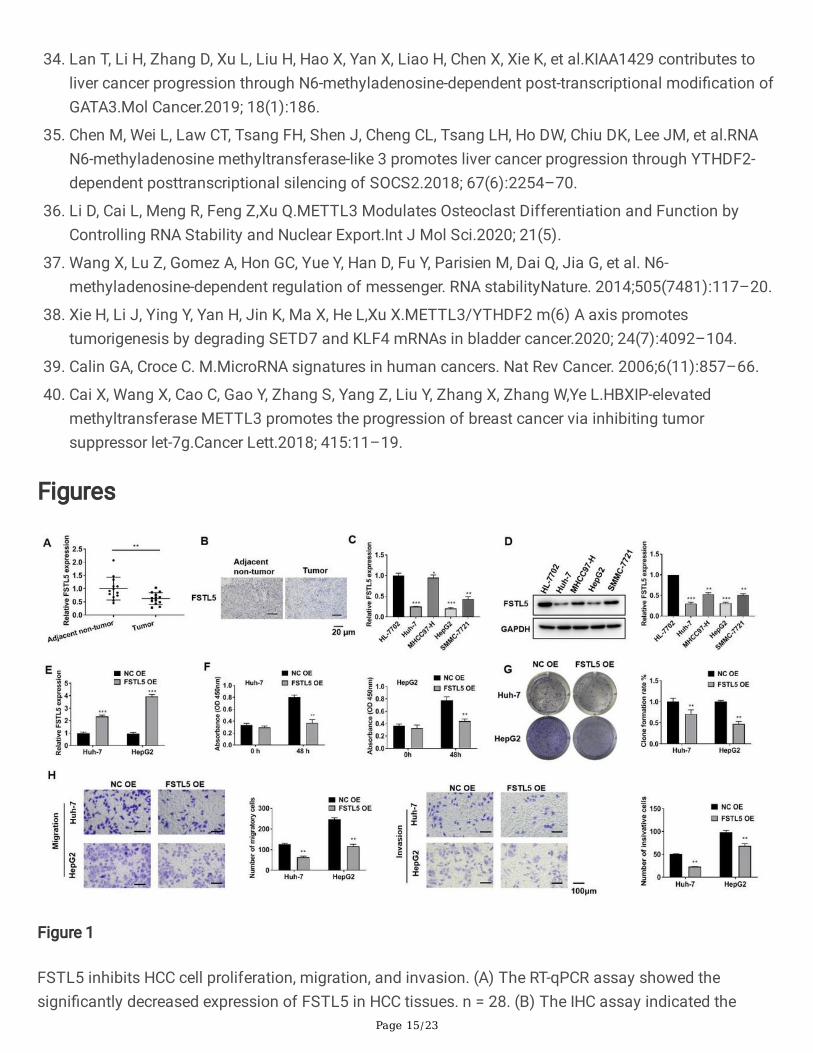
Figure 1
FSTL5 inhibits HCC cell proliferation, migration, and invasion. (A) The RT-qPCR assay showed thesigni�cantly decreased expression of FSTL5 in HCC tissues. n = 28. (B) The IHC assay indicated the

Page 16/23
downregulated FSTL5 level in HCC tissues. Bar = 20 μm. (C) The RT-qPCR and (D) western blot assaysindicated that FSTL5 expression was downregulated in HCC cell lines Huh-7, MHCC97-H, HepG2, andSMMC-7721. (E) The transfection e�ciency of the FSTL5 overexpression plasmid (FSTL5 OE) wascon�rmed by RT-qPCR assay. (F) Cell viability, (G) clone formation, (H) migration, and invasion wereinhibited by FSTL5 overexpression. Bar = 100 μm. The data represent three separate experiments and areexpressed as mean ± SEM. *p < 0.05, **p < 0.01, ***p < 0.001, compared with the control group.
Figure 2
METTL3 promotes FSTL5 mRNA degradation. (A)The RNA stability assay showed the FSTL5 mRNA half-life (t1/2) in HepG2 cells. (B) The total m6A level of adjacent non-tumor tissues and tumor tissues. n =28. (C) The METTL3, METTL14, FTO, and ALKBH5 levels were detected in HCC tissues by RT-qPCR. (D)The MeRIP qPCR assay was performed to assess the effect of METTL3 targeting the FSTL5 mRNA onm6A modi�cation. (E) The RNA stability assay showed the FSTL5 mRNA half-life (t1/2) in HepG2 cellswith METTL3 knockdown. (F) RT-qPCR and (G) western blot assays showed that METTL3 negativelyregulated the FSTL5 expression. (H) Luciferase reporter assay showed the relationship between METTL3and FSTL5. (I) RT-qPCR and (J) IHC assays indicated that the expression of METTL3 was upregulated inHCC tissues. n = 28. Bar = 20 μm. (K) Pearson’s correlation assay showed the negative correlationbetween the METTL3 and FSTL5 expression in HCC. The data represent three separate experiments andare expressed as mean ± SEM. *p < 0.05, **p < 0.01, ***p < 0.001, compared with the control group.
Page 17/23
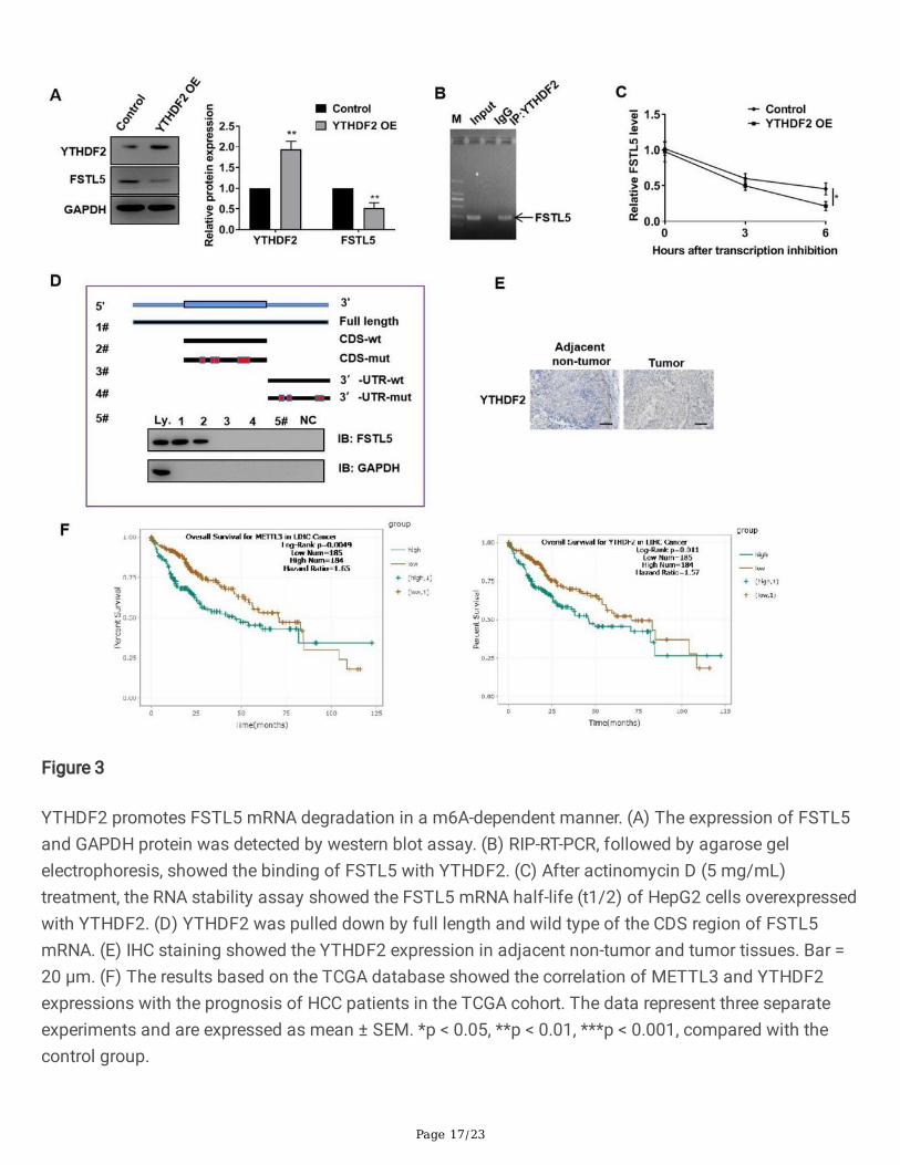
Figure 3
YTHDF2 promotes FSTL5 mRNA degradation in a m6A-dependent manner. (A) The expression of FSTL5and GAPDH protein was detected by western blot assay. (B) RIP-RT-PCR, followed by agarose gelelectrophoresis, showed the binding of FSTL5 with YTHDF2. (C) After actinomycin D (5 mg/mL)treatment, the RNA stability assay showed the FSTL5 mRNA half-life (t1/2) of HepG2 cells overexpressedwith YTHDF2. (D) YTHDF2 was pulled down by full length and wild type of the CDS region of FSTL5mRNA. (E) IHC staining showed the YTHDF2 expression in adjacent non-tumor and tumor tissues. Bar =20 μm. (F) The results based on the TCGA database showed the correlation of METTL3 and YTHDF2expressions with the prognosis of HCC patients in the TCGA cohort. The data represent three separateexperiments and are expressed as mean ± SEM. *p < 0.05, **p < 0.01, ***p < 0.001, compared with thecontrol group.

Page 18/23
Figure 4
Downregulated METTL3 inhibits HCC cell proliferation, migration, and invasion by up-regulating FSTL5.(A) The transfection e�ciency of sh-METTL3-1 and sh-FSTL5-1 in Huh-7 and HepG2 cells was con�rmedby RT-qPCR assay. (B) CCK-8 and (C) colony formation assays were used to detect cell proliferation. (D)Transwell assay was performed to detect cell migrations and invasion. Bar = 100 μm. The data representthree separate experiments and are expressed as mean ± SEM. ***p < 0.001, *p < 0.05, **p < 0.01.

Page 19/23
Figure 5
Downregulated METTL3 inhibits HCC tumor growth by up-regulating FSTL5 in vivo. A tumor xenograftmodel was constructed through the subcutaneous injection of 1 × 10⁷ HepG2 cells. The knockdown ofMETTL3 and FSTL5 was performed using the METTL3 knockdown lentivirus and FSTL5 knockdownlentivirus (GenePharmA, Shanghai, China), respectively, by the intratumoral injection of 50 μL of virus (4 ×10⁷ IU/mL) after the tumor cell injection. (A) Pictures of the isolated tumors of the indicated group. n = 5.
Page 20/23
(B) The tumor of mice in the indicated group is shown. n = 5. (C) The expression of METTL3, FSTL5, andGAPDH in the tumor tissues were detected by western blot assay. (D) HE and IHC staining were used toassess tumor in�ltration and Ki67 expression. Bar = 200 μm. The data represent three separateexperiments and are expressed as mean ± SEM. ***p < 0.001, *p < 0.05, **p < 0.01.
Figure 6
MiR-186-5p inhibits HCC cell proliferation, migration, and invasion. (A) The correlation between theMETTL3 protein and mRNA expression in HCC tissues is shown. n = 28. (B) Online databases Targetscan,miRWalk, and Starbase were used to predict the possible miRNAs. (C) The miR-186-5p level in HCCtissues was con�rmed by RT-qPCR assay. n = 28. miR-186-5p was overexpressed or knocked down inHuh-7 and HepG2 cells by treating the cells with miR-186-5p mimic or inhibitor for 24 h. (D) Thetransfection e�ciency was veri�ed by RT-qPCR. (E) Clone formation was inhibited by miR-186-5poverexpression and increased miR-186-5p knockdown cells. (F) Transwell assay showed that cellinvasion and migration were inhibited by miR-186-5p overexpression and increased miR-186-5p

Page 21/23
knockdown cells. Bar = 100 μm. The data represent three separate experiments and are expressed asmean ± SEM. *** p < 0.001, *p < 0.05, **p < 0.01.
Figure 7
METTL3 is a direct target of miR-186-5p. (A) The HepG2 cells were treated with miR-186-5p mimic orinhibitor for 24 h. The total m6A level in the cells of the indicated group is shown. (B) The METTL3,METTL14, ALKBH5, FTO, and GAPDH protein expression levels were detected by western blot assay. (C)The potential binding sites of miR-186-5p and 3’UTR of METTL3 mRNA. (D) Luciferase reporter assaywas performed for 48 h, with luciferase reporter plasmid containing the wild type or mutant METTL3 3′UTR and NC, miR-186-5p mimic, and miR-186-5p inhibitor. (E) The protein expression of FSTL5 wasassessed by western blot assay. (F) MeRIP qPCR assay was used to evaluate the m6A level of FSTL5mRNA. The data represent three separate experiments and are expressed as mean ± SEM. *p < 0.05, **p <0.01, ***p < 0.001.

Page 22/23
Figure 8
MiR-186-5p inhibits HCC cell proliferation, migration, and invasion by downregulating METTL3. The Huh-7 and HepG2 cells were treated with miR-186-5p mimic, with or without transfection with METTL3overexpression lentivirus for 24 h. (A) The cell viability and (B) clone formation were inhibited by miR-186-5p overexpression, which was reversed by METTL3 overexpression. (C) Transwell assay showed that cellinvasion and migration were inhibited by miR-186-5p overexpression, which was reversed by METTL3overexpression. Bar = 100 μm. The data represent three separate experiments and are expressed as mean± SEM. *p < 0.05, **p < 0.01, ***p < 0.001.

Page 23/23
Figure 9
The mechanism of the regulatory network miR-186-5p/METTL3/YTHDF2/FSTL5 axis.
Supplementary Files
This is a list of supplementary �les associated with this preprint. Click to download.
Supplementary.docx