expression and clinical prognostic value of m6a rna
TRANSCRIPT

RESEARCH Open Access
Expression and clinical prognostic value ofm6A RNA methylation modification inbreast cancerFangchao Zheng1, Feng Du2, Haili Qian3, Jiuda Zhao4, Xue Wang5, Jian Yue5, Nanlin Hu1, Yiran Si1, Binghe Xu1 andPeng Yuan1,5*
Abstract
Background: N6-methyladenosine(m6A) methylation modification affects the tumorigenesis, progression, andmetastasis of breast cancer (BC). However, the expression characteristics and prognostic value of m6A modificationin BC are still unclear. We aimed to evaluate the relationship between m6A modification and clinicopathologicalcharacteristics, and to explore the underlying mechanisms.
Methods: Three public cohorts and our clinical cohort were included: 1091 BC samples and 113 normal samplesfrom the TCGA database, 1985 BC samples from the METABRIC database, 1764 BC samples from the KM Plotterwebsite, and 134 BC samples of our clinical cohort. We collected date from these cohorts and analyzed the geneticexpression, gene-gene interactions, gene mutations, copy number variations (CNVs), and clinicopathological andprognostic features of 28 m6A RNA regulators in BC.
Results: This study demonstrated that some m6A regulators were significantly differenially expressed in BCs andtheir adjacent tissues, and also different in various molecular types. All 28 studied m6A regulators exhibitedinteractions. KIAA1429 had the highest mutation frequency. CNVs of m6A regulators were observed in BC patients.The expression of the m6A regulators was differentially associated with survival of BC. Higher CBLL1 expression wasassociated with a better prognosis in BC than lower CBLL1 expression. Functional analysis showed that CBLL1 wasrelated to the ESR1-related pathway, apoptosis-related pathway, cell cycle pathway and immune-related pathway inBC.
Conclusions: m6A RNA modification modulated gene expression and thereby affected clinicopathological featuresand survival outcomes in BC. CBLL1 may be a promising prognostic biomarker for BC patients.
Keywords: N6-methyladenosine modification, Methylation, Breast cancer, CBLL1, Prognosis
© The Author(s). 2021 Open Access This article is licensed under a Creative Commons Attribution 4.0 International License,which permits use, sharing, adaptation, distribution and reproduction in any medium or format, as long as you giveappropriate credit to the original author(s) and the source, provide a link to the Creative Commons licence, and indicate ifchanges were made. The images or other third party material in this article are included in the article's Creative Commonslicence, unless indicated otherwise in a credit line to the material. If material is not included in the article's Creative Commonslicence and your intended use is not permitted by statutory regulation or exceeds the permitted use, you will need to obtainpermission directly from the copyright holder. To view a copy of this licence, visit http://creativecommons.org/licenses/by/4.0/.The Creative Commons Public Domain Dedication waiver (http://creativecommons.org/publicdomain/zero/1.0/) applies to thedata made available in this article, unless otherwise stated in a credit line to the data.
* Correspondence: [email protected] of Medical Oncology, National Cancer Center/National ClinicalResearch Center for Cancer/Cancer Hospital, Chinese Academy of MedicalSciences and Peking Union Medical College, No. 17 Panjiayuan Nanli, Beijing100021, China5Department of VIP Medical Services, National Cancer Center/NationalClinical Research Center for Cancer/Cancer Hospital, Chinese Academy ofMedical Sciences and Peking Union Medical College, Beijing 100021, ChinaFull list of author information is available at the end of the article
Zheng et al. Biomarker Research (2021) 9:28 https://doi.org/10.1186/s40364-021-00285-w

Highlights
1. m6A RNA methylation is associated with thedifferential gene expression, CNV, molecular typing,and clinicopathological and prognostic features ofbreast cancer.
2. CBLL1 is correlated with better prognosis in breastcancer.
3. This is the first study to report m6A RNAexpression characteristics and prognostic value inbreast cancers based on three public cohorts andour clinical cohort.
IntroductionBreast carcinoma (BC) is the most commonly diagnosedmalignant tumor and the leading cause of cancer-relateddeaths annually in women annually; thus, it is a seriousthreat to the health of females [1]. The outcome for BCpatients worldwide have improved because of the devel-opment of medical therapy, surgical therapy, and noveltherapeutic interventions and the use of newer molecu-lar typing techniques. Despite these efforts, BC con-tinues to have a 15% of cancer-related death ratefollowing treatment according to GLOBOCAN2018, andthe mortality is still not optimistic [1]. In addition,WHO Cancer Tomorrow predicted that approximately817, 361 females will die from BC by 2030. That is, theprognosis of BC still remains dismal. Thus, there is anurgent need exists to develop new treatment options toimprove survival for BC patients, particularly those withdisease or recurrent metastasis.RNA modification, especially N6-methyladenosine
(m6A) modification, have provided a more effectivemethod and a new prospects in the treatment of BC [2].The m6A modification is a highly abundant and conser-vative messenger RNA modification in mammals thatconsists of three vital components, as follows: writers,which are also termed methyltransferases; erasers, whichare demethylases that remove m6A modifications; andreaders, which recognize m6A-modified sites and regu-late m6A modifications [2]. In general, m6A RNA modi-fications regulate RNA termination codons, 5’capstructure, and the 3′ untranslated region (UTR), posi-tively or negatively affecting tumorigenesis, tumor differ-entiation, tumor proliferation, tumor invasion andtumor metastasis of BC [2, 3].Growing evidence has demonstrated that m6A modifi-
cation is closely associated with tumorigenesis, tumordifferentiation, tumor proliferation, tumor invasion andworse survival in BC patients, including METTL3,METTL14, WTAP, ALKBH5, IGF2BP2, IGF2BP3, andFTO [3–14]. In addition, the m6A regulator IGF2BP3was associated with reduced cell apoptosis, larger tumorsizes, higher grade, higher clinical stage, necrosis, and
CK5/6 expression and further worsened the DFS and OSof BC patients [4, 15, 16]. However, the m6A regulatorEIF3A had no association with age, tumor size, or differ-entiation grade. Unfortunately, data published on m6Amodification in BC are partly conflicting and highly het-erogeneous. Most studies have investigated partial mech-anisms and capabilities, and the gene is relativelysimplistic. Some studies evaluated the prognostic valueof m6A regulators in BC patients, but significantly fewertissue samples were included.In the present study, we clarified the biological mech-
anism of all 28 m6A regulators and evaluated the associ-ations with the clinicopathological features andprognosis in BC patients based on The Cancer GenomeAtlas (TCGA), Molecular Taxonomy of Breast CancerInternational Consortium (METABRIC), KM Plotterwebsite, and one clinical cohort. The aim of this study isto identify potential new therapeutic targets and improvethe prognosis of BC patients.
Materials and methodsData acquisitionThe TCGA data were downloaded from the TCGAbreast cancer cohort within the Genomic Data Common(GDC) data portal (https://portal.gdc.cancer.gov/) Thedataset contains clinical data, genetic mutation, copynumber variation (CNV), and m6A regulator expressiondata. For validation, we obtained independent gene ex-pression and survival data from the METABRIC (http://www.cbioportal.org/), Kaplan-Meier Plotter websites(https://kmplot.com/analysis/) and one clinic cohort. Inthe TCGA cohort, 1091 BC samples and 113 normal ad-jacent tissues were enrolled. Besides, a total of 1985 BCsamples from the METABRIC cohort, 1764 BC samplesfrom the KM plotter website cohort and 134 BC samplesfrom one clinical cohort were enrolled and analyzed inthe validation sets. (see Fig. 1) The detailed clinicopatho-logical parameters of the BC patients are shown inTable 1.Referring to the relevant literature on m6A methyla-
tion modification, 34 alternative m6A regulators werefirst included to be studied, and then 6 regulators wereexcluded since they did not coexist in the enrolled co-horts. (31 m6A regulators in the TCGA cohort and 28m6A regulators in the METABRIC cohort, Fig. 1) Thus,a total of 28 m6A regulators were enrolled in this ana-lysis, including 10 writers, 1 eraser and 17 readers, re-spectively [2, 17–22].
Tissue microarray from clinic cohortThe BC tissue microarray (#HBreD140Su07, clinical co-hort) was purchased from Shanghai Outdo Biotech CO.,Ltd. (Shanghai, China) and immunohistochemically(IHC) stained for CBLL1. Specifically, the CBLL1
Zheng et al. Biomarker Research (2021) 9:28 Page 2 of 13

immunohistochemical expression in the cytoplasm was(−), (+), (++), and (+++).
Statistical analysisAll these data and figures were analysed by using SPSS24.0 (IBM, Chicago, USA), GraphPad Prism 8.0 (Graph-Pad Software, La Jolla, CA, USA) and R software (ver-sion 3.6.1). The associations between an m6A regulatorCNV and the clinicopathological characteristics of thepatients were analyzed with ANOVA test to conduct dif-ference comparisons among three or more groups.Kaplan-Meier curves and the log-rank test were used toevaluate the prognostic value of m6A genes. All statis-tical results were significantly different at P value < 0.05.
ResultsM6A expression in BCIn this study, three public cohort datasets and one clin-ical dataset were included, which were as follows: 1091BC cases and 113 non-tumor normal cases from theTCGA date, 1985 cases from the METABRIC date, BCcases from the KM Plotter website, and 134 BC casesfrom one clinical dataset.In this study, the following 28 m6A regulators were
studied: 10 writers were METTL3, METTL14, METTL16, WTAP, ZC3H13, RBM15B, RBM15, CBLL1,KIAA1429, and NSUN2; 1 eraser: ALKBH5; and 17readers: YTHDC1, YTHDC2, YTHDF1, YTHDF2,
YTHDF3, FXR2, FXR1, EIF3A, EIF4G2, IGF2BP1,IGF2BP2, IGF2BP3, ELAVL1, G3BP1, HNRNPA2B1,LRPPRC, and ABCF1.
Genetic differences between BC and adjacent normaltissuesBased on the TCGA data cohort, the analysis resultsdemonstrated that 21 identified genes were differentiallyexpressed between BC tissues and adjacent normal tis-sues, including 14 up-regulated genes and 7 down-regulated genes (Fig. 2a). These up-regulated geneswere IGF2BP1, IGF2BP3, RBM15, CBLL1, KIAA1429,FXR1, ELAVL1, NSUN2, ABCF1, LRPPRC, YTHDF2,YTHDF1, HNRNPA2B1, and EIF4G2. These down-regulated genes were IGF2BP2, METTL14, METTL16,ZC3H13, YTHDC1, WTAP, and EIF3A. In addition,no significant differences were observed for the otherseven genes (Fig. 2a).
Genetic differences between different types of BCBased on the TCGA cohort, the ANOVA showed thatthe expression of 28 m6A regulators in various BCtypes was significantly different (Fig. 2b). The resultsshowed that the expression of m6A regulators wassignificantly correlated with different molecular typingof BC, except EIF3A and YTHDF2 (Fig. 2b, supple-mentary Figure 1). Specifically, IGF2BP1, IGF2BP2,and IGF2BP3 were highly expressed in TNBC, while
Fig. 1 Flow chart showing the m6A modification selection and analysis process.
Zheng et al. Biomarker Research (2021) 9:28 Page 3 of 13
they were expressed at low levels in HER2+, luminalA, and luminal B BC (Fig. 2b).
Interaction of m6A regulatorsBy analyzing the TCGA cohort, the results indicated thatall enrolled 28 studied genes exhibited gene-gene inter-actions. The results also showed negative gene-gene ex-pression interaction among IGF2BP1, IGF2BP2, andIGF2BP3, and strong positive gene-gene expression in-teractions among the remaining 25 genes (Fig. 2c).
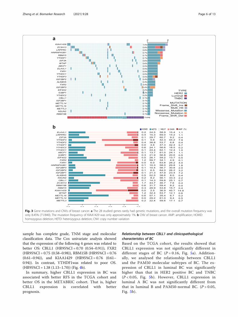
Genetic mutation and copy number variations (CNVs) ofthe m6A regulatorsAccording to the TCGA data cohort, the results showedthat these enrolled 28 studied genes rarely had geneticmutations, and the overall mutation frequency was only
8.45% (71/840). The statistical analysis indicated that themutation frequency of KIAA1429, which had the highestfrequency, was only approximately 1%, and as for theother genes, the frequencies were below 1% (Fig. 3a).We also used COSMIC (the Catalogue Of Somatic Mu-tations In Cancer, https://cancer.sanger.ac.uk/cosmic/)to explore the variants of m6A regulators in BC. The re-sults demonstrated that the mutation frequency ofIGF2BP2 was 6.3%, IGF2BP3 was 6.0% and LRPPRC was4.2%. The mutation frequencies of the remaining m6Aregulators remained between 0.5% and 2.9%.In this study, we also analyzed the following CNVs of
m6a regulators: homozygous deletion (HOMD), hetero-zygous deletion (HETD), neural (NEUT), gain, and amp-lification (AMP). The top three genes with CNV gainswere KIAA1429, YTHDF3, and YTHDF1; the top three
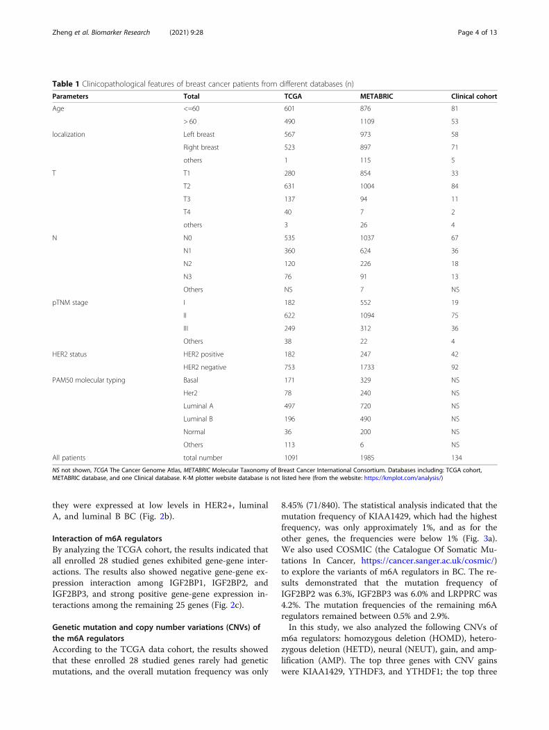
Table 1 Clinicopathological features of breast cancer patients from different databases (n)
Parameters Total TCGA METABRIC Clinical cohort
Age <=60 601 876 81
> 60 490 1109 53
localization Left breast 567 973 58
Right breast 523 897 71
others 1 115 5
T T1 280 854 33
T2 631 1004 84
T3 137 94 11
T4 40 7 2
others 3 26 4
N N0 535 1037 67
N1 360 624 36
N2 120 226 18
N3 76 91 13
Others NS 7 NS
pTNM stage I 182 552 19
II 622 1094 75
III 249 312 36
Others 38 22 4
HER2 status HER2 positive 182 247 42
HER2 negative 753 1733 92
PAM50 molecular typing Basal 171 329 NS
Her2 78 240 NS
Luminal A 497 720 NS
Luminal B 196 490 NS
Normal 36 200 NS
Others 113 6 NS
All patients total number 1091 1985 134
NS not shown, TCGA The Cancer Genome Atlas, METABRIC Molecular Taxonomy of Breast Cancer International Consortium. Databases including: TCGA cohort,METABRIC database, and one Clinical database. K-M plotter website database is not listed here (from the website: https://kmplot.com/analysis/)
Zheng et al. Biomarker Research (2021) 9:28 Page 4 of 13

genes with HOMD mutations were ZC3H13 andFXR2, and WTAP; the top three genes with HETDmutations were FXR2, METT16, and ALKBH5; andthe top three genes with AMP mutations wereKIAA1429, YTHDF3, and IGF2BP1 (Fig. 3b). More-over, the results indicated a clear relationship betweenm6A regulators and CNVs. (Supplementary Figure 2)Supplementary Figure 2 also showes that the expres-sion trend of some genes was from low to high. Fur-thermore, the expression of ABCF1, ALKBH5, ELAVL1 and FXR1 expression was gradually increased fromdeletion, loss, neural, gain, to amplification (Supple-mentary Figure 2a-d).
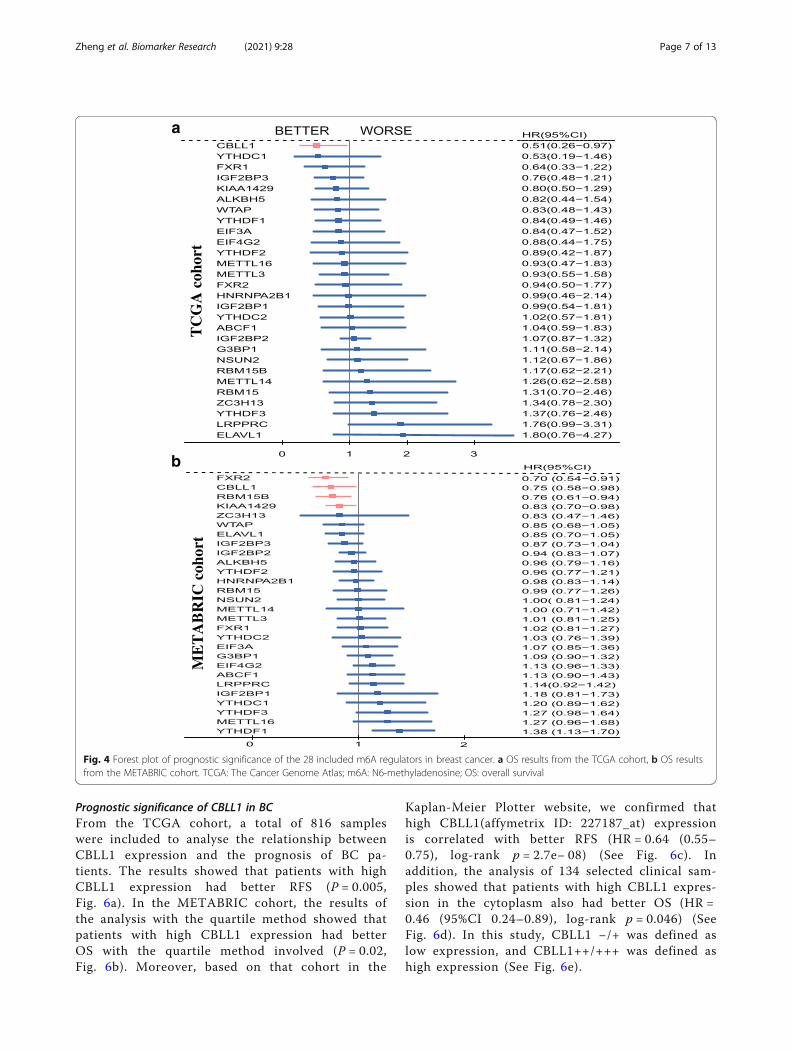
Prognostic significance of m6A regulators in BCPrognostic significance from TCGA data and METABRICdataIn the TCGA cohort, 816 eligible samples were selectedaccording to the following inclusion criteria: 1. overallsurvival (OS) > 6 months; and 2. enrolled sample hascomplete stage, molecular classification and relapse-freesurvival (RFS) data. The Cox univariate analysis showedthat BC patients with CBLL1 high expression had a bet-ter RFS than those with low CBLL1 expression.(HR95%CI = 0.51(0.26–0.97)) (Fig. 4a).In the METABRIC cohort, the inclusion criteria of the
population were as follows: 1. OS > 12months; 2. The
Fig. 2 m6A regulator expression of m6A modification in breast cancer. a Gene expression between breast cancer tissues and adjacent normaltissues according to TCGA. b Genetic differences of different PAM50 molecular typing of breast cancer. c Correlations among the 28 enrolledm6A regulators. *P < 0.05, **P < 0.01, ***P < 0.001, ****P < 0.0001, #P > 0.05. TCGA: The Cancer Genome Atlas; m6A: N6-methyladenosine
Zheng et al. Biomarker Research (2021) 9:28 Page 5 of 13
sample has complete grade, TNM stage and molecularclassification data. The Cox univariate analysis showedthat the expression of the following 4 genes was related tobetter OS: CBLL1 (HR95%CI = 0.70 (0.54–0.91)), FXR2(HR95%CI = 0.75 (0.58–0.98)), RBM15B (HR95%CI = 0.76(0.61–0.94)), and KIAA1429 (HR95%CI = 0.76 (0.61–0.94)). In contrast, YTHDF1was related to poor OS.(HR95%CI = 1.38 (1.13–1.70)) (Fig. 4b).In summary, higher CBLL1 expression in BC was
associated with better RFS in the TCGA cohort andbetter OS in the METABRIC cohort. That is, higherCBLL1 expression is correlated with betterprognosis.
Relationship between CBLL1 and clinicopathologicalcharacteristics of BCBased on the TCGA cohort, the results showed thatCBLL1 expression was not significantly different indifferent stages of BC (P = 0.16, Fig. 5a). Addition-ally, we analysed the relationship between CBLL1and the PAM50 molecular subtypes of BC. The ex-pression of CBLL1 in luminal BC was significantlyhigher than that in HER2 positive BC and TNBC(P < 0.05, Fig. 5b). However, CBLL1 expression inluminal A BC was not significantly different fromthat in luminal B and PAM50-normal BC. (P > 0.05,Fig. 5b).
Fig. 3 Gene mutations and CNVs of breast cancer. a The 28 studied genes rarely had genetic mutations, and the overall mutation frequency wasonly 8.45% (71/840). The mutation frequency of KIAA1429 was only approximately 1%. b CNV of breast cancer. AMP: amplification; HOMD:homozygous deletion; HETD: heterozygous deletion; CNV: copy number variation
Zheng et al. Biomarker Research (2021) 9:28 Page 6 of 13
Prognostic significance of CBLL1 in BCFrom the TCGA cohort, a total of 816 sampleswere included to analyse the relationship betweenCBLL1 expression and the prognosis of BC pa-tients. The results showed that patients with highCBLL1 expression had better RFS (P = 0.005,Fig. 6a). In the METABRIC cohort, the results ofthe analysis with the quartile method showed thatpatients with high CBLL1 expression had betterOS with the quartile method involved (P = 0.02,Fig. 6b). Moreover, based on that cohort in the
Kaplan-Meier Plotter website, we confirmed thathigh CBLL1(affymetrix ID: 227187_at) expressionis correlated with better RFS (HR = 0.64 (0.55–0.75), log-rank p = 2.7e− 08) (See Fig. 6c). Inaddition, the analysis of 134 selected clinical sam-ples showed that patients with high CBLL1 expres-sion in the cytoplasm also had better OS (HR =0.46 (95%CI 0.24–0.89), log-rank p = 0.046) (SeeFig. 6d). In this study, CBLL1 −/+ was defined aslow expression, and CBLL1++/+++ was defined ashigh expression (See Fig. 6e).
Fig. 4 Forest plot of prognostic significance of the 28 included m6A regulators in breast cancer. a OS results from the TCGA cohort, b OS resultsfrom the METABRIC cohort. TCGA: The Cancer Genome Atlas; m6A: N6-methyladenosine; OS: overall survival
Zheng et al. Biomarker Research (2021) 9:28 Page 7 of 13

Functional analysis of CBLL1 in luminal BCIn a previous study, Hakai, as a coregulator of oestrogenreceptor alpha, was found to play a negative role in thedevelopment and progression of BC cells [23]. Thus, weperformed functional analysis of CBLL1 in ER- or PR-positive BC. Based on the TCGA cohort, a total of 597ER- or PR-positive and HER2-negative BC patients pa-tients were included according to the inclusion criteria.Altogether, 521 genes with differential gene expression(log FC > 0.8 or < − 0.3) were included in the analysis.A total of 597 patients was were divided into the
CBLL1-high (CBLL1-H) and CBLL1-low (CBLL1-L)groups according to the median value of CBLL1 expres-sion. The heat map compared the pathological featuresand signalling pathway features of BC in the CBLL1high-expression group with those in the CBLL1-low ex-pression group. The following parameters were observedin these two groups: pTNM staging, PAM50, PIK3CA,GATA3, apoptosis-related pathways, ESR1-related path-ways, and immune-related pathways (Fig. 7a). The ex-pression of the following apoptosis regulators was
significantly different in the CBLL1-H and CBLL1-L group, asfollows: POMK, NRIP1, GTF2I, SEMA3C, VPS13C, RAB27B,PIK3C2A, ITPR2, HIPK2, and ADAM9. In addition, the ex-pression of ESR1 regulators,including ZNF770, AFF3, PRLR,SLC7A2, and CLSTN2, was also significantly different inCBLL1-H and CBLL1-L group. Furthermore, immune regula-tors, including CALML5, ISG15, S100A8, LTB, CYBA,S100A9, TNFRSF4, SCT, and IFI27, were significantly differ-ent between the CBLL1-H and CBLL1-L groups (Fig. 7a).Gene set enrichment analysis (GSEA) demonstrated
that the CBLL1-related differentially expressed geneswere significantly enriched in the ESR1-related signallingpathway, cell apoptosis related pathway and immune-related pathway. In particular, CBLL1 expression pro-moted the upregulation of genes related to ESR1 and cellapoptosis-related pathways (Fig. 7b). Moreover, single-sample gene set enrichment analysis (ssGSEA) demon-strated that low CBLL1 expression increased the occur-rence of tamoxifen resistance and tumor-associatedhypoxia. High CBLL1 expression induced apoptosis, mi-totic cell death and the upregulation of ESR1 (Fig. 7c).
Fig. 5 The relationship between CBLL1 and the clinicopathological characteristics of breast cancer. a CBLL1 with stage. b CBLL1 with PAM50molecular typing
Zheng et al. Biomarker Research (2021) 9:28 Page 8 of 13

DiscussionBased on the TCGA cohort, the METABRIC cohort, theK-M plotter website cohort and one clinical cohort, thisarticle first analysed the expression of m6A regulators inBC and the effects of m6A modification on the biologicalbehaviour of BC. Research has shown that m6A RNAmodification regulates certain signalling pathways viamethylation transferases, demethylation enzymes, andreader effectors and thus affects the differential expressionof genes between human BC and normal tissue, as well asthe molecular typing, genetic mutations, and genomeCNVs of BC. In addition, m6A regulator expression wascorrelated with the clinicopathological characteristics,tumor drug sensitivity, RFS and OS of BC. Importantly,CBLL1 was a protective factor of prognosis in BC.This study demonstrated that the expression of some
m6A regulators expressions was significantly differentbetween BC tissues and adjacent normal tissues. Previ-ous studies have shown that m6A modification plays animportant role in a variety of tumors, such as gastriccancer, glioblastoma, kidney cancer, and BC [23–26]. In
this study, 21 m6A regulators were differentiallyexpressed, including 14 upregulated genes and 7 down-regulated genes. The expression of CBLL1 expressionwas upregulated in lung cancer and BC compared withadjacent tissues [27]. Consistent with the findings of theabove study, our research also confirmed that CBLL1was up-regulated in BC. KIAA1429 expression was up-regulated in BC tissue and regulated tumor proliferationand tumor differentiation [28]. This stydy also confirmedthat KIAA1429 was upregulated in BC. Previous studiesshowed that METTL3 was downregulated in BC celllines and tissues [7]. However, METTL3 expression wasno differentially expressed between cancer tissues andnormal tissues. ALKBH5 and G3BP1 have been found tobe overexpressed in BC; however, they did not show dif-ferential expression in our study [12, 29]. To date, theexpression of m6A regulator is controversial. The pos-sible reasons are as follows:1. fewer samples were in-cluded in previous studies; 2. most previous sampleswere cell lines; and 3. no specific molecular typing classi-fication was used.
Fig. 6 Prognostic significance of CBLL1 in breast cancer. a Association of CBLL1 RNA expression with RFS in the TCGA cohort. b Association ofCBLL1 RNA expression with OS in the METABRIC cohort. c Association of CBLL1 RNA expression with RFS in the K-M plotter cohort. d Associationof CBLL1 (immunohistochemistry staining) with OS in the clinical dataset. e CBLL1Immunohistochemistry, including (−), (+), (++), (+++); (−)/(+):low expression; (++)/(+++): high expression. OS: overall survival; RFS: relapse-free survival; TCGA: The Cancer Genome Atlas
Zheng et al. Biomarker Research (2021) 9:28 Page 9 of 13

In addition, m6A RNA regulators exhibit gene-geneinteractions and have different gene expression patternsin different molecular types of BC. Previous studies haveshown that crosslinks among different m6A regulators,affect tumor pathogenesis and differentiation [2]. Wealso observed that m6A regulators, except EIF3A andYTHDF2, were clearly correlated with different molecu-lar types of BC. Of note, IGF2BP1, IGF2BP2, andIGF2BP3 expressions were upregulated in TNBC anddownregulated in HER2+, luminal A, and luminal B BC.CBLL1 expression was upregulated in ER-positive breastcancer [23]. In other words, the above relevant regula-tors may participate in the regulation of moleculartyping.Futhermore, our research indicated that the mutation
frequencies of m6A regulators were low in BC, but theCNV mutation frequencies of m6A regulators were highand related to gene expression. Rui et al. confirmed thatthe mutation frequencies of all m6A regulators werelower than 1.1% in glioma [25]. The results of one pan-cancer study also confirmed that the mutation frequen-cies of m6A regulators were very low in tumors, rangingfrom 0.02 to 8.07% [30]. Similarly, the mutation frequen-cies of the 28 included genes were less than 1%, and thatof the top gene KIAA1429 was only 1.0%. Based on thedata presented in this research, gene mutation have littleimpacts on m6A modifications. However, another studyof renal cancer confirmed that the mutation frequency
of the m6A regulator YTHDC2 was 55.11% and that ofMETTL3 was 30.11% [24]. That is, gene mutations weredifferent in various cancers. In gastric cancer, m6A mod-ifications are mediated by the CNV deletions of ELAVL1, YTHDF2 and FMR1, which then affects tumor for-mation [26]. As described above, the m6A regulators ofBC also had a high frequency of CNVs. For example,KIAA1429, YTHDF3, and YTHDF1 had higher CNVgain frequencies of over 40%. The CNV HETD frequen-cies of FXR2, METT16, and ALKBH5 were all higherthan 50%. In addition, the expression of m6A regulatorscorrelated with CNVs. In particular, the expression ofABCF1, ALKBH5, ELAVL1 and FXR1 increased with in-creasing CNVs. Therefore, based on former conclusions,we can speculate that genomic CNVs play a strongerrole in m6A modification compared with genemutations.Meanwhile, this result indicated that the m6A regula-
tor CBLL1 had no correlation with TNM stage in BC.Similarly, CBLL1 had no correlation with TNM stage inlung cancer [27]. This result may indicate that CBLL1does not affect tumor stage. However, WTAP, RBM15,YTHDF, and ALBKH5 had strong correlations withtumor stage and 1p/19q codeletion in glioma [25]. Thism6A regulator was correlated with the nuclear grade ofkidney cancer [24]. Of course, further experiments areneeded to verify whether CBLL1 can regulate the malig-nant phenotype of tumor cells.
Fig. 7 Functional Analysis of CBLL1. a Relationship between CBLL1 and signalling pathways. b GSEA of CBLL1. c ssGSEA of CBLL1. GSEA: gene setenrichment analysis; ssGSEA: single-sample gene set enrichment analysis
Zheng et al. Biomarker Research (2021) 9:28 Page 10 of 13

This study found that m6A regulator is related toprognosis in BC. CBLL1 expression correlated with poorprognosis in lung cancer [27]. However, CBLL1 was cor-related with good prognosis in BC, according to theTCGA, METABRIC, K-M plotter and one clinical co-horts. Similarly, previous studies have found that CBLL1,as an E3 ubiquitin ligase, inhibits ER pathway activity bybinding to an ER coactivator and then further inhibitsthe proliferation and differentiation of BC cells [23]. Thepotential reason may be that CBLL1 inhibited tumormetastasis, enhanced cell apoptosis or increased drugsensitivity. It is also important to note that CBLL1 haslow tissue specificity, according to data from the HumanPro t e in At l a s (h t tp s : / /www.p ro t e ina t l a s . o rg /ENSG00000105879-CBLL1). Certainly, further work isneeded to enhance the accuracy of this inference. Never-theless, high CBLL1 expression is a protective factor forBC patients. In addition, some m6A regulators were as-sociated with prognosis. For example, IGF2BP1 expres-sion implies a poor prognosis in ovarian, liver and lungcancers [31–33]. METTL3 expression correlated withbrain metastasis and worse prognosis in lung cancer[34].Furthermore, functional analysis indicated that CBLL1
may affect the occurrence, development and drug resist-ance of BC by regulating various pathways. CBLL1 ex-pression was associated with apoptosis-related pathways,ESR1-related pathways, and immune-related pathways.That is, CBLL1 plays a vital role in regulating these sig-nalling pathways or their related genes, such as mediat-ing POMK and NRIP1 of the apoptosis-related pathway,ZNF770 and AFF3 of the ESR1-related pathway, andCALML5 and ISG15 of the immune-related pathway. Inthe CBLL1 high expression group, the ESR1-relatedpathway was upregulated. These results are not consist-ent with the results of previous studies showing thatCBLL1 inhibits ER pathway activity by binding to ERcoactivators [23], probably because of these genes areregulated by different cancer pathways. In addition, theapoptotic pathway was also upregulated in the CBLLhigh expression group and may regulate the cell cycleand apoptosis of BC. Nevertheless, in addition to CBLL1,which indicates a better prognosis, we suspect that theactivation of the above pathways might be the reason forthe prolonged survival time of patients. Furthermore,our study demonstrated that low CBLL1 expression wasrelated to the tamoxifen resistance pathway; that is, re-sistance to tamoxifen was more likely to occur inCBLL1-low patients. Therefore, other nonsteroidal endo-crine therapies should be considered in CBLL1-low ex-pression patients. Therefore, CBLL1 could be a suitableand attractive target for new cancer therapies.Of course, the study also has some limitations, as fol-
lows: Only 28 m6A regulators were analysed in the
current cohorts. However, in this study, multiple cohortswere firstly used to study the correlation between m6Amodification and BC. The genes involved in thisstudyare not comprehensive; for example, one eraser of m6A,FTO, was excluded due to a lack of detection in the en-rolled cohorts. This research focused on survival, andmore studies are needed to further explore the specificmechanisms and detailed pathways.
ConclusionsIn conclusion, this study showed that m6A modificationsaffect the tumorigenesis, molecular typing, genetic muta-tions, CNVs, and prognosis of BC patients. HigherCBLL1 expression was correlated with better prognosisin BC. CBLL1 can be considered a novel and excellenttarget for antitumor therapy.
AbbreviationsBC: Breast carcinoma or breast cancer; M6A: N6-methyladenosine; TCGA: TheCancer Genome Atlas; METABRIC: Molecular Taxonomy of Breast CancerInternational Consortium; CNV: Copy number variation; UTR: Untranslatedregion; NEUT: Neural; HOMD: Homozygous deletion; HETD: Heterozygousdeletion; ssGSEA: Single-sample gene set enrichment analysis; GSEA: Geneset enrichment analysis; DFS: Disease-free survival; RFS: Relapse-free survival;OS: Overall survival
Supplementary InformationThe online version contains supplementary material available at https://doi.org/10.1186/s40364-021-00285-w.
Additional file 1: Supplementary figure 1. The relationship between28 m6A regulators and different PAM50 molecular typing of breastcancer. PAM50 molecular typing including normal, basal, luminal A,luminal B, HER2. M6A: N6-methyladenosine.
Additional file 2: Supplementary figure 2. The relationship between28 m6A regulators and CNV of breast cancer. CNV: Copy NumberVariation. (a-q) we listed 16 genes of 28 enrolled m6A regulators. M6A:N6-methyladenosine.
AcknowledgementsNot applicable.
Authors’ contributionsConception and design: Peng Yuan, Fangchao Zheng, and Feng Du.Acquisition and analysis of data (provided tissue microarray, statisticalanalysis, biostatistics, etc.): Fangchao Zheng, Feng Du, and Jiuda Zhao.Writing, review, and/or revision of the manuscript: Fangchao Zheng, FengDu, Haili Qian, Jiuda Zhao, Xue Wang, Jian Yue, Nanli Hu, Yiran Si, Binghe Xu,and Peng Yuan. Study supervision: Peng Yuan, Feng Du, and Haili Qian. Theauthors read and approved the final manuscript.
FundingThis work was supported by the National Key R&D Program of China(2018YFC0115204), National Natural Science Foundation of China (81672634),CSCO Pilot Oncology Research Fund (Y-2019AZMS-0377) and Capital HealthDevelopment Research Project (2018–2-4023).
Availability of data and materialsThree public datasets were obtained from TCGA (https://portal.gdc.cancer.gov/), METABRIC (http://www.cbioportal.org/), and Kaplan-Meier Plotter web-sites (https://kmplot.com/analysis/). The clinic cohort data was analyzed byShanghai Outdo Biotech CO., Ltd. (Shanghai, China).
Zheng et al. Biomarker Research (2021) 9:28 Page 11 of 13
Declarations
Ethics approval and consent to participateThis article was approved by the medical ethics committee of NationalCancer Center/Cancer Hospital, Chinese Academy of Medical Sciences andPeking Union Medical College.
Consent for publicationWritten informed consent was obtained from all participants.
Competing interestsThe authors declare that they have no conflicts of interest.
Author details1Department of Medical Oncology, National Cancer Center/National ClinicalResearch Center for Cancer/Cancer Hospital, Chinese Academy of MedicalSciences and Peking Union Medical College, No. 17 Panjiayuan Nanli, Beijing100021, China. 2Key Laboratory of Carcinogenesis and Translational Research(Ministry of Education/Beijing), The VIPII Gastrointestinal Cancer Division ofMedical Department, Peking University Cancer Hospital and Institute, Beijing100021, China. 3State Key Laboratory of Molecular Oncology, CancerHospital/Institute, Chinese Academy of Medical Sciences and Peking UnionMedical College, Beijing 100021, China. 4Breast Disease Diagnosis andTreatment Center, Affiliated Hospital of Qinghai University & Affiliated CancerHospital of Qinghai University, Xining 810000, China. 5Department of VIPMedical Services, National Cancer Center/National Clinical Research Centerfor Cancer/Cancer Hospital, Chinese Academy of Medical Sciences andPeking Union Medical College, Beijing 100021, China.
Received: 11 January 2021 Accepted: 15 April 2021
References1. Bray F, Ferlay J, Soerjomataram I, Siegel RL, Torre LA, Jemal A. Global cancer
statistics 2018: GLOBOCAN estimates of incidence and mortality worldwidefor 36 cancers in 185 countries. CA Cancer J Clin. 2018;68(6):394–424.https://doi.org/10.3322/caac.21492.
2. He L, Li H, Wu A, Peng Y, Shu G, Yin G. Functions of N6-methyladenosineand its role in cancer. Mol Cancer. 2019;18(1):1–15.
3. Chen B, Li Y, Song R, Xue C, Xu F. Functions of RNA N6-methyladenosinemodification in cancer progression. Mol Biol Rep. 2019;46(1):1383–91.https://doi.org/10.1007/s11033-018-4471-6.
4. Kim HY, Ha Thi HT, Hong S. IMP2 and IMP3 cooperate to promote themetastasis of triple-negative breast cancer through destabilization ofprogesterone receptor. Cancer Lett. 2018;415:30–9. https://doi.org/10.1016/j.canlet.2017.11.039.
5. McMullen ER, Gonzalez ME, Skala SL, Tran M, Thomas D, Djomehri SI, et al.CCN6 regulates IGF2BP2 and HMGA2 signaling in metaplastic carcinomas ofthe breast. Breast Cancer Res Treat. 2018;172(3):577–86. https://doi.org/10.1007/s10549-018-4960-2.
6. Toyama T, Kondo N, Endo Y, Sugiura H, Yoshimoto N, Iwasa M, et al. Highexpression of microRNA-210 is an independent factor indicating a poorprognosis in Japanese triple-negative breast cancer patients. Jpn J ClinOncol. 2012;42(4):256–63. https://doi.org/10.1093/jjco/hys001.
7. Wang H, Xu B, Shi J. N6-methyladenosine METTL3 promotes the breastcancer progression via targeting Bcl-2. Gene. 2020;722:1–6.
8. Wu L, Wu D, Ning J, Liu W, Zhang D. Changes of N6-methyladenosinemodulators promote breast cancer progression. BMC Cancer. 2019;19(1):326.https://doi.org/10.1186/s12885-019-5538-z.
9. Yamaga R, Ikeda K, Horie-Inoue K, Ouchi Y, Suzuki Y, Inoue S. RNAsequencing of MCF-7 breast cancer cells identifies novel estrogen-responsive genes with functional estrogen receptor-binding sites in thevicinity of their transcription start sites. Horm Cancer. 2013;4(4):222–32.https://doi.org/10.1007/s12672-013-0140-3.
10. Yi D, Wang R, Shi X, Xu L, Yilihamu Y, Sang J. METTL14 promotes themigration and invasion of breast cancer cells by modulatingN6methyladenosine and hsamiR146a5p expression. Oncol Rep. 2020;43(5):1375–86. https://doi.org/10.3892/or.2020.7515.
11. Zang XP, Pento JT, Tari AM. Wilms’ tumor 1 protein and focal adhesionkinase mediate keratinocyte growth factor signaling in breast Cancer cells.Anticancer Res. 2008;28:133–8.
12. Zhang C, Zhi WI, Lu H, Samanta D, Chen I, Gabrielson E, et al. Hypoxia-inducible factors regulate pluripotency factor expression by ZNF217- andALKBH5-mediated modulation of RNA methylation in breast cancer cells.Oncotarget. 2016;7(40):64527–42. https://doi.org/10.18632/oncotarget.11743.
13. Huang H, Weng H, Chen J. M (6) a modification in coding and non-codingRNAs: roles and therapeutic implications in Cancer. Cancer Cell. 2020;37(3):270–88. https://doi.org/10.1016/j.ccell.2020.02.004.
14. Shulman Z, Stern-Ginossar N. The RNA modification N (6)-methyladenosineas a novel regulator of the immune system. Nat Immunol. 2020;21(5):501–12. https://doi.org/10.1038/s41590-020-0650-4.
15. Ohashi R, Sangen M, Namimatsu S, Takei H, Naito Z. IMP3 contributes topoor prognosis of patients with metaplastic breast carcinoma: aclinicopathological study. Ann Diagn Pathol. 2017;31:30–5. https://doi.org/10.1016/j.anndiagpath.2017.05.015.
16. Sjekloca N, Tomic S, Mrklic I, Vukmirovic F, Vuckovic L, Lovasic IB, et al.Prognostic value of IMP3 immunohistochemical expression in triplenegative breast cancer. Medicine (Baltimore). 2020;99(7):e19091. https://doi.org/10.1097/MD.0000000000019091.
17. Chen XY, Zhang J, Zhu JS. The role of m (6) a RNA methylation in humancancer. Mol Cancer. 2019;18(1):103. https://doi.org/10.1186/s12943-019-1033-z.
18. Dai D, Wang H, Zhu L, Jin H, Wang X. N6-methyladenosine links RNAmetabolism to cancer progression. Cell Death Dis. 2018;9(2):124. https://doi.org/10.1038/s41419-017-0129-x.
19. Edupuganti RR, Geiger S, Lindeboom RGH, Shi H, Hsu PJ, Lu Z, et al. N (6)-methyladenosine (m (6) a) recruits and repels proteins to regulate mRNAhomeostasis. Nat Struct Mol Biol. 2017;24(10):870–8. https://doi.org/10.1038/nsmb.3462.
20. He L, Li J, Wang X, Ying Y, Xie H, Yan H, et al. The dual role of N6-methyladenosine modification of RNAs is involved in human cancers. J CellMol Med. 2018;22(10):4630–9. https://doi.org/10.1111/jcmm.13804.
21. Ianniello Z, Fatica A. N6-Methyladenosine Role in Acute Myeloid Leukaemia.Int J Mol Sci. 2018;19(8):2345.
22. Sun T, Wu R, Ming L. The role of m6A RNA methylation in cancer. BiomedPharmacother. 2019;112:108613. https://doi.org/10.1016/j.biopha.2019.108613.
23. Makdissi FB, Machado LV, Oliveira AG, Benvenuti TT, Katayama ML, et al.Expression of E-cadherin, snail and Hakai in epithelial cells isolated from theprimary tumor and from peritumoral tissue of invasive ductal breastcarcinomas. Braz J Med Biol Res. 2009;42(12):1128–37. https://doi.org/10.1590/S0100-879X2009001200002.
24. Zhou J, Wang J, Hong B, Ma K, Xie H, Li L, et al. Gene signatures andprognostic values of m6A regulators in clear cell renal cell carcinoma – aretrospective study using TCGA database. Aging. 2019;11(6):1633–47.https://doi.org/10.18632/aging.101856.
25. Chai R-C, Wu F, Wang Q-X, Zhang S, Zhang K-N, Liu Y-Q, et al. m6A RNAmethylation regulators contribute to malignant progression and haveclinical prognostic impact in gliomas. Aging. 2019;11(4):1204–25. https://doi.org/10.18632/aging.101829.
26. Zhang B, Wu Q, Li B, Wang D, Wang L, Zhou YL. m (6) A regulator-mediatedmethylation modification patterns and tumor microenvironment infiltrationcharacterization in gastric cancer. Mol Cancer. 2020;19(1):53.
27. Hui L, Zhang S, Wudu M, Ren H, Xu Y, Zhang Q, et al. CBLL1 is highlyexpressed in non-small cell lung cancer and promotes cell proliferation andinvasion. Thorac Cancer. 2019;10(6):1479–88. https://doi.org/10.1111/1759-7714.13097.
28. Qian JY, Gao J, Sun X, Cao MD, Shi L, Xia TS, et al. KIAA1429 acts as anoncogenic factor in breast cancer by regulating CDK1 in an N6-methyladenosine-independent manner. Oncogene. 2019;38(33):6123–41.https://doi.org/10.1038/s41388-019-0861-z.
29. Zhang CH, Wang JX, Cai ML, Shao R, Liu H, Zhao WL. The roles andmechanisms of G3BP1 in tumour promotion. J Drug Target. 2019;27(3):300–5. https://doi.org/10.1080/1061186X.2018.1523415.
30. Li Y, Xiao J, Bai J, Tian Y, Qu Y, Chen X, et al. Molecular characterization andclinical relevance of m (6) a regulators across 33 cancer types. Mol Cancer.2019;18(1):137. https://doi.org/10.1186/s12943-019-1066-3.
31. Gong F, Ren P, Zhang Y, Jiang J, Zhang H. MicroRNAs-491-5p suppressescell proliferation and invasion by inhibiting IGF2BP1 in non-small cell lungcancer. Am J Transl Res. 2016;8(2):485–95.
32. Muller S, Bley N, Glass M, Busch B, Rousseau V, Misiak D, et al. IGF2BP1enhances an aggressive tumor cell phenotype by impairing miRNA-directed
Zheng et al. Biomarker Research (2021) 9:28 Page 12 of 13

downregulation of oncogenic factors. Nucleic Acids Res. 2018;46(12):6285–303. https://doi.org/10.1093/nar/gky229.
33. Hammerle M, Gutschner T, Uckelmann H, Ozgur S, Fiskin E, Gross M, et al.Posttranscriptional destabilization of the liver-specific long noncoding RNAHULC by the IGF2 mRNA-binding protein 1 (IGF2BP1). Hepatology. 2013;58(5):1703–12. https://doi.org/10.1002/hep.26537.
34. Wang H, Deng Q, Lv Z, Ling Y, Hou X, Chen Z, et al. N6-methyladenosineinduced miR-143-3p promotes the brain metastasis of lung cancer viaregulation of VASH1. Mol Cancer. 2019;18(1):181. https://doi.org/10.1186/s12943-019-1108-x.
Publisher’s NoteSpringer Nature remains neutral with regard to jurisdictional claims inpublished maps and institutional affiliations.
Zheng et al. Biomarker Research (2021) 9:28 Page 13 of 13