lung - inflammation · lung – inflammation exudate may contain fibroblasts, fibrous connective...
TRANSCRIPT
Lung – Inflammation
1
Lung – Inflammation
2
Lung – Inflammation
3
Lung – Inflammation
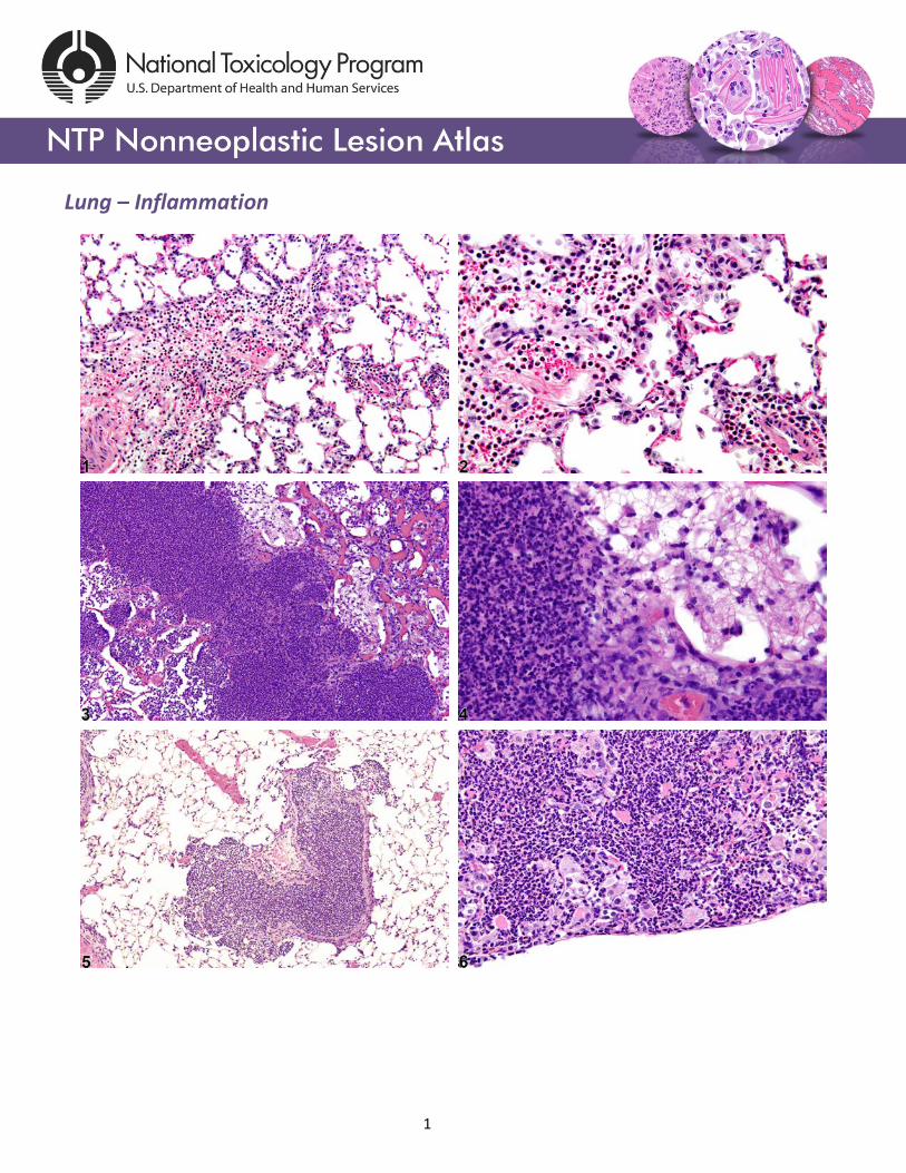
Figure Legend: Figure 1 Lung - Inflammation, Acute in a male Wistar Han rat from a subchronic
study. The majority of the inflammatory cells are neutrophils; there is also a small amount of
hemorrhage. Figure 2 Lung - Inflammation, Acute in a male Wistar Han rat from a subchronic study
(higher magnification of Figure 1). The majority of the inflammatory cells are neutrophils, but there are
also mononuclear cells, including alveolar macrophages. Figure 3 Lung - Inflammation, Suppurative in
a male Wistar Han rat from a chronic study. Large numbers of degenerate neutrophils fill and replace
alveoli. Figure 4 Lung - Inflammation, Suppurative in a male Wistar Han rat from a chronic study
(higher magnification of Figure 3). Abundant necrotic debris is admixed with the degenerate
neutrophils. Figure 5 Lung, Bronchiole - Inflammation, Suppurative in a male B6C3F1/N mouse from a
chronic study. Degenerate neutrophils fill the bronchiole. Figure 6 Lung - Inflammation, Suppurative in
a female B6C3F1/N mouse from a chronic study. There are numerous large, foamy, activated alveolar
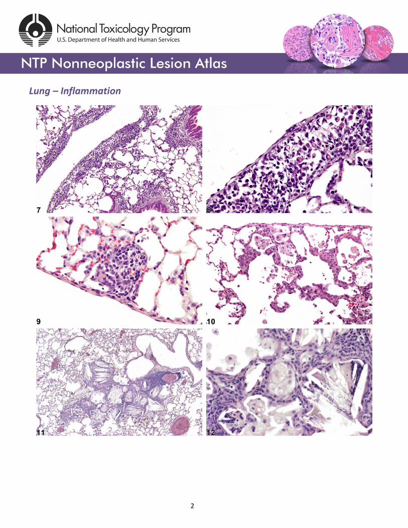
macrophages amid the degenerate neutrophils. Figure 7 Lung - Inflammation, Chronic in a female
B6C3F1/N mouse from a chronic study. The mononuclear inflammatory cells are largely perivascular,
peribronchiolar, and subpleural. Figure 8 Lung - Inflammation, Chronic in a female B6C3F1/N mouse
from a chronic study (higher magnification of Figure 7). Mott cells are visible amid the lymphocytes and
plasma cells in the subpleural region. Figure 9 Lung - Inflammation, Chronic in a male F344/N rat from
a subchronic study. These focal, subpleural lesions are a common background finding. Figure 10 Lung
- Inflammation, Chronic in a male F344/N rat from a subchronic study. This is a slightly more severe
example of the common background lesion shown in Figure 9, with thickening of the alveolar septa.
Figure 11 Lung - Inflammation, Chronic in a female B6C3F1/N mouse from a chronic study. Numerous
4
Lung – Inflammation
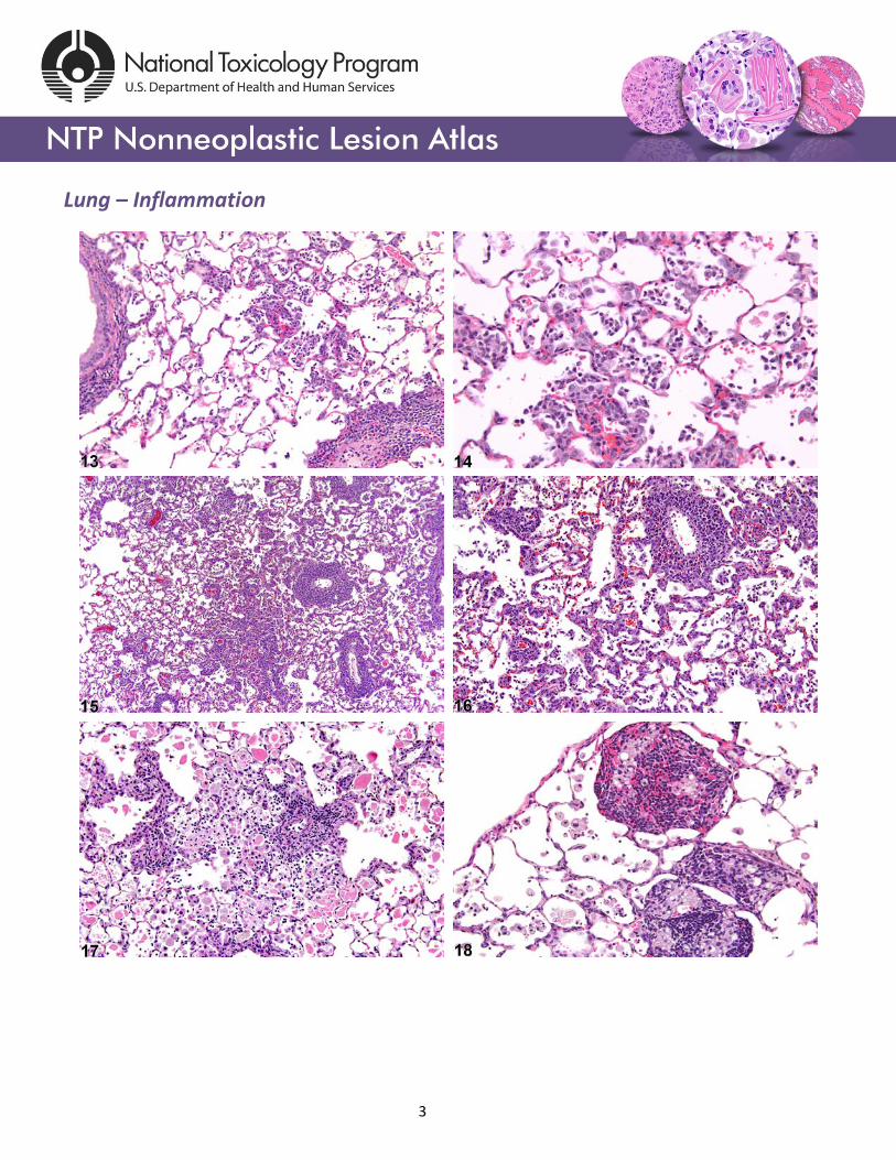
cholesterol clefts and pigmented macrophages are present in this inflammatory lesion. Figure 12 Lung
- Inflammation, Chronic in a female B6C3F1/N mouse from a chronic study (higher magnification of
Figure 11). The macrophages are large, foamy, and activated. Figure 13 Lung - Inflammation, Chronic
active in a male F344/N rat from a subchronic study. There is a mixture of lymphocytes, macrophages,
and neutrophils. Figure 14 Lung - Inflammation, Chronic active in a male F344/N rat from a subchronic
study (higher magnification of Figure 13). There is a mixture of lymphocytes, macrophages, and
neutrophils, with a small amount of alveolar hemorrhage. Figure 15 Lung - Inflammation, Chronic
active in a male F344/NTac rat from a subchronic study. The perivascular and interstitial inflammation
in this control rat is consistent with Pneumocystis carinii infection (formerly rat respiratory virus). Figure 16 Lung - Inflammation, Chronic active in a male F344/NTAC rat from a subchronic study (higher
magnification of Figure 15). The lesion is consistent with Pneumocystis carinii infection (formerly rat
respiratory virus). Figure 17 Lung - Inflammation, Chronic active in a male B6C3F1/N mouse from a
subchronic study. There is a mixture of inflammatory cell types, including neutrophils, and alveolar
proteinosis. Figure 18 Lung - Inflammation, Granulomatous in a male Wistar Han rat from a chronic
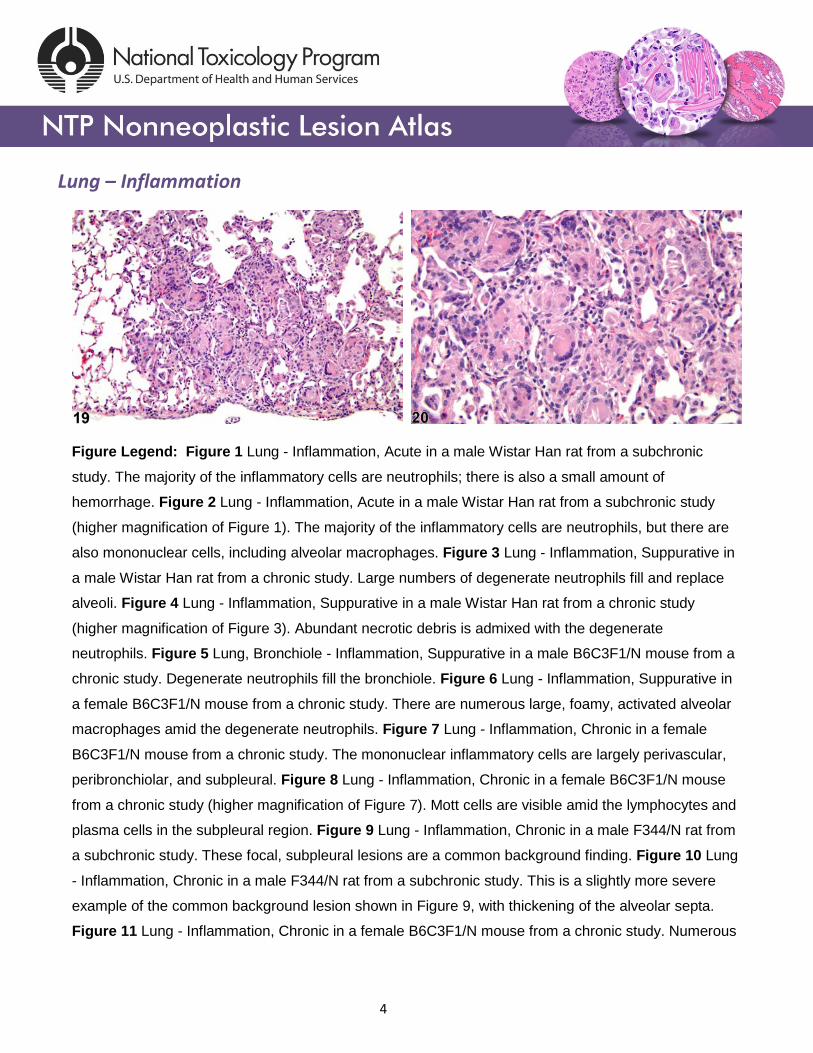
study. Clusters of large, foamy macrophages are surrounded by mononuclear cells. Figure 19 Lung -
Inflammation, Granulomatous in a female F344/NTAC rat from a subchronic study. There are numerous
multinucleated giant cells in this lesion. Figure 20 Lung - Inflammation, Granulomatous from a female
F344/NTac rat in a subchronic study (higher magnification of Figure 19). The multinucleated giant cells
are of the Langhans type.
Comment: Inflammation of the lungs is one of the most common lesions seen in inhalation studies. In
NTP studies, there are five standard categories of inflammation: acute, suppurative, chronic, chronic
active, and granulomatous. In acute inflammation (Figure 1 and Figure 2), the predominant infiltrating
cell is the neutrophil, though fewer macrophages and lymphocytes may also be present. There may
also be evidence of edema or hyperemia. The neutrophil is also the predominant infiltrating cell type in
suppurative inflammation (Figure 3, Figure 4, Figure 5, and Figure 6), but they are aggregated, and
many of them are degenerate (suppurative exudate). Cell debris, from both the resident cell populations
and infiltrating leukocytes, and proteinaceous fluid containing fibrin, fewer macrophages, occasional
lymphocytes or plasma cells, and, possibly, an infectious agent may also be present within the exudate.
Grossly, these lesions would be characterized by the presence of pus. The tissue surrounding the
5
Lung – Inflammation
exudate may contain fibroblasts, fibrous connective tissue, and mixed inflammatory cells, depending on
the chronicity of the lesion. Lymphocytes predominate in chronic inflammation (Figure 7, Figure 8,
Figure 9, Figure 10, Figure 11, and Figure 12). Lymphocytes also predominate in chronic active
inflammation (Figure 13, Figure 14, Figure 15, Figure 16, and Figure 17), but there are also a significant
number of neutrophils. Both lesions may contain macrophages. Granulomatous inflammation (Figure
18, Figure 19, and Figure 20) is another form of chronic inflammation, but this diagnosis requires the
presence of a significant number of aggregated, large, activated macrophages, epithelioid
macrophages, or multinucleated giant cells. In all forms of inflammation, there may also be edema,
hemorrhage, fibrin, proteinosis, degeneration, or necrosis. Inflammation is differentiated from cellular
infiltrates by the presence of other changes, such as edema, hemorrhage, degeneration, necrosis, or
other evidence of tissue damage.
A test agent may stimulate epithelial cells and resident macrophages to secrete cytokines, thus
inducing an inflammatory response. Alternatively, a test agent may cause tissue damage, which
secondarily results in inflammation. Several compartments within the lung may be inflamed, including
the airways (bronchi and bronchioles), the alveoli or alveolar septa (interstitium), the terminal
bronchiole/alveolar duct region (acinar region), perivascular areas, and the pleura. Occasionally, focal,
minimal inflammation in the lung is seen as a background lesion in mice and rats (Figure 9 and Figure
10), particularly in the subpleural region. Small aggregates of alveolar histiocytes can also be seen (see
Lung – Infiltration cellular, Histiocyte) as a background lesion and must be differentiated from
inflammation. Inflammation can also be caused by a number of infectious agents, but these are rare
under current husbandry practices. Systemic bacterial infections affecting the pulmonary interstitium via
the bloodstream often produce a suppurative alveolitis, whereas viral infections tend to induce
suppurative or mononuclear perivascular inflammation. Inflammation can also be associated with
pulmonary neoplasms.
Recommendation: Whenever present, Lung - Inflammation should be diagnosed and assigned a
severity grade. A site modifier (e.g., perivascular, interstitial, bronchial, bronchiolar, pleural, or
subpleural) should be included in the diagnosis to indicate the location of the lesion. Also, the type of
inflammation (e.g., acute, chronic) should be included in the diagnosis as a modifier. If the inflammation
6
Lung – Inflammation
affects more than one site, the site modifier may be omitted and the affected locations identified in the
pathology narrative. The term “inflammation” should be used when the inflammatory cells are
accompanied by other changes indicative of inflammation, such as vascular changes (which may result
in hemorrhage or edema), necrosis or degeneration of cells, or disruption of the normal architecture.
Lesions that are considered part of the inflammatory process, such as edema and hemorrhage, need
not be diagnosed separately unless warranted by severity but should be described in the narrative.
Necrosis or degeneration of cells may be primary, inciting an inflammatory response, or it may be
secondary to the inflammation. Therefore, it can be very difficult to determine which lesion (necrosis or
inflammation) is primary and which is secondary. The pathologist should use his or her judgment in
determining whether to diagnose these lesions separately or to combine these related lesions into a
single diagnosis. If they are combined into a single diagnosis, all components of the lesions should be
thoroughly described in the narrative. A small, focal accumulation of inflammatory cells with no other
evidence of inflammation (e.g., edema, hemorrhage, cell swelling, degeneration, or necrosis, alveolar
septal thickening, fibrin deposition), should be diagnosed as “infiltration cellular” rather than
inflammation. When present as a secondary finding to a neoplasm, the inflammation need not be
diagnosed separately but should be described in the pathology narrative.
References: Boorman GA, Eustis SL. 1990. Lung. In: Pathology of the Fischer Rat: Reference and Atlas (Boorman GA, Eustis SL, Elwell MR, Montgomery CA, MacKenzie WF, eds). Academic Press, San Diego, CA, 339-367.
Dixon D, Herbert RA, Sills RC, Boorman GA. 1999. Lungs, pleura, and mediastinum. In: Pathology of the Mouse: Reference and Atlas (Maronpot RR, Boorman GA, Gaul BW, eds). Cache River Press, Vienna, IL, 293-332.
Dungworth DL, Ernst H, Nolte T, Mohr U. 1992. Nonneoplastic lesions in the lungs. In: Pathobiology of the Aging Rat (Mohr U, Dungworth DL, Capen CC, eds). ILSI Press, Washington, DC, 143-160.
Plopper CG, Dungworth DL. 197. Structure, function, cell injury and cell renewal of bronchiolar and alveolar epithelium. In: Lung Carcinomas (McDowell EM, ed). Churchill Livingstone, Edinburgh, 94-128.
Renne R, Brix A, Harkema J, Herbert R, Kittel K, Lewis D, March T, Nagano K, Pino M, Rittinghausen S, Rosenbruch M, Tellier P, Wohrmann T. 2009. Proliferative and nonproliferative lesions of the rat and mouse respiratory tract. Toxicol Pathol 37(suppl):5S-73S. Abstract: http://www.ncbi.nlm.nih.gov/pubmed/20032296
7
Lung – Inflammation
Authors: Mark F. Cesta, DVM, PhD, DACVP Staff Scientist/NTP Pathologist NTP Pathology Group National Toxicology Program National Institute of Environmental Health Sciences Research Triangle Park, NC
Darlene Dixon, DVM, PhD, DACVP Group Leader Molecular Pathogenesis Group National Toxicology Program National Institute of Environmental Health Sciences Research Triangle Park, NC
Ronald A. Herbert, DVM, PhD Group Leader/NTP Pathologist Pathology Support Group National Toxicology Program National Institute of Environmental Health Sciences Research Triangle Park, NC
Lauren M. Staska, DVM, PhD, DACVP Senior Pathologist WIL Research Hillsborough, NC
8