lung inflammation and lack of genotoxicity in the comet
TRANSCRIPT
RESEARCH Open Access
Lung inflammation and lack of genotoxicityin the comet and micronucleus assays ofindustrial multiwalled carbon nanotubesGraphistrength© C100 after a 90-day nose-onlyinhalation exposure of ratsDaniela Pothmann1, Sophie Simar2, Detlef Schuler1, Eva Dony3, Stéphane Gaering1, Jean-Loïc Le Net4,Yoshi Okazaki5, Jean Michel Chabagno6, Cécile Bessibes6, Julien Beausoleil6, Fabrice Nesslany2,7
and Jean-François Régnier8*
Abstract
Background: Graphistrength© C100 multiwalled carbon nanotubes (MWCNT) provide superior electrical andmechanical properties for various applications. The evaluation of the intrinsic hazard properties of Graphistrength©
C100 is an essential step for safe use. A general feature of multiwalled carbon nanotubes after inhalation orintratracheal exposures is the induction of an inflammatory reaction in the lungs sometimes associated with localgenotoxic effects.
Methods: After investigating different parameters for the aerosol generation and performing a 5-day inhalationrange finding study, male and female Wistar rats were exposed nose-only for 90 days to target concentrations of0.05, 0.25 and 5.0 mg/m3 air of Graphistrength© C100 and sacrificed 24 h and 90 days after the last exposure.Broncho-alveolar lavage fluid (BALF) was also collected and analyzed for inflammatory parameters. Twenty-fourhours post-exposure, chromosomal aberrations in the bone marrow cells were evaluated by the micronucleus testand DNA damages in the lung, kidney and liver cells by both the standard and the human 8-oxoguanine DNAN-glycosylase 1 (hOGG1)-modified comet assay. All studies were performed according to the OECD test guidelines.
Results: An inflammatory lung reaction and the release of inflammatory factors in the BALF were observed in allrats exposed to 5.0 mg/m3, associated with changes in the differential white blood cells counts. The slight changesin BALF parameters at 0.25 mg/m3 recovered and signs of lung clearance of the MWCNT were observed. Nopathological changes were observed on the pleura. Neither increase in the number of micronucleatedpolychromatic erythrocytes nor increase in percent DNA damage were observed at any concentration.
Conclusions: Lung inflammation characteristic of an overload with insoluble particles was observed after a 90-dayexposure to 5.0 mg/m3 of Graphistrength© C100. Clear signs of clearance and recovery were observed at 0.25 mg/m3. Nogenotoxicity was detected locally in lung and distally in bone marrow, liver and kidney. Therefore, Graphistrength© C100appears of low concern in term of local and systemic genotoxicity and a No-Observed Adverse Effect Concentration(NOAEC) of 0.25 mg/m3 (0.28 mg/m3 as actual concentration) was established for the repeated-dose toxicity.
Keywords: Multiwalled carbon nanotubes, Comet assay, Micronucleus assay, Genotoxicity, Subchronic, Inhalation, Toxicity,NOAEC
* Correspondence: [email protected] France, Département Toxicologie et Environnement, 420 rue d’Estienne d’ Orves, 92705 Colombes, FranceFull list of author information is available at the end of the article
© 2015 Pothmann et al. This is an Open Access article distributed under the terms of the Creative Commons AttributionLicense (http://creativecommons.org/licenses/by/4.0), which permits unrestricted use, distribution, and reproduction in anymedium, provided the original work is properly credited. The Creative Commons Public Domain Dedication waiver (http://creativecommons.org/publicdomain/zero/1.0/) applies to the data made available in this article, unless otherwise stated.
Pothmann et al. Particle and Fibre Toxicology (2015) 12:21 DOI 10.1186/s12989-015-0096-2

BackgroundMultiwalled carbon nanotubes (MWCNT) are emergingnew materials intended for use in aeronautics, automo-tive, electronics, and many other fields. The outstandingperformance of MWCNT in applications like electricalconduction or mechanical improvements makes themvaluable in the development of new light materials withimproved properties [1, 2]. There is not just a singlekind of MWCNT. There are diverse materials whosephysico-chemical and toxicological properties depend onseveral factors: number of walls, diameter, length, shape(needle-like shape or flexible and contorted), states ofagglomeration and aggregation, surface chemistry, me-tallic impurities, to name a few. Determination of thepotential health effects of MWCNT has become a focusof attention in the scientific and regulatory community.The possible impact on the health of workers involvedin manufacture and handling and the development ofwide spread use including a number of consumer uses[3] prompted studies of various materials using differentroutes of exposure. The similarity in the shape and di-mension of some kinds of MWCNT and asbestos wasreported by Poland in 2008 [4]. Toxicology studies con-firmed the induction of inflammation, granulomas and/or mesotheliomas by a long, thick and rigid (needle-like)type of MWCNT (MWCNT-7 from Mitsui, diameter70-170 nm, length ca. 5 μm) after intraperitoneal (i.p.)injection to p53+/- mice [5, 6] and Fischer 344/BrownNorway F1 hybrids rats [7], injection into the pleuralspace of C57Bl/6 mice [8], and intrascrotal injection toFischer 344 rats [9]. Inhalation exposure of maleB6C3F1 mice to MWCNT-7 for 15 days at a concentra-tion of 5 mg/m3 promoted bronchioloalveolar adenomaand carcinoma induced by a single i.p. injection of meth-ylcholanthrene [10]. MWCNT-7 was also found to in-duce oxidative DNA damages and gene mutations in thelung cells of mice after a single intratracheal (i.t.) instilla-tion [11]. In contrast, no fibrotic lesions were observed[8] after the intrapleural injection to C57Bl/6 mice ofshort and straight (diameter 20-30 nm, length 0.5-2 μm,from Nanostructured & Amorphous Materials, Inc.) orcurled/tangled (diameter ca. 15 nm, length 1-5 μm anddiameter ca. 10 nm, length 5-20 μm, both from NanoLabInc.) MWCNT. Moreover, no increased incidence ofmesothelioma and other tumors was recorded by Mulleret al. [12] in male Wistar rats two years after a single i.p.injection of MWCNT (diameter ca 10 nm, length0.7 μm, from Namur University) with or without struc-tural defects. No induction of malignant mesotheliomain the peritoneal cavity [13] was also observed when ratswere followed for up to 3 years after two i.p. injectionsof a tangled form of MWCNT (diameter 15 nm, length3 μm, from Showa Denko [7]). Therefore, it seems thatthe rigidity, diameter, length, surface properties and
possibly contaminant metals are key factors when consid-ering the potential for a carcinogenic effect of MWCNT[13]. In view of the lack of coherent evidence across thevarious distinct MWCNT, the IARC Monograph WorkingGroup [14] specifically classified MWCNT-7 as possiblycarcinogenic to humans (Group 2B) and the other types ofMWCNT were categorized as not classifiable in respect totheir carcinogenicity to humans (Group 3).Graphistrength© C100 is one of the industrial MWCNT
referenced in the sponsorship program for the safety test-ing of nanomaterials by the Organization for EconomicCooperation and Development (OECD) [15]. There are anumber of publications [16–25] reporting studies onGraphistrength© C100 under the coded named NM 402or JRCNM04002a, sample from the repository of theEuropean Commission Joint Research Centre (EU-JRC)[26]. This EU-JRC Graphistrength© C100 was produced byArkema in a pilot production unit, whereas, the Graphis-trength© C100 used in the present 90-day study was froman industrial production unit. Nevertheless, these two unitsused the same process of synthesis and there are no signifi-cant physico-chemical differences between the productsfrom both production units.Graphistrength© C100 is formed of large MWCNT ag-
glomerates with a mean particle size of about 400 μm andcontains a residual amount (<0.23 %) of small agglomerates(<15 μm) [27]. These small particles are comparable tothose observed by R’mili et al. [28] in the atmosphere ofour high safety laboratory dedicated to scientific experi-ments with MWCNT, indicating a possible inhalation ex-posure to these small particles. Therefore, conducting aninhalation subchronic toxicity study was judged to be a keyfeature in the safety assessment of Graphistrength© C100.However, Graphistrength© C100 does not contain sufficientquantities of these small agglomerates to directly providethe test material necessary for an experimental inhalationstudy [27]. Thus an aerosol generation procedure, as re-ported in the additional file 1, was developed in order toperform a valid study which fulfils the requirement of theinhalation specific OECD test guidelines [29]. The micron-isation process is an enrichment of the small particle frac-tion and is allowing the worst case material to be used asexpected by the regulatory authorities [30]. Another im-portant criterion was ensuring that the administered aero-sol has physico-chemical properties similar to the originalmaterial. After a careful evaluation, the defined technicalconditions for the generation of Graphistrength© C100aerosols were assessed in a 5-day range finding inhalationtoxicity study in rats with a 28-day recovery period. Then, a90-day inhalation toxicity study was performed in rats. Itincluded a 90-day recovery period and an evaluation of thepulmonary inflammation parameters. This subchronicstudy also provided the opportunity to perform a micronu-cleus assay on the bone marrow cells, as well as, a standard
Pothmann et al. Particle and Fibre Toxicology (2015) 12:21 Page 2 of 28

and a hOGG1-modified comet assay on the lung, liver andkidney cells of the exposed rats. The hOGG1-modifiedcomet assay was chosen because it is more specific than theFPG (formamidopyrimidine glycosilase) comet assay for theidentification of oxidative DNA damage [31]. Thus, thegenotoxic potential was evaluated in the cells directly incontact with Graphistrength© C100, and at a distance incase material was translocated from the lungs.
ResultsPhysico-chemical analysisGraphistrength© C100 used in the 5-day (batch no. 8287)and the 90-day (batch no. 110329-018) studies have respect-ively a median agglomerate size distribution of 376 and418 μm, an ash content of about 8.6 and 8.2 %, an apparentdensity of 0.085 and 0.106 g/cm3, a specific surface area of187 and 225.6 m2/g and metal contents (from the catalyst)of 3.2 and 3.0 % for Al and 2.7 and 2.7 % for Fe. MWCNTconstituents of Graphistrength© C100 have respectively11 ± 3 and 12 ± 4 walls with an outer mean diameter of11.8 and 12.1 nm, and a length of 1.05 ± 0.67 and 1.07 ±1.10 μm. The surface to volume ratio of the material usedfor the 90-day study was 2.4 × 10⁷ m-1. The other physico-chemical data are presented in Additional file 1: Table S3.The physico-chemical characterizations of Graphis-
trength© C100 (batch no. 110329-018) after a 12-h mill-ing under argon and after aerosol generation (samplescollected at the exhaust of the elutriator just before theinhalation chamber) are detailed in the Additional file 1:Table S3 and Additional file 1: Figure S4 and showed min-imal changes between the starting material and the ballmilled and micronized samples.
Characterization of the aerosol during exposuresThe gravimetrically determined mean achieved aerosolconcentrations of 0.066, 0.26 and 1.30 mg/m3 air duringthe 5-day exposure study were close to the targets of0.05, 0.25 and 1.25 mg/m3 (Additional file 1: Table S4), re-spectively. Over the 90-day exposure, the mean achievedaerosol concentrations of 0.06, 0.28 and 4.84 mg/m3 airwere also close to the target concentrations of 0.05, 0.25and 5.0 mg/m3, respectively (Table 1).The mean mass median aerodynamic diameters (MMAD)
and geometric standard deviations (GSD) by impactor/gravi-metric determinations during the 5-day and 90-day expo-sures were within the target ranges (Additional file 1: TableS4 and Table 1). The count median aerodynamic diameters(CMAD) determined by Wide Range Particle Spectrometer©
(WPRS) analysis (Table 1) were similar between the exposedgroups of the 90-day exposure study. All together these datashowed that the generated aerosols were within the respir-able range for rats (MMAD< 3 μm).Temperature, relative humidity and oxygen parameters
were consistent during both treatment periods (Additional
file 1: Table S4 and Table 1, respectively). In addition,values for temperature and oxygen concentration weresimilar across all groups. Dried air was used for aerosolgeneration and, accordingly, the relative humidity valueswere below 8 % for all groups. Differences between thegroups were considered to be negligible at this level.Therefore, the exposure conditions were considered to besatisfactory for this type of studies.
Ante-mortem animal observations5-day exposure with a 28-day recovery periodAll animals survived the 5-day exposure and 28-day recov-ery periods without showing clinical signs (data not shown).The food intake was similar across all groups during thestudy and there were no effects on body weight that wereconsidered to be related to exposure to Graphistrength©
C100. Stagnation of body weight gain or marginal bodyweight loss was noted between day 1 and day 5 of treatmentfor all groups including controls. It is not unusual in inhal-ation studies and was considered to be due to the restrainingof the animals in the tubes during the nose-only exposureprocedure and not related to treatment with the test mater-ial. Normal body weight gain was observed during the 28-day recovery period across all groups (data not shown).
90-day exposure with a 90-day recovery periodAll animals survived the 90-day exposure and recovery pe-riods. There were no test-item related clinical signs in anygroup. Hair loss, scabs, erythema and localized swellingwere recorded. These signs are commonly seen in animalsof this age and strain and are, therefore, considered to beincidental. No effects on food consumption were observedduring the 90-day treatment. Increased food intake was re-corded in male rats exposed to 0.05 and 5.0 mg/m3 duringthe first week of the 90-day recovery and several weeksthereafter. In addition increased food intake was recordedduring the first two weeks of recovery in females exposedto 5.0 mg/m3 (data not shown). Slightly reduced bodyweight gains were seen in males and females exposed to0.25 and 5.0 mg/m3 during several weeks of exposure. How-ever, the mean body weights of these animals remainedsimilar to the control group during the exposure period. At0, 0.05, 0.25, and 5.0 mg/m3, the body weights (means ±SDs) were at the commencement of study (day 0): 266.8 ±11.9 g, 269.7 ± 9.9 g, 268.5 ± 12.2 g and 270.4 ± 11.0 g inmales (35 rats per group), respectively and 170.7 ± 19.1 g,170.8 ± 16.0 g, 174.3 ± 19.4 g and 177.9 ± 22.7 g in females(35 per group), respectively. At the end of the 90-day expos-ure period, the respective body weights were 401.7 ± 26.9 g,407.3 ± 35.9 g, 405.3 ± 37.9 g and 411.2 ± 29.4 g in males (30per group) and 244.7 ± 21.9 g, 240.3 ± 20.3 g, 248.0 ± 25.4 gand 245.9 ± 25.3 g in females (30 per group). Increased bodyweight gains in males and females and body weights inmales were observed during recovery in animals exposed to
Pothmann et al. Particle and Fibre Toxicology (2015) 12:21 Page 3 of 28

5.0 mg/m3. At 0, 0.05, 0.25, and 5.0 mg/m3, the respectivebody weights (means ± SDs) at the end of the 90-day recov-ery period were 494.5 ± 44.1 g, 529.0 ± 53.5 g, 518.8 ± 55.0 gand 539.3 ± 40.6 g in males (20 per group) and 297.1 ±33.9 g, 290.2 ± 30.0 g, 298.5 ± 39.8 g and 302.4 ± 45.5 g in fe-males (20 per group). None of these changes were consid-ered to be adverse effects.There were no effects of exposure on grip strength,
body temperature, landing foot splay and locomotors ac-tivity. There were no differences in blood pressure thatwere considered to be related to the exposure. High sys-tolic/diastolic blood pressures (ca. 140/100 mmHg) wereobserved in one male of each control and treatedgroups. No effect was recorded during ophthalmoscopicexamination (data not shown).The statistically significant changes observed in
hematological parameters are summarized in Table 2.An increase in relative and absolute neutrophil countsand a slight decrease of the relative (but not absolute)lymphocyte counts were recorded in males and femalesexposed to 5.0 mg/m3 at the end of the 90-day exposureand recovery periods. The other statistically significantchanges (relative eosinophil counts, prothrombin time,and platelets) were all in the range of the historical con-trol data (HCD) of the laboratory (Table 2), not-dose-related, observed only in one sex and not observed afterthe 90-day treatment free period and therefore were notconsidered to be treatment-related.The statistically significant changes of the blood chem-
istry parameters are summarized in Table 3. Increasedpotassium values were recorded in males exposed to5.0 mg/m3 (9 %) and in all treated groups of females (15,21 and 11 % at 0.05, 0.25 and 5.0 mg/m3, respectively) at
the end of the treatment period but not after the 90-daytreatment free period. Considering the low magnitude ofthis hyperkalemia and the variability of the potassiumlevels in the rats [32], these changes were not consideredto be treatment related and/or adverse. The other statisti-cally significant changes (creatinine, triglycerides, sodium,chloride, calcium, and proteins) were not considered to betreatment-related as the values were all in the range of theHCD data (Table 3), not dose-related, observed only inone sex, and not correlated with histological findings.Urinalysis and estrus cycles parameters were unre-
markable (data not shown).
Post-mortem animal observationsBronchoalveolar lavage fluid (BALF) analysisDetailed results of cellular, biochemical and cytokinesmeasurements are displayed in Additional file 1: TablesS5 and Additional file 1: Table S6 for the 5-day studyand Tables 4, 5, 6 for the 90-day study. As the methodsused to collect of the BALF were slightly different be-tween the 5-day (use of the full lung) and the 90-day(use of only the left lobe) studies, and to also allow acomparison with the recovery data, changes in BALF pa-rameters presented in Additional file 1: Figure S5 (5-daystudy) and Figs. 1 and 2 (90-day study) were normalizedrelative to the time-matched, concurrent control group.
5-day exposure with a 28-day recovery periodTwenty-four hours post exposure, the differential cellcount in BALF revealed statistically significantly in-creased polymorphonuclear neutrophils (PMN) levels at1.25 mg/m3 (x9 compare to control group) as detailed inAdditional file 1: Table S5 and Additional file 1: Figure
Table 1 Target and achieved aerosol concentrations and particles size of Graphistrength© C100. Temperature, relative humidity andoxygen concentration measured over the 90-day exposure period
Groups Control Low Mid High
Target aerosol concentrations (mg/m3 air) 0 0.05 0.25 5.0
Achieved aerosol concentrations (mg/m3 air) - 0.06 ± 0.04 0.28 ± 0.06 4.84 ± 0.41
Mean mass median aerodynamic diameter (MMAD, μm) (gravimetric determination) - nda 1.62 ± 0.39 2.30 ± 0.34
(n = 5) (n = 14)
Mean GSD (gravimetric determination) - nda 4.67 ± 4.81 2.47 ± 0.26
Mean percentage of particles < 3 μm (gravimetric determination) - nda 74.10 ± 14.00 63.49 ± 6.23
Count median aerodynamic diameter (CMAD, nm) (WPRS determination) - 196.2 ± 54.7 231,5 ± 65.1 208.0 ± 62.0
(n = 14) (n = 14) (n = 14)
Mean temperature (°C) 23.2 ± 0.9 23.3 ± 0.7 23.6 ± 0.7 23.8 ± 0.6
Mean relative humidity (%) 5.8 ± 1.5 6.1 ± 1.7 6.1 ± 1.7 6.3 ± 1.6
Mean oxygen concentration (%) 20.8 ± 0.0 20.8 ± 0.0 20.8 ± 0.0 20.8 ± 0.0
nd: not determineda at 0.05 mg/m3 due to the very low concentration, the particle size could not be determined by gravimetry at an air flow rate of 1 L/min. The aerosolconcentrations at 0.05 and 0.25 mg/m3 were achieved by serial dilution with compressed, filtered, dry air of the 0.25 and 5.0 mg/m3 concentrations, respectively.Therefore, the MMAD and GSD at 0.05 mg/m3 are expected to be of the same order as at 0.25 mg/m3. This is also confirmed by particle size data from the 5-daystudy with sampling at an air flow rate of 9 L/min (see Additional file 1: Table S4 for details)
Pothmann et al. Particle and Fibre Toxicology (2015) 12:21 Page 4 of 28

S5 panel A. After the 28-day recovery period the PMNvalue was similar to controls (Additional file 1: Table S5and Additional file 1: Figure S5 panel B). Twenty-fourhours post exposure, the macrophages in all treatedgroups contained phagocytized test material with adose-related increased incidence (Additional file 1: TableS5). At the end of the recovery period, the incidence sig-nificantly decreased at all concentrations (Additional file1: Table S5).Biochemical analysis of the BALF 24 h post-exposure
resulted in statistically significantly increased γ-glutamyltransferase (GGT) levels at 1.25 mg/m3 (x3.5)and increased protein values at 0.25 (x23) and 1.25(x20) mg/m3 (Additional file 1: Table S6 and Additionalfile 1: Figure S5 panel A). After the 28-day recovery
period, differences to controls diminished and statisticalsignificance was only observed for changes in proteinvalues at 0.25 (x3) and 1.25 (x3.5) mg/m3 (Additionalfile 1: Table S6, Additional file 1: Figure S5 panel B). Allother changes between exposed and control groups andbetween the 24-h-and 28-day sacrifice times are justbiological variations.
90-day exposure with a 90-day recovery periodTwenty-four hours post exposure, there were no changesin BALF total cell count and viability in males (Table 4,Fig. 1 panel a). Nevertheless, it must be mentioned thatthese 2 parameters have been determined in only 4 outof 10 males rats exposed at 5.0 mg/m3 due to the largeamount of black particles and MWCNT-laden BAL cells
Table 2 Statistically significant changesa in hematological parameters 24 h and 90 days after a 90-day exposure of male and femalerats to Graphistrength© C100
Concentration Total leucocytes Neutrophils Eosinophils Lymphocytes Prothrombin time Platelets
(mg/m3) G/L rel. 1 (G/l) rel. 1 (G/l) rel. 1 (G/l) rel. 1 (sec) G/l
MALE
HCD b, c 3.74 - 9.53 0.119 - 0.339 0.01 - 0.035 0.611 - 0.842 0.70 – 0.97 708 - 1168
(0.66 - 2.29) (0.05 - 0.22) (2.59 - 7.39) (14.4 - 29.9)
24 h post exposure
0 6.43 0.197 (1.28) 0.009 (0.06) 0.742 (4.78) 0.73 (23.4) 879
0.05 7.01 0.220 (1.79) 0.013 (0.10) 0.716 (4.79) 0.73 (23.4) 973
0.25 7.30 0.211 (1.71) 0.013* (0.10) 0.727 (5.15) 0.76 (22.8) 972
5.0 8.13 0.327** (2.63**) 0.015* (0.12) 0.608** (5.05) 0.77* (22.5) 1003*
90 days post exposure
0 7.07 0.237 (1.79) 0.012 (0.09) 0.707 (4.88) 0.85 (23.7) 903
0.05 7.47 0.223 (1.62) 0.014 (0.11) 0.716 (5.44) 0.86 (23.4) 914
0.25 7.14 0.283 (2.23) 0.015 (0.11) 0.664 (4.55) 0.85 (23.7) 822
5.0 8.23 0.374* (2.96**) 0.015 (0.12) 0.577* (4.84) 0.89 (22.8) 901
FEMALE
HCDb, c 1.91 - 6.14 0.099 - 0.343 0.009 - 0.045 0.598 - 0.860 0.70 – 0.98 723 - 1235
(0.34-1.31) (0.03 - 0.15) (1.30 - 4.86) (13.5 - 38.2)
24 h post exposure
0 4.07 0.146 (0.61) 0.011 (0.04) 0.809 (3.29) 0.73 (23.5) 1144
0.05 4.05 0.152 (0.61) 0.006 (0.04) 0.808 (3.27) 0.75 (23.0) 1087
0.25 3.69 0.170 (0.62) 0.016 (0.06) 0.779 (2.87) 0.79** (22.2**) 1042
5.0 4.96 0.261** (1.21**) 0.012 (0.06) 0.698** (3.54) 0.77 (22.6) 1099
90 days post exposure
0 4.17 0.175 (0.67) 0.017 (0.06) 0.772 (3.27) 0.87 (23.2) 883
0.05 3.75 0.220 (0.84) 0.019 (0.07) 0.729 (2.71) 0.88 (22.8) 933
0.25 3.32 0.238* (0.74) 0.018 (0.05) 0.714 (2.40) 0.86 (23.5) 841
5.0 4.90 0.382** (1.81**) 0.013 (0.07) 0.564** (2.85) 0.89 (22.7) 914
* p < 0.05, ** p < 0.01; statistical significant differences to controlsa No statistically significant changes were observed at any concentrations and time-points on red blood cell, hemoglobin, hematocrit, mean corpuscular volume,mean corpuscular hemoglobin, mean corpuscular hemoglobin concentration, reticulocytes, basophils, monocytes and partial thromboplastine timeb HCD Historical control data, 95 % tolerance limitsc Changes statistically significant outside the HCD are in italic characters
Pothmann et al. Particle and Fibre Toxicology (2015) 12:21 Page 5 of 28

in the cellular suspension. In females, the total cell countwas increased at 5.0 mg/m3 (+78 %) while viability didnot show any differences (Table 4, Fig. 1 panel b. Thecytodifferentiation of BALF cells (Table 4, Fig. 1) showeddecreases in macrophages and increases in neutrophilsand lymphocytes in males and females exposed to 0.25and 5.0 mg/m3, respectively.Statistically significant increases in protein (x4.3 and
x3.7), phospholipids (x6.6 and x4.8), LDH (x6.6 andx6.2), GGT (x3.3 and x 3.1) and ALP (x5.2 and 4.1) wererecorded in males and females exposed at 5.0 mg/m3, re-spectively (Table 5, Fig. 1). In addition phospholipids inmales (x2.2) and GGT in males (x2.1) and females (x2.4)were also increased at 0.25 mg/m3.A statistically and biologically significant increase
(values higher than mean + 2 SD of the correspondingcontrol group) of tumor necrosis factor alpha (TNF-α)
was observed in BALF of male and female rats exposedto 0.25 (x3.4 and x8.5, respectively) and 5.0 mg/m3 (x6.1and x10, respectively) and of IL-1β levels (x1.8) in fe-males exposed to 5.0 mg/m3 (Table 6, Fig. 1). Detectablelevels of IL-5 and IL-1α (data not shown) were measuredin some single animals (including controls) without adose-response relationship.Ninety days post-exposure, there was an increase in
the total cell counts in male and female rats exposed to5.0 mg/m3 (x3.5 and x2.4, respectively) but viabilitieswere not affected (Table 4, Fig. 2). Differential countswere still changed with a similar intensity as 24-h post-exposure. In males exposed to 0.25 mg/m3, an increaseof the total cell count (+58 %) was observed, but it wasnot present 24 h post-exposure. As this increase wasnot associated with changes in differential counts, bio-chemistry parameters, and lung pathological effects, it
Table 3 Statistically significant changesa in blood chemistry parameters 24 h and 90 days after a 90-day exposure of male andfemale rats to Graphistrength© C100
Concentration(mg/m3)
Creatinineμmol/l
Triglyceridesmmol/l
Sodiummmol/l
Potassiummmol/l
Chloridemmol/l
Calciummmol/l
Total proteing/l
MALE
HCDb, c 21.9 - 35.0 0.20 - 1.08 138. 5- 149.2 3.22 - 4.47 99.9 - 109.2 2.55 - 2.97 62.10 - 73.54
24 h post exposure
0 23.7 0.48 142.8 4.13 102.3 2.68 66.15
0.05 24.6 0.38* 142.9 4.20 102.1 2.67 65.39
0.25 23.6 0.37* 143.9 4.25 103.0 2.69 67.02
5.0 22.4 0.41 144.2 4.52** 103.0 2.69 66.97
90 days post exposure
0 27.9 0.77 145.5 4.52 103.4 2.75 69.08
0.05 25.5* 0.68 145.7 4.59 103.2 2.77 68.42
0.25 24.4** 0.75 145.9 4.58 104.4 2.71 67.41
5.0 26.3 0.79 145.8** 4.73 104.5 2.74- 68.24
FEMALE
HCDb, c 25.0 - 41.7 0.18 - 0.57 137.8 - 147.8 2.73 - 3.90 100.6 - 110.3 2.53 - 2.99 63.62 - 79.36
24 h post exposure
0 30.4 0.32 143.5 3.43 102.0 2.75 72.27
0.05 27.7 0.31 144.8 3.95** 103.8 2.76 71.50
0.25 28.3 0.35 144.0 4.17** 103.4 2.80 72.87
5.0 27.3* 0.30 145.8 3.81* 105.3** 2.72 69.27*
90 days post exposure
0 28.8 0.72 143.5 3.40 101.7 2.78 77.11
0.05 31.9 0.60 146.4** 3.41 104.1 2.76 74.74
0.25 30.1 0.54* 144.9 3.21 101.9 2.72* 74.78
5.0 30.6 0.47** 145.1 3.54 104.0 2.74 72.29**
* p < 0.05, ** p < 0.01; statistical significant differences to controlsa No statistically significant changes were observed at any concentrations and time-points on glucose, urea, bilirubin, cholesterol, triglycerides, phospholipids, as-partate aminotransferase (ASAT), alanine aminotransferase (ALAT), lactate dehydrogenase (LDH), alkaline phosphatase (ALP), gamma-glutamyltransferase (GGT),creatine kinase (CK), phosphorus, albumin and globulinb HCD Historical control data, 95 % tolerance limitsc Changes statistically significant outside the HCD are in italic characters
Pothmann et al. Particle and Fibre Toxicology (2015) 12:21 Page 6 of 28

was thought to be of no toxicological significance andrelated to the clearance process.The changes in the biochemical parameters (Table 5,
Fig. 2) and cytokines levels (Table 6, Fig. 2) observed at5.0 mg/m3 were of similar intensity as at 24-h post-exposure. No change of the biochemical parameters wasobserved at 0.05 and 0.25 mg/m3 except a persistent in-crease of TNF-α levels in males and females exposed at0.25 mg/m3 (x2.4 and x2.1, respectively) related to theresidual amount of black particles in the lungs.
Organs weights and macroscopic examination5-day exposure with a 28-day recovery periodThere were no macroscopic lesions that could be attrib-uted to treatment with Graphistrength© C100. No changewas observed in lung weight at either sacrifice times (datanot shown). The absolute kidney weight was statisticallysignificantly reduced just after the 5-day exposure in malesexposed to 0.25 and 1.25 mg/m3 (1.71 and 1.77 g vs.2.00 g in control, respectively, p < 0.01) as well as thekidney to body weight ratio at 0.25 mg/m3 (0.56 % vs.0.61 % in control, p < 0.05). However, in the absence ofhistological correlates, effects in females and after the 28-
day recovery period, the kidney weight change was notconsidered to be treatment-related. There were no furthereffects on organ weights which were considered to be pos-sibly related to treatment (data not shown).
90-day exposure with a 90-day recovery periodBlack brown foci in the lung and black brown discolor-ation of the bronchial lymph nodes were recorded in allor most animals 24 h and 90 days after 90 days of expos-ure to 5.0 mg/m3. No test item-related macroscopicfindings were observed in animals exposed to 0.05 and0.25 mg/m3.Twenty-hour hours post-exposure, absolute and
relative (to body weight) lung weights were increasedin males (+47 and +50 %, respectively) and females(+45 and +50 %, respectively) exposed to 5.0 mg/m3
(Table 7). Lung weight (absolute and relative) werestill increased in males (+62 and +53 %, respectively)and females (+45 and +36 %, respectively) exposed to5.0 mg/m3 after 90 days of recovery (Table 7). Allother statistically significant changes in organ weightswere incidental and not considered to be treatment-related (data not shown).
Table 4 Cell analysis in the BALF of male and female rats 24 h and 90 days after a 90-day exposure to Graphistrength© C100
Concentration (mg/m3) Total cell Count Viability Macrophages Eosinophils Lymphocytes Neutrophils Epithelial cells
106 % % % % %
MALE
24 h post exposure
0 1.74 84.40 93.9 0.0 4.4 1.5 0.1
0.05 1.62 84.65 91.8 0.0 5.1 3.1 0.0
0.25 2.24 89.00 64.8** 0.0 9.7* 25.2** 0.3
5.0 2.01 88.00 31.5** 0.0 12.8** 55.6** 0.1
90 days post exposure
0 1.80 90.10 96.5 0.0 2.1 1.2 0.2
0.05 2.21 91.35 96.9 0.0 1.9 0.9 0.3
0.25 2.85* 91.35 91.2 0.1 3.8 4.7 0.3
5.0 6.23** 89.86 44.6** 0.0 20.4** 39.8** 0.3
FEMALE
24 h post exposure
0 1.67 91.80 96.9 0.0 2.5 0.4 0.1
0.05 1.27 92.55 94.5 0.0 4.4 1.0 0.1
0.25 2.16 92.55 80.9** 0.0 4.1 15.0** 0.0
5.0 2.98** 95.50 34.7** 0.0 11.5** 53.8** 0.0
90 days post exposure
0 1.67 86.39 96.1 0.0 3.1 0.5 0.2
0.05 1.66 85.40 95.8 0.0 3.4 0.8 0.0
0.25 1.86 87.55 93.9 0.0 3.3 2.8 0.0
5.0 4.02** 88.80 46.7** 0.0 13.0** 40.1** 0.0
* p < 0.05, ** p < 0.01; statistical significant differences to controls
Pothmann et al. Particle and Fibre Toxicology (2015) 12:21 Page 7 of 28

Fig. 1 BALF parameters of male (a) and female (b) rats 24 h after a 90-day exposure to Graphistrength© C100. Changes are shown as x-folddifferences compared to controls using a logarithmic scaling. Abbreviations: ALP: alkaline phosphatase, GGT: γ-glutamyltransferase, LDH: lactatedehydrogenase, IL-1β: interleukin 1β, TNF-α: Tumor Necrosis Factor α
Pothmann et al. Particle and Fibre Toxicology (2015) 12:21 Page 8 of 28

Fig. 2 BALF parameters of male (a) and female (b) rats 90 days after a 90-day exposure to Graphistrength© C100. Changes are shown as x-folddifferences compared to controls using a logarithmic scaling. Abbreviations: ALP: alkaline phosphatase, GGT: γ-glutamyltransferase, LDH: lactatedehydrogenase, IL-1β: interleukin 1β, TNF-α: Tumor Necrosis Factor α
Pothmann et al. Particle and Fibre Toxicology (2015) 12:21 Page 9 of 28

Microscopic examination5-day exposure with a 28-day recovery periodFindings observed in the lungs are summarized inAdditional file 1: Table S7 and typical lesions areshown in Additional file 1: Figure S6. Twenty-fourhours post exposure, hypertrophy of the bronchial andbronchiolar epithelial cells (Additional file 1: Figure S6panel B) and an increased severity of the infiltration ofmacrophages were noted in several animals exposed to1.25 mg/m3. Black inclusions in the cytoplasm of theinfiltrated macrophages (Additional file 1: Figure S6 panelC) were observed in a concentration-dependent mannerin all exposed animals confirming the observations donein the BALF.At the end of the 28-day recovery period, all these find-
ings were still present at a similar level, however, the inci-dence and the severity of hypertrophy of the bronchial andbronchiolar epithelial cells tended to be lower (only 4 ani-mals affected, all with a minimal grade) (Additional file 1:Figure S6 panel D). The other microscopic findings were
incidental lesions or within the range of normal back-ground lesions for animals of this strain and age.
90-day exposure with a 90-day recovery periodResults of the microscopic examinations are presentedin Tables 8, 9 and 10 for lungs, tracheobronchial lymphnodes and nasal cavity and larynx, respectively. Typicallesions in lungs and tracheobronchial lymph nodes areshown in Fig. 3.Twenty-four hours post-exposure to Graphistrength©
C100, minimal to marked concentration-related depos-ition of black particles were recorded in the lungs of allexposed rats (Table 8, Fig. 3 panel a; for comparison apicture of the lung of a control rat is displayed in Add-itional file 1: Figure S6 panel A). After 90 days of recov-ery, the mean severity decreased at 0.05 and 0.25 mg/m3, indicating partial clearance of the black particles atthese two lower concentrations; however, at 5.0 mg/m3,the mean severity score were overall similar indicatingincomplete clearance during this timeframe in theselungs overloaded with Graphistrength© C100 particles.At both sacrifice times, this deposition was associatedwith minimal to moderate concentration-related infil-tration of alveolar macrophages at 0.25 and 5.0 mg/m3
and with alveolar eosinophilic material (considered tobe the result of macrophages membrane cell rupture,Fig. 3 panel b), minimal infiltration of neutrophils in thealveoli (Fig. 3 panel c), and minimal to slight interstitialinflammation (Fig. 3 panel a) at 5.0 mg/m3 only. Note-worthy differences at the end of the 90-day recoveryperiod were the presence of minimal increased intersti-tial collagen fibers (Fig. 3 panel d) in 3 males and 2females exposed to 5.0 mg/m3 while the minimal bron-chiolar cell hypertrophy/hyperplasia and increased lym-phocytes in the bronchus associated lymphoid tissue(BALT) occasionally observed just post-exposure werenot recorded anymore. Minimal deposition of black par-ticles was also seen at the tracheal bifurcation in someanimals exposed at 5.0 mg/m3 at both sacrifice times.Minimal to moderate dose-related deposition of black
particles in the tracheobronchial lymph nodes was thehistological correlate of the black discoloration recordedat necropsy. This finding was recorded 24 h post-exposurein animals exposed to 0.25 and 5.0 mg/m3 (Table 9, Fig. 3panel e) and was associated with increased lymphocyteswithin the cortex/paracortex and vacuolation of the endo-thelial cells lining the high endothelial venules. After90 days of recovery, the mean severity score of black par-ticle deposition was similar at 0.25 mg/m3 and slightly in-creased at 5.0 mg/m3 (Table 9, Fig. 3 panel f), consistentwith continuous drainage of black particles from the lungsafter the end of the treatment. In the meantime, the otherassociated changes disappeared at 0.25 mg/m3 and theirintensities decreased at 5.0 mg/m3.
Table 5 Biochemistry parameters in the BALF of male andfemale rats 24 h and 90 days after a 90-day exposure toGraphistrength© C100
Concentration(mg/m3)
Phospholipidsmmol/L
LDHU/L
ALPU/L
GGTU/L
Proteinmg/L
MALE
24 h post exposure
0 0.16 196.8 37.6 5.9 92.7
0.05 0.21 168.6 38.8 6.7 84.2
0.25 0.36* 275.4 64.7 12.5** 115.4
5.0 1.05** 1306.2** 195.4** 19.5** 395.0**
90 days post exposure
0 0.26 225.7 76.4 5.6 115.4
0.05 0.27 174.8 78.2 6.3 79.8
0.25 0.42 237.2 108.5 9.5 102.3
5.0 1.18** 1353.2** 330.3** 16.1** 347.4**
FEMALE
24 h post exposure
0 0.17 163.6 34.9 5.1 78.8
0.05 0.18 163.0 28.8 6.6 91.2
0.25 0.30 312.7 63.4 12.5** 146.4
5.0 0.82** 1020.2** 144.1** 15.9** 292.6**
90 days post exposure
0 0.23 147.9 55.2 5.0 103.5
0.05 0.22 159.0 45.3 4.8 90.8
0.25 0.31 194.5 63.0 7.8 103.5
5.0 0.76** 566.2** 117.8** 15.8** 300.2**
* p < 0.05, ** p < 0.01; statistical significant differences to controls
Pothmann et al. Particle and Fibre Toxicology (2015) 12:21 Page 10 of 28
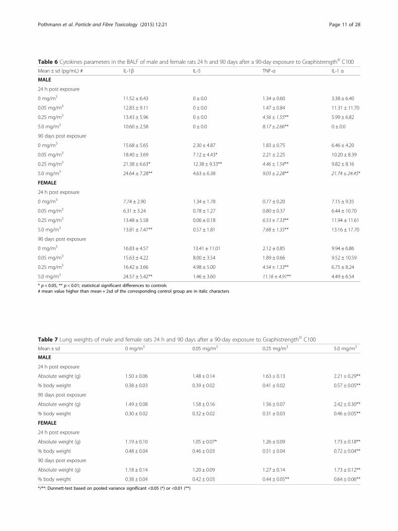
Table 6 Cytokines parameters in the BALF of male and female rats 24 h and 90 days after a 90-day exposure to Graphistrength© C100
Mean ± sd (pg/mL) # IL-1β IL-5 TNF-α IL-1 α
MALE
24 h post exposure
0 mg/m3 11.52 ± 6.43 0 ± 0.0 1.34 ± 0.60 3.38 ± 6.40
0.05 mg/m3 12.83 ± 9.11 0 ± 0.0 1.47 ± 0.84 11.31 ± 11.70
0.25 mg/m3 13.43 ± 5.96 0 ± 0.0 4.56 ± 1.55** 5.99 ± 6.82
5.0 mg/m3 10.60 ± 2.58 0 ± 0.0 8.17 ± 2.66** 0 ± 0.0
90 days post exposure
0 mg/m3 15.68 ± 5.65 2.30 ± 4.87 1.83 ± 0.75 6.46 ± 4.20
0.05 mg/m3 18.40 ± 3.69 7.12 ± 4.43* 2.21 ± 2.25 10.20 ± 8.39
0.25 mg/m3 21.38 ± 6.63* 12.38 ± 9.33** 4.46 ± 1.54** 9.82 ± 8.16
5.0 mg/m3 24.64 ± 7.28** 4.63 ± 6.38 9.03 ± 2.28** 21.74 ± 24.45*
FEMALE
24 h post exposure
0 mg/m3 7.74 ± 2.90 1.34 ± 1.78 0.77 ± 0.20 7.15 ± 9.35
0.05 mg/m3 6.31 ± 3.24 0.78 ± 1.27 0.80 ± 0.37 6.44 ± 10.70
0.25 mg/m3 13.48 ± 5.58 0.06 ± 0.18 6.53 ± 7.33** 11.94 ± 11.61
5.0 mg/m3 13.81 ± 7.47** 0.57 ± 1.81 7.68 ± 1.35** 13.16 ± 17.70
90 days post exposure
0 mg/m3 16.83 ± 4.57 13.41 ± 11.01 2.12 ± 0.85 9.94 ± 6.86
0.05 mg/m3 15.63 ± 4.22 8.00 ± 3.54 1.89 ± 0.66 9.52 ± 10.59
0.25 mg/m3 16.42 ± 3.66 4.98 ± 5.00 4.54 ± 1.33** 6.75 ± 8.24
5.0 mg/m3 24.57 ± 5.42** 1.46 ± 3.60 11.16 ± 4.91** 4.49 ± 6.54
* p < 0.05, ** p < 0.01; statistical significant differences to controls# mean value higher than mean + 2sd of the corresponding control group are in italic characters
Table 7 Lung weights of male and female rats 24 h and 90 days after a 90-day exposure to Graphistrength© C100
Mean ± sd 0 mg/m3 0.05 mg/m3 0.25 mg/m3 5.0 mg/m3
MALE
24 h post exposure
Absolute weight (g) 1.50 ± 0.06 1.48 ± 0.14 1.63 ± 0.13 2.21 ± 0.29**
% body weight 0.38 ± 0.03 0.39 ± 0.02 0.41 ± 0.02 0.57 ± 0.05**
90 days post exposure
Absolute weight (g) 1.49 ± 0.08 1.58 ± 0.16 1.56 ± 0.07 2.42 ± 0.30**
% body weight 0.30 ± 0.02 0.32 ± 0.02 0.31 ± 0.03 0.46 ± 0.05**
FEMALE
24 h post exposure
Absolute weight (g) 1.19 ± 0.10 1.05 ± 0.07* 1.26 ± 0.09 1.73 ± 0.18**
% body weight 0.48 ± 0.04 0.46 ± 0.03 0.51 ± 0.04 0.72 ± 0.04**
90 days post exposure
Absolute weight (g) 1.18 ± 0.14 1.20 ± 0.09 1.27 ± 0.14 1.73 ± 0.12**
% body weight 0.38 ± 0.04 0.42 ± 0.03 0.44 ± 0.05** 0.64 ± 0.06**
*/**: Dunnett-test based on pooled variance significant <0.05 (*) or <0.01 (**)
Pothmann et al. Particle and Fibre Toxicology (2015) 12:21 Page 11 of 28

Cytoplasmic eosinophilic globules (inclusions) in therespiratory and olfactory epithelial cells were observedwith increased incidence/severity at 5.0 mg/m3 in males
and females at both sacrifice time, albeit to lower magni-tude after 90 days of recovery indicating partial revers-ibility (Table 10). Such findings are frequently observed
Table 8 Microscopic findings in the lungs of male and female rats 24 h and 90 days after a 90-day exposure to Graphistrength© C100
Males Females
Target concentration (mg/m3) 0 0.05 0.25 5.0 0 0.05 0.25 5.0
Number examined 10/10 10/10 10/10 10/10 10/10 10/10 10/10 10/10
(24 h/90 days)
Black particle deposition
trace - -/10 - - - -/10 - -
minimal - 9/- -/10 - - 10/- 2/10 -
slight - - 10/- - - - 8/- -
moderate - - - -/2 - - - 4/3
marked - - - 10/8 - - - 6/7
Mean severitya - 0.9/0.5 2.0/1.0 4.0/3.8 - 1.0/0.5 1.8/1.0 3.6/3.7
Alveolar macrophages
minimal - - 8/10 -/1 - - 9/10 -
slight - - - -/3 - - - 1/8
moderate - - - 10/6 - - - 9/2
Mean severitya - 0.0 0.8/1.0 3.0/2.5 - - 0.9/1.0 2.9/2.2
Alveolar eosinophilic material
minimal - - - 6/8 - - - 4/4
slight - - - 2/2 - - - 4/5
moderate - - - 1/- - - - 1/-
Mean severitya - - - 1.3/1.2 - - - 1.5/1.4
Alveolar granulocyte infiltration
minimal - - - 10/8 - - - 10/7
Mean severitya - - - 1.0/0.8 - - - 1.0/0.7
Interstitial inflammation
minimal - - - -/1 - - - 4/5
slight - - - 10/9 - - - 6/5
Mean severitya - - - 2.0/1.9 - - - 1.6/1.5
Bronchiolar cell hypertrophy/hyperplasia
minimal - - - 10/- - - - 10/-
Mean severitya - - - 1.0/- - - - 1.0/-
Focal/multifocal collagen depositions, alveolar septa
minimal - - - - - - - -/2
slight - - - -/2 - - - -
Mean severitya - - - -/0.4 - - - -/0.2
Increased lymphocytes, BALTb
minimal - - - 2/- - - - 2/-
slight - - - 1/- - - - -
Mean severity - - - 0.4/- - - - 0.2/-
Histopathological findings were graded in severity using a five point system of minimal (grade 1), slight (grade 2), moderate (grade 3), marked (grade 4) or severe(grade 5). An additional grading unit, trace (grade 0.5), was used to describe a trace amount of black particle deposition in the lungsa: mean severity is ∑ number of animals x severity / number of examined organs in the groupbbronchus associated lymphoid tissue- : no animal affected
Pothmann et al. Particle and Fibre Toxicology (2015) 12:21 Page 12 of 28

in inhalation studies [33] and are considered to be evi-dences of irritation [34].Twenty-four hours post exposure, minimal squamous
metaplasia was observed in the larynx in 2 males ex-posed to 0.25 mg/m3 and 4 males and 5 females exposedto 5.0 mg/m3. At the end of the 90-day recovery period,minimal squamous metaplasia was still recorded in thelarynx of 1 male exposed to 5.0 mg/m3 (Table 10). Thisfinding, restricted to the ventral larynx at the base of the
epiglottis, is a common reaction to inhaled material and,when of minimal severity, is considered to be a non-adverse adaptive change [35].No microscopic changes were observed in the respira-
tory tract of rats exposed to 0.05 mg/m3. All other find-ings observed at 5.0 mg/m3 were those commonly seenas spontaneous changes in the rat and bore no relation-ship to the exposure to Graphistrength© C100. Specific-ally, no histological lesions were observed in pleura
Table 9 Microscopic findings in the tracheobronchial lymph nodes of male and female rats 24 h and 90 days after a 90-dayexposure to Graphistrength© C100
Males Females
Target concentration (mg/m3) 0 0.05 0.25 5.0 0 0.05 0.25 5.0
Number examined 10/10 10/8 10/9 10/10 10/8 9/10 10/10 10/10
(24 h/90 days)
Black particle deposition
minimal - - 8/5 - - - 5/3 1
slight - - -/2 6/3 - - - 7/1
moderate - - - 4/7 - - - 1/9
Mean severitya - - 0.8/0.9 2.4/2.7 - - 0.5/0.3 1.8/2.9
Increased lymphocytes, cortex/paracortex
minimal - - 3/- 4/7 - - 1/- 3/6
slight - - - 3/2 - - - 7/-
moderate - - - 3/- - - - -
Mean severitya - - 0.3/- 1.9/1.1 - - 0.1/- 1.7/0.6
Endothelial vacuolation, high endothelial venule
minimal - - 1/- 1/2 - - - 2/5
slight - - 1/- 7/1 - - - 5/-
Mean severitya - - 0.3/- 1.5/0.4 - - - 1.2/0.5
Histopathological findings were graded in severity using a five point system of minimal (grade 1), slight (grade 2), moderate (grade 3), marked (grade 4) or severe(grade 5)a: mean severity is ∑ number of animals x severity / number of examined organs in the groupa: mean severity is ∑ number of animals x severity / number of examined organs in the group - : no animal affected
Table 10 Microscopic findings in the nasal cavity and larynx of male and female rats 24 h and 90 days after a 90-day exposure toGraphistrength© C100
Males Females
Target concentration (mg/m3) 0 0.05 0.25 5.0 0 0.05 0.25 5.0
Number examined 10/10 10/0 10/10 10/10 10/10 10/0 10/10 10/10
(24 h/90 days)
Nasal cavity epithelium, eosinophilic globules
minimal 4/2 - 2/3 2/6 -/3 - 3/2 1/4
slight -/1 - - 3/2 - - - 6/2
moderate - - - 5/2 - - - 2/3
Mean severitya 0.4/0.4 - 0.2/0.3 2.3/1.6 -/0.3 - 0.3/0.2 1.9/1.7
Larynx, squamous metaplasia
minimal - - 2/- 4/- - - - 5/1
Mean severitya - - 0.2/- 0.4/- - - - 0.5/0.1
Histopathological findings were graded in severity using a five point system of minimal (grade 1), slight (grade 2), moderate (grade 3), marked (grade 4) or severe(grade 5)a: mean severity is ∑ number of animals x severity / number of examined organs in the groupa: mean severity is ∑ number of animals x severity / number of examined organs in the group - : no animal affected
Pothmann et al. Particle and Fibre Toxicology (2015) 12:21 Page 13 of 28

Fig. 3 Microscopic appearance of lungs around terminal bronchiole and tracheochonchial lymph nodes 24 h or 90 days after a 90-day exposureto 5.0 mg/m3 of Graphistrength© C100. a End of exposure. Presence of black particles within the alveolar macrophages (blue arrow) and tissuemacrophages (red arrow). Note the interstitial inflammation around the alveolar duct with macrophages arranged as a small nodule-like structure(black arrow), forming concentric layers around black particles. b End of exposure. Presence of black particles within alveolar macrophages (blue arrow)or free within the alveolar lumen, admixed with the eosinophilic material (black arrow). c End of exposure. Presence of black particles within thealveolar macrophages (blue arrow) and tissue macrophages (red arrow) around a blood vessel. Note the intact pleura (black arrow). d End of recovery.Presence of interstitial collagen fibers protruding within the alveolar lumen (blue arrow) (e) End of exposure. Presence of black particles (blue arrow) intracheobronchial lymph node associated with increased lymphocytes in the cortex/paracortex. f End of recovery. Tracheobronchial lymph node withlarge deposit of black particles (blue arrow). Original lens magnification: (a), (b), (c) and (d): 40-fold; (e): 6.3-fold; (f): 25-fold
Pothmann et al. Particle and Fibre Toxicology (2015) 12:21 Page 14 of 28

(Fig. 3, panel c) and olfactory bulb and no deposit ofMWCNT aggregate was observed in the liver, kidneysand bone marrow and other organs of these exposedanimals.
Seminology and spermatid countTwenty-four hours and 90 days post-exposure, there wereno adverse and/or treatment related effects on the spermcounts, motility and morphology (data not shown).
Micronucleus testNo increase in the frequency of micronucleated poly-chromatic erythrocytes (PCE) and no signs of medullartoxicity were observed in male and female rats 24 h after90 days of exposure to 0.05, 0.25 and 5.0 mg/m3 ofGraphistrength© C100 (Table 11). Statistically significantincreases in the frequency of PCEs with micronucleiwere observed in males and females treated with thepositive control CPA.
Comet assayNo increase in the tail intensity (mean of median), in ab-sence and presence of hOGG1, was observed in isolatedlung, liver and kidney cells of the male rats 24 h after90 days of exposure to 0.05, 0.25 and 5.0 mg/m3 ofGraphistrength© C100 (Table 12). Statistically significantincreases in percent of DNA in tail were observed inlung, liver and kidney cells of male rats treated with thepositive control MMS.
DiscussionInhalation toxicity studiesThese 5-day and 90-day inhalation toxicity and geno-toxicity studies on Gaphistrength© C100 were performedafter a careful tuning of the conditions for the gener-ation of a respirable aerosol which respect the physico-chemical properties of the MWCNT and allow exposureof all relevant regions of the respiratory tract (MMAD <3 μm). The procedure that was considered to be mostappropriate for this type of agglomerated MWCNT con-sisted of ball milling and aerosol generation from thesieved material using an aerosol type generator (dust dis-perser) that minimizes the physical stress to the test ma-terial. This approach was subsequently refined bymilling the original Gaphistrength© C100 under anargon atmosphere to minimize the surface oxidation. Anextensive set of physico-chemical investigations was per-formed and there were generally no relevant modifica-tions between the raw Graphistrength© C100 and theaerosol generated from milled and sieved Graphis-trength© C100. The few changes that occurred wereconsidered to be secondary to the reduction of the par-ticle size and the sieving process. These changes wereminor and within acceptable limits considering the
procedure used for aerosol generation. In contrast, weobserved markedly elevated levels of oxygen surface con-tent in aerosol samples from a previous 5-day inhalationtoxicity study with Graphistrength© C100 [16] (see the1). This observation was accompanied by marked surfacedamages (lace-like appearance) of the MWCNT noticedby TEM which was not apparent by SEM. The principaldifference in preparation of the test material consisted inthe use of a rotating brush generator which may havescratched the surface of the MWCNT. Surface proper-ties [36, 37] and structural defects [38, 39] being an im-portant factor governing biological effects of MWCNT,the higher levels of the BALF toxicity markers and sever-ity of the lung microscopic changes reported in thisstudy [16] than those observed in our 5-day study arethought to be the consequence of the alteration of theMWCNT by the use of a rotating brush generator.In both the 5-day and 90-day exposure studies,
concentration-related increases of black inclusions(regarded to be MWCNT) were observed in the cyto-plasm of infiltrated macrophages, indicating an ad-equate exposure of the lungs. After 5 days of exposure,the principal findings were limited to the lungs and es-pecially a minimal to slight hypertrophy of the bronchialand bronchiolar epithelial cells and an infiltration ofmacrophages at 1.25 mg/m3. Partial recovery was notedafter a 28-day treatment free period. These changeswere considered to be a normal physiological responseto insoluble particles overload and not adverse.Twenty-four hours after 90 days of exposure to Gra-
phistrength© C100 and also after 90 days of recovery,the signs of systemic effects were limited to an in-crease in neutrophil counts and a concomitant de-crease in lymphocyte counts in blood of rats exposedto 5.0 mg/m3 but without change in the total WBCcounts. These effects, also observed in a 90-day inhalationstudy with MWCNT NC 7000 from Nanocyl [40, 41],were most probably secondary to the inflammatory re-sponse observed in the lungs of the exposed animals.The microscopic examinations of the heart and aorta,the plasma cholesterol and measurement of the bloodpressure after 90 days of exposure did not suggest anycardiovascular changes which could be associated withaccelerated progression of atherosclerosis as suggestedby Cao et al. [17] after 5 weekly i.t. instillations of Gra-phistrength© C100 in wild type and atherosclerosis-prone ApoE−/− transgenic mice. As well, the anemic andprocoagulant effects reported in Bmal1 (brain andmuscle ARNT-like protein-1) knockout (Bmal1−/−) miceafter 5 weekly oropharyngeal aspirations of Graphis-trength© C100 [25] were not observed in the rats ex-posed 90 days by inhalation. The microscopicexamination of olfactory bulb and brain sections of therats exposed to Graphistrength© C100 did not suggest
Pothmann et al. Particle and Fibre Toxicology (2015) 12:21 Page 15 of 28
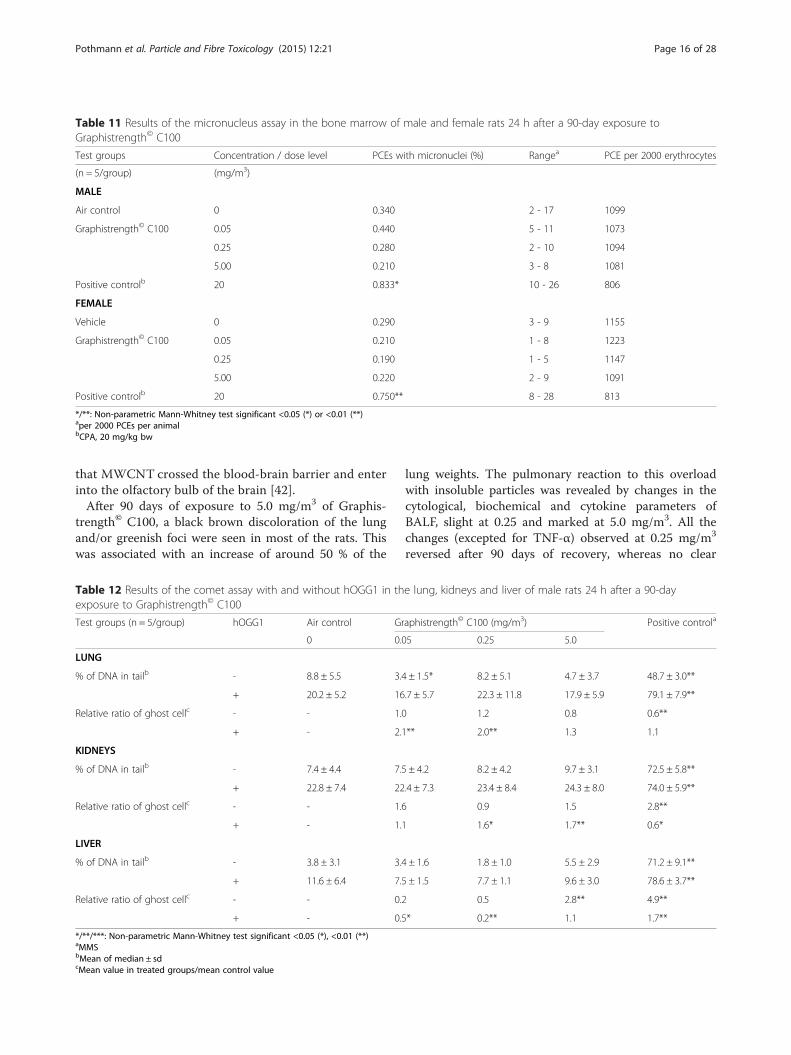
that MWCNT crossed the blood-brain barrier and enterinto the olfactory bulb of the brain [42].After 90 days of exposure to 5.0 mg/m3 of Graphis-
trength© C100, a black brown discoloration of the lungand/or greenish foci were seen in most of the rats. Thiswas associated with an increase of around 50 % of the
lung weights. The pulmonary reaction to this overloadwith insoluble particles was revealed by changes in thecytological, biochemical and cytokine parameters ofBALF, slight at 0.25 and marked at 5.0 mg/m3. All thechanges (excepted for TNF-α) observed at 0.25 mg/m3
reversed after 90 days of recovery, whereas no clear
Table 11 Results of the micronucleus assay in the bone marrow of male and female rats 24 h after a 90-day exposure toGraphistrength© C100
Test groups Concentration / dose level PCEs with micronuclei (%) Rangea PCE per 2000 erythrocytes
(n = 5/group) (mg/m3)
MALE
Air control 0 0.340 2 - 17 1099
Graphistrength© C100 0.05 0.440 5 - 11 1073
0.25 0.280 2 - 10 1094
5.00 0.210 3 - 8 1081
Positive controlb 20 0.833* 10 - 26 806
FEMALE
Vehicle 0 0.290 3 - 9 1155
Graphistrength© C100 0.05 0.210 1 - 8 1223
0.25 0.190 1 - 5 1147
5.00 0.220 2 - 9 1091
Positive controlb 20 0.750** 8 - 28 813
*/**: Non-parametric Mann-Whitney test significant <0.05 (*) or <0.01 (**)aper 2000 PCEs per animalbCPA, 20 mg/kg bw
Table 12 Results of the comet assay with and without hOGG1 in the lung, kidneys and liver of male rats 24 h after a 90-dayexposure to Graphistrength© C100
Test groups (n = 5/group) hOGG1 Air control Graphistrength© C100 (mg/m3) Positive controla
0 0.05 0.25 5.0
LUNG
% of DNA in tailb - 8.8 ± 5.5 3.4 ± 1.5* 8.2 ± 5.1 4.7 ± 3.7 48.7 ± 3.0**
+ 20.2 ± 5.2 16.7 ± 5.7 22.3 ± 11.8 17.9 ± 5.9 79.1 ± 7.9**
Relative ratio of ghost cellc - - 1.0 1.2 0.8 0.6**
+ - 2.1** 2.0** 1.3 1.1
KIDNEYS
% of DNA in tailb - 7.4 ± 4.4 7.5 ± 4.2 8.2 ± 4.2 9.7 ± 3.1 72.5 ± 5.8**
+ 22.8 ± 7.4 22.4 ± 7.3 23.4 ± 8.4 24.3 ± 8.0 74.0 ± 5.9**
Relative ratio of ghost cellc - - 1.6 0.9 1.5 2.8**
+ - 1.1 1.6* 1.7** 0.6*
LIVER
% of DNA in tailb - 3.8 ± 3.1 3.4 ± 1.6 1.8 ± 1.0 5.5 ± 2.9 71.2 ± 9.1**
+ 11.6 ± 6.4 7.5 ± 1.5 7.7 ± 1.1 9.6 ± 3.0 78.6 ± 3.7**
Relative ratio of ghost cellc - - 0.2 0.5 2.8** 4.9**
+ - 0.5* 0.2** 1.1 1.7**
*/**/***: Non-parametric Mann-Whitney test significant <0.05 (*), <0.01 (**)aMMSbMean of median ± sdcMean value in treated groups/mean control value
Pothmann et al. Particle and Fibre Toxicology (2015) 12:21 Page 16 of 28

improvement was observed at 5.0 mg/m3. These changesin BALF correlated with a concentration-related depos-ition of black particle in the lungs, which decreased at0.05 and 0.25 mg/m3 after 90 days of recovery. However,at 5.0 mg/m3, the mean severity scores of black particledeposition were overall similar at both time points, indi-cating a significant overload of the lungs. The increaseof alveolar macrophages and the changes in the tracheo-bronchial lymph nodes at 0.25 and 5.0 mg/m3 were con-sistent with the drainage of the black particles from thelungs [43] and a retention halftime of 375 days estimatedin rats exposed for 90 days to 6 mg/m3 of MWCNTBaytubes [44]. In animals exposed to 5.0 mg/m3, slightinflammatory changes were observed in the lungs atboth sacrifice times. The interstitial inflammation mainlyaround the alveolar ducts at the bronchiole-alveolarjunction and the cell hypertrophy/hyperplasia in theterminal and respiratory bronchioles observed justpost-exposure were most likely reactive changes to thesurrounding inflammatory process. After the 90-dayrecovery, additional findings were the presence of min-imal or slight focal collagen deposition within alveolarseptae in a few rats. No microscopic changes were ob-served in pleura. The minimal squamous metaplasia inthe larynx observed 24 h post-exposure to 0.25 and5.0 mg/m3 was fully reversible at 0.25 mg/m3 and per-sisted in one rat at 5.0 mg/m3 90 days post exposure.The presence of epithelial eosinophilic globules in thenasal cavity at 5.0 mg/m3 was only partially reversible.All these changes in the respiratory tract by exposureto Graphistrength© C100 were qualitatively consistentwith those reported in previous 90-day inhalation tox-icity studies in rats [40, 41, 44] with two other types ofthin and tangled MWCNT produced as large agglom-erates like Graphistrength© C100. All the pulmonarychanges induced by Graphistrength© C100 were sig-nificantly less severe than those induced in rats ex-posed for 90 days to 1.0 and 5.0 mg/m3 of the longneedle-like MWCNT-7 [45], notably the induction ofinflammatory and fibrotic effects in the pleura whichwere not observed in our study.Pauluhn [43] concluded that a post-exposure period of
6 months (c.a. 0.5 t½) would be suitable to reveal anyappreciable reversibility in the lungs after MWCNT ex-posure. However, fibrotic response was found to developand persist about one year after an inhalation exposure(4 times/week for 3 weeks) of male C57BL/6 J mice to5.0 mg/m3 of MWCNT-7 [46]. Therefore, the evolutionof the inflammatory reaction in the lungs of the rats ex-posed to Graphistrength© C100 for 90 days is still underevaluation over a 1-year recovery period.The effects of Graphistrength© C100 on the lungs of
mice were reported by Tabet et al. [47] after a single i.t.instillation as micrometric agglomerates suspended in
DMEM. The Balb/C mice were then monitored for upto 6 months. BALF analysis showed a dose-dependentincrease in total cell count and a significant influx ofneutrophils and macrophages only 24 h post-instillation.The MWCNT were internalized in macrophages be-tween 1 day and 1 month after instillation. Histology ofthe lungs 24 h after instillation showed the presence ofwidespread micrometric MWCNT agglomerates, whichwere mainly located in the bronchiolar lumen and alveo-lar ducts. After one week, clusters of cells surroundingvisible MWCNT agglomerates were seen near the ter-minal bronchioles, the alveolar ducts and alveoli in thelungs. However, no modification in mRNA expression ofvarious genes implicated in oxidative stress (SOD-2 andHO-1), inflammation (CXCL2, TNF-α) and fibrosis(αcollagen-1 and αcollagen-3) was quantified in lung ho-mogenates and no evidence of fibrosis was found6 months post exposure. Ronzani et al. [48] have alsoassessed the inflammation and airway remodeling inBALF or lung tissue of mice induced by surfactant-dispersed Graphistrength© C100, 24 h after a single(6.25 μg/mouse) and 7 days after repeated (1.5, 6.25 and25 μg/mouse once a week over 3 weeks) intranasal instil-lation(s). MWCNT distributed all throughout the mouseairways and were observed in alveolar macrophages, epi-thelial cells, and in infiltrated neutrophils. Mice that re-ceived a single administration of MWCNT showedneutrophils infiltrate and greater concentrations of TNF-α, keratinocyte-derived chemokine (KC) and IL-17 inBALF when compared to controls. After repeated Gra-phistrength© C100 administrations, increases in macro-phage number, KC and tumor growth factor (TGF)-β1levels in BALF, and collagen deposition and mucushyperplasia in lung tissue were observed. Cao et al. [17]exposed female wild-type C57BL/6 N Tac mice to totaldoses of 32 and/or 128 μg Graphistrength© C100/mouseadministered by i.t. instillation once a week over 5 weeks.Pulmonary inflammation was demonstrated one and/or28 days after the last exposure by increased influx ofneutrophils and higher levels of cytokines (IL1β, IL6,IL12, G-CSF, KC, CCL2, MIP1β, CCL5 and TNF) inBALF, and increased levels of 8-isoprostanes in lung tis-sue. Even if there are significant methodological differ-ences (rat vs. mice, inhalation vs. i.t., 90-day exposure vs.single or short-term administrations), consistently thesestudies tend to show the same inflammatory reaction inlungs to insoluble particles.
Genotoxicity studiesThis subchronic inhalation toxicity study gave us the op-portunity to evaluate the genotoxic potential of Graphis-trength© C100 in in vivo studies as recommended by theREACH guidance [49] when the set of the available gen-otoxicity data doesn’t allow a definitive conclusion. The
Pothmann et al. Particle and Fibre Toxicology (2015) 12:21 Page 17 of 28

results of in vitro studies are conflicting. Graphis-trength© C100 was negative in a battery of standard invitro genotoxicity assays [50] performed according to thecurrent OECD test guidelines no. 471 [51], 476 [52] and473 [53] to assess the potential induction of gene muta-tions in bacteria (Ames test) and mouse lymphoma cells,and chromosomal aberrations in human lymphocytes, re-spectively. Using nonstandard protocols, Kermanizadeh etal. [18, 19] showed that exposure of human hepatoblas-toma C3A and HK-2 cells at sub-lethal concentrations ofGraphistrength© C100 resulted in weak DNA damage inthe FPG-modified Comet assay. Conversely, Jackson et al.[20] did not observe an increase of the level of DNAstrand breaks in the comet assay with FE1-Muta© Mouselung epithelial cell cultures exposed to Graphistrength©
C100, even if the product was able to generate ROS andthe highest tested concentration induced a decrease of thecell proliferation.In the EU Nanogenotox Joint Action [21], Graphis-
trength© C100 was reported to induce a weak increase(≤2-fold compared to negative control) in the micronu-cleated binucleated cell (MNBC) frequency in a primaryculture of human lymphocytes [22, 23], in adenocarci-nomic human alveolar basal epithelial A549 and bron-chial epithelial BEAS 2B cell lines [23], but not in thebronchial epithelial 16 HBE and epithelial colorectaladenocarcinoma Caco-2 cell lines [23]. Graphistrength©
C100 was also tested in the in vitro comet assay usingBEAS 2B, 16 HBE, A549 and Caco-2 cell lines [23]. InBEAS 2B and Caco-2 cells, Graphistrength© C100 wasstudied using the FpG-modified comet assay. The cometassay was negative for all the cell lines with or withoutFpG.The results of the in vivo genotoxicity studies pub-
lished on Graphistrength© C100 are scarce and also notconclusive. Cao et al. [17] reported a small increase (<2-fold) of the level of DNA strand breaks, without effectson the level of FPG sensitive sites, in lung tissue of dysli-pemic C57BL/6 N-Apoe tm1 (ApoE-/-) mice exposed toGraphistrength© C100 by i.t. instillation once a week for5 weeks at total dose of 128 μg/mouse. This effect wasassociated with a lung inflammation as demonstrated byan influx of neutrophils in BALF. A 6-fold increase ofthe mRNA expression of the DNA repair oxoguanineDNA glycosylase 1 (Ogg1) enzyme was also observed inlungs of the same exposed animals. The authors have sug-gested, according to Risom et al. [54, 55], that after re-peated exposures to Graphistrength© C100, up-regulationof DNA repair counteracts the increased rate of 8-oxodGformation leaving the steady state level of 8-oxodG inDNA unchanged, whereas oxidative DNA damage couldbe induced after a short-term exposure. However, this hy-pothesis was not supported by the results of the FPG-modified comet assay performed in the frame of the
Nanogenotox Joint Action [24]. Three to six hours after 3daily i.t. instillations up to 320 μg/rat/day of Graphis-trength© C100 to Wistar rats, no statistically significant in-crease in the percentage of tail DNA was noticed in thelung cells with and without FPG enzyme. According toPant et al. [56] a 2- to 10-fold variation of the backgroundlevels of DNA damage is not unusual in the comet assay,therefore, it is difficult to ascribe the 2-fold increase re-ported by Cao et al. [17] to Graphistrength© C100 expos-ure. In our study, 24 h after a 90-day inhalation exposurewith Graphistrength© C100, even in the presence of a clearinflammatory reaction in the lungs, no primary andhOGG1-sensitive oxidative DNA damage was detected bythe comet assay, either in the lung cells directly in contactwith the MWCNT or systemically in the liver and kidneycells and no micronucleus induction was observed in thebone marrow cells.The genotoxic effects of MWCNT may result from
primary or secondary mechanisms [57]. Primary geno-toxicity refers to the elicitation of genetic damage in theabsence of inflammation, either by a direct interactionwith genomic DNA or associated components that de-termine its integrity, or indirectly through the enhancedproduction of ROS by cellular constituents in responseto their interaction with particles or through the deple-tion of antioxidants within the cell which can lead to theinduction of oxidative DNA damage [58]. Secondarygenotoxicity could be driven by inflammatory cells suchas macrophages and polymorphonuclear neutrophilicleukocytes and in situations of chronic inflammation thiscan lead to persistent oxidative stress and repeated DNAinsults [58]. Exposure to some MWCNT has been asso-ciated with depletion of antioxidants, increased intracel-lular production of ROS and pro-inflammatory signalingin cultured cells [59]. In the case of Graphistrength©
C100 there is no clear evidence of ROS production. Anincrease was observed in human hepatoblastoma C3Acells [18], but not in human renal [19] or FE1-Muta©
Mouse lung epithelial [20] cells. After i.t. instillation(s)of Graphistrength© C100 in the lung of mice, Tabet etal. [47] did not see a modification in mRNA expressionof genes implicated in oxidative stress, whereas Cao etal. [17] reported an increased expression of Hmox1.Even if the ROS formation was not specifically evaluatedin our study, the lung inflammation, macrophage phago-cytosis, TNF-α secretion in BALF and elevated numbersof neutrophils in blood observed at 5.0 mg/m3 are indi-cations of an oxidative stress. The negative comet resultin the lung cells exposed to Graphistrength© C100 couldbe related to the ROS scavenger capability of carbonnanotubes [60], which might limit the effects of the oxi-dative stress, and could also limit the DNA damage. Fur-thermore, the apparent absence of translocation ofGraphistrength© C100 from the lungs to the other
Pothmann et al. Particle and Fibre Toxicology (2015) 12:21 Page 18 of 28

organs could explain the lack of genotoxic effects in theliver, kidney and bone marrow cells.Therefore, the lack of in vivo genotoxicity of Graphis-
trength© C100 MWCNT indicates a toxicological profilesignificantly different compared to the genotoxic longneedle-like MWCNT-7 [11, 61], classified by IARC [14] aspossibly carcinogenic to humans (Group 2B) and to someother genotoxic thin and tangled MWCNT [62–64], evenif in these later cases, a role of residual cobalt catalyst,which is a known genotoxin [65], could not be excluded.
ConclusionFive-day and 90-day rat inhalation toxicity studies wereperformed with MWCNT Graphistrength© C100. Themilling procedure and the dust disperser used as solidaerosol generator produced an aerosol which retainedthe physico-chemical integrity of the original product inthe test aerosols. This contrasts with a previous inhal-ation study with the same MWCNT product [16], usingan aerosol produced with a rotating brush generator inwhich the structure of the MWCNT was affected. In thepresent study, principal health findings were limited tothe lungs. The infiltration of phagocytizing macrophagesis thought to be a trigger and results from the host reac-tion towards foreign bodies [66]. The inflammation mayalso deteriorate the alveolar barrier function whichincreased particle translocation to the draining lymphnodes of the lung [44]. Bronchial and alveolar epitheliawere influenced secondarily. During an inhalation study,at a certain time point, a lung burden is reached that ex-ceeds the macrophage clearance capacity and results inoverload effects [66]. It seems to be the case at 5.0 mg/m3,as Graphistrength© C100 deposition persisted in the lungwithout apparent signs of decrease after a 90-daytreatment-free period, whereas at 0.25 mg/m3 clear signsof clearance and recovery were observed. Prolonged TNF-α release in BALF was observed at 0.25 and 5.0 mg/m3
which was associated only at 5.0 mg/m3 with an increasedcollagen staining like that reported by Pauluhn [44] withMWCNT Baytubes. The lack of genotoxicity in the lungcells and microscopic change in the pleura indicated alung reaction to Graphistrength© C100 exposure quite dif-ferent than that of the asbestos-like MWCNT-7 and couldbe related to the absence of internalization of Graphis-trength© C100 by the alveolar or mesothelial cells asshown by Tabet et al. [67] with human epithelial A549and mesothelial MeT5A cell lines cultures. Overall, theseeffects are consistent with a normal physiological and notadverse response to the overload of the lung with insolubleparticles [44]. Considering the limited and reversibleeffects on the BALF parameters, the lack of patho-logical changes in the lungs and the clearance of theGraphistrength© C100 observed at 0.25 mg/m3, thisconcentration can be considered as a No-observed
Adverse Effect Concentration (NOAEC). In spite of theinflammatory response, neither primary nor oxidativeDNA damages were observed locally in lung. The lackof DNA damage or chromosomal aberration remotelyin the liver, kidneys and bone marrow was most prob-ably related to the lack of bioavailability via a systemictranslocation of the MWCNT from the lungs. There-fore, Graphistrength© C100 appears of low concern interm of local and systemic genotoxicity and a NOAECof 0.25 mg/m3 (0.28 mg/m3 as actual concentration)was established for the repeated-dose toxicity.
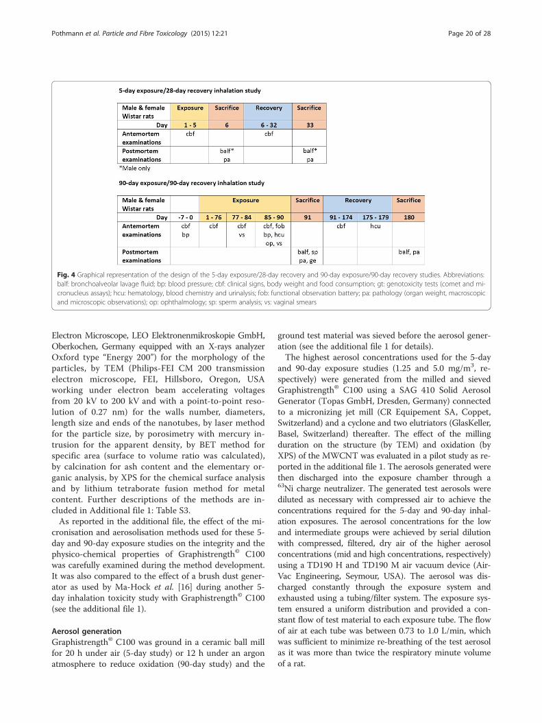
MethodsGeneralThe present studies were conducted according to theOECD Principles of Good Laboratory Practice [68] andthe OECD test guidelines no. 413 [29], 474 [69] and 489[70]. The design of the 90-day inhalation toxicity studywas developed taken into account the OECD recom-mendations [30] for the revision of the tests guidelinesapplicable to the inhalation toxicity testing of nanoma-terials. A graphical representation of the design of the 5-and 90-day studies is presented in Fig. 4. The studieswere performed in an AAALAC-accredited laboratory inaccordance with the Swiss Animal Protection Law.
Test materialsGraphistrength© C100, are exclusively produced byArkema France, Colombes, France. SEM and TEM im-ages (Fig. 5) show that Graphistrength© C100 is made oftightly bound agglomerates constituted with entangledMWCNT. These agglomerates can be spherical, ovoid orirregular shaped and have a granulometry centred on400 μm, with fragments of pellets of less than 15 μm repre-senting a volume of under 0.23 % [27]. The MWCNT ofGraphistrength© C100 are synthesized at high temperatureusing a fluidized bed with ethylene as a carbon feedstockand iron oxide (Fe2O3, ≤5 %, [1309-37-1]) on alumina(Al2O3, ≤7 %, [1344-28-1]) as catalytic source. Batches nos.8287 and 110329-018 of Graphistrength© C100 were usedfor the 5-day and the 90-day exposure studies, respectively.Positive control substances for the genotoxicity assays
were selected as recommended by the OECD test guide-lines [69, 70]. Cyclophosphamide monohydrate (CPA,batch A0302605, purity 97 %) from Fisher ScientificGmbH for the micronucleus assay and methyl methane-sulfonate (MMS, batch MKBL6789V, purity 99.9 %) fromSigma-Aldrich for the comet assay.
Physico-chemical characterisationsThe original Graphistrength© C100 batches and samplestaken at different steps of our aerosol generation processduring the method development (see the additional file 1)were analyzed by SEM (ZEISS LEO 1530 VP Scanning
Pothmann et al. Particle and Fibre Toxicology (2015) 12:21 Page 19 of 28
Electron Microscope, LEO Elektronenmikroskopie GmbH,Oberkochen, Germany equipped with an X-rays analyzerOxford type “Energy 200”) for the morphology of theparticles, by TEM (Philips-FEI CM 200 transmissionelectron microscope, FEI, Hillsboro, Oregon, USAworking under electron beam accelerating voltagesfrom 20 kV to 200 kV and with a point-to-point reso-lution of 0.27 nm) for the walls number, diameters,length size and ends of the nanotubes, by laser methodfor the particle size, by porosimetry with mercury in-trusion for the apparent density, by BET method forspecific area (surface to volume ratio was calculated),by calcination for ash content and the elementary or-ganic analysis, by XPS for the chemical surface analysisand by lithium tetraborate fusion method for metalcontent. Further descriptions of the methods are in-cluded in Additional file 1: Table S3.As reported in the additional file, the effect of the mi-
cronisation and aerosolisation methods used for these 5-day and 90-day exposure studies on the integrity and thephysico-chemical properties of Graphistrength© C100was carefully examined during the method development.It was also compared to the effect of a brush dust gener-ator as used by Ma-Hock et al. [16] during another 5-day inhalation toxicity study with Graphistrength© C100(see the additional file 1).
Aerosol generationGraphistrength© C100 was ground in a ceramic ball millfor 20 h under air (5-day study) or 12 h under an argonatmosphere to reduce oxidation (90-day study) and the
ground test material was sieved before the aerosol gener-ation (see the additional file 1 for details).The highest aerosol concentrations used for the 5-day
and 90-day exposure studies (1.25 and 5.0 mg/m3, re-spectively) were generated from the milled and sievedGraphistrength© C100 using a SAG 410 Solid AerosolGenerator (Topas GmbH, Dresden, Germany) connectedto a micronizing jet mill (CR Equipement SA, Coppet,Switzerland) and a cyclone and two elutriators (GlasKeller,Basel, Switzerland) thereafter. The effect of the millingduration on the structure (by TEM) and oxidation (byXPS) of the MWCNT was evaluated in a pilot study as re-ported in the additional file 1. The aerosols generated werethen discharged into the exposure chamber through a63Ni charge neutralizer. The generated test aerosols werediluted as necessary with compressed air to achieve theconcentrations required for the 5-day and 90-day inhal-ation exposures. The aerosol concentrations for the lowand intermediate groups were achieved by serial dilutionwith compressed, filtered, dry air of the higher aerosolconcentrations (mid and high concentrations, respectively)using a TD190 H and TD190 M air vacuum device (Air-Vac Engineering, Seymour, USA). The aerosol was dis-charged constantly through the exposure system andexhausted using a tubing/filter system. The exposure sys-tem ensured a uniform distribution and provided a con-stant flow of test material to each exposure tube. The flowof air at each tube was between 0.73 to 1.0 L/min, whichwas sufficient to minimize re-breathing of the test aerosolas it was more than twice the respiratory minute volumeof a rat.
Fig. 4 Graphical representation of the design of the 5-day exposure/28-day recovery and 90-day exposure/90-day recovery studies. Abbreviations:balf: bronchoalveolar lavage fluid; bp: blood pressure; cbf: clinical signs, body weight and food consumption; gt: genotoxicity tests (comet and mi-cronucleus assays); hcu: hematology, blood chemistry and urinalysis; fob: functional observation battery; pa: pathology (organ weight, macroscopicand microscopic observations); op: ophthalmology; sp: sperm analysis; vs: vaginal smears
Pothmann et al. Particle and Fibre Toxicology (2015) 12:21 Page 20 of 28

Fig. 5 Electron microscopic images of Graphistrength© C100. SEM of the commercial Graphistrength© C100. (a) Magnification: 22 fold. (b) Magnification:120 fold. (c) Magnification: 10’000 fold
Pothmann et al. Particle and Fibre Toxicology (2015) 12:21 Page 21 of 28

Animals and husbandryHealthy male and female Rats, RccHan©: WIST(SPF)were supplied by Harlan Laboratories, B.V. (5961 NMHorst, The Netherlands). After an acclimatization periodof at least 7 days, the animals were 11 and 8 weeks oldat the start of 5-day and 90-day exposures, respectively.On the first day of the 5-day and 90-day exposures, thebody weights ranged from 291 to 347 g and 243 to 296 g formales and from 176 to 214 g and 135 to 228 g for females,respectively. The rats were randomly allocated by sex to thecontrol and the test groups in groups of maximally five inMakrolon type-4 cages with wire mesh tops and sterilizedstandard softwood bedding (“Lignocel” J. Rettenmaier &Söhne GmbH & Co. KG, 73494 Rosenberg, Germany) in-cluding paper enrichment (Enviro-dri from Lillico, Biotech-nology, Surrey, UK). The animal room was air-conditionedwith 10-15 air changes/h, a 12 h light-12 h dark cycle, thetemperature ranging from 20 to 24 °C with relative hu-midity ranging from 30 to 70 %. Except during expos-ure, pelleted standard Harlan Teklad 2914C rodentmaintenance diet (Provimi Kliba AG, 4303 Kaiseraugst,Switzerland) and water were provided ad libitum.
Animal exposureGroups of 20 male or 10 female rats or 35 male and 35 fe-male rats were exposed nose-only 6 h per day for 5 daysor 5 days per week for 90 days, respectively. Target con-centrations were 0.05, 0.25 and 1.25 mg/m3 air and 0.05,0.25 and 5.0 mg/m3 air of Graphistrength© C100 as low,mid and high concentrations for the 5-day and 90-day in-halation exposures, respectively. The choice of the con-centrations for the 5-day exposure study was based onpublished evidence on other MWCNT [40, 44]. Consider-ing the limited effects observed in the 5-day range-findingstudy, the same low and mid concentrations were used forthe 90-day exposure study and the high concentration wasincreased up to the top concentrations tested in previous90-day exposure studies [40, 44].Animals of control groups (0 mg/m3) were exposed to
compressed air under the same conditions as animals ex-posed to Graphistrength© C100. During nose-only inhal-ation exposure, rats have a breathing pattern that results ina more realistic internal exposure than a single high dose ini.t. instillation and oropharyngeal aspiration or possible add-itional oral exposure in whole-body exposure chambers. In-halation exposure was performed using a flow-past system.The animals were confined separately in restraint tubeswhich were positioned radially around the flow-past, nose-only exposure chamber as described by Cannon et al. [71].As positive control groups for the genotoxicity assays,
5 males and 5 females were treated once by oral gavagewith 20 mg/kg bw cyclophosphamide (CPA) approxi-mately 24 h before tissue sampling for the micronucleusassay, and 5 males were treated by oral gavage with
methyl methane sulfonate (MMS) two times with 100 mg/kg bw and then once with 70 mg/kg at approximately 24 hintervals for the comet assay.
Monitoring and characterization of the test aerosolThe oxygen concentration, relative humidity andtemperature in the exposure device were measured con-tinuously during each exposure using a calibrated device.The nominal concentration was determined for the highconcentrations by weighing the generator reservoir con-taining the test material before and after exposure anddividing the used amount by the total air-flow volume.These data were used to monitor the performance of thegeneration system. During the 5-day and 90-day expo-sures, gravimetric determinations of the aerosol concen-trations were respectively performed once and 4 to 7times daily for high concentrations and twice and 1 to 3times daily for the low and mid concentrations usingMillipore® durapore filters, type HVLP, loaded in a47 mm in-line stainless steel filter sampling device. Thesampling flow rates were respectively 3 to 4 and 1 L/minper exposure port. For the low concentration of the 90-day exposure study, filters were taken at the inlet pipewith airflow of 3 to 4 L/min. The same device was usedto collect aerosol samples for TEM analysis.For both exposure periods, the cumulative particle size
distribution of the test aerosol was determined usingMercer cascade impactors (In-Tox Products, Moriarty,USA). The test aerosol was impacted at each stage (cov-ered with grease) and the particle size distribution of thetest material in the generated aerosol was measured bygravimetric analysis of the test material deposited oneach stage of the cascade impactor.During the 5-day exposure, measurements were per-
formed twice for the low and high concentrations andonce for the mid concentration using an impactorModel 200 at a sampling rate of 9 L/min in order to col-lect sufficient material for this short study period.During the 90-day exposure, measurements were per-
formed at least once per week for the high concentrationand five times for the mid concentrations using a stand-ard impactor Model 02-005 at a sampling rate of 1 L/min, matching the air flow rate at the animal port toavoid a possible imbalance of the aerosol flow within theexposure chamber. Accordingly, impactor samples forthe mid concentration of the 90-day exposure were col-lected over several days. No samples were collected forthe low concentration of the 90-day exposure as theaerosol concentration was too low to obtain reliable re-sults with an air flow rate of 1 L/min.The mass median aerodynamic diameter (MMAD)
and the geometric standard deviation (GSD) were calcu-lated on the basis of the results from the impactors,using Microsoft Excel® software (Microsoft Corporation,
Pothmann et al. Particle and Fibre Toxicology (2015) 12:21 Page 22 of 28

USA). In addition, during the 90-day inhalation expos-ure, the aerosol was analyzed once per week with a WideRange Particle Spectrometer© (WRPS, Model 1000XP,MSP Corporation, Shoreview, USA) in the size range of5 nm to 10 μm to determine the count median aero-dynamic diameter (CMAD). The sampling airflow ratewas approximately 1 L/min.
Ante mortem observationsCage-side clinical observations were recorded twice dailybefore and after exposures. During the 90-day study, acareful clinical examination of each animal was per-formed once weekly in a standard arena. Food consump-tions and body weights were recorded twice during the5-day exposure and weekly during the 28-day recoveryand the 90-day exposure and 90-day recovery periods.Functional observation battery, locomotor activity, grip
strength, landing foot splay, body temperature and oph-thalmoscopic investigations were recorded during thelast week of the 90-day exposure period. In animalsfasted in metabolism cages for approximately 18 h,hematology, blood chemistry and urinalysis (parametersas per OECD test guideline no. 413 [29] and listed in Ta-bles 2 and 3) were performed during the last week of the90-day exposure. Hematology and blood chemistry wererepeated during the last week of the 90-day recoveryperiod. Tail cuff blood pressure was measured duringacclimatization and during the last week of the 90-dayexposure period. Vaginal smears for estrous cycle evalu-ation were taken for 14 days from all females duringtreatment weeks 11 and 12.
Post mortem observationsTwenty-four hours and 28 days after the 5-day exposure,5 rats/sex/group were anaesthetized by i.p. injection ofpentobarbitone and killed by exsanguination for histo-logical examinations and another 5 males/group servedfor BALF investigations on both occasions. Twenty-fourhours and 90 days after the 90-day exposure, 10 rats/sex/groups were sacrificed for organ weights, andmacroscopic, histological and BALF examinations.
Bronchoalveolar lavage fluid analysisFor broncho-alveolar lavage sampling, the completelungs (5-day study) or the left lobes of the lungs (90-daystudy) were washed six times with physiological saline(4.0 ml for the complete lungs or 1 x 2.5 mL + 5 x1.5 mL for the left lobes) at room temperature by slowinstillation and withdrawal of fluid. The lavage fluid ofanimals from the positive control group for the cometassay was discarded. After washing, the left lung lobeswere preserved in mincing solution at 5 ± 3 °C for thecomet assay. The lavage fluids from the first (5-daystudy) or the first two lavages (90-day study) were
collected in a centrifugation tube on ice. The recoveredvolume from this wash was recorded. Approximately0.75 or 1 mL of the fluid was placed into a second tubecontaining 37.5 or 50 μL of 20 % BSA in PBS, respect-ively. Both tubes were centrifuged at approximately300 g for 10 min between 2 and 8 ° C. Thereafter, thesupernatant (without BSA) was taken in one tube andimmediately set on ice and stored at 2-8 ° C until ana-lysis. The first aliquot (without BSA) was used for thedetermination of the enzymatic activity of lactate de-hydrogenase (LDH), alkaline phosphatase (ALP), and γ-glutamyltransferase (GGT) as well as for the determin-ation of total protein and/or phospholipids (storage at2-8 ° C until analysis). The second aliquot from the 90-day exposed and 90-day recovery animals was split intothree tubes, two of them containing approximately0.1 mL each, the third one containing 0.3 mL and wereanalyzed for contents of natural immunity mediator cy-tokines (TNF-α, IL-1α and IL-1β) and adaptive immun-ity mediator cytokines (IL-5). IL-5, IL-1-β and TNF-αwere analyzed using the Proinflammatory Panel 1 (rat)Kit V-PlexTM from MSD. IL-α was analyzed using theMSD® Multi-Spot Assay System, Rat Demonstration 7-Plex Ultra-sensitive Kit.The lavage fluid from the four further lavages was
pooled separately for each animal in a centrifugationtube, and centrifuged at approximately 300 g for 10 minbetween 2 and 8 °C. The supernatant was discarded.The cell pellets were each suspended with physiologicalsaline and pooled with the suspended cell pellet fromthe first two lavages. The total volume was gentlyshaken for mixing. Aliquots of the cell suspension weretaken for total cell count, cell viability and differentialcell counting. A total cell count was performed by dilut-ing an aliquot of the cell suspension in Türk’s stain(Diagnostik Merck No. 9277, Merck AG, Darmstadt,Germany). An aliquot of this mixture was introducedinto a hemocytometer chamber, and the cells werecounted (at least 1 mm3). A differential cell count wasperformed. According to the results of the total cellcount, an aliquot of cell suspension was diluted withphysiological saline solution to give an end concentra-tion of at least 106 cells/ml. From this suspension asmear was prepared using a Cytocentrifuge (Shandon,Instrument Gesellschaft AG, Switzerland) and stainedwith Diff-Quick (Baxter Dade, Switzerland). At least500 cells per smear were counted by light microscopy.The number of each cell type (macrophages, neutro-phils, lymphocytes, eosinophils, epithelial cells, andother cells) was counted.
Macroscopic and organ weight examinations at necropsyAll animals were weighed at necropsy and descriptionsof all macroscopic abnormalities were recorded. The
Pothmann et al. Particle and Fibre Toxicology (2015) 12:21 Page 23 of 28

weights of adrenal glands, heart including auricles,kidneys, liver, lungs, and spleen were determined in allanimals exposed for 5 days and 90 days (including re-covery animals). In addition, weights of brain, epididy-mis, ovaries, testes, thymus and uterus were recorded inall animals exposed for 90 days (including recovery ani-mals). The organ to terminal body weight ratios wascalculated.
HistopathologyAlmost all organs were collected from all rats exposed for5-day (including recovery animals). All gross lesions, allorgans as per OECD test guideline no. 413 [29], olfactorybulb and pleura were collected from all rats exposed for90 days (including recovery animals). Lung lobes wereinstilled via trachea with neutral phosphate buffered 4 %formaldehyde solution at approximately 30 cm H2O pres-sure. The organs were fixed in formalin (excepted testesfixed in modified Davidson’s solution), processed, embed-ded and cut at an approximate thickness of 2 - 4 μm andstained with hematoxylin and eosin.For the 5-day exposure study, all gross lesions, heart
including auricles, kidneys, larynx, liver, spleen, medias-tinal lymph nodes, nasal cavity, nasopharyngeal duct andpharynx, spleen and trachea (adjacent to larynx and car-ina and bifurcation) of the main and recovery rats fromthe control and 1.25 mg/m3 groups were examinedmicroscopically. Lungs from all main and recovery ratsfrom all groups were examined.For the 90-day exposure study, the microscopic exam-
ination of the respiratory tract and associated lymphnodes was performed on all control and exposed animalsat both sacrifice periods (except the nasal cavity and lar-ynx of the recovery rats exposed to 0.05 mg/m3). Theother organs, as per OECD test guideline no. 413 [29],were examined for the control and 5.0 mg/m3 groupsrats sacrificed 24 h post-exposure (this microscopicexamination was not performed on the 90-day recoveryrats as no microscopic changes was observed in the 24-hsacrificed rats).A part of left lobe of lungs (also used for BALF), a
caudal part of kidney and the middle lobe of liver ofmale rats exposed for 90 days were preserved for thecomet assay.
Semiology and spermatid countSperm motility, spermatid count in testis and spermcount in cauda epididymis were performed on all malesexposed for 90 days and sacrificed 24 h and 90 days postexposure (expect the sperm count in recovery males ex-posed to 0.05 and 0.25 mg/m3). Sperm morphology wasanalyzed on all males at the 24-h post exposure sacrificeand on control and 5.0 mg/m3 exposed males after the90-day recovery period.
Micronucleus testThe 5 last male and female rats (in the numbering order)out of 10 of sacrificed 24 h after the 90-day exposure wereused for the micronucleus assay. The femora were re-moved, the epiphyses cut off and the marrow was flushedout with fetal calf serum, using a syringe (3 mL per femur).The nucleated cells were separated from the erythrocytesusing the method of Romagna [72]. Briefly, the cell suspen-sions were passed through a column consisting of α-Cellulose (Sigma) and Cellulose (Sigmacell type 50) (1:1mixture). The columns were then washed with Hank΄sbuffered saline. The cell suspensions were centrifuged at1500 rpm (390 × g) for 10 min and the supernatant wasdiscarded. The pellet was suspended in a small drop of fetalcalf serum and spread on slides. The smears were air-dried,fixed in methanol. The slides were stained with May-Grünwald/Giemsa. For staining, the slides were first incu-bated one minute in a 5 % May-Grünwald solutionfollowed by one minute in a 1:1 mixture of May-Grünwald/phosphate buffer (pH 7.4). After 20 min in 14 % Giemsa so-lution the slides were washed twice in phosphate bufferand 10 s in deionized water. Cover slips were mountedwith EUKITT (Kindler, 79110 Freiburg, Germany). Atleast one slide was made from each bone marrow sam-ple. The slides were coded using a computer generatedcoding list. Evaluation of the slides was performed usingNIKON microscopes with 100x oil immersion objec-tives. Six thousand polychromatic erythrocytes (PCE)were analyzed per animal for micronuclei.The test is considered to be positive if there is either a
dose-related increase in the number of micronucleatedpolychromatic erythrocytes or a statistically significantpositive response for at least one of the test points. In theabsence of a dose-related increase in the number of micro-nucleated polychromatic erythrocytes or a statistically sig-nificant positive response at any of the test points thesubstance is considered non-genotoxic in this system. Todescribe a cytotoxic effect the ratio between polychro-matic and normochromatic erythrocytes was determinedin the same sample and expressed in polychromatic eryth-rocytes per 2000 erythrocytes.
Comet assayAs suggested by the OECD test guideline no. 489 [70],when there was no difference in toxicity between malesand females, the comet assay could be performed in eithersex. The 5 first male rats (in the numbering order) out of10 sacrificed 24 h after the 90-day exposure were assignedfor the comet assay. Three to 6 h after the last treatmentfor positive control group or 22 to 26 h after the last ex-posure for air control and Graphistrength© C100 exposedgroups, the lung (left lobe used for BALF), the kidney(caudal part) and the liver (middle lobe) were collectedfrom each animal and maintained in cold mincing buffer
Pothmann et al. Particle and Fibre Toxicology (2015) 12:21 Page 24 of 28

(Mg2+ and Ca2+ free Hanks’ Balanced Salt Solution (Gibco,CA, USA) with 20 mM Na EDTA (EDTA) (Sigma, USA)and 10 % (v/v) dimethylsulfoxide (DMSO) (Sigma)). Im-mediately after dissection, the organs were minced usingfine scissors in cold mincing buffer. The cell suspensionwas stored on ice for 15-30 s to allow large clumps to set-tle, and the supernatant was used to prepare comet slides.An aliquot of single cell suspension (3 x 10⁴ cells) wasmixed per 75 μL of 0.5 % low melting agarose (Invitrogen,USA) and spread on comet assay slides (Travigen, USA).The slides were immersed in cold lysis solution (2.5 MNaCl, 100 mM Na2EDTA, 10 mM Tris-base, 10 % DMSO,1 % Triton-X (pH 10)) overnight. For the hOGG1-modified comet assay, following lysis as described, slideswere washed two times for 5 min with the hOGG1 incu-bation buffer (40 mM Hepes, 100 mM KCl, 0.5 mMEDTA and 0.2 mg/mL BSA) at room temperature andthen incubated for 10 min at 37 °C with 0.12 x 10⁻³ U/slideof hOGG1 (Biolabs) in the hOGG1 incubation buffer.After this incubation period, slides were placed in electro-phoresis solution (0.3 M NaOH, 1 mM EDTA (pH > 13))for 20 min to allow for DNA unwinding. Subsequently,electrophoresis was conducted at 25 V, 300 mA for20 min. The slides were immersed in a neutralizationsolution (0.4 M Tris-base, pH 7.5) for at least 5 min anddehydrated with absolute ethanol to fix. The cells werestained with propidium iodide (20 μg/mL) (Invitrogen). Allslides were independently coded before the microscopicanalysis. The comet was observed via fluorescence micro-scope (Leica Microsystems SAS - DM 2000, Heerbrugg,Switzerland) at magnification of x200 and analyzed byCOMETASSAY IV software (Perceptive Instruments, UK).For each sample (animal/tissue), fifty comets per slide wereanalyzed, with 3 slides scored per sample.A positive response is defined as a statistically signifi-
cant change in the % tail DNA in at least one dose groupin comparison with the vehicle control value. The posi-tive control should produce a statistically significant in-crease. Cytotoxicity was evaluated by the enumeration ofhedgehogs. Indeed, important fragmentation of the DNAcan be induced not only by genotoxicity but also duringthe process of cell death, i.e. apoptosis and necrosis.
Statistical analysisThe food consumption, blood pressure, grip strength,landing foot play, body temperature, body weight, food con-sumption, blood clinical laboratory and BALF parameters,organ weights, and sperm parameters were analyzed forstatistical significance by the Dunnett-test (many to one t-test) based on a pooled variance estimate, if the variablescan be assumed to follow a normal distribution for thecomparison of the treated groups and the control groupsfor each sex. The Steel-test (many-one rank test) was ap-plied for the locomotor activity, urinalysis, and BALF
biochemical parameters instead of the Dunnett-test whenthe data cannot be assumed to follow a normal distribu-tion. Fisher’s exact test was applied to the ophthalmoscopyand macroscopic findings. Nonparametric Mann-Whitneytest was applied to the micronucleus, comet and to cyto-kines test results as data did not follow a normaldistribution.
Additional file
Additional file 1: Figure S1. Electron microscopic images ofGraphistrength© C100 aerosols generated by the rotating brushgenerator [16]. (A) TEM of the original material batch no. 6097,Magnification: 135’000 fold. (B) and (C) TEM of aerosol samples.Magnification: 135’000 fold. (D) SEM of aerosol sample, Magnification:25’000 fold. Figure S2: Scanning (SEM) and transmission (TEM) electronmicroscopic images of Graphistrength© C100 aerosols generated by the dustdisperser and collected directly into TEM grids. Millipore® durapore filter, TypeHVLP as used in the 5-day inhalation study. (A) SEM, Magnification: 2’000 fold.(B) SEM, Magnification: 29’500 fold. (C) TEM, Magnification: 350’000 fold.Figure S3: Particle size distribution after different milling durations ofGraphistrength© C100. Figure S4: Scanning (SEM) and transmission (TEM)microscopic images of Graphistrength© C100 using the refined aerosolgeneration procedure used during the 13-week exposure study. (A) TEM oforiginal Graphistrength© C100 batch no. 110329-018, 350’000 fold. (B) TEM ofmilled Graphistrength© C100 under argon for 12 h and sieved (63 μm),350’000 fold. (C) TEM of aerosol sample, 350’000 fold. (D) SEM of aerosolsample, 50’000 fold. Figure S5: BALF parameters of male rats 24 h (A) and4 weeks (B) after a 5-day exposure to Graphistrength© C100. Changes areshown as x-fold differences compared to controls using a logarithmic scaling.Abbreviations: ALP: alkaline phosphatase, GGT: γ-Glutamyltransferase, LDH:lactate dehydrogenase. Figure S6: Microscopic appearance of lungs aroundterminal bronchiole in rats after a 5-day exposure to Graphistrength© C100(A) Control. (B) Graphistrength© C100, 1.30 mg/m3, after a 5-day inhalationexposure: Terminal bronchiolar epithelium showing hypertrophic appearance(asterisks). No inflammatory and granulomatous lesions are found. (C)Graphistrength© C100, 1.30 mg/m3, after a 5-day inhalation exposure: Blackishparticle laden alveolar macrophages (arrows). (D) Graphistrength© C100,1.30 mg/m3, after a 4-week recovery period: Histologic appearance ofthe bronchiolar epithelium recovered partially. Original lens magnification:(A), (B) and (D) Magnification: 10 fold; (C) Magnification: 40 fold. Table S1:Oxygen surface content by XPS in Graphistrength© C100 aerosols from therotating brush dust generator [16]. Table S2: Effect by XPS of the millingduration and milling atmosphere (air or argon) on the oxygen surfacecontent in Graphistrength© C100. Table S3: Physico-chemicalcharacterization of Graphistrength© C100 before and after aerosolgeneration. Table S4: Target and achieved aerosol concentrations andparticles size of Graphistrength© C100. Temperature, relative humidity andoxygen concentration measured over the 5-day exposure study. Table S5:Cellular analysis of BALF of male rats 24 h and 4 weeks after a 5-dayexposure to Graphistrength© C100. Table S6: Biochemical analysis of BALFof male rats 24 h and 4 weeks after a 5-day exposure to Graphistrength©
C100. Table S7: Main microscopic findings in the lungs of male and femalerats 24 h and 4 weeks after a 5-day exposure to Graphistrength© C100.
AbbreviationsAAALAC: Association for Assessment and Accreditation of Laboratory AnimalCare; ALP: Alkaline phosphatase; BALF: Broncho-alveolar lavage fluid;BET: Brunauer, Emmett and Teller (method); BSA: Bovine serum albumin;CCL2: Chemokine ligand 2; CCL5: Chemokine (C-C motif) ligand 5;CMAD: Count median aerodynamic diameter; CPA: Cyclophosphamide;CXCL2: Macrophage inflammatory protein-2; DMEM: Dulbecco’s modifiedEagle medium; DNA: Deoxyribonucleic acid; ESCA: Electron spectroscopy forchemical analysis; FPG: Formamidopyrimidine DNA glycosylase; GGT:γ-glutamyltransferase; GPX-1: Glutathione peroxidase-1; G-CSF: Granulocytecolony-stimulating factor; GSD: Geometric standard deviation; HCD: Historicalcontrol data; hOGG1: Human 8-hydroxyguanine DNA-glycosylase;
Pothmann et al. Particle and Fibre Toxicology (2015) 12:21 Page 25 of 28

HO-1: Heme oxygenase-1; IARC: International Agency for Research on Cancer;IL: Interleukin; KC: Keratinocyte-derived chemokine; LDH: Lactatedehydrogenase; MIP1β: Macrophage inflammatory protein 1β; MMAD: Massmedian aerodynamic diameter; MMS: Methyl methanesulfonate;MWCNT: Multiwall carbon nanotubes; OECD: Organization for EconomicCooperation and Development; PBS: Phosphate buffered saline;PCE: Polychromatic erythrocytes; PMN: Polymorphonuclear neutrophils;ROS: Reactive oxygen species; SEM: Scanning electron microscopy;SOD: Superoxide dismutase; TEM: Transmission electron microscopy;TNF-α: Tumor necrosis factor alpha; WBC: White blood cells; WPRS: WideRange Particle Spectrometer©; XPS: X-ray photoelectron spectroscopy.
Competing interestsThis study was financially supported by Arkema France and technicallyperformed by the co-authors from Harlan Laboratories Ltd (Switzerland),Institut Pasteur de Lille (France), Harlan Cytotest Cell Research GmbH(Germany), Le Net Pathology Consulting (France), AnaPath GmbH(Switzerland) and Arkema France.
Authors’ contributionsDP carried out the 90-day inhalation toxicity study and was involved indrafting the manuscript. SS carried out the comet assay and was involved indrafting the manuscript. DS conducted the method development and the5-day inhalation study, participated in the design of the 90-day toxicity studyand was involved in drafting the manuscript. SG carried out the test materialpreparation and the atmosphere generation. ED carried out the micronucleusassay. JLLN and YO carried out the histological examinations. JMC, CB and JBperformed the physico-chemical, SEM and TEM characterizations of the testmaterials and aerosol samples. FN was involved in the design of the cometassay and participated to the analysis of the data. JFR was in charge of theconception, design and monitoring of the toxicity and genotoxicity studies,participated to the analysis and interpretation of the data and has draftedthe manuscript. All authors read and approved the final manuscript.
Authors’ informationDS is actually employee of Schuler Toxicology Consulting GmbH,Schafackertr. 40/1, 79576 Weil am Rhein, Germany.
AcknowledgementsThe authors wish to thank Emmanuel Laux for excellent technical assistancein performing the inhalation study, Margrit Degert, Diana Hahnke, NicoleMojzes and Mirjiam Schmid for undertaking the histopathology work, ReginaDraheim, Monika Gisin and Conni Büschlen for performing the BALFinvestigations and Klaus Weber for carrying out the histopathological peerreview. They are also grateful to Gwendoline Dewaele, Smaïl Talahari andEmilie Nicolas for their technical assistance in performing the comet assay,Maria Köditz and Sigrid Schneck which did the micronucleus preparation,Peter Kunzendorf for performing SEM and Sandi Murphy for the editorialreview of the manuscript.
Author details1Harlan Laboratories Ltd, Zelgliweg 1, 4452 Itingen, Switzerland. 2Laboratoirede Toxicologie Génétique, Institut Pasteur de Lille, 1 rue du Pr. Calmette,59019 Lille, Cedex, France. 3Harlan Cytotest Cell Research GmbH, In denLeppsteinwiesen 19, 64380 Rossdorf, Germany. 4Le Net Pathology Consulting,18 rue Henry Dunant, 37400 Amboise, France. 5AnaPath GmbH, Buchsweg56, 4625 Oberbuchsiten, Switzerland. 6Arkema France, Groupement deRecherches de Lacq (GRL), Laboratoires d’analyse de surface et microscopieet de chimie analytique, 64170 Lacq, France. 7UDSL, EA 4483, DépartementToxicologie-Santé Publique-Environnement, Faculté de Pharmacie, 1 rue duPr. Laguesse, 59019 Lille, France. 8Arkema France, Département Toxicologieet Environnement, 420 rue d’ Estienne d’ Orves, 92705 Colombes, France.
Received: 21 January 2015 Accepted: 17 June 2015
References1. Piccione PM, Bordere S, Corpart J, Bounia NE, Gaillard P, Passade-Boupat N,
Plée D: Industrial production and applications of carbon nanotubes. InNanopolymers 2007. Rapra Conference Proceedings. The 1st InternationalConference: 12-13 June 2007; Berlin, Germany. Shawbury, Shrewsbury:
Smithers Rapra Ltd. [also available at http://www.graphistrength.com/ex-port/sites/graphistrength/.content/medias/downloads/literature/General-in-formation-on-carbon-nanotubes.pdf]
2. De Volder MFL, Tawfick SH, Baughman RH, Hart J. Carbon Nanotubes:present and future commercial applications. Science. 2013;339(1):535–9.
3. European Chemical Agency (ECHA): Justification for the selection of acandidate CoRAP substance. (MWCNT), synthetic graphite in tubular shape.March 2015 [http://echa.europa.eu/information-on-chemicals/evaluation/community-rolling-action-plan/corap-table]
4. Poland CA, Duffin R, Kinloch I, Maynard A, Wallace WAH, Seaton A, et al.Carbon nanotubes introduced into the abdominal cavity of mice showasbestos like pathogenicity in a pilot study. Nat Nanotechnol. 2008;3:423–8.
5. Takagi A, Hirose A, Nishimura T, Fukumori N, Ogata A, Ohashi N, et al.Induction of mesothelioma in p53+/- mouse by intraperitoneal applicationof multi-walled carbon nanotubes. J Toxicol Sci. 2008;33:105–16.
6. Takagi A, Hirose A, Futakuchi M, Tsuda H, Kanno J. Dose-dependentmesothelioma induction by intraperitoneal administration of multi-wallcarbon nanotubes in p53 heterozygous mice. Cancer Sci. 2012;103:1440–4.
7. Nagai H, Okazaki Y, Chew SH, Misawa N, Yamashita Y, Akatsuka S, et al.Diameter and rigidity of multiwalled carbon nanotubes are critical factors inmesothelial injury and carcinogenesis. Proc Natl Acad Sci U S A.2011;108(49):1330–8.
8. Murphy FA, Poland CA, Duffin R, Al Jamal KT, Ali Boucetta H, Nunes A, et al.Length-dependent retention of carbon nanotubes in the pleural space ofmice initiates sustained inflammation and progressive fibrosis on theparietal pleura. Amer J Pathol. 2011;178:2587–600.
9. Sakamoto Y, Nakae D, Fukumori N, Tayama K, Maekawa A, Imai K, et al.Induction of mesothelioma by a single intra-scrotal administration ofMulti-walled carbon nanotubes in intact male Fisher 344 rats. J Toxicol Sci.2009;34:65–76.
10. Sargent LM, Porter DW, Staska LM, Hubbs AF, Lowry DT, Battelli L, SiegristKJ, Kashon ML, Mercer RR, Bauer AK, Chen BT, Salisbury JL, Frazer D,McKinney W, Andrew M, Tsuruoka S, Endo M, Fluharty KL, Castranova V,Reynolds SH: Promotion of lung adenocarcinoma following inhalationexposure to multi-walled carbon nanotubes. Part Fibre Toxicol 2014, 11: 3[http://www.particleandfibretoxicology.com/content/11/1/3]
11. Kato T, Totsuka Y, Ishino K, Matsumoto Y, Tada Y, Nakae D, et al.Genotoxicity of multi-walled carbon nanotubes in both in vitro and in vivoassay systems. Nanotoxicology. 2013;7(4):452–61.
12. Muller J, Delos M, Panin N, Rabolli V, Huaux F, Lison D. Absence ofcarcinogenic response to multi-walled carbon nanotubes in a 2-yearbioassay in the peritoneal cavity of the rat. Toxicol Sci. 2009;110:442–8.
13. Nagai H, Okazaki Y, Chew SH, Misawa N, Miyata Y, Shinohara H, et al.Intraperitoneal administration of tangled multiwalled carbon nanotubes of15 nm in diameter does not induce mesothelial carcinogenesis in rats.Pathol Int. 2013;63:457–62.
14. Grosse Y, Loomis D, Guyton KZ et al.: Carcinogenicity of fluoro-edenite, siliconcarbide fibres and whiskers, and carbon nanotubes. The Lancet Oncology, EarlyOnline Publication, 31 October 2014, doi: 10.1016/S1470-2045(14)71109-X [http://www.thelancet.com/journals/lanonc/article/PIIS1470-2045(14)71109-X/fulltext#]
15. Organisation for Economic Cooperation and Development (OECD): WPMNwork on the safety of manufactured nanomaterials. OECD Environment,Health and Safety Division (EHS). OECD Environment Directorate, Paris, France[http://www.oecd.org/chemicalsafety/nanosafety/testing-programme-manufactured-nanomaterials.htm], consulted on 25th June 2015.
16. Ma-Hock L, Strauss V, Treumann S, Küttler K, Wohlleben W, Hofmann T, et al.Comparative inhalation toxicity of multi-wall carbon nanotubes, graphene,graphite nanoplatelets and low surface carbon black. Part Fibre Toxicol.2013;10:23.
17. Cao Y, Jacobsen NR, Danielsen PH, Lenz AG, Stoeger T, Loft S, et al. Vasculareffects of multiwalled carbon nanotubes in dyslipidemic ApoE-/- mice andcultured endothelial cells. Toxicol Sci. 2014;138(1):104–16.
18. Kermanizadeh A, Gaiser BK, Hutchison GR, Stone V: An in vitro liver model-assessing oxidative stress and genotoxicity following exposure of hepatocytesto a panel of engineered nanomaterials. Part Fibre Toxicol 2012, 9:28, [http://www.particleandfibretoxicology.com/content/9/1/28]
19. Kermanizadeh A, Vranic S, Boland S, Moreau K, Baeza-Squiban A, Gaiser BK,Andrzejczuk LA, Stone V: An in vitro assessment of panel of engineerednanomaterials using a human renal cell line: cytotoxicity, pro-inflammatoryresponse, oxidative stress and genotoxicity. BMC Nephrology2013, 14:96. [http://www.biomedcentral.com/1471-2369/14/96]
Pothmann et al. Particle and Fibre Toxicology (2015) 12:21 Page 26 of 28
20. Jackson P, Kling K, Jensen KA, Clausen PA, Madsen AM, Wallin H, et al.Characterization of genotoxic response to 15 multiwalled carbon nanotubeswith variable physicochemical properties including surfacefunctionalizations in the FE1-Muta(TM) mouse lung epithelial cell line.Environ Mol Mutagen. 2015;56:183–203.
21. Nanogenotox Joint Action: Facilitating the safety evaluation ofmanufactured nanomaterials by characterising their potential genotoxichazard [http://www.nanogenotox.eu] Consulted on 25th June 2015.
22. Tavares AM, Louro H, Antunes S, Quarré S, Simar S, De Temmerman PJ,et al. Genotoxicity evaluation of nanosized titanium dioxide, syntheticamorphous silica and multi-walled carbon nanotubes in humanlymphocytes. Toxicol in Vitro. 2014;28(1):60–9.
23. Norppa H, Siivola K, Fessard V, Tarantini A et al.: WP 5: In vitro methods forgenotoxicity. Deliverable 5: In vitro testing strategy for nanomaterialsincluding database. Report no.: Grant Agreement n° 2009 21 01.Nanogenotox Joint Action, 2013-05-04. [http://www.nanogenotox.eu/files/PDF/Deliverables/nanogenotox_deliverable_wp5. Pdf]
24. Nanogenotox Joint Action: Characterisation of manufactured nanomaterialsfor their clastogenic/aneugenic effects or DNA damage potentials andcorrelation analysis. Final report, March 2013 [http://www.nanogenotox.eu/files/PDF/DELIVRABLES2/nanogenotox%20deliverable6_bis.pdf] Consultedon 3rd November 2014.
25. Luyts K, Smulders S, Napierska D, van kerckhoven S, Poels K, Scheers H, et al.Pulmonary and hemostatic toxicity of multi-walled carbon nanotubes andzinc oxide nanoparticles after pulmonary exposure in Bmal1 knockout mice.Part Fibre Toxicol. 2014;11:61 [http://www.particleandfibretoxicology.com/content/11/1/61].
26. Rasmussen K, Mast J, De Temmerman P-J, Verleysen E, Waegeneers N, VanSteen J, Pizzolon J-C: Multi-walled Carbon Nanotubes, NM-400, NM-401, NM-402, NM-403: Characterisation and Physico-Chemical Properties. JRC Repository:NM-series of Representative Manufactured Nanomaterials, 2014, Report EUR26796 EN [https://ec.europa.eu/jrc/sites/default/files/mwcnt-online.pdf]
27. Le Bihan OLC, Ustache A, Bernard D et al. Experimental Study of theAerosolization from a CarbonNanotube Bulk by a Vortex Shaker. Journal ofNanomaterials, 2014, Article ID 193154, http://www.hindawi.com/journals/jnm/2014/193154/
28. R’Mili B, Dutouquet C, Sirven JB, et al. Analysis of particle release using LIBS(laser-induced breakdown spectroscopy) and TEM (transmission electronmicroscopy) samplers when handling CNT (carbon nanotube) powders.J Nanoparticle Res. 2011;13(2):563–77.
29. Organisation for Economic Cooperation and Development (OECD). OECDguidelines for testing of chemicals, Section 4: Health Effects, Test No. 413:Subchronic Inhalation Toxicity: 90-day Study. Paris: Organisation forEconomic Cooperation and Development; 1998.
30. Organisation for Economic Cooperation and Development (OECD).Inhalation toxicity testing: expert meeting on potential revisions to OECDtest guidelines and guidance document. Series Safety ManufacturedNanomaterials. 2012;35:[ENV/JM/MONO(2012)14].
31. Smith CC, O’Donovan MR, Martin EA. hOGG1 recognizes oxidative damageusing the comet assay with greater specificity than FPG or ENDOIII.Mutagenesis. 2006;21(3):185–90.
32. Carakostas MC, Banerjee AK. Interpreting rodent clinical laboratory data insafety assessment studies: biological and analytical components of variation.Fundam Appl Toxicol. 1990;15:744–53.
33. Renne R, Brix A, Harkema J, Herbert R, Kittel B, Lewis D, et al. Proliferativeand nonproliferative lesions of the rat and mouse respiratory tract. ToxicolPathol. 2009;37(7 Suppl):5S–73.
34. Greaves P. Respiratory tract. In: Histopathology of Preclinical Toxicity Studies.3rd ed. 2007. p. 215–69. Elsiever, Amsterdam, The Netherlands.
35. Kaufmann W, Bader R, Ernst H, Harada T, Hardisty J, Kittel B, et al. 1stinternational ESTP expert workshop: “Larynx squamous metaplasia”. Are-consideration of morphology and diagnostic approaches in rodentstudies and its relevance for human risk assessment. Exp Toxicol Pathol.2009;61:591–603.
36. Bussy C, Pinault M, Cambedouzou J, Landry MJ, Jegou P, Mayne-L’hermiteM, et al. Critical role of surface chemical modifications induced by lengthshortening on multi-walled carbon nanotubes-induced toxicity. Part FibreToxicol. 2012;9:46.
37. Vittorio O, Raffa V, Cuschieri A. Influence of purity and surface oxidation oncytotoxicity of multiwalled carbon nanotubes with human neuroblastomacells. Nanomedicine. 2009;5(4):424–31.
38. Muller J, Huaux F, Fonseca A, Nagy JB, Moreau N, Delos M, et al.Structural defects play a major role in the acute lung toxicity ofmultiwall carbon nanotubes: toxicological aspects. Chem Res Toxicol.2008;21:1698–705.
39. Fenoglio I, Greco J, Tomatis M, Muller J, Raymundo-Piñero E, Béguin F, et al.Structural defects play a major role in the acute lung toxicity of multiwallcarbon nanotubes: physicochemical aspects. Chem Res Toxicol.2008;21:1690–7.
40. Ma-Hock L, Treumann S, Strauss V, Brill S, Luizi F, Mertler M, et al. Inhalationtoxicity of multi-wall carbon nanotubes in rats exposed for 3 months.Toxicol Sci. 2009;112(2):468–81.
41. Treumann S, Ma-Hock L, Gröters S, Landsiedel R, van Ravenzwaay B.Additional histopathologic examination of the lungs from a 3-monthinhalation toxicity study with multiwall carbon nanotubes in rats.Toxicol Sci. 2013;134(1):103–10.
42. Yu KO, Braydich-Stolle LK, Mattie DM, Schlager JJ, Hussain SM: In vitroand in vivo models for nanotoxicity testing. In: Sahu SC and CascianoDA, editors. Nanotoxicity: From in vivo and in vitro models to health risks.John Wiley & Sons, Ltd., Chichester, United Kingdom; 2009, 1st Edition.p. 335-348.
43. Pauluhn J. Comparative pulmonary response to inhaled nanostructures:considerations on test design and endpoints. Inhal Toxicol.2009;21 suppl 1:40–54.
44. Pauluhn J. Subchronic 13-week inhalation exposure of rats to multiwalledcarbon nanotubes: Toxic effects are determined by density of agglomeratestructures, not fibrillar structures. Toxicol Sci. 2010;113(1):226–42.
45. Kasai T, Umeda Y, Ohnishi M, Kondo H, Takeuchi T, Aiso S, et al. Thirteen-week study of toxicity of fiber-like multi-walled carbon nanotubes withwhole-body inhalation exposure in rats. Nanotoxicology. 2014;17:1–10.
46. Mercer RR, Scabilloni JF, Hubbs AF, Battelli LA, McKinney W, Friend S, et al.Distribution and fibrotic response following inhalation exposure tomulti-walled carbon nanotubes. Part Fibre Toxico. 2013;30:10–33.
47. Tabet L, Bussy C, Setyan A, Simon-Deckers A, Rossi MJ, Boczkowski J, et al.Coating carbon nanotubes with a polystyrene based polymer protectsagainst pulmonary toxicity. Part Fibre Toxicol. 2011;8:3.
48. Ronzani C, Spiegelhalter C, Vonesch JL, Lebeau L, Pons F. Lung depositionand toxicological responses evoked by multi-walled carbon nanotubesdispersed in a synthetic lung surfactant in the mouse. Arch Toxicol.2012;86(1):137–49.
49. European Chemicals Agency (ECHA). Guidance on information requirementsand Chemical Safety Assessment Chapter R.7a: Endpoint specific guidance.Version 2.1. August 2013 [http://echa.europa.eu/documents/10162/13632/information_requirements_r7a_en.pdf]
50. European Chemical Agency (ECHA): ECHA’s public database withinformation on registered substances. [http://echa.europa.eu/web/guest/information-on-chemicals/registered-substances] Registration no. 01-2119875125-36-0000, consulted on 28th October 2014.
51. Organisation for Economic Cooperation and Development (OECD). OECDguidelines for testing of chemicals, Section 4: Health Effects, Test No. 471:Bacterial Reverse Mutation Test. Paris: Organisation for EconomicCooperation and Development; 1997.
52. Organisation for Economic Cooperation and Development (OECD). OECDguidelines for testing of chemicals, Section 4: Health Effects, Test No. 476.In: In vitro Mammalian Cell Gene Mutation Test. Paris: Organisation forEconomic Cooperation and Development; 1997.
53. Organisation for Economic Cooperation and Development (OECD). ECDguidelines for testing of chemicals, Section 4: Health Effects, Test No. 473.In: In vitro Mammalian Chromosome Aberration Test. Paris: Organisation forEconomic Cooperation and Development; 1997.
54. Risom L, Dybdahl M, Bornholdt J, Vogel U, Wallin H, Møller P, et al.Oxidative DNA damage and defence gene expression in the mouse lungafter short-term exposure to diesel exhaust particles by inhalation.Carcinogenesis. 2003;24(11):1847–52.
55. Risom L, Dybdahl M, Møller P, Wallin H, Haug T, Vogel U, et al. Repeatedinhalations of diesel exhaust particles and oxidatively damaged DNA inyoung oxoguanine DNA glycosylase (OGG1) deficient mice. Free Radic Res.2007;41(2):172–81.
56. Pant K, Springer S, Bruce S, Lawlor T, Hewitt N, Aardema MJ. Vehicle andpositive control values from the in vivo rodent comet assay andbiomonitoring studies using human lymphocytes: historical database andinfluence of technical aspects. Environ Mol Mutagen. 2014;55(8):633–42.
Pothmann et al. Particle and Fibre Toxicology (2015) 12:21 Page 27 of 28
57. Schins RP, Knaapen AM. Genotoxicity of poorly soluble particles. InhalToxicol. 2007;19 Suppl 1:189–98.
58. Donaldson K, Poland CA, Schins RP. Possible genotoxic mechanisms ofnanoparticles: criteria for improved test strategies. Nanotoxicology.2010;4:414–20.
59. Møller P, Christophersen DV, Jensen DM, Kermanizadeh A, Roursgaard M,Jacobsen NR, et al. Role of oxidative stress in carbon nanotube-generatedhealth effects. Arch Toxicol. 2014;88(11):1939–64.
60. Crouzier D, Follot S, Gentilhomme E, Flahaut E, Arnaud R, Dabouis V, et al.Carbon nanotubes induce inflammation but decrease the production ofreactive oxygen species in lung. Toxicology. 2010;272:39–45.
61. Finnish Institute of Occupational Health: Evaluation of the health effects ofcarbon nanotubes. Final report on project number 109137 of the Finnishwork environment fund. 2013 [http://www.tsr.fi/c/document_library/get_file?folderId=13109&name=DLFE-9367.pdf]
62. Kim JS, Sung JH, Choi BG, Ryu HY, Song KS, Shin JH, et al. In vivogenotoxicity evaluation of lung cells from Fischer 344 rats following 28 daysof inhalation exposure to MWCNTs, plus 28 days and 90 days post-exposure. Inhal Toxicol. 2014;26(4):222–34.
63. Kim JS, Sung JH, Song KS, Lee JH, Kim SM, Lee GH, et al. Persistent DNAdamage measured by comet assay of Sprague Dawley rat lung cells afterfive days of inhalation exposure and 1 month post-exposure to dispersedmulti-wall carbon nanotubes (MWCNTs) generated by new MWCNT aerosolgeneration system. Toxicol Sci. 2012;128(2):439–48.
64. Muller J, Decordier I, Hoet PH, Lombaert N, Thomassen L, Huaux F, et al.Clastogenic and aneugenic effects of multi-walled carbon nanotubes inepithelial cells. Carcinogenesis. 2008;29:427–33.
65. IARC Working Group on the Evaluation of Carcinogenic Risks to Humans:Cobalt in Hard Metals and Cobalt Sulfate, Gallium Arsenide, IndiumPhosphide and Vanadium Pentoxide. IARC monographs on the evaluationof carcinogenic risks to humans 2006, 86:39-155 [http://monographs.iarc.fr/ENG/Monographs/vol86/mono86.pdf]
66. International Life Science Institute (ILSI). The relevance of the rat lungresponse to particle overload for human risk assessment: A workshopconsensus report. Inhal Toxicol. 2000;12:1–17.
67. Tabet L, Bussy C, Amara N, Setyan A, Grodet A, Rossi MJ, et al. Adverseeffects of industrial multiwalled carbon nanotubes on human pulmonarycells. J Toxicol Environ Health A. 2009;72(2):60–73.
68. Organisation for Economic Cooperation and Development (OECD). OECDSeries on Principles of Good Laboratory Practice and ComplianceMonitoring, No. 1, OECD Principles on Good Laboratory Practice (as revisedin 1997). OECD. Paris, France: Environment Directorate; 1998.
69. Organisation for Economic Cooperation and Development (OECD). OECDguidelines for testing of chemicals, Section 4: Health Effects, Test No. 474:Mammalian Erythrocyte Micronucleus Test. Paris: Organisation for EconomicCooperation and Development; 2014.
70. Organisation for Economic Cooperation and Development (OECD). OECDguidelines for testing of chemicals, Section 4: Health Effects, Test No. 489.In: In Vivo Mammalian Alkaline Comet Assay. Paris: Organisation forEconomic Cooperation and Development; 2014.
71. Cannon WC, Blanton EF, McDonald KE. The flow-past chamber: an improvednose-only exposure system for rodents. Am Ind Hyg Assoc J. 1983;344:923–8.
72. Romagna F, Staniforth CD. The automated bone marrow micronucleus test.Mutat Res. 1989;213:91–104.
Submit your next manuscript to BioMed Centraland take full advantage of:
• Convenient online submission
• Thorough peer review
• No space constraints or color figure charges
• Immediate publication on acceptance
• Inclusion in PubMed, CAS, Scopus and Google Scholar
• Research which is freely available for redistribution
Submit your manuscript at www.biomedcentral.com/submit
Pothmann et al. Particle and Fibre Toxicology (2015) 12:21 Page 28 of 28