loss of function of def selectively up-regulates 113p53
TRANSCRIPT

Loss of function of def selectivelyup-regulates �113p53 expressionto arrest expansion growth of digestiveorgans in zebrafishJun Chen,1,5 Hua Ruan,1,4,5 Sok Meng Ng,1 Chuan Gao,4 Hui Meng Soo,1 Wei Wu,1 Zhenhai Zhang,1
Zilong Wen,2 David P. Lane,3 and Jinrong Peng1,4,6
1Laboratory of Functional Genomics, 2Laboratory of Molecular and Developmental Immunology, 3Laboratory of Control ofp53 Pathway, Institute of Molecular and Cell Biology, Proteos, Singapore 138673; 4Department of Biological Sciences,National University of Singapore, Singapore 117543
Transcription factor p53 forms a network with associated factors to regulate the cell cycle and apoptosis inresponse to environmental stresses. However, there is currently no direct genetic evidence to show if or howthe p53 pathway functions during organogenesis. Here we present evidence to show that the zebrafish def(digestive-organ expansion factor) gene encodes a novel pan-endoderm-specific factor. A loss-of-functionmutation in def confers hypoplastic digestive organs and selectively up-regulates the expression of �113p53,counterpart to a newly identified isoform of p53 produced by an alternative internal promoter in intron 4 ofthe p53 gene in human. The increased �113p53 expression is limited to within the mutant digestive organs,and this increase selectively induces the expression of p53-responsive genes to trigger the arrest of the cellcycle but not apoptosis, resulting in compromised organ growth in the mutant. Our data demonstrate that,while induction of expression of p53 and/or its isoforms is crucial to suppress abnormal cell growth, �113p53is tightly regulated by an organ/tissue-specific factor Def, especially during organogenesis, to prevent adverseinhibition of organ/tissue growth.
[Keywords: Def (digestive-organ expansion factor); endoderm organogenesis; p53; zebrafish]
Supplemental material is available at http://www.genesdev.org.
Received August 18, 2005; revised version accepted September 27, 2005.
In a normal cell, levels of p53 increase rapidly followingstresses such as UV treatment and ionizing radiation(Levine 1997). An increase in the level of p53 in responseto environmental stresses will both initiate and suppressthe expression of target genes to regulate cell cycle andcell death in order to eliminate abnormal, potentiallycancer-predisposing cells and to maintain genome stabil-ity (Vogelstein et al. 2000; Schumacher et al. 2005). Thisproperty makes p53 a well-known tumor-suppressorgene (Greenblatt et al. 1994). However, there is littlegenetic evidence to show if p53 is necessary for organo-genesis during embryogenesis. Studies of loss-of-func-tion mutants in mouse, zebrafish, Drosophila, and Cae-norhabditis elegans showed that all p53 mutants sup-pressed stress-induced (e.g., radiation) apoptosis, and, inthe case of mouse and zebrafish, mutants were also sus-
ceptible to developing tumors. Further detailed studiesshowed that a subset of embryos of p53−/− female micedisplayed defects in neural tube closure resulting in over-growth of neural tissue in the region of the mid-brain, acondition known as exencephaly (Armstrong et al. 1995;Sah et al. 1995). Otherwise, in general, p53 mutants wereviable and did not confer other obvious developmentaldefects during embryogenesis (Donehower et al. 1992;Jin et al. 2000; Ollmann et al. 2000; Derry et al. 2001;Berghmans et al. 2005). In contrast, overexpression ofp53, either ectopically or in transgenic animals, causedvarious developmental abnormalities. Ectopic overex-pression of Drosophila Dmp53 in the eye caused celldeath and led to a rough and small eye phenotype (Jin etal. 2000; Ollmann et al. 2000). Overproduction of C. el-egans p53 (CEP-1) in the GLD-1 mutant, which encodesa translational repressor of CEP-1, led to the elevation ofp53-mediated germ cell apoptosis in response to DNAdamage (Schumacher et al. 2005). In mouse, loss of func-tion of mdm2 caused embryonic lethality, whilemdm2−/− p53−/− double-mutant mice developed nor-mally, suggesting that the activity of p53 in mdm2−/−
5These authors contributed equally to this work.6Corresponding author.E-MAIL [email protected]; FAX 65-67791117.Article and publication are at http://www.genesdev.org/cgi/doi/10.1101/gad.1366405.
2900 GENES & DEVELOPMENT 19:2900–2911 © 2005 by Cold Spring Harbor Laboratory Press ISSN 0890-9369/05; www.genesdev.org
Cold Spring Harbor Laboratory Press on March 29, 2022 - Published by genesdev.cshlp.orgDownloaded from

embryos is the likely cause of lethality (Montes de Ocaet al. 1995). In zebrafish, increase in p53 by knock-downof mdm2 using mdm2-specific morpholinos caused se-vere developmental arrest during embryogenesis. Coin-jection of p53-specific morpholinos rescued the mor-phant phenotype to normal (Langheinrich et al. 2002).Apparently, an organism has to develop systems to keepp53 at a low level to protect normal development. In fact,although p53 is ubiquitously expressed, the p53 proteinis kept at low levels in normal cells by Mdm2, an E3ligase that targets p53 for degradation via the ubiquitin-mediated 26S proteasome pathway (Momand et al. 1992).In addition, the fact that p53 shows cell-autonomoushaploinsufficiency implies that p53 transcription mustbe also very tightly regulated to ensure the correct activ-ity of the p53 pathway (Clarke et al. 1993). However,very little is known about if or how p53 expression, es-pecially at the transcription level, is regulated in a spe-cific organ/tissue during organogenesis. Furthermore, ithas very recently been shown that p53, as found for p63and p73, exists in multiple isoforms derived from eitheralternative splicing or products initiated by an alterna-tive promoter (Benard et al. 2003; Melino et al. 2003;Bourdon et al. 2005). One of the p53 isoforms, �133p53,is derived from an alternative promoter in intron 4 of p53gene. However, little is known about how �133p53 ex-pression is regulated and what kind of biological func-tion it plays. In mice expressing an N-terminally trun-cated fragment of p53 as well as the full-length protein,an accelerated aging phenotype has been reported thatseems to be due to excess activation of p53 function(Tyner et al. 2002), whereas in transfection-based sys-tems, such truncated fragments can act as dominant-negative inhibitors of p53 (Bourdon et al. 2005).
The vertebrate alimentary tracts are derived from acommon primitive gut tube that originates from the en-dodermal layer (Wells and Melton 1999). In mammals,the primitive gut tube is defined into fore-, mid-, andhindgut regions (Kiefer 2003). The liver, lung, thyroid,and the ventral rudiment of the pancreas are all origi-nated from the ventral foregut endoderm, while theesophagus, stomach, dorsal pancreas, and duodenumarise from the dorsal endoderm of the foregut and midgut(Wells and Melton 1999; Edlund 2002; Zaret 2002;Horne-Badovinac et al. 2003; Ober et al. 2003). In ze-brafish, endodermal progenitor cells are located aroundthe margin, the region where the blastoderm meets theyolk cells at the yolk syncytial layer (YSL) at the mid-blastula stage, and begin to involute during gastrulation.The endodermal cells then form a sparse but uniformmonolayer by the end of gastrulation (10 h post-fertiliza-tion [hpf]) (Warga and Nusslein-Volhard 1999). Later, en-dodermal cells move medially to form a solid rod thatgives rise to the endodermal components of the alimen-tary canal and its derived organs, such as liver, gallblad-der, pancreas, and swimbladder (Field et al. 2003; Ober etal. 2003; Wallace and Pack 2003; Wallace et al. 2005). By50 hpf, both liver and pancreatic buds become obviousorgans connected to the gut tube (Field et al. 2003; Oberet al. 2003; Wallace and Pack 2003).
Although several factors have been identified as mas-ter controls for the initiation, development, and differ-entiation of digestive organs, very little is known abouthow the fundamental mechanisms of cell division,growth, and movement are coordinated with these spe-cific factors to control the development of digestive or-gans to reach the final size, shape, and position in thebody. In this report, we present our studies on the def(digestive-organ expansion factor) gene and the defhi429
mutant in zebrafish. The expression of def is enriched inthe digestive organs at the later stage of organogenesis.Histological analysis and in situ hybridization showedthat the initiation and early development of digestiveorgans are not obviously altered in the defhi429 mutant.However, at a later stage, all digestive organs displayhypoplasia. Studies using organ-specific markers showedthat cell differentiation does occur but organ expansionand maturation are compromised in the mutant. Surpris-ingly, detailed studies showed that the expression of�113p53, a counterpart to the human isoform �133p53initiated by an alternative promoter in intron 4 (Bourdonet al. 2005), is selectively up-regulated in the defhi429
mutant. More interestingly, this increase in �113p53 ex-pression was restricted within the mutant endoderm or-gans, and this expression pattern phenocopies the ex-pression pattern of the wild-type def gene. The increasedlevel of �113p53 induces the expression of p53 responsegenes and causes obvious arrest of cell proliferation inthe mutant digestive organs from 3 d post-fertilization(dpf). Thus, Def acts as a pan-endoderm factor to coordi-nate the expansion growth of the entire digestive systemthrough negatively regulating �113p53 expression in ze-brafish.
Results
Major digestive organs in the defhi429 mutantare severely hypoplastic
The hi429 line was originally identified through screen-ing progenies derived from retrovirus insertional muta-genesis (Golling et al. 2002) and was outcrossed threetimes with the wild-type progenitor ABtü before used forcharacterization in this study. The homozygous mutantappeared normal up to 3 dpf. At 5.5 dpf, the mutant fishcould be easily distinguished from other siblings byshowing a large unabsorbed yolk (Fig. 1A,B). In addition,the mutant fish had an underexpanded anterior intestineand a smaller liver, pancreas, and swimbladder whenviewed under a dissecting microscope (Fig. 1A,B). PhenolRed injection showed that, in addition to the defects ob-served for the intestine, the gallbladder was also smallerin size in the mutant fish (Fig. 1C,D). In zebrafish, thereare seven pairs of branchial arches along the mouth thatare derived from the endoderm (Neuhauss et al. 1996). Inthe hi429 mutant, branchial arches 2–7 are reduced insize and the cartilage has an irregular shape comparedwith the wild type (Fig. 1E,F). The mutant dies between8 and 11 dpf. On the other hand, alkaline phosphatase(AP) staining showed that the development of pronephric
Def represses �113p53 for organogenesis
GENES & DEVELOPMENT 2901
Cold Spring Harbor Laboratory Press on March 29, 2022 - Published by genesdev.cshlp.orgDownloaded from

ducts at 3 dpf (Fig. 1G,H) and blood vessels at 4 dpf (Fig.1I,J), two mesoderm-derived organs, appeared normal inthe mutant. Other unaffected structures and organs in-clude somites and body size (Fig. 1B,J). These results sug-gest that the main function of the wild-type gene productis limited to the digestive organs. Because hi429 exhibitshypoplasia in the digestive organs, we renamed this geneas def, and the mutant is designated as defhi429.
Def is essential for the intestine expansion growthbut not the endoderm–intestine transition
Examination of expression of the early endoderm mark-ers foxa1 (Fig. 2A,B), foxa3, and shh (data not shown) didnot reveal discernible differences between mutant andwild-type embryos before 2 dpf, suggesting that defmight not play a major role during the early stage ofendodermal organogenesis. In the zebrafish, the endo-derm–intestine transition happens ∼60 hpf and is markedmorphologically by the formation of columnar epithe-lium with highly organized brush border microvilli andmolecularly by expression of gut-specific proteins suchas intestine fatty acid-binding protein (ifabp) and AP(Mayer and Fishman 2003). Transmission electron mi-croscope (TEM) analysis revealed that the mutant intes-tine tube did form a columnar epithelium and brush bor-der (data not shown). However, the mutant anterior in-testine tube was underexpanded, and the columnarepithelium did not fold properly, likely due to a greatlyreduced number of epithelial cells (Fig. 2C,D). Whole-mount in situ hybridization and histochemical assayshowed that, although at much lower levels when com-pared with the wild type, both ifabp (Fig. 2E,F) and AP(Fig. 2G,H) are expressed in the mutant intestine. Basedon these results, it seems that the defhi429 mutation doesnot abolish cell differentiation but mainly affects organgrowth/expansion.
Def is required for expansion growth of the exocrinebut not the endocrine pancreas
The zebrafish pancreas originates from two anlagen, thefirst (posterior one) initiating at ∼24 hpf and the second(anterior one) at ∼40 hpf. These two buds merge at 52 hpfto form the morphologically identifiable pancreas (Fieldet al. 2003; Ober et al. 2003). pdx1 staining did not revealdiscernible defects in the defhi429 mutant at 2 dpf, sug-gesting that def is not essential for the initiation andbudding of the pancreas (Fig. 2I,J). Whole-mount in situhybridization using the exocrine pancreas-specificmarker trypsin showed that the mutant pancreas did ex-press trypsin; however, the size of exocrine pancreasmarked by trypsin expression was significantly smallerthan that in the wild type at 3 dpf (Fig. 2K,L). In contrast,examination of the expression of insulin, an endocrinepancreas-specific marker, revealed that the islet size andsignal intensity of insulin in mutant fish were indistin-guishable from that in the wild type (Fig. 2M,N). Histo-logical sectioning and in situ hybridization clearlyshowed that the mutant pancreatic islet appeared nor-mal; however, it was surrounded only by a thin layer ofexocrine cells that clearly expressed trypsin (Fig. 2O,P).This observation suggests that def regulates the expan-sion growth of exocrine pancreas rather than the processof cell differentiation.
Def is required for liver expansion growth
The wild-type zebrafish liver bud appears at ∼44 hpf(Ober et al. 2003; Wallace and Pack 2003) and undergoesrapid expansion to form the left and right two lobes be-tween 3 and 5 dpf (Mayer and Fishman 2003). Examina-tion of liver development using prox1 as marker did notreveal discernible differences between the defhi429 mu-tant and wild type up to 2 dpf (Fig. 2Q,R), suggesting thatdef is not essential for the initiation and budding of liver.
Figure 1. The defhi429 mutant exhibits hypo-plastic digestive organs. (A,B) At 5.5 dpf, thedefhi429 mutant had an unabsorbed york sacand exhibited smaller liver and swimbladderand much thinner intestine as seen by dissec-tion microscopes. The liver is outlined withgreen, the gut with red, the swimbladder withpurple, and the york sac with yellow. (mu)Mutant; (wt) wild-type. (C,D) At 5 dpf, PhenolRed injection revealed that the defhi429 mu-tant had a smaller gallbladder (g) and under-expanded anterior intestine (in). The gut fromanterior to posterior (left to right) is outlinedwith a black line. (E,F) At 5 dpf, alcian bluestaining showed that the branchial arches 2–7in the defhi429 mutant (mu) were deformed.Branchial arches 1–7 in wild type are indi-cated in E. (G–J) Histochemical staining of al-kaline phosphatase showed that pronephricducts (G,H) at 3 dpf and blood vessels (I,J) at 4dpf in wild type (wt) and the defhi429 mutant(mu) were indistinguishable. (bv) Blood ves-sels; (pd) pronephric ducts.
Chen et al.
2902 GENES & DEVELOPMENT
Cold Spring Harbor Laboratory Press on March 29, 2022 - Published by genesdev.cshlp.orgDownloaded from
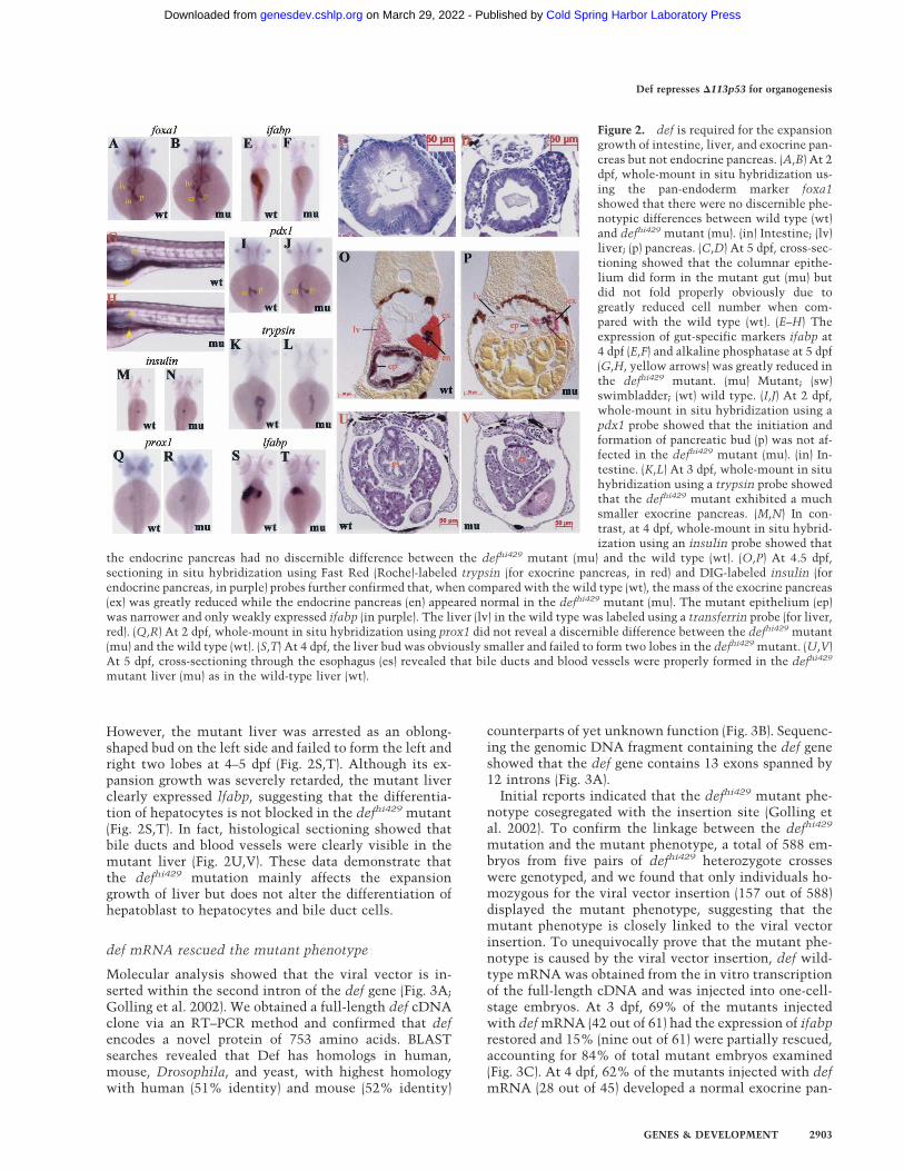
However, the mutant liver was arrested as an oblong-shaped bud on the left side and failed to form the left andright two lobes at 4–5 dpf (Fig. 2S,T). Although its ex-pansion growth was severely retarded, the mutant liverclearly expressed lfabp, suggesting that the differentia-tion of hepatocytes is not blocked in the defhi429 mutant(Fig. 2S,T). In fact, histological sectioning showed thatbile ducts and blood vessels were clearly visible in themutant liver (Fig. 2U,V). These data demonstrate thatthe defhi429 mutation mainly affects the expansiongrowth of liver but does not alter the differentiation ofhepatoblast to hepatocytes and bile duct cells.
def mRNA rescued the mutant phenotype
Molecular analysis showed that the viral vector is in-serted within the second intron of the def gene (Fig. 3A;Golling et al. 2002). We obtained a full-length def cDNAclone via an RT–PCR method and confirmed that defencodes a novel protein of 753 amino acids. BLASTsearches revealed that Def has homologs in human,mouse, Drosophila, and yeast, with highest homologywith human (51% identity) and mouse (52% identity)
counterparts of yet unknown function (Fig. 3B). Sequenc-ing the genomic DNA fragment containing the def geneshowed that the def gene contains 13 exons spanned by12 introns (Fig. 3A).
Initial reports indicated that the defhi429 mutant phe-notype cosegregated with the insertion site (Golling etal. 2002). To confirm the linkage between the defhi429
mutation and the mutant phenotype, a total of 588 em-bryos from five pairs of defhi429 heterozygote crosseswere genotyped, and we found that only individuals ho-mozygous for the viral vector insertion (157 out of 588)displayed the mutant phenotype, suggesting that themutant phenotype is closely linked to the viral vectorinsertion. To unequivocally prove that the mutant phe-notype is caused by the viral vector insertion, def wild-type mRNA was obtained from the in vitro transcriptionof the full-length cDNA and was injected into one-cell-stage embryos. At 3 dpf, 69% of the mutants injectedwith def mRNA (42 out of 61) had the expression of ifabprestored and 15% (nine out of 61) were partially rescued,accounting for 84% of total mutant embryos examined(Fig. 3C). At 4 dpf, 62% of the mutants injected with defmRNA (28 out of 45) developed a normal exocrine pan-
Figure 2. def is required for the expansiongrowth of intestine, liver, and exocrine pan-creas but not endocrine pancreas. (A,B) At 2dpf, whole-mount in situ hybridization us-ing the pan-endoderm marker foxa1showed that there were no discernible phe-notypic differences between wild type (wt)and defhi429 mutant (mu). (in) Intestine; (lv)liver; (p) pancreas. (C,D) At 5 dpf, cross-sec-tioning showed that the columnar epithe-lium did form in the mutant gut (mu) butdid not fold properly obviously due togreatly reduced cell number when com-pared with the wild type (wt). (E–H) Theexpression of gut-specific markers ifabp at4 dpf (E,F) and alkaline phosphatase at 5 dpf(G,H, yellow arrows) was greatly reduced inthe defhi429 mutant. (mu) Mutant; (sw)swimbladder; (wt) wild type. (I,J) At 2 dpf,whole-mount in situ hybridization using apdx1 probe showed that the initiation andformation of pancreatic bud (p) was not af-fected in the defhi429 mutant (mu). (in) In-testine. (K,L) At 3 dpf, whole-mount in situhybridization using a trypsin probe showedthat the defhi429 mutant exhibited a muchsmaller exocrine pancreas. (M,N) In con-trast, at 4 dpf, whole-mount in situ hybrid-ization using an insulin probe showed that
the endocrine pancreas had no discernible difference between the defhi429 mutant (mu) and the wild type (wt). (O,P) At 4.5 dpf,sectioning in situ hybridization using Fast Red (Roche)-labeled trypsin (for exocrine pancreas, in red) and DIG-labeled insulin (forendocrine pancreas, in purple) probes further confirmed that, when compared with the wild type (wt), the mass of the exocrine pancreas(ex) was greatly reduced while the endocrine pancreas (en) appeared normal in the defhi429 mutant (mu). The mutant epithelium (ep)was narrower and only weakly expressed ifabp (in purple). The liver (lv) in the wild type was labeled using a transferrin probe (for liver,red). (Q,R) At 2 dpf, whole-mount in situ hybridization using prox1 did not reveal a discernible difference between the defhi429 mutant(mu) and the wild type (wt). (S,T) At 4 dpf, the liver bud was obviously smaller and failed to form two lobes in the defhi429 mutant. (U,V)At 5 dpf, cross-sectioning through the esophagus (es) revealed that bile ducts and blood vessels were properly formed in the defhi429
mutant liver (mu) as in the wild-type liver (wt).
Def represses �113p53 for organogenesis
GENES & DEVELOPMENT 2903
Cold Spring Harbor Laboratory Press on March 29, 2022 - Published by genesdev.cshlp.orgDownloaded from
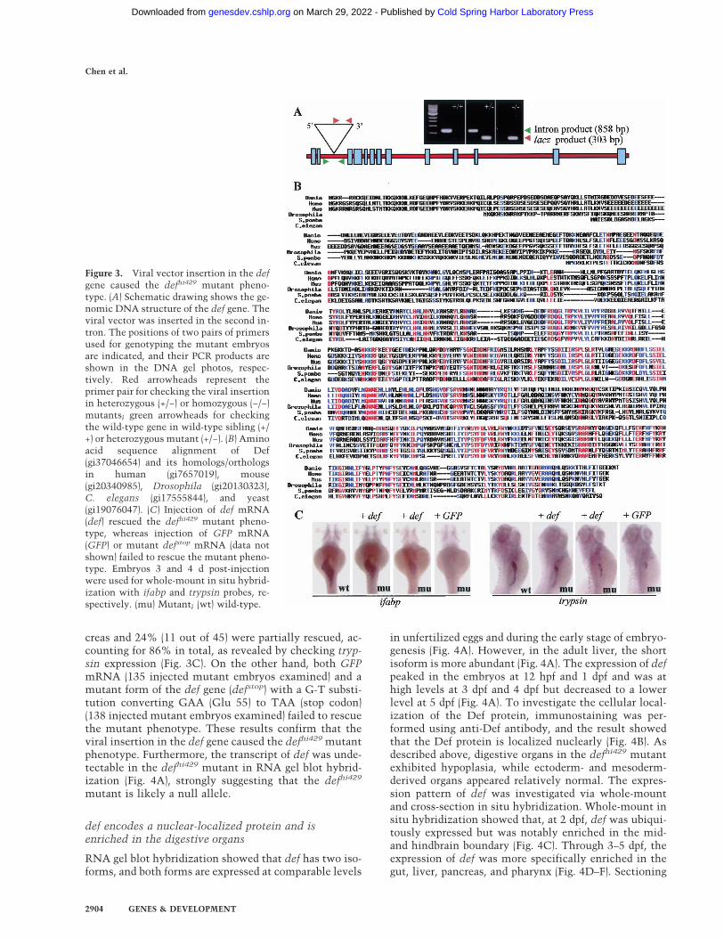
creas and 24% (11 out of 45) were partially rescued, ac-counting for 86% in total, as revealed by checking tryp-sin expression (Fig. 3C). On the other hand, both GFPmRNA (135 injected mutant embryos examined) and amutant form of the def gene (defstop) with a G-T substi-tution converting GAA (Glu 55) to TAA (stop codon)(138 injected mutant embryos examined) failed to rescuethe mutant phenotype. These results confirm that theviral insertion in the def gene caused the defhi429 mutantphenotype. Furthermore, the transcript of def was unde-tectable in the defhi429 mutant in RNA gel blot hybrid-ization (Fig. 4A), strongly suggesting that the defhi429
mutant is likely a null allele.
def encodes a nuclear-localized protein and isenriched in the digestive organs
RNA gel blot hybridization showed that def has two iso-forms, and both forms are expressed at comparable levels
in unfertilized eggs and during the early stage of embryo-genesis (Fig. 4A). However, in the adult liver, the shortisoform is more abundant (Fig. 4A). The expression of defpeaked in the embryos at 12 hpf and 1 dpf and was athigh levels at 3 dpf and 4 dpf but decreased to a lowerlevel at 5 dpf (Fig. 4A). To investigate the cellular local-ization of the Def protein, immunostaining was per-formed using anti-Def antibody, and the result showedthat the Def protein is localized nuclearly (Fig. 4B). Asdescribed above, digestive organs in the defhi429 mutantexhibited hypoplasia, while ectoderm- and mesoderm-derived organs appeared relatively normal. The expres-sion pattern of def was investigated via whole-mountand cross-section in situ hybridization. Whole-mount insitu hybridization showed that, at 2 dpf, def was ubiqui-tously expressed but was notably enriched in the mid-and hindbrain boundary (Fig. 4C). Through 3–5 dpf, theexpression of def was more specifically enriched in thegut, liver, pancreas, and pharynx (Fig. 4D–F). Sectioning
Figure 3. Viral vector insertion in the defgene caused the defhi429 mutant pheno-type. (A) Schematic drawing shows the ge-nomic DNA structure of the def gene. Theviral vector was inserted in the second in-tron. The positions of two pairs of primersused for genotyping the mutant embryosare indicated, and their PCR products areshown in the DNA gel photos, respec-tively. Red arrowheads represent theprimer pair for checking the viral insertionin heterozygous (+/−) or homozygous (−/−)mutants; green arrowheads for checkingthe wild-type gene in wild-type sibling (+/+) or heterozygous mutant (+/−). (B) Aminoacid sequence alignment of Def(gi37046654) and its homologs/orthologsin human (gi7657019), mouse(gi20340985), Drosophila (gi20130323),C. elegans (gi17555844), and yeast(gi19076047). (C) Injection of def mRNA(def) rescued the defhi429 mutant pheno-type, whereas injection of GFP mRNA(GFP) or mutant defstop mRNA (data notshown) failed to rescue the mutant pheno-type. Embryos 3 and 4 d post-injectionwere used for whole-mount in situ hybrid-ization with ifabp and trypsin probes, re-spectively. (mu) Mutant; (wt) wild-type.
Chen et al.
2904 GENES & DEVELOPMENT
Cold Spring Harbor Laboratory Press on March 29, 2022 - Published by genesdev.cshlp.orgDownloaded from

in situ hybridization confirmed that def expression wasspecifically enriched in the liver, gut, and exocrine pan-creas but was excluded from the islet (Fig. 4G,H). Thisexpression pattern coincides with the observation thatislet development is not affected in the defhi429 mutant(Fig. 2M,N).
Expression of �113p53 but not the wild-type p53 isdrastically up-regulated in the mutant digestive organs
To identify genes downstream of def, RNA samples wereprepared from wild-type and mutant whole fish, respec-tively, and were used to compare the expression profilesbetween the wild type and the defhi429 mutant at 5 dpfusing the Affymetrix zebrafish GeneChip carrying
14,900 unigenes. Analysis of results obtained from fiveindependent hybridizations showed that the expressionof 141 genes was down-regulated at least twofold in thedefhi429 mutant compared with the wild type (Supple-mentary Table 1). Extensive database search revealedthat 122 of these down-regulated genes each can be as-signed a biochemical function and the majority of themare well-known for their relative specific expression indigestive organs (Supplementary Table 1). On the otherhand, only 23 genes are specifically up-regulated (greaterthan or equal to twofold) in the defhi429 mutant (Supple-mentary Table 2). Surprisingly, the tumor-suppressorgene p53 and its response genes mdm2 and cyclin G1 areamong these 23 up-regulated genes (SupplementaryTable 2).
To confirm the result obtained from the microarrayhybridization, RNA gel blot hybridization was per-formed to compare the p53 transcripts in the defhi429
mutant and wild-type control. In the wild type, p53 ex-pression peaks at 1 dpf, then decreases gradually to alower level at 5 dpf (Supplementary Fig. S1A). Surpris-ingly, at 5 dpf, a short form of p53 transcripts in thedefhi429 mutant showed very high levels, whereas thisshort form was almost undetectable in the wild-type em-bryos from 1 dpf to 5 dpf (Supplementary Fig. S1A). Toinvestigate the nature of this short-form p53 transcript,we performed 5�-RACE using the RLM-RACE (Ambion)kit to ensure amplifying cDNA only from full-length,capped mRNA. Two 5�-RACE products were observedwhen a primer derived from exon 6 was used for the5�-RACE reaction (Fig. 5A). Sequencing analysis showedthat the longer product corresponded to wild-type p53(data not shown). The short-form p53 contains exons5–12 with 155 base pairs (bp) derived from intron 4 im-mediately adjacent to exon 5 (Fig. 5B), suggesting thatthe short form is initiated from an alternative internalpromoter in intron 4. The short-form transcript encodesfor an N-truncated p53 protein initiated at codon 113(named �113p53), and this product corresponds to�133p53 found in human (Bourdon et al. 2005; Supple-mentary Fig. S2). To further confirm that �113p53 butnot the wild-type p53 is, indeed, increased in the mutant,RNA samples were prepared from wild-type embryos,from wild-type embryos injected with an effective defantisense morpholino (def-MO) (Supplementary Fig. S3),and from the defhi429 mutant embryos, respectively.RNA gel blot hybridization using probes specific for de-tection of wild-type p53 (using P2 probe), �113p53 (P3probe), or both isoforms (P1 probe) showed that �113p53is, indeed, up-regulated only in the defhi429 mutant anddef-MO morphants (Fig. 5C). Because the �113p53-spe-cific probe is derived from the intron 4 sequence only,this result confirms the previous report that �133p53 isa transcribed product initiated by the alternative pro-moter in intron 4 (Bourdon et al. 2005). Furthermore,quantitative real-time PCR using primer pairs specificfor p53 and �113p53 showed that levels of �113p53 tran-scripts in the mutant increased ∼12-, 8.6-, and 7.7-fold at3, 4, and 5 dpf, respectively, whereas the levels of p53 didnot show significant change between the wild type and
Figure 4. def encodes a nuclear protein, and its expression isenriched in the endoderm organs. (A, top panel, lanes 2–7) RNAgel blot hybridization revealed that def had two transcribedforms that peaked at 12 hpf and 1 dpf and then gradually reducedto a lower level at 5 dpf. (Lane 9) The shorter form was moreabundant in the adult liver (AL). (Lane 8) def expression is un-detectable in the defhi429 mutant. (elf1a) elongation factor 1a.(B) Immunostaining using anti-Def antibody showed that Def isa nuclear-localized protein. (C–F) Whole-mount in situ hybrid-ization using a def riboprobe to examine the expression pat-terns of def at 2 dpf (C), 3 dpf (D), 4 dpf (E), and 5 dpf (F). defexpression was specifically enriched in the digestive organsfrom 3 to 5 dpf. (in) Intestine; (lv) liver; (p) pancreas; (sw) swim-bladder. (G,H) Sectioning in situ hybridization showed that defexpression is specifically enriched in the intestine (in), liver (lv),and exocrine pancreas (ex) but is excluded from the endocrinepancreas (en) at 3 and 4 dpf.
Def represses �113p53 for organogenesis
GENES & DEVELOPMENT 2905
Cold Spring Harbor Laboratory Press on March 29, 2022 - Published by genesdev.cshlp.orgDownloaded from

the defhi429 mutant (Fig. 6C). Taken together, our datademonstrated that loss of function of def selectively up-regulated the expression of �113p53 but not p53.
Next, we investigated if the increase of �113p53 iscorrelated to the phenotype observed in the defhi429 mu-tant. Whole-mount in situ hybridization using the probedetecting both p53 and �113p53 transcripts (P1 probe)showed that p53 is ubiquitously expressed in the wild-type embryos (Fig. 5D, left panels). However, the sameprobe detected high levels of gene expression that ap-peared to be specifically restricted in the digestive organsincluding pharynx, liver, pancreas, and intestine in thedefhi429 mutant at 3, 4, and 5 dpf, respectively (Figs. 5D[right panel], 6C; Supplementary Fig. S1B). Because mo-lecular analyses have shown that the expression of�113p53 but not p53 was selectively elevated in the mu-tant (Figs. 5C, 6C) and because p53 is normally ubiqui-tously expressed, it is reasonable to conclude that thehigh levels of gene expression in the mutant digestiveorgans detected by the P1 probe reflected the levels of�113p53. Indeed, in situ hybridization using a �113p53-specific probe derived from intron 4 revealed that the�113p53 transcripts were specifically enriched in the di-gestive organs (Supplementary Fig. S1C). Cross-sectionin situ hybridization revealed that the increase of�113p53 expression is specifically within the intestinaltube, pancreas, and liver, but excluded from the islet (Fig.
5E,F), displaying a pattern closely resembled that of thedef gene (Fig. 4G,H), which suggests that there is a strongcorrelation between the defhi429 mutation and the el-evated �113p53 expression in the digestive organs.
Knock-down of p53 and �113p53 levels rescuedmutant phenotype to normal
Expression of �113p53 was up-regulated in the defhi429
mutant, suggesting that Def might be, directly or indi-rectly, a negative regulator of �113p53 expression andmight exert its function through the p53 pathway. Toanswer this question, two p53 antisense morpholinos,one specifically targeting the splicing site of exon 5 andintron 5 of the p53 transcript (p53-MOspl) and the othertargeting the p53 start codon ATG to block the transla-tion of p53 protein (p53-MOATG), were designed and in-jected into mutant embryos at the one-cell stage. RT–PCR showed that p53-MOspl created aberrant splicingproducts in morphants (Fig. 6A). Examination of p53-MOspl morphants 70 h post-injection using trypsin ex-pression showed that the size of the mutant pancreaswas restored in 24 out of 55 total mutants examined(44%) (Fig. 6B). On the other hand, only 23% of the mu-tants (14 out of 62 mutant embryos examined) were res-cued by p53-MOATG. Injection of the standard controlmorpholino (against human �-globin) failed to rescue the
Figure 5. Loss of function of def selectively up-regu-lates the expression of �113p53 in the defhi429 mutant.(A) 5�-RACE reaction identified two p53 isoforms. Thelonger isoform corresponds to the wild-type p53 (p53)and was predominant in the wild-type embryos. Theshort isoform (�113p53) was drastically increased in thedefhi429 mutant. (B) The short isoform contains 155 bpderived from intron 4 (letters in lowercase) immediatelyadjacent to exon 5 and encodes for an N-truncated p53protein initiated at codon 113 of the wild-type p53. (C)RNA gel blot hybridization using probes for detectingboth p53 and �113p53 isoforms (P1 probe, top panel),p53 only (P2 probe, second panel), and �113p53 only (P3probe, third panel) showed that �113p53 but not p53was selectively increased in the def-MO morphants(def-MO) and defhi429 mutant (mu), respectively, whencompared with the standard control MO morphants(SC-MO) and the wild-type control (wt). (Bottom panel)28S rRNA was used as the loading control. (D) Whole-mount in situ hybridization using the P1 probe showedthat �113p53 expression in the defhi429 mutant (mu,right panels) was increased at 3 dpf, 4 dpf, and 5 dpf, andthis increase was specifically limited to within diges-tive organs when compared with the expression of p53in the wild type (left panels). (in) Intestine; (lv) liver; (p)pancreas; (sw) swimbladder. (E,F) At 4 dpf, sectioning insitu hybridization further confirmed that p53 expres-sion was increased in the intestine (in), liver (data notshown), and exocrine pancreas (ex) but not in the endo-crine pancreas (en) in the defhi429 mutant.
Chen et al.
2906 GENES & DEVELOPMENT
Cold Spring Harbor Laboratory Press on March 29, 2022 - Published by genesdev.cshlp.orgDownloaded from
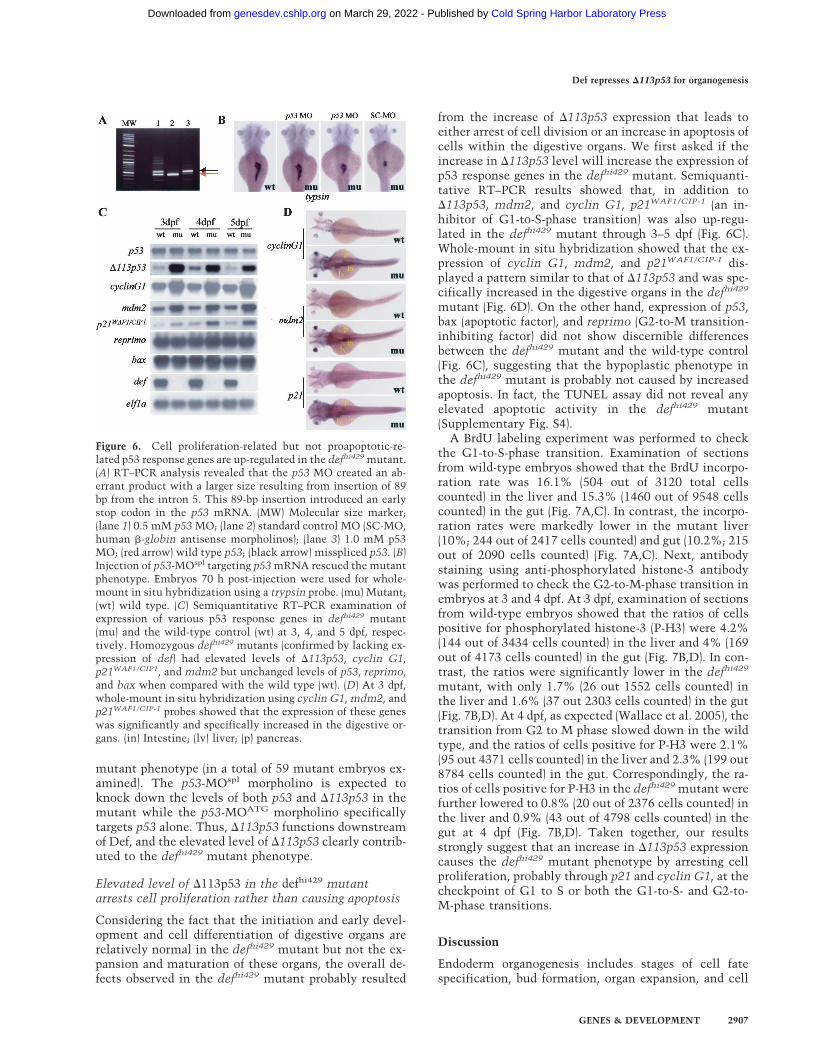
mutant phenotype (in a total of 59 mutant embryos ex-amined). The p53-MOspl morpholino is expected toknock down the levels of both p53 and �113p53 in themutant while the p53-MOATG morpholino specificallytargets p53 alone. Thus, �113p53 functions downstreamof Def, and the elevated level of �113p53 clearly contrib-uted to the defhi429 mutant phenotype.
Elevated level of �113p53 in the defhi429 mutantarrests cell proliferation rather than causing apoptosis
Considering the fact that the initiation and early devel-opment and cell differentiation of digestive organs arerelatively normal in the defhi429 mutant but not the ex-pansion and maturation of these organs, the overall de-fects observed in the defhi429 mutant probably resulted
from the increase of �113p53 expression that leads toeither arrest of cell division or an increase in apoptosis ofcells within the digestive organs. We first asked if theincrease in �113p53 level will increase the expression ofp53 response genes in the defhi429 mutant. Semiquanti-tative RT–PCR results showed that, in addition to�113p53, mdm2, and cyclin G1, p21WAF1/CIP-1 (an in-hibitor of G1-to-S-phase transition) was also up-regu-lated in the defhi429 mutant through 3–5 dpf (Fig. 6C).Whole-mount in situ hybridization showed that the ex-pression of cyclin G1, mdm2, and p21WAF1/CIP-1 dis-played a pattern similar to that of �113p53 and was spe-cifically increased in the digestive organs in the defhi429
mutant (Fig. 6D). On the other hand, expression of p53,bax (apoptotic factor), and reprimo (G2-to-M transition-inhibiting factor) did not show discernible differencesbetween the defhi429 mutant and the wild-type control(Fig. 6C), suggesting that the hypoplastic phenotype inthe defhi429 mutant is probably not caused by increasedapoptosis. In fact, the TUNEL assay did not reveal anyelevated apoptotic activity in the defhi429 mutant(Supplementary Fig. S4).
A BrdU labeling experiment was performed to checkthe G1-to-S-phase transition. Examination of sectionsfrom wild-type embryos showed that the BrdU incorpo-ration rate was 16.1% (504 out of 3120 total cellscounted) in the liver and 15.3% (1460 out of 9548 cellscounted) in the gut (Fig. 7A,C). In contrast, the incorpo-ration rates were markedly lower in the mutant liver(10%; 244 out of 2417 cells counted) and gut (10.2%; 215out of 2090 cells counted) (Fig. 7A,C). Next, antibodystaining using anti-phosphorylated histone-3 antibodywas performed to check the G2-to-M-phase transition inembryos at 3 and 4 dpf. At 3 dpf, examination of sectionsfrom wild-type embryos showed that the ratios of cellspositive for phosphorylated histone-3 (P-H3) were 4.2%(144 out of 3434 cells counted) in the liver and 4% (169out of 4173 cells counted) in the gut (Fig. 7B,D). In con-trast, the ratios were significantly lower in the defhi429
mutant, with only 1.7% (26 out 1552 cells counted) inthe liver and 1.6% (37 out 2303 cells counted) in the gut(Fig. 7B,D). At 4 dpf, as expected (Wallace et al. 2005), thetransition from G2 to M phase slowed down in the wildtype, and the ratios of cells positive for P-H3 were 2.1%(95 out 4371 cells counted) in the liver and 2.3% (199 out8784 cells counted) in the gut. Correspondingly, the ra-tios of cells positive for P-H3 in the defhi429 mutant werefurther lowered to 0.8% (20 out of 2376 cells counted) inthe liver and 0.9% (43 out of 4798 cells counted) in thegut at 4 dpf (Fig. 7B,D). Taken together, our resultsstrongly suggest that an increase in �113p53 expressioncauses the defhi429 mutant phenotype by arresting cellproliferation, probably through p21 and cyclin G1, at thecheckpoint of G1 to S or both the G1-to-S- and G2-to-M-phase transitions.
Discussion
Endoderm organogenesis includes stages of cell fatespecification, bud formation, organ expansion, and cell
Figure 6. Cell proliferation-related but not proapoptotic-re-lated p53 response genes are up-regulated in the defhi429 mutant.(A) RT–PCR analysis revealed that the p53 MO created an ab-errant product with a larger size resulting from insertion of 89bp from the intron 5. This 89-bp insertion introduced an earlystop codon in the p53 mRNA. (MW) Molecular size marker;(lane 1) 0.5 mM p53 MO; (lane 2) standard control MO (SC-MO,human �-globin antisense morpholinos); (lane 3) 1.0 mM p53MO; (red arrow) wild type p53; (black arrow) misspliced p53. (B)Injection of p53-MOspl targeting p53 mRNA rescued the mutantphenotype. Embryos 70 h post-injection were used for whole-mount in situ hybridization using a trypsin probe. (mu) Mutant;(wt) wild type. (C) Semiquantitative RT–PCR examination ofexpression of various p53 response genes in defhi429 mutant(mu) and the wild-type control (wt) at 3, 4, and 5 dpf, respec-tively. Homozygous defhi429 mutants (confirmed by lacking ex-pression of def) had elevated levels of �113p53, cyclin G1,p21WAF1/CIP1, and mdm2 but unchanged levels of p53, reprimo,and bax when compared with the wild type (wt). (D) At 3 dpf,whole-mount in situ hybridization using cyclin G1, mdm2, andp21WAF1/CIP-1 probes showed that the expression of these geneswas significantly and specifically increased in the digestive or-gans. (in) Intestine; (lv) liver; (p) pancreas.
Def represses �113p53 for organogenesis
GENES & DEVELOPMENT 2907
Cold Spring Harbor Laboratory Press on March 29, 2022 - Published by genesdev.cshlp.orgDownloaded from

differentiation (Wells and Melton 1999; Zaret 2002). Ex-amination of major endoderm organs including intes-tine, pancreas, and liver in the defhi429 mutant usingorgan-specific molecular markers and histological analy-sis showed that these organs appeared normal till 2 dpf,suggesting that def is not involved in or does not play amajor role for the initiation and budding of digestive or-gans. However, digestive organs start to become hypo-
plastic from 3 dpf, including an underdeveloped gut tubeand a smaller liver, gall bladder, pancreas, and swimblad-der, whereas mesoderm-derived organs includingsomites, blood vessels, and pronephric ducts appearednormal. The underdevelopment of the entire digestivesystem in the defhi429 mutant could result from the ar-rest of cell differentiation as found for the nil per osemutant (Mayer and Fishman 2003). If this is the case,digestive organs are probably mainly composed of pro-genitor cells. Although at a much lower level when com-pared with the wild-type control, molecular markers spe-cific to fully differentiated intestinal epithelial cells(ifabp and alkaline phosphatase), hepatocytes (lfabp andtransferin), and exocrine pancreas (trypsin) are expressedin the mutant fish, demonstrating that cell differentia-tion is compromised but not abolished by the defhi429
mutation. Whole-mount in situ hybridization showedthat def expression is enriched in the entire digestiveducts and organs including pharynx, pancreas, liver,swimbladder, and intestine. Furthermore, sectioning insitu hybridization showed that def expression is ex-cluded from the islet. The expression pattern of def per-fectly matched the phenotype displayed by the defhi429
mutant and explains why the endocrine pancreas appearsnormal in the mutant. The evidence provided stronglysuggests that Def functions as a cell-autonomous factorto regulate the expansion growth of various digestive or-gans except the endocrine pancreas at the later stage ofendoderm organogenesis.
The def gene encodes a nuclear-localized novel pro-tein. In order to study how Def controls the developmentof digestive organs, we compared the gene expressionprofiles between the defhi429 mutant and the wild-typecontrol. As expected, the expression of a large number ofenzyme genes that are known to be enriched in the in-testine, liver, and pancreas is significantly decreased inthe defhi429 mutant due to great loss of organ mass andcompromised cell differentiation. Surprisingly, p53 ex-pression showed a drastic increase in the mutant em-bryos. Further molecular analysis revealed that �113p53,a newly identified p53 isoform initiated from an alterna-tive promoter in intron 4 (Bourdon et al. 2005), ratherthan the wild-type p53, selectively increased in the mu-tant. In situ hybridization showed that the increased�113p53 expression is specifically limited to within thedigestive organs of the mutant embryo but not in otherorgans. This expression pattern of �113p53 in the mu-tant embryo phenocopies the pattern displayed by thedef gene in the wild type, suggesting that Def might be anegative regulator of �113p53 in the digestive organs andthe increase in �113p53 might contribute to causing themutant phenotype. This hypothesis is supported by ourdata showing that injection of p53-MOspl targeting both�113p53 and p53 exhibited a much higher rescue rate ofthe defhi429 mutant phenotype than did injection of p53-specific morpholinos p53-MOATG. Thus, Def functions,at least in part, through regulating the p53 pathway tocontrol the expansion growth of digestive organs. p53 isknown to both initiate and suppress the expression oftarget genes to regulate cell cycle and cell death. Exami-
Figure 7. The defhi429 mutant phenotype is caused by compro-mised cell proliferation. (A,C) BrdU labeling revealed that, at 4dpf, the number and ratio of cells entering the G1-to-S-phasetransition was significantly reduced in the digestive organs ofthe defhi429 mutant (mu) when compared with the wild type(wt). (Top panel) Cross-sectioning through liver (lv) and intes-tine (in). (Bottom panel) Cross-sectioning through pancreas andintestine (in). The BrdU incorporation ratios shown in C wereobtained by counting BrdU-labeled cells versus total cells in aspecific organ (e.g., liver) in sections from seven wild-type andseven mutant embryos, respectively. (en) Endocrine pancreas;(ex) exocrine pancreas. (B,D) At 3 and 4 dpf, histochemical stain-ing using anti-phosphorylated histone 3 (anti-P-H3) revealedthat the number and ratio of cells entering G2 to M phase wasdrastically reduced in the digestive organs of the defhi429 mutant(mu) when compared with the wild type (wt). The ratios ofP-H3-positive cells shown in D were obtained by countingP-H3-positive cells versus total cells in a specific organ (e.g.,liver) in sections from three wild-type and three mutant em-bryos, respectively.
Chen et al.
2908 GENES & DEVELOPMENT
Cold Spring Harbor Laboratory Press on March 29, 2022 - Published by genesdev.cshlp.orgDownloaded from

nation of p53 response genes showed that the expressionof p21WAF1/CIP-1 and cyclin G1, two inhibiting factors ofcell proliferation (Ball 1997; Zhao et al. 2003), were in-creased in the defhi429 mutant. On the other hand, theexpression of bax, a proapoptotic factor (Reed 1999), andreprimo, an inhibitor of G2-to-M-phase transition (Ohkiet al. 2000), remained unchanged in the defhi429 mutant.These results strongly suggest that (1) �113p53 selec-tively activates the p53 response genes, and (2) the hy-poplastic phenotype in the defhi429 mutant is probablydue to an arrest of cell proliferation but not due to in-creased apoptosis. This hypothesis is further strength-ened by the observation that the TUNEL assay did notreveal any obvious elevated apoptotic activities in thedefhi429 mutant. In contrast, both a BrdU labeling andimmunochemical staining using anti-P-H3 antibodyshowed that the cell division index is greatly reduced inthe defhi429 mutant, demonstrating that the arrest of cellproliferation is the main cue to cause the hypoplasticphenotype in defhi429 mutant.
In zebrafish, Nodal signaling plays a decisive role inendoderm specification (Ober et al. 2003). gata5 (Reiteret al. 1999), hhex (Wallace et al. 2001), hnf1 (Sun andHopkins 2001), hnf6 (Matthews et al. 2004), mnr2a(Wendik et al. 2004), notch signaling (Lorent et al. 2004),pdx1 (Huang et al. 2001), prox1 (Liu et al. 2003), ptf1a(Lin et al. 2004), shh (diIorio et al. 2002), and their coun-terparts in other vertebrates (Edlund 2002; Zaret 2002)play crucial roles in controlling endoderm organ devel-opment in zebrafish. While some of these factors func-tion relatively specifically only in certain endoderm or-gan(s) (e.g., hhex in liver, pdx1 and ptfia in pancreas),others are known to be general factors (e.g., shh andnotch signaling). Apparently, the final size and shape ofan organ and its proper position in the body can be per-fectly achieved only when the interaction of differen-tially expressed organ-specific factors with general fac-tors is precisely regulated. However, little is knownabout the involvement of any pan-endoderm-specific fac-tors to coordinate the expansion growth of the entiredigestive system at the later stage. Here we propose thatDef, like ni per os (Mayer and Fishman 2003), is one ofsuch factors that acts as a pan-endoderm-specific factorto negatively regulate the expression of a general factor,namely, �113p53, to coordinate the proliferation of cellswithin digestive system. Thus, we have shown in thiswork that, in addition to acting as a checkpoint control-ler to suppress abnormal cell growth, p53 and its iso-forms are closely watched and tightly controlled by or-gan/tissue-specific factors during organogenesis, espe-cially at the stage wherein fast cell proliferation isneeded, to prevent any adverse effect on organ/tissuegrowth. The remaining intriguing questions includewhether Def directly or indirectly regulates �113p53 ex-pression, because currently we cannot exclude the pos-sibility that the absence of Def could induce a general-ized stress response that could indirectly up-regulate theexpression of �113p53. In addition, it is also interestingto know how �113p53 differentially regulates the tran-scription of a subset of p53 response genes (e.g., cyclin
G1 and p21WAF1/CIP-1) but not others (e.g., reprimo andbax). These studies provide overwhelming genetic sup-port for the physiological significance of the newly dis-covered p53 isoforms and establish that they can play akey role in development. Detailed biochemical studies ofthese isoform interactions will be needed to understandthese dramatic observations. The consequences of thisnew understanding of p53 function and control in devel-opment for interpreting its role in neoplasia and agingare very exciting.
Materials and methods
defhi429 mutant
Zebrafish line hi429 was obtained from a large-scale insertionalmutagenesis screen using mouse retroviral vectors as the mu-tagen (Golling et al. 2002) and was kindly provided by ProfessorNancy Hopkins at Massachusetts Institute of Technology(Cambridge, MA). Zebrafish were raised and maintained accord-ing to standard procedures (Mayer and Fishman 2003). All mu-tant embryos used for characterization in this report were con-firmed by genotyping using two pairs of primers, one pair de-rived from the LacZ gene (P1: 5�-ATCCTCTAGACTGCCATGG-3�; P2: 5�-ATCGTAACCGTGCATCTG-3�) harbored bythe viral vector for confirmation of insertion and the other fromintron II of the def genomic sequence (P1: 5�-TATTGCCTTACGACAGTTT-3�; P2: 5�-CAAGCGTTTGACATTAGAGT-3�)flanking the viral vector insertion site for confirmation of thedef gene (Fig. 3A).
Whole-mount RNA in situ hybridization
Whole-mount RNA in situ hybridizations were performed asdescribed (Mayer and Fishman 2003). Probes were labeled withdigoxigenin (DIG). foxa1 (AL911576), lfabp (AL926262),p21WAF1/CIP-1 (AL912410), and p53 (AL922791) cDNA cloneswere from our own EST collection (Lo et al. 2003). For transfer-rin and ifabp (Mudumana et al. 2004), pdx1 (NM_131443), def(nucleotides 336–1027), cyclin G1 (BC052125), and mdm2(AF356346), primers were designed based on available sequencedata, and RT–PCR products were cloned into the pGEM-T EasyVector, respectively. Photos were taken under a Leica M216optics.
Mutant phenotype rescue
For mRNA rescue, 1.0 ng of in vitro transcribed def mRNA,mutant defstop mRNA, and GFP mRNA were injected into theyolk of 1-cell-stage embryos, respectively. Embryos 3 and 4 dpost-injection were used for whole mount in situ. p53-MOspl
(5�-AAAATGTCTGTACTATCTCCATCCG-3�) was designedto target the splice junction between exon 5 and intron 5, andp53-MOATG was designed corresponding to the start codonATG (5�-GCGCCATTGCTTTGCAAGAATTG-3�) (Langhein-rich et al. 2002). Morpholinos were supplied by Gene Tools and1 nL (0.5 mM) was injected into the yolk of one-cell-stage em-bryos each time. Human �-globin antisense morpholino (5�-CCTCTTACCTCAGTTACAATTT-3�) was used as the stan-dard control.
Affimetrix array
Total RNA was extracted from 5 dpf wild-type and defhi429 mu-tant embryos, respectively, using TRIzol (GIBCO-BRL, USA)and treating with DNase I. cDNA synthesis, RNA probe label-
Def represses �113p53 for organogenesis
GENES & DEVELOPMENT 2909
Cold Spring Harbor Laboratory Press on March 29, 2022 - Published by genesdev.cshlp.orgDownloaded from

ing, target hybridization, washing, and staining were performedfollowing the manufacturer’s instructions (Affymetrix).GeneChip arrays were scanned on an Affymetrix probe arrayscanner. Data were analyzed using the statistics softwareMAS5.0 from Affymetrix.
Histological and immunohistochemical analysis
All embryos were anesthetized using 3-aminobenzoic acid ethylester, and the tail was clipped for genotyping. All photos weretaken under a Zeiss Axiophot 2 optics. For hematoxylin andeosin staining, sectioning (3.5 µm) was done on paraffin-embed-ded embryos processed as described (Wallace et al. 2005). Em-bryo fixation and cryo-section in situ hybridization were per-formed as described (Wendl et al. 2002). For BrdU in vivo label-ing, 5-bromo-2�-deoxy-uridine (BrdU) (1 nL, 10 mM; Roche) wasmicroinjected into the peritoneal cavity of 4-dpf embryos. Em-bryos were incubated for 4 h at 28.5°C and then fixed at 4% PFAfor 24 h before being used for immunohistochemical study asdescribed (Wallace et al. 2005). Cells positive for P-H3 weredetected using the polyclonal anti-P-H3 antibody (Santa Cruz)as the first antibody (1:200) and anti-rabbit horseradish POD(1:150) as the secondary antibody with diaminobenzidine as thesubstrate for color reaction. All slides were mounted with amount medium containing DAPI (Vector). Methods for alcianblue staining and alkaline phosphatase staining were as de-scribed (Neuhauss et al. 1996; Mayer and Fishman 2003). For thenuclear-localization experiment, 1.0 ng of def mRNA was in-jected into the yolk of 1-cell-stage embryos, and embryos werecollected at 10 hpf and fixed by 4% PFA at 4°C overnight andincubated with anti-Def antibody (against amino acids 32–178raised in rabbit). Alexa Fluor 488 goat anti-rabbit IgG antibody(Molecular probes) conjugated with GFP was used as the secondantibody for detection. The embryos were then deyolked andsoaked in DAPI mounting medium for visualization and pho-tography.
TUNEL assay
Cryo-sections (8 µM) were fixed in 4% PFA for 20 min, washed30 min with PBS, and incubated in permeabilization solution(0.1% Triton X-100, 0.1% sodium citrate) for 2 min on ice. TheTUNEL assay was carried out with the In Situ Cell Death De-tection Kit, TMR red (Roche).
RNA gel blot hybridization, 5�-RACE, and RT–PCR
Total RNA was extracted using TRIzol (GIBCO-BRL). Poly(A)+
mRNA was obtained from total RNA using the Oligotex mRNAMidi kit (QIAGEN). DIG-labeled probes (def: nucleotides 2108–2466; elf1a: nucleotides 18–706) were used for RNA gel blothybridization as described (Wen et al. 2005). The P1 probe fordetecting both p53 and �113p53 transcripts was derived fromnucleotides 44–1165 of the p53 gene. The P2 probe specific forthe p53 transcript was derived from nucleotides 7–294 of thep53 gene, and the P3 probe specific for �113p53 transcript wasderived from the transcribed sequence nucleotides 13–155 origi-nated from intron 4 (Fig. 5B). 5�-RACE was performed using theRLM-RACE kit (RNA Ligase Mediated Rapid Amplification ofcDNA Ends; Ambion) according to the protocol provided by themanufacturer.
For Real-Time RT–PCR, embryos were collected at 3, 4, and5 dpf and genotyped. RNA of individual embryos was extractedusing the RNAeasy 96 kit (QIAGEN) according to the manufac-turer’s protocol. Ten individual wild-type and mutant embryoswere pooled, respectively, and treated with DNase I. RNA was
reverse-transcribed using Expand Reverse Transcriptase(Roche). The amount of transcribed cDNAs was normalizedbased on elongation factor 1a (elf1a) as a control with theRealtime LightCycler (Roche). The amount of template and thenumber of PCR cycles were optimized to ensure that the reac-tions were in the linear range of amplification. The primer pairsand detailed PCR conditions used to amplify each of these genesare listed in Supplementary Table 3.
Acknowledgments
We thank Professor N. Hopkins for providing the defhi429 mu-tant line. We thank Qi Zeng, Ke Guo, Jie Li, and Binqi Gan forhistological analysis. We thank Honghui Huang, Jeremy Wu,and Lin Guo for their suggestions and technical support. Thiswork is supported by the Agency for Science, Technology andResearch in Singapore.
References
Armstrong, J.F., Kaufman, M.H., Harrison, D.J., and Clarke,A.R. 1995. High-frequency developmental abnormalities inp53-deficient mice. Curr. Biol. 5: 931–936.
Ball, K.L. 1997. p21: Structure and functions associated withcyclin–CDK binding. Prog. Cell Cycle Res. 3: 125–134.
Benard, J., Douc-Rasy, S., and Ahomadegbe, J.C. 2003. TP53family members and human cancers. Hum. Mutat. 21: 182–191.
Berghmans, S., Murphey, R.D., Wienholds, E., Neuberg, D., Ku-tok, J.L., Fletcher, C.D., Morris, J.P., Liu, T.X., Schulte-Merker, S., Kanki, J.P., et al. 2005. tp53 mutant zebrafishdevelop malignant peripheral nerve sheath tumors. Proc.Natl. Acad. Sci. 102: 407–412.
Bourdon, J.-C., Fernandes, K., Murray-Zmijewski, F., Liu, G.,Xirodimas, D.P., Saville, M.K., and Lane, D.P. 2005. p53 iso-forms can regulate p53 transcriptional activity. Genes &Dev. 19: 2122–2137.
Clarke, A.R., Purdie, C.A., Harrison, D.J., Morris, R.G., Bird,C.C., Hooper, M.L., and Wyllie, A.H. 1993. Thymocyte ap-optosis induced by p53-dependent and independent path-ways. Nature 362: 849–852.
Derry, W.B., Putzke, A.P., and Rothman, J.H. 2001. Caenorhab-ditis elegans p53: Role in apoptosis, meiosis, and stress re-sistance. Science 294: 591–595.
diIorio, P.J., Moss, J.B., Sbrogna, J.L., Karlstrom, R.O., and Moss,L.G. 2002. Sonic hedgehog is required early in pancreaticislet development. Dev. Biol. 244: 75–84.
Donehower, L.A., Harvey, M., Slagle, B.L., McArthur, M.J.,Montgomery Jr., C.A., Butel, J.S., and Bradley, A. 1992. Micedeficient for p53 are developmentally normal but susceptibleto spontaneous tumours. Nature 356: 215–221.
Edlund, H. 2002. Pancreatic organogenesis—Developmentalmechanisms and implications for therapy. Nat. Rev. Genet.3: 524–532.
Field, H.A., Ober, E.A., Roeser, T., and Stainier, D.Y. 2003. For-mation of the digestive system in zebrafish. I. Liver morpho-genesis. Dev. Biol. 253: 279–290.
Golling, G., Amsterdam, A., Sun, Z., Antonelli, M., Maldonado,E., Chen, W., Burgess, S., Haldi, M., Artzt, K., Farrington, S.,et al. 2002. Insertional mutagenesis in zebrafish rapidly iden-tifies genes essential for early vertebrate development. Nat.Genet. 31: 135–140.
Greenblatt, M.S., Bennett, W.P., Hollstein, M., and Harris, C.C.1994. Mutations in the p53 tumor suppressor gene: Clues tocancer etiology and molecular pathogenesis. Cancer Res.
Chen et al.
2910 GENES & DEVELOPMENT
Cold Spring Harbor Laboratory Press on March 29, 2022 - Published by genesdev.cshlp.orgDownloaded from

54: 4855–4878.Horne-Badovinac, S., Rebagliati, M., and Stainier, D.Y. 2003. A
cellular framework for gut-looping morphogenesis in ze-brafish. Science 302: 662–665.
Huang, H., Liu, N., and Lin, S. 2001. Pdx-1 knockdown reducesinsulin promoter activity in zebrafish. Genesis 30: 134–136.
Jin, S., Martinek, S., Joo, W.S., Wortman, J.R., Mirkovic, N., Sali,A., Yandell, M.D., Pavletich, N.P., Young, M.W., and Levine,A.J. 2000. Identification and characterization of a p53 homo-logue in Drosophila melanogaster. Proc. Natl Acad. Sci.97: 7301–7306.
Kiefer, J.C. 2003. Molecular mechanisms of early gut organo-genesis: A primer on development of the digestive tract. Dev.Dyn. 228: 287–291.
Langheinrich, U., Hennen, E., Stott, G., and Vacun, G. 2002.Zebrafish as a model organism for the identification andcharacterization of drugs and genes affecting p53 signaling.Curr. Biol. 12: 2023–2028.
Levine, A.J. 1997. p53, the cellular gatekeeper for growth anddivision. Cell 88: 323–331.
Lin, J.W., Biankin, A.V., Horb, M.E., Ghosh, B., Prasad, N.B.,Yee, N.S., Pack, M.A., and Leach, S.D. 2004. Differentialrequirement for ptf1a in endocrine and exocrine lineages ofdeveloping zebrafish pancreas. Dev. Biol. 274: 491–503.
Liu, Y.W., Gao, W., Teh, H.L., Tan, J.H., and Chan, W.K. 2003.Prox1 is a novel coregulator of Ff1b and is involved in theembryonic development of the zebra fish interrenal primor-dium. Mol. Cell Biol. 23: 7243–7255.
Lo, J., Lee, S., Xu, M., Liu, F., Ruan, H., Eun, A., He, Y., Ma, W.,Wang, W., Wen, Z., et al. 2003. 15000 unique zebrafish ESTclusters and their future use in microarray for profiling geneexpression patterns during embryogenesis. Genome Res.13: 455–466.
Lorent, K., Yeo, S.Y., Oda, T., Chandrasekharappa, S., Chitnis,A., Matthews, R.P., and Pack, M. 2004. Inhibition of Jagged-mediated Notch signaling disrupts zebrafish biliary develop-ment and generates multi-organ defects compatible with anAlagille syndrome phenocopy. Development 131: 5753–5766.
Matthews, R.P., Lorent, K., Russo, P., and Pack, M. 2004. Thezebrafish onecut gene hnf-6 functions in an evolutionarilyconserved genetic pathway that regulates vertebrate biliarydevelopment. Dev. Biol. 274: 245–259.
Mayer, A.N. and Fishman, M.C. 2003. Nil per os encodes aconserved RNA recognition motif protein required for mor-phogenesis and cytodifferentiation of digestive organs in ze-brafish. Development 130: 3917–3928.
Melino, G., Lu, X., Gasco, M., Crook, T., and Knight, R.A. 2003.Functional regulation of p73 and p63: Development and can-cer. Trends Biochem. Sci. 28: 663–670.
Momand, J., Zambetti, G.P., Olson, D.C., George, D., andLevine, A.J. 1992. The mdm-2 oncogene product forms acomplex with the p53 protein and inhibits p53-mediatedtransactivation. Cell 69: 1237–1245.
Montes de Oca, L.R., Wagner, D.S., and Lozano, G. 1995. Rescueof early embryonic lethality in mdm2-deficient mice by de-letion of p53. Nature 378: 203–206.
Mudumana, S.P., Wan, H., Singh, M., Korzh, V., and Gong, Z.2004. Expression analyses of zebrafish transferrin, ifabp, andelastaseB mRNAs as differentiation markers for the threemajor endodermal organs: Liver, intestine, and exocrine pan-creas. Dev. Dyn. 230: 165–173.
Neuhauss, S.C., Solnica-Krezel, L., Schier, A.F., Zwartkruis, F.,Stemple, D.L., Malicki, J., Abdelilah, S., Stainier, D.Y., andDriever, W. 1996. Mutations affecting craniofacial develop-ment in zebrafish. Development 123: 357–367.
Ober, E.A., Field, H.A., and Stainier, D.Y. 2003. From endodermformation to liver and pancreas development in zebrafish.Mech. Dev. 120: 5–18.
Ohki, R., Nemoto, J., Murasawa, H., Oda, E., Inazawa, J.,Tanaka, N., and Taniguchi, T. 2000. Reprimo, a new candi-date mediator of the p53-mediated cell cycle arrest at the G2phase. J. Biol. Chem. 275: 22627–22630.
Ollmann, M., Young, L.M., Di Como, C.J., Karim, F., Belvin, M.,Robertson, S., Whittaker, K., Demsky, M., Fisher, W.W.,Buchman, A., et al. 2000. Drosophila p53 is a structural andfunctional homolog of the tumor suppressor p53. Cell101: 91–101.
Reed, J.C. 1999. Dysregulation of apoptosis in cancer. J. Clin.Oncol. 17: 2941–2953.
Reiter, J.F., Alexander, J., Rodaway, A., Yelon, D., Patient, R.,Holder, N., and Stainier, D.Y. 1999. Gata5 is required for thedevelopment of the heart and endoderm in zebrafish. Genes& Dev. 13: 2983–2995.
Sah, V.P., Attardi, L.D., Mulligan, G.J., Williams, B.O., Bronson,R.T., and Jacks, T. 1995. A subset of p53-deficient embryosexhibit exencephaly. Nat. Genet. 10: 175–180.
Schumacher, B., Hanazawa, M., Lee, M.H., Nayak, S., Volk-mann, K., Hofmann, R., Hengartner, M., Schedl, T., andGartner, A. 2005. Translational repression of C. elegans p53by GLD-1 regulates DNA damage-induced apoptosis. Cell120: 357–368.
Sun, Z. and Hopkins, N. 2001. vhnf1, the MODY5 and familialGCKD-associated gene, regulates regional specification ofthe zebrafish gut, pronephros, and hindbrain. Genes & Dev.15: 3217–3229.
Tyner, S.D., Venkatachalam, S., Choi, J., Jones, S., Ghebranious,N., Igelmann, H., Lu, X., Soron, G., Cooper, B., Brayton, C.,et al. 2002. p53 mutant mice that display early ageing-asso-ciated phenotypes. Nature 415: 45–53.
Vogelstein, B., Lane, D., and Levine, A.J. 2000. Surfing the p53network. Nature 408: 307–310.
Wallace, K.N. and Pack, M. 2003. Unique and conserved aspectsof gut development in zebrafish. Dev. Biol. 255: 12–29.
Wallace, K.N., Yusuff, S., Sonntag, J.M., Chin, A.J., and Pack, M.2001. Zebrafish hhex regulates liver development and diges-tive organ chirality. Genesis 30: 141–143.
Wallace, K.N., Akhter, S., Smith, E.M., Lorent, K., and Pack, M.2005. Intestinal growth and differentiation in zebrafish.Mech. Dev. 122: 157–173.
Warga, R.M. and Nusslein-Volhard, C. 1999. Origin and devel-opment of the zebrafish endoderm. Development 126: 827–838.
Wells, J.M. and Melton, D.A. 1999. Vertebrate endoderm devel-opment. Annu. Rev. Cell Dev. Biol. 15: 393–410.
Wen, C., Zhang, Z., Ma, W., Xu, M., Wen, Z., and Peng, J.R.2005. Genome-wide identification of female-enriched genesin zebrafish. Dev. Dyn. 232: 171–179.
Wendik, B., Maier, E., and Meyer, D. 2004. Zebrafish mnx genesin endocrine and exocrine pancreas formation. Dev. Biol.268: 372–383.
Wendl, T., Lun, K., Mione, M., Favor, J., Brand, M., Wilson,S.W., and Rohr, K.B. 2002. Pax2.1 is required for the devel-opment of thyroid follicles in zebrafish. Development129: 3751–3760.
Zaret, K.S. 2002. Regulatory phases of early liver development:Paradigms of organogenesis. Nat. Rev. Genet. 3: 499–512.
Zhao, L., Samuels, T., Winckler, S., Korgaonkar, C., Tompkins,V., Horne, M.C., and Quelle, D.E. 2003. Cyclin G1 hasgrowth inhibitory activity linked to the ARF-Mdm2-p53 andpRb tumor suppressor pathways. Mol. Cancer Res. 1: 195–206.
Def represses �113p53 for organogenesis
GENES & DEVELOPMENT 2911
Cold Spring Harbor Laboratory Press on March 29, 2022 - Published by genesdev.cshlp.orgDownloaded from

10.1101/gad.1366405Access the most recent version at doi: 19:2005, Genes Dev.
Jun Chen, Hua Ruan, Sok Meng Ng, et al. arrest expansion growth of digestive organs in zebrafish
expression to113p53∆ selectively up-regulates defLoss of function of
Material
Supplemental
http://genesdev.cshlp.org/content/suppl/2005/12/02/19.23.2900.DC1
References
http://genesdev.cshlp.org/content/19/23/2900.full.html#ref-list-1
This article cites 50 articles, 18 of which can be accessed free at:
License
ServiceEmail Alerting
click here.right corner of the article or
Receive free email alerts when new articles cite this article - sign up in the box at the top
Cold Spring Harbor Laboratory Press
Cold Spring Harbor Laboratory Press on March 29, 2022 - Published by genesdev.cshlp.orgDownloaded from