long-term effects of enriched environment on ... · consequences to trauma: ... environmental...
TRANSCRIPT

PHYSIOLOGICAL RESEARCH • ISSN 0862-8408 (print) • ISSN 1802-9973 (online) 2015 Institute of Physiology v.v.i., Academy of Sciences of the Czech Republic, Prague, Czech Republic
Fax +420 241 062 164, e-mail: [email protected], www.biomed.cas.cz/physiolres
Physiol. Res. 64: 129-145, 2015
Long-Term Effects of Enriched Environment on Neurofunctional Outcome and CNS Lesion Volume After Traumatic Brain Injury in Rats M. MAEGELE1,2, M. BRAUN1, A. WAFAISADE1,2, N. SCHÄFER1, M. LIPPERT-GRUENER3, C. KREIPKE4, J. RAFOLS4, U. SCHÄFER5, D. N. ANGELOV6, E. K. STUERMER1 1Institute for Research in Operative Medicine (IFOM), University of Witten-Herdecke (Campus Cologne-Merheim), Cologne, Germany, 2Department for Traumatology and Orthopedic Surgery, Cologne-Merheim Medical Center (CMMC), University Witten-Herdecke (Campus Cologne-Merheim), Cologne, Germany, 3Department for Neurosurgery, University of Cologne Medical Center, Cologne, Germany, 4Department of Anatomy, Wayne State University School of Medicine, Detroit, Michigan, USA, 5Experimental Neurotraumatology, University Hospital for Neurosurgery, Medical University Graz, Graz, Austria, 6Institute for Anatomy, University of Cologne, Cologne, Germany
Received September 23, 2013
Accepted May 30, 2014
On-line September 5, 2014
Summary
To determine whether the exposure to long term enriched
environment (EE) would result in a continuous improvement of
neurological recovery and ameliorate the loss of brain tissue after
traumatic brain injury (TBI) vs. standard housing (SH). Male
Sprague-Dawley rats (300-350 g, n=28) underwent lateral fluid
percussion brain injury or SHAM operation. One TBI group was
held under complex EE for 90 days, the other under SH.
Neuromotor and sensorimotor dysfunction and recovery were
assessed after injury and at days 7, 15, and 90 via Composite
Neuroscore (NS), RotaRod test, and Barnes Circular Maze (BCM).
Cortical tissue loss was assessed using serial brain sections. After
day 7 EE animals showed similar latencies and errors as SHAM in
the BCM. SH animals performed notably worse with differences
still significant on day 90 (p<0.001). RotaRod test and NS
revealed superior results for EE animals after day 7. The mean
cortical volume was significantly higher in EE vs. SH animals
(p=0.003). In summary, EE animals after lateral fluid percussion
(LFP) brain injury performed significantly better than SH animals
after 90 days of recovery. The window of opportunity may be
wide and also lends further credibility to the importance of long
term interventions in patients suffering from TBI.
Key words
Traumatic brain injury Enriched environment Controlled
cortical impact Neurobehavioral Functional recovery
Corresponding author
M. Maegele, Department of Traumalogy and Orthopedic Surgery,
University of Witten/Herdecke, Cologne-Merheim Medical Center
(CMMC), Institute for Research in Operative Medicine (IFOM),
Ostmerheimerstr. 200, D-51109 Cologne, Germany. Fax: +49
221 989 5721. E-mail: [email protected]
Introduction
Traumatic brain injury (TBI) continues to be the
leading cause of death and long-term disability world-
wide (Waxweiler and Thurman 1995, Bruns and Hauser
2003, Langlois and Rutland-Brown 2006). In the United
States an estimated number of 1.6 million persons sustain
a TBI each year with 52,000 deaths and 80,000 patients
suffering from permanent neurological impairment (Sosin
et al. 1991, Bruns and Hauser 2003). Only 1/4 patients of
the total may reach good recovery with no or only

130 Maegele et al. Vol. 64
minimal deficits. Thus, TBI represents a highly relevant
medical and socioeconomic burden for modern societies
(Murray and Lopez 1997, Ghajar 2000).
The main features of the central nervous system
(CNS) response to traumatic brain injury (TBI) have been
principally elucidated. Using the lateral fluid percussion
(LFP) model in rats (McIntosh and Vink 1989, Dietrich et
al. 1996, Pierce and Trojanowski 1996), numerous
investigations describe the major histopathological
consequences to trauma: lesion-induced vascular
perturbations (Cortez and McIntosh 1989, Schmidt and
Grady 1993, Fukida et al. 1995), glial hypertrophy and
proliferation (Hill and Barbarese 1996), and neuronal
necrosis (Soares et al. 1992, 1995, Dietrich et al.
1994a,b, Hicks and Soares 1996, McIntosh and Vink
1989). These reports also confirm that, causing profound
cell death (necrotic and apoptotic) and axonal
degeneration throughout the brain (Cortez and McIntosh
1989, Dietrich et al. 1994a, Soares et al. 1995, Hicks and
Soares 1996, Conti et al. 1998), the LFP model is the one
that most closely mirrors postlesional events associated
with TBI in humans (McIntosh and Vink 1989, Dietrich
et al. 1996, Pierce and Trojanowski 1996, Graham et al.
2000).
Voluminous experimental work has been
conducted to characterize new neurobiological events
after TBI (Saatman et al. 2001, Stein et al. 2002),
unknown effect(s) of different pharmacological trials
(Wahl et al. 2000, Belayev et al. 2001, Bentzer et al.
2001, Faden et al. 2001, 2003, LaPlaca et al. 2001,
Marklund et al. 2001, Alessandri et al. 2002), and various
post-traumatic treatments (Dietrich et al. 1994a, Bramlett
and Dietrich 1997, Philips et al. 2001, Knoblach and
Faden 2002, Hicks and Zhang 2002, Rice et al. 2002).
Only some studies have focused on the concept of
environmental enrichment (EE) after TBI, first described
by Hepp et al. (1947), and then further developed by
Diamond and Krech (1964), Rosenzweig (1966) and
Dobbing (1970). To date, a series of behavioral, cellular,
and molecular studies have revealed significant effects of
EE on rodents and other species, and provided new
insights into the mechanisms of experienced-dependent
plasticity, including adult neurogenesis and synaptic
plasticity (Nithianantharajah and Hannan 2006). EE has
been reported to lead to enhanced expression of trophic
factors and neurogenesis and to increase the number of
dendrites, synapses, glia cells and blood vessels
(Falkenberg and Mohammed 1992, Kempermann and
Kuhn 1997, Nilsson and Perfilieva 1999). At the
behavioral level, EE enhances learning and memory
(Myslivecek and Hassmannova 1987, Moser et al. 1997,
Rampon et al. 2000a,b, Tang et al. 2001, Schrijver et al.
2002, Lee et al. 2003), reduces memory decline (Bennett
et al. 2006), decreases anxiety, and increases exploratory
activity (Chapillon et al. 1999, Roy et al. 2001,
Benaroya-Milshtein 2004, Friske and Gammie 2005).
To date, the maximum observation periods after
TBI and exposure to EE have been restricted to four to
eight weeks following impact only. Thus, the
regeneration potential of the experimentally lesioned
brain beyond this time window including ways to trigger
these potentials for improved outcome is still scarce.
Further, it is not known whether there exist specific time
windows within the post-injury sequalae beyond
traditional survival times in which the lesioned brain is
more receptive for external clues that may be translated
into central reorganization and improved function. In
considering a standardized experimental approach and the
key aspects of EE, i.e. environmental complexity and
novelty (Nithianantharajah and Hannan 2006), the present
research proposal aimed to further investigate the benefits
associated with EE, that have been observed up to
30 days post-injury (Maegele and Lippert-Gruener
2005a,b, Lippert-Gruener et al. 2006a,b), but now on an
extended time scale up to three months after injury.
Together with previous findings results may be translated
into optimizing current clinical stimulation concepts in
the rehabilitation of brain trauma patients (Lippert-
Gruener and Terhaag 2000, Lippert-Gruener et al. 2002,
2003).
Materials and Methods
Overview of experimental animal groups
Male Sprague-Dawley rats (300-350 g, n=28)
were obtained from Harlan-Winkelmann (Borchen,
Germany) and housed in individual cages. After
acclimatization, animals were randomized into the two
housing paradigms: in standard housing (SH) or in an EE
for a total of 90 days after surgery. Each group consisted
of 14 animals, 4 sham-operated animals as well as
10 traumatized animals. For analysis SHAM animals
from both housing paradigms were pooled into one group
(n=8) a priory as we assumed no significant effect of
housing paradigm on our outcome parameters.
Furthermore, this group size increased the statistical
power. All experimental procedures confirmed with the
guidelines of the Witten-Herdecke University and the
2015 Multimodal Stimulation and Enriched Environment 131
Local Animal Ethics Committee.
Operative procedures and lateral fluid percussion brain
injury (LFP)
The LFP brain injury model is one of the most
widely used and well characterized models of
experimental traumatic brain injury (Laurer and
Lenzlinger 2000) and has been described previously
(McIntosh and Vink 1989). In brief, under anesthesia
with sodium pentobarbital (60 mg/kg BW i.p.), animals
were fixated in a stereotaxic frame (Kopf Instruments,
Tujunga, CA, USA). After incision of the scalp, the
temporal muscles were reflected and a 4.8 mm
craniotomy, 2.5 mm lateral to the sagital sinus and
centered between bregma and lambda was drilled,
keeping the dura mater intact. To this a hollow female
Luer-Lok fitting was fixated using dental cement. TBI
was induced with the fluid percussion device. Prior to
induction of trauma a connection between the female
Luer-Lok, anchored in the rats skull and the male one on
the fluid percussion device was made, creating a closed
system filled with isotonic saline in connection with the
dura. For the induction of trauma, a metal pendulum was
released from a pre-selected height, striking the other end
of the Plexiglas piston. The induced rapid injection of
saline into the closed cranial cavity thus created a pulse
of increased intracranial pressure of 21-23 ms duration.
This caused a brief displacement and deformation of
neural tissue. The pressure pulse was recorded using
a computer oscilloscope emulation program (RC
Electronics, Santa Barbara, CA, USA) via a transducer
(Gould) housed in the injury device. The injury was
induced at a moderate level (2.1 atm). Afterwards the
cemented Luer-Lok was removed from the scull, the
incision closed by interrupted 4.0 silk sutures and the
animals placed onto a heated pad for 1 h following
surgery. Sham operated animals underwent surgical
procedures as described above without being subject to
LFP brain injury.
Standard housing and enriched environment
After surgery rats held in the standard housing
paradigm remained in standard cages (425 x 266 x
185 mm; polycarbonate, Techniplast, Buguggiate, Italy)
with no specific stimulation. Rats subjected to EE were
placed in specifically designed cages, experiencing group
living. The EE consisted of three cages, 610 x 435 x
215 mm of size, connected in a row via tunnels. The EE
furthermore consisted of horizontal and inclining
platforms, climbing ladders, balls, tunnels, bridges,
hanging ropes, bells. Objects and toys were randomly
circulated as some were removed and others were added
within the course of the experiment. In both housing
paradigms food and water were available ad libitum;
temperature was 22 °C and a 12 h light-dark-cycle. The
sham operated animals were randomized into one of
either housing paradigms. Wherever scientifically
feasible their results were pooled to reduce the total
number of animals used. All trials were performed by an
investigator blinded to housing paradigm and injury
status.
Neurofunctional evaluation (Composite Neuroscore)
Evaluation of neuromotor impairment after TBI
by using a Composite Neuroscore (NS) test has been
described previously (Okiyama et al. 1992, Sinson et al.
1995) and results correlate with injury severity (Sullivan
et al. 1976, McIntosh and Vink 1989). Scoring for each
animal ranged from 0 (severely impaired) to 4 (normal
strength and function) for each of the following
modalities: (1) left and (2) right forelimb flexion during
suspension by the tail; (3) left and (4) right hind limb
flexion with the forelimbs remaining on a flat surface as
the hind limbs are lifted up and down by the tail; (5)
ability to resist lateral pulsion to the left and (6) right; (7)
ability to stand on an inclined plane in the left; (8) right
and (9) vertical position. Inclined plane scoring (0-4) is
determined by the animal´s ability to stand at an angle up
to 45 degrees (4=45, 3=42.5, 2=40, 1=37.5, 0≤37.5). The scores for (7), (8), and (9) were averaged, and
a composite neurological motor score (0-28) was
calculated for each animal from the summation of
individual test scores. Baseline composite neuromotor
scores were calculated 24 h prior to injury. The degree of
acute neurological impairment after trauma and prior to
the beginning of either SH or EE was assessed in all
animals at 24 h post-injury; the recovery of neuromotor
functions was evaluated blinded to the injury status at
days post-injury 7, 15, and 90 days.
Sensorimotor coordination (Rota-Rod test)
Sensorimotor coordination was assessed using
the Rota-Rod test (IITC Life Science, Woodland Hills,
USA) (Dunham and Miya 1957, Jones and Roberts
1968). For the test a series of three trials with at least
5 min of rest was performed. All animals were placed on
a cylinder which then gradually began to rotate with an
increasing speed of 0 to 30 rpm within 60 s. The animals

132 Maegele et al. Vol. 64
were placed on textured drums to avoid slipping. The
system provided individual timers for measuring the time
the animals stayed on the rod. Animal falls were detected
by light-beam sensors mounted into each compartment.
Each trial was terminated if an animal fell or jumped of
the cylinder or remained on it for >90 s. The mean
duration (in s), distance (in m), and maximum speed (in
rpm) were recorded for a series of three consecutive
trials. Animals were allowed to recover in their home
cages for at least 3 min before a new trial was started.
Baseline values were recorded 24 h prior to injury. The
extent of sensorimotor impairment was surveyed at 24 h
post-injury and at days post injury 7, 15, and 90 after
trauma the extent of recovery was assessed.
Spatial reference memory (Barnes Circular Maze)
The Barnes Circular Maze (Barnes 1979) has
been adapted to assess spatial reference memory
following TBI (Maegele et al. 2005). The maze
represents an efficient and proven alternative to the
commonly used water-maze-test with less stress to the
animal, less physical demand, and fewer trials over fewer
days for satisfactory training (Fox et al. 1998). During
the Barnes Circular Maze procedure, the animals had to
locate a dark escape chamber, hidden underneath one of
a series of holes around the perimeter of a bright disc.
This disc was illuminated by four overhead lamps to
create a low-level adverse stimulus. Our maze was
manufactured from white acrylic plastic to form a disk
1.5 cm thick and 122 cm in diameter, with 18 evenly
spaced holes, 7 cm in diameter, at its periphery. All trials
were recorded by a video camera above the maze to
measure the distance covered by each animal using an
electronic tracking system. Animals had to perform two
trials per day for five consecutive days (day 85-89 after
TBI). Trials were ended after the animal had entered the
escape chamber or when a pre-determined time (300 s)
had elapsed, whichever occurred first. All surfaces were
cleaned before each trial to eliminate possible olfactory
cues from preceding animals.
Forelimb sensorimotor function (Limb-use asymetry test,
Cylinder test)
Experimental LFP brain injury is known to be
associated with reduced contralateral forelimb motor
function and coordination (McIntosh and Vink 1989,
Pierce and Smith 1998, Schallert and Fleming 2000,
DeBow et al. 2003, Grow and Liu 2003). When placed in
the cylinder, animals will usually rear and explore the
cylinder walls with their forepaws, allowing three
categories of placements to be recorded: i) independent
ipsilateral limb use, ii) independent contralateral limb
use, and iii) both movements, when the animal uses both
paws in unison or in quick succession. Symmetry of
forelimb use was assessed by videotaping rats for 3 min
while exploring a transparent glass cylinder (25 cm in
diameter, 30 cm in height). To facilitate scoring of
movements while the animal was facing away from the
camera, a mirror was placed behind the cylinder. Animals
were tested using a red lamp in the dark during the
animals’ light phase, to encourage exploratory behavior
and rearing. Scoring of forelimb use was done blinded to
housing and surgery status. The scored behaviors were
calculated in percentage use of the forelimb used
(ipsilateral, contralateral, and both) in relation to the total
number of limb use observed. Values were recorded 24 h
prior and 24 h post-injury. Recovery was evaluated at
days 7, 15, and 90 post injury.
Tissue preparation
At day 90 post-injury, the animals were
re-anesthetized and transcardially perfused with 0.9 %
NaCl in distilled water for 60 s followed by fixation with
4 % paraformaldehyde (PFA) in 0.1 m phosphate buffer
(pH 7.4). After removal, brains were stored in 4 % PFA
until further processing. For quantification of lesion
volume four animals from each group were selected at
random. Brains for immunocytochemistry were placed in
a cryoprotective solution for 24 h (20 % sucrose in 4 %
PFA). Brains were then precut using a brain slicer matrix
(Zivic Instruments, Pittsburgh, PA, USA). The rostral
border of the resection was defined by the demarcation of
the infundibular recess (Bregma –0.3 mm) the caudal cut
was made approximately 8 mm dorsally (Bregma
–6.8 mm). The resulting brain-slice was embedded in
TFM™ tissue freezing medium (Triangle Biomedical
Sciences, Inc., Durham, NC, USA). After freezing
at –80 °C, 40 µm-thick sections were cut on a Cryostat
and stored in 1x PBS (P5493, Sigma-Aldrich CO, St.
Louis, MO, USA) until further processing.
Immunocytochemistry
Immunocytochemistry was performed on
40 µm-thick, free-floating sections. After sections were
washed in 1xPBS quenching of endogenous peroxidase
was performed using 500 µl per section of 3 % H2O2 in
water (Sigma-Aldrich CO, St. Louis, MO, USA) for
5 min, followed by thorough washing in 1xPBS. Sections

2015 Multimodal Stimulation and Enriched Environment 133
were then incubated in 500 µl 0.6 % Triton-X 100 in
1xPBS (Sigma-Aldrich CO, St. Louis, MO, USA)
containing 5 % NGS (Normal Goat Serum, Sigma-
Aldrich CO, St. Louis, MO, USA) and primary antibody,
GFAP (1:150, G3893, Sigma-Aldrich CO, St. Louis, MO,
USA). Sections were placed on a rocker (Shaker 25,
Labnet International Inc., Woodbridge, NJ, USA) for
24 h. After 3 washes in 1xPBS, sections were processed
using the Vectastain Elite Kit (Vector Laboratories, Inc.,
Burlingame, CA, USA). Biotinylated secondary antibody
was used at 1:200 in 1xPBS, A and B reagents were used
at 1:66 each. The incubation time was 30 min each.
Visualization was performed with 3.3’-DAB
(SigmafastTM, D4293, Sigma-Aldrich CO, St. Louis, MO,
USA). For controls, sections incubated with the omission
of primary antibody yielded blank sections. Sections were
then mounted on FisherbrandTM slides (Fisher Scientific,
Pittsburgh, PA, USA), air-dried, cleaned in Xylenes and
coverslipped using Permount® (Fisher Scientific,
Pittsburgh, PA, USA).
Quantification of cortical tissue loss
The assessment of differences in tissue loss
between both groups was performed by measuring
cortical thickness from Bregma –1.3 to Bregma –6.3. As
previously demonstrated, regions with greatest cortical
damage are located in between those two planes at time
points between one month and one year after LFP
induced TBI (Cortez and McIntosh 1989, Hicks and
Soares 1996, Smith and Chen 1997). In accordance to the
fractionators sampling strategy we stained every 20th
coronal section throughout the pre-cut brain (a total of
6 equidistant sections) using GFAP-staining. The cortical
volume for each group was calculated according to the
Cavalieri method (Gundersen et al. 1988), multiplying
the total lesioned area (µm2) by the mean section
thickness (40 µm). Detailed accounts of the principles
and the procedures for calculating coefficients of error
are given in Gundersen and Jensen (1987) and West and
Gundersen (1990). In brief, the cortical volume (V) was
estimated using an exhaustive set of parallel slices
through the brain at a known mean slice separation (d).
The position of the first slice must be uniform random in
the interval 0-d. It is important that only one face of each
section is measured. The slice areas of the appropriate
faces were estimated by randomly superimposing
a systematic array of test points on each face in turn. The
points falling on all of the section faces, P were counted.
The estimated object volume could be calculated by the
formula: V = A d = Pa(p) d. In this calculation a(p) is the
areal equivalent of one test point on the scale of the
specimen (=brain). Previously, it was shown that efficient
estimates could be obtained from few as five to six slices
per object or brain, respectively (Mayhew 1991). Cortical
thickness was measured in µm to estimate the total lesion
area in µm2. Image acquisition for quantification of
cortical thickness was performed using a Nikon D3 with
AF-S Micro Nikkor 60 mm lens (Nikon Europe B.V.).
Image calibration and analysis was performed using
AxioVision 4.8 software (Carl Zeiss Inc.).
Statistical analysis
All statistical analysis was performed using
SPSS 13.0 software. Data surveyed from all experimental
groups was tested for differences using one-way
descriptive statistics and one-way ANOVA as well as
post-hoc analysis. A level of significance of p<0.05 was
used for all analyses.
Results
Neurofunctional evaluation (Composite Neuroscore)
In the Composite Neuroscores (NS) no
differences were observed between intact animals
pre-injury with respect to forelimb flexion, lateral
pulsation, hind limb function and baseline angles in
the angle-board test. At 24 h post-injury, all animals
subjected to LFP brain injury showed a similar level of
severe neurofunctional impairment compared to their
uninjured SHAM counterparts irrespective of housing
conditions (SHAM: 28±0.0, SH: 12.2±1.0, EE: 11.7±1.1,
both p<0.005 vs. SHAM ). Within the first week after
injury, EE animals recovered significantly some
neurological function (EE 15.2±1.4) while animals held
under standard housing displayed a worsening with
respect to their neurofunction (SH 10.2±0.9) (Fig. 1).
Within the further sequalae of the experiment until the
end of the observation period (day 90) both groups
(SH and EE groups) recovered a considerable amount of
neurofunction but with EE animals being always
significantly superior (p<0.005) over SH animals at all
time points studied (SH 19.3±1.1, EE 22.4±1.7).
Sensorimotor coordination (Rota-Rod Test)
Within 90 days of the trial SHAM rats showed
excellent results in the three categories of the
sensomotoric test (dpi-1: 2.49±1.42 m, 23.07±447 rpm,
49.02±14.9 s, dpi+90: 3.04±1.5 m, 26.74±4.99 rpm,

134 Maegele et al. Vol. 64
55.80±15.51 s). In the Rota-Rod test at 24 h post-injury,
all animals subjected to LFP brain injury showed
a significant decline in all three dimensions of the test,
i.e. time, distance and maximum speed sustained
compared to their sham counterparts (p<0.005 vs.
SHAM), but the difference between SH and EE animals
was not significant, indicating a similar level of inflicted
brain injury. The SH rats always showed the highest
neurological deficit (dpi+1: 0.4±0.32 m, 9.05±3.64 rpm,
16.65±7.51 s). Recovery in EE animals was more
pronounced and statistically significant (p=0.04) for
distance (EE: 1.08 m, SH: 0.59 m) and time (EE:
31.54±7.66 s, SH: 21.61±8.85 s) at day 15 post injury
while the amount of recovery in SH animals was not
(Fig. 2). Both injured groups showed stagnating
sensorimotor coordination in the RotaRod test from day 7
to day 15 and exhibited diminishing performance
in all three dimensions at day 90 (EE: 0.71±0.36 m,
12.82±3.84 rpm, 24.46±7.85 s, SH: 0.66±0.61 m,
11.81±5.57 rpm, 22.36±11.51 s) compared to days 7
and 15. On day 90 post injury no influence of EE on
healthy rats could be detected (SHAM+SH: 2.05±0.67 m,
25.09±3.38 rpm, 50.33±7.51 s, SHAM+EE: 2.72±0.82 m,
28.76±1 rpm, 52.72±7.79 s).
Spatial reference memory (Barnes Circular Maze)
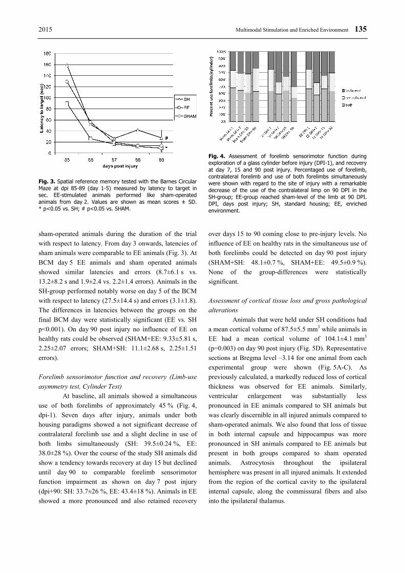
At Barnes Circular Maze (BMC) on day 1
animals in EE had found the escape chamber after
89.8±86.1 s while animals in SH animals after
158.8±92.0 s. Sham operated animals had located the
escape box after 129.3±113.7 s of search. Errors made in
EE- and SH- animals were comparable (7.4±5.2 vs.
7.6±5.3), sham-operated animals made 9.8±6.1 errors on
average. An apparent learning curve for spatial reference
memory was evident in both injured groups as well as in
sham-operated animals over the 5 day training period.
Animals housed in EE performed better than SH and
Fig. 1. Composite Neuroscore. Evaluation of neuromotor functionand recovery over a 90 day period. All injured animals showeda comparable decline in neuromotor function 24 h post injurywith faster recovery after EE. dpi, days post injury; SH, standard housing; EE, enriched environment. Values are shown as meanscores ± SD. * p<0.05 vs. SH.
Fig. 2. RotaRod test to evaluate sensorimotor coordination and recovery at baseline, 24 h post injury (dpi 1) and dpi 7, 15, 30 and 90. Injured animals show a similar level of decline (dpi 1) compared to baseline with faster recovery after EE. Values are shown as mean ± SD for time (seconds), distance traveled (meter) and speed (rounds per minute, rpm). * p<0.05 vs. SH.
2015 Multimodal Stimulation and Enriched Environment 135
sham-operated animals during the duration of the trial
with respect to latency. From day 3 onwards, latencies of
sham animals were comparable to EE animals (Fig. 3). At
BCM day 5 EE animals and sham operated animals
showed similar latencies and errors (8.7±6.1 s vs.
13.2±8.2 s and 1.9±2.4 vs. 2.2±1.4 errors). Animals in the
SH-group performed notably worse on day 5 of the BCM
with respect to latency (27.5±14.4 s) and errors (3.1±1.8).
The differences in latencies between the groups on the
final BCM day were statistically significant (EE vs. SH
p<0.001). On day 90 post injury no influence of EE on
healthy rats could be observed (SHAM+EE: 9.33±5.81 s,
2.25±2.07 errors; SHAM+SH: 11.1±2.68 s, 2.25±1.51
errors).
Forelimb sensorimotor function and recovery (Limb-use
asymmetry test, Cylinder Test)
At baseline, all animals showed a simultaneous
use of both forelimbs of approximately 45 % (Fig. 4,
dpi-1). Seven days after injury, animals under both
housing paradigms showed a not significant decrease of
contralateral forelimb use and a slight decline in use of
both limbs simultaneously (SH: 39.5±0.24 %, EE:
38.0±28 %). Over the course of the study SH animals did
show a tendency towards recovery at day 15 but declined
until day 90 to comparable forelimb sensorimotor
function impairment as shown on day 7 post injury
(dpi+90: SH: 33.7±26 %, EE: 43.4±18 %). Animals in EE
showed a more pronounced and also retained recovery
over days 15 to 90 coming close to pre-injury levels. No
influence of EE on healthy rats in the simultaneous use of
both forelimbs could be detected on day 90 post injury
(SHAM+SH: 48.1±0.7 %, SHAM+EE: 49.5±0.9 %).
None of the group-differences were statistically
significant.
Assessment of cortical tissue loss and gross pathological
alterations
Animals that were held under SH conditions had
a mean cortical volume of 87.5±5.5 mm3 while animals in
EE had a mean cortical volume of 104.1±4.1 mm3
(p=0.003) on day 90 post injury (Fig. 5D). Representative
sections at Bregma level –3.14 for one animal from each
experimental group were shown (Fig. 5A-C). As
previously calculated, a markedly reduced loss of cortical
thickness was observed for EE animals. Similarly,
ventricular enlargement was substantially less
pronounced in EE animals compared to SH animals but
was clearly discernible in all injured animals compared to
sham-operated animals. We also found that loss of tissue
in both internal capsule and hippocampus was more
pronounced in SH animals compared to EE animals but
present in both groups compared to sham operated
animals. Astrocytosis throughout the ipsilateral
hemisphere was present in all injured animals. It extended
from the region of the cortical cavity to the ipsilateral
internal capsule, along the commissural fibers and also
into the ipsilateral thalamus.
Fig. 3. Spatial reference memory tested with the Barnes CircularMaze at dpi 85-89 (day 1-5) measured by latency to target insec. EE-stimulated animals performed like sham-operated animals from day 2. Values are shown as mean scores ± SD.* p<0.05 vs. SH; # p<0.05 vs. SHAM.
Fig. 4. Assessment of forelimb sensorimotor function during exploration of a glass cylinder before injury (DPI-1), and recovery at day 7, 15 and 90 post injury. Percentaged use of forelimb, contralateral forelimb and use of both forelimbs simultaneously were shown with regard to the site of injury with a remarkable decrease of the use of the contralateral limp on 90 DPI in the SH-group; EE-group reached sham-level of the limb at 90 DPI. DPI, days post injury; SH, standard housing; EE, enrichedenvironment.

136 Maegele et al. Vol. 64
Fig. 5. Rat brain sections at Bregma –3.14 from a representative sham-operated animal (A), a SH (B) and an EE animal (C) after LFP brain injury. Notably difference between SH and EE brains in structural damage of the hippocampus, the external and internal capsule on the injured side. (D) Mean cortical volume after LFP brain injury in mm3 for animals housed in SH and EE (n=4 per group) calculated according to the Cavalieri method (Gundersen et al. 1988). Boxplots show median ± SD; whiskers show extrema. * p<0.05 vs. SH.
Discussion
Environmental enrichment has been shown to
have numerous beneficial effects on brain and behavior.
Unfortunately, current knowledge from these
investigations is restricted to short-term survival periods
(14-30 days) only leaving the potential mid- to long-term
benefits associated with EE unclear. Because there is an
ongoing loss of brain tissue and impairment of
neurological functioning as assessed by the Morris Water
Maze and Composite Neuroscore that seem to be
progressive for up to one year after LFP brain injury
(Smith and Chen 1997, Pierce and Smith 1998) it is of
great interest what continuous EE can do. The results for
up to 30 days post injury showed promising improvement
on functional outcome and tissue integrity following TBI
(Maegele et al. 2005). The present study investigating the
potential of long-term effects associated with EE after
LFP brain injury in rats underlines the findings of
Kovesdi et al. (2011) after 71 days of EE and show
a decelerated, but continuous recovery of brain function.
In general, animals after subjected to LFP brain

2015 Multimodal Stimulation and Enriched Environment 137
injury followed by exposure to EE behaved better in three
out of four standardized tests to assess both sensorimotor
or neuromotor function and spatial reference memory. SH
animals also showed a clear learning-curve but performed
worse than injured animals from EE and sham-operated
animals. The advantage for EE vs. SH-housed animals
with respect to spatial learning as described previously
for short-term survival (Hamm and Temple 1996,
Maegele et al. 2005) thus seems to be sustained over
longer periods post injury as well. The results obtained
from the cylinder test for forelimb sensorimotor function
and recovery showed also a trend in favor of EE animals
however without reaching statistical significance. Histo-
morphologically, the results from neurofunctional testing
were reflected by the preservation of cortical thickness at
the lesion site. Although none of the animals in the EE
group showed full recovery when compared to baseline
and sham-operated animals, the pattern of recovery was
similar to that with shorter survival periods as previously
reported by our group (Pierce and Smith 1998). Both
groups continued to improve their scores markedly from
days 30 to 90 post injury.
Voluminous experimental work has been
conducted to characterize new neurobiological events
after TBI (Saatman et al. 2001, Stein et al. 2002),
unknown effect(s) of different pharmacological trials
(Wahl et al. 2000, Belayev et al. 2001, Bentzer et al.
2001, LaPlaca et al. 2001, Marklund et al. 2001, Faden et
al. 2001, 2003, Alessandri et al. 2002), and various post-
traumatic treatments (Dietrich et al. 1994a, Bramlett and
Dietrich 1997, Philips et al. 2001, Knoblach and Faden
2002, Hicks and Zhang 2002, Hicks et al. 2002, Rice et
al. 2002).
To date, a series of behavioral, cellular and
molecular studies have revealed significant effects of EE
on rodents and other species, and provided new insights
into the mechanisms of experienced-dependent plasticity,
including adult neurogenesis and synaptic plasticity.
Interestingly, EE seemed to improve the outcome even if
it is applied 15 days before TBI (Johnson et al. 2013).
At the behavioral level, EE enhances learning
and memory (Moser et al. 1997, Rampon et al. 2000a,b,
Tang et al. 2001, Schrijver et al. 2002, Lee et al. 2003),
reduces memory decline (Bennett et al. 2006), decreases
anxiety, and increases exploratory activity (Chapillon et
al. 1999, Roy et al. 2001, Benaroya-Milshtein 2004,
Friske and Gammie 2005).
Similarly, several studies have investigated the
effects of EE of functional recovery after experimental
models of TBI. Meanwhile, there is a body of evidence
that that EE following TBI enhances functional outcome
and attenuates both motor and cognitive deficits
(Schwartz 1964, Will et al. 1976, Whishaw et al. 1984,
Kolb and Gibb 1991, Hamm and Temple 1996, van
Rijzingen et al. 1997, Passineau and Green 2001, Wagner
et al. 2002, Kozlowski et al. 2004), in which early, but
not immediate onset seemed to be of advantage (Matter et
al. 2011). A recent study combined EE with
a transplantation of murine embryonic stem cells 7 days
after brain injury in rats and pointed out a further
advantage compared to sole EE stimulation (Peruzzaro et
al. 2013).
Early experiments in wild-type rodents
investigating the effects of differential housing showed
that EE altered cortical weight and thickness (Bennett et
al. 1969, Diamond et al. 1972, 1976, van Praag et al.
2000). Enrichment following TBI has been shown to
have beneficial effects on the brain, such as preservation
of tissue integrity, decreasing lesion size (Passineau and
Green 2001), enhancing dendritic branching and the size
of synapses (Greenough and Volkmar 1973, Greenough
et al. 1973, 1985, Connor et al. 1982, Turner and
Greenough 1985, Kolb and Gibb 1991, Rampon et al.
2000a,b, Faherty et al. 2003, Leggio et al. 2005),
promoting the survival of progenitor cells (Gaulke et al.
2005), increasing neurotrophins (BDNF, NGF)-that both
play integral roles in neuronal signaling (Torasdotter et
al. 1998, Pham et al. 1999, Ickes et al. 2000, Chen et al.
2005), and decreasing DAT levels (Wagner et al. 2005).
Furthermore, EE has been associated with increased
neurogenesis and integration of these newly born cells
into functional circuits (Kempermann and Kuhn 1997,
Kempermann et al. 1998a,b, 2002, van Praag et al. 2000,
Bruel-Jungermann et al. 2005). In both experimental
groups, a loss of cortical tissue was observed at 90 days
post injury, but EE was comparably associated with
a substantial preservation of cortical thickness at the
lesion site. Animals in the SH paradigm did exhibit
a markedly more pronounced loss of hippocampal tissue
compared to them in EE. Possible explanations for this
may be that EE attenuated the acute and delayed tissue
damage having neuroprotective properties (Pierce and
Trojanowski 1996, Bramlett and Dietrich 1997, Passineau
and Green 2001, Hicks and Zhang 2002, Hicks et al.
2002). It enhanced the regenerative plasticity responses
of the brain to injury and thus promoted not only
functional but also histomorphological recovery (Nilsson
and Perfilieva 1999, Passineau and Green 2001). The

138 Maegele et al. Vol. 64
limitation of this study is certainly the qualitative
description of ventricular enlargement, astrocytosis and
hippocampus tissue loss without quantifying these
changes.
Conclusion
In the present study, animals after LFP brain
injury stimulated by EE performed significantly better in
three out of four standardized tests to assess sensorimotor
and neuromotor function, as well as spatial reference
memory after 90 days of recovery. Early observation time
points may overestimate the amount of sustained
recovery and it may be prudent to include longer survival
times into future studies concerning the outcome after
treatments of TBI to account for the phenomenon of
progressive worsening of neurofunctional and
histological outcome parameters over time. Although
effective treatments in the acute setting are clearly
needed, this study shows that the window of opportunity
may be wide and also lends further credibility to the
importance of long term interventions in patients
suffering from TBI. Continuous cognitive and physical
training (physiotherapy, ergotherapy, etc.) on the basis of
social integration and supporting medication may to lead
to a slow but continuous recovery which is even effective
in long-term duration.
Conflict of Interest There is no conflict of interest.
References
ALESSANDRI B, RICE AC, LEVASSEUR J, DEFORD M, HAMM RJ, BULLOCK MR: Cyclosporin A improves
brain tissue oxygen consumption and learning/memory performance after lateral fluid percussion injury in rats.
J Neurotrauma 19: 829-841, 2002.
ANGELOV D, NEISS WF, GUNKEL A, GUNTINAS-LICHIUS O, STENNERT O: Axotomy induces intranuclear
immunolocalization of neuron-specific enolase in facial and hypoglossal neurons of the rat. J Neurocytol 23:
218-233, 1994.
ANGELOV D, CEYNOVA M, GUNTINAS-LICHIUS O, STREPPEL M, GROSHEVA M, KIRYAKOVA SI,
SKOURAS E, MAEGELE M, IRINTCHEV A, NEISS W, SINIS N, ALVANOU A, DUNLOP SA:
Mechanical stimulation of paralyzed vibrissal muscles following facial nerve injury in adult rat promotes full
recovery of whisking. Neurobiol Dis 26: 229-242, 2007.
ARMSTEAD WM: Role of endothelin-1 in age-dependent cerebrovascular hypotensive responses after brain injury. Am
J Physiol 277: H1884-H1894, 1999.
ARMSTEAD WM, KURTH CD: Different cerebral hemodynamic responses following fluid percussion brain injury in
the newborn and juvenile pig. J Neurotrauma 11: 487-497, 1994.
BARNES CA: Memory deficits associated with senescence: A neurophysiological and behavioral study in the rat.
J Comp Physiol Psychol 93: 74-104, 1979.
BELYAEV L, ALONSO OF, LIU Y, CHAPPELL AS, ZHAO W, GINSBERG MD, BUSTO R: Talampanel, a novel
noncompetetive AMPA antagonist, is neuroprotective after traumatic brain injury in rats. J Neurotrauma 18:
1031-1038, 2001.
BENAROYA-MILSHTEIN N: Environmental enrichment in mice decreases anxiety, attenuates stress responses and
enhances natural killer cell activity. Eur J Neurosci 20: 1341-1347, 2004.
BENNETT EL, ROSENZWEIG MR, DIAMOND M: Rat brain: effects of environmental enrichment on wet and dry
weights. Science 163: 825-826, 1969.
BENNETT JC, MCRAE PA, LEVY LJ, FRICK KM: Long-term continuous, but not daily, environmental enrichment
reduces spatial memory decline in aged male mice. Neurobiol Learn Mem 85: 139-152, 2006.
BENTZER P, MATTIASSON G, MCINTOSH TK, WIELOCH T, GRANDE PO: Infusion of prostacyclin following
experimental brain injury in the rat reduces cortical lesion volume. J Neurotrauma 18: 275-285, 2001.
BLINZINGER K, KREUTZBERG G: Displacement of synaptic terminals from regenerating motoneurons by
microglial cells. Z Zellforsch Mikrosk Anat 85: 145-157, 1968.
BRAMLETT HM, DIETRICH WD: Chronic histopathological consequences of fluid-percussion brain injury in rats:
effects of post-traumatic hypothermia. Acta Neuropathol (Berl) 93: 190-199, 1997.
2015 Multimodal Stimulation and Enriched Environment 139
BROOK G, SCHMITT AB, NASCIMIENTO W, WEIS J, SCHRÖDER JM, NOTH J: Distribution of B-50 (GAP-43)
mRNA and protein in the normal adult human spinal cord. Acta Neuropathol 95: 378-386, 1998.
BRUEL-JUNGERMAN E, LAROCHE S, RAMPON C: New neurons in the dentate gyrus are involved in the
expression of enhanced long-term memory following environmental enrichment. Eur J Neurosci 21: 513-521,
2005.
BRUNS J, HAUSER WA: The epidemiology of traumatic brain injury: a review. Epilepsia 44 (Suppl 10): 2-10, 2003.
CHAPILLON P, MANNECHE C, BELZUNG C, CASTON J: Rearing environmental enrichment in two inbred strains
of mice: 1. Effects on emotional reactivity. Behav Genet 29: 41-46, 1999.
CHEN X, LI Y, KLINE AE, DIXON CE, ZAFONTE RD, WAGNER AK: Gender and environmental effects on
regional brain-derived neurotrophic factor expression after experimental traumatic brain injury. Neuroscience
135: 11-17, 2005.
CONNOR JR, WANG EC, DIAMOND MC: Increased length of terminal dendritic segments in old adult rats’
somatosensory cortex: an environmentally induced response. Exp Neurol 78: 466-470, 1982.
CONTI AC, RAGUPATHI R, TROJANOWSKI JQ, MCINTOSH TK: Experimental brain injury induces regionally
distinct apoptosis during the acute and delayed post-traumatic period. J Neurosci 18: 5663-5672, 1998.
CORTEZ SC, MCINTOSH TK: Experimental fluid percussion brain injury: vascular disruption and neuronal and glial
alterations. Brain Res 482: 271-282, 1989.
DEBOW SB, DAVIES ML, CLARKE HL, COLBOURNE F: Constraint-induced movement therapy and rehabilitation
exercises lessen motor deficits and volume of brain injury after striatal hemorrhagic stroke in rats. Stroke 34:
1021-1026, 2003.
DIAMOND MC, KRECH D: The effects of an enriched environment on the histology of the rat cerebral cortex. J Comp
Neurol 123: 111-120, 1964.
DIAMOND MC, ROSENZWEIG MR, BENNETT EL, LINDNER B, LYON L: Effects of environmental enrichment
and impoverishment on rat cerebral cortex. J Neurobiol 3: 47-64, 1972.
DIAMOND MC, INGHAM CA, JOHNSON RE, BENNETT EL, ROSENZWEIG MR: Effects of environment on
morphology of rat cerebral cortex and hippocampus. J Neurobiol 7: 75-85, 1976.
DIETRICH WD, ALONSO O, BUSTO R, GLOBUS MY, GINSBERG MD: Post-traumatic brain hypothermia reduces
histopathological damage following concussive brain injury in the rat. Acta Neuropathol 87: 250-258, 1994a.
DIETRICH WD, ALONSO O, HALLEY M: Early microvascular and neuronal consequences of traumatic brain injury:
a light and electron microscopic study in rats. J Neurotrauma 11: 289-301, 1994b.
DIETRICH WD, ALONSO O, HALLEY M: Delayed posttraumatic brain hyperthermia worsens outcome after fluid
percussion brain injury: a light and electron microscopic study in rats. Neurosurgery 38: 533-541, 1996.
DOBBING J: Undernutrition and the developing brain. In: Developmental Neurobiology. HIMWICH WA (ed),
Thomas, Springfield, Illinois, 1970, pp 253-286.
DUNHAM NW, MIYA TS: A note on a simple apparatus for detecting neurological deficit in rats and mice. J Am
Pharmacol Assoc 46: 208-209, 1957.
EMERY DL, FULP CT, SAATMAN KE, FISCHER I, GRADY S, MCINTOSH TK: Bilateral growth-related protein
expression suggests a transient increase in regenerative potential following brain trauma. J Comp Neurol 424:
521-531, 2000.
EMERY DL, RAGUPATHI R, SAATMAN KE, SCHUTZ C, NEUGEBAUER E, MCINTOSH TK: Newly born
granule cells in the dentate gyrus rapidly extend axons into the hippocampal CA3 region following
experimental brain injury. J Neurotrauma 22: 978-988, 2005.
FADEN AI, O´LEARY DM, FAN L, BAO W, MULLINS PG, MOVSESYAN VA: Selective blockade of the mGluR1
receptor reduces traumatic neuronal injury in vitro and improves outcome after brain trauma. Exp Neurol 167:
435-444, 2001.
FADEN AI, KNOBLACH SM, CERNAK I, FAN L, VINK R, ARALDI GL, FRICKE ST, ROTH BL, KOZIKOWSKI
AP: Novel diketopiperazine enhances motor and cognitive recovery after traumatic brain injury in rats and
shows neuroprptection in vitro and in vivo. J Cerbr Blood Flow Metab 23: 342-354, 2003.
FAHERTY CJ, KERLEY D, SMEYNE R: A Golgi-Cox morphological analysis of neuronal changes induced by
environmental enrichment. Brain Res Dev Brain Res 141: 55-61, 2003.
140 Maegele et al. Vol. 64
FALKENBERG T, MOHAMMED AK: Increased expression of brain-derived neurotrophic factor mRNA in rat
hippocampus is associated with improved spatial memory and enriched environment. Neurosci Lett 138:
153-156, 1992.
FOX GB, FAN L, LEVASSEUR RA, FADEN AI: Effect of traumatic brain injury on mouse spatial and nonspatial
learning in the Barnes circular maze. J Neurotrauma 15: 1037-1046, 1998.
FRISKE JE, GAMMIE SC: Environmental enrichment alters plus maze, but not maternal defense performance in mice.
Physiol Behav 85: 187-194, 2005.
FUKIDA K, TANNO H, OKIMURA Y, NAKAMURA M, YAMAURA A: The blood-brain barrier disruption to
circulating proteins in the early period after fluid percussion brain injury in rats. J Neurotrauma 12: 315-325,
1995.
GAULKE LJ, HORNER PJ, FINK AJ, MCNAMARA CL, HICKS RR: Environmental enrichment increases progenitor
cell survival in the dentate gyrus following lateral fluid percussion injury. Brain Res Mol Brain Res 141:
138-150, 2005.
GHAJAR J: Traumatic brain injury. Lancet 356: 923-929, 2000.
GRAHAM DI, RAGHUPATHI R, SAATMAN KE, MEANEY D, MCINTOSH TK: Tissue tears in the white matter
after lateral fluid percussion brain injury in the rat: relevance to human brain injury. Acta Neuropathol (Berl)
99: 117-124, 2000.
GREENOUGH WT, VOLKMAR FR: Pattern of dendritic branching in occipital cortex of rats reared in complex
environments. Exp Neurol 40: 491-504, 1973.
GREENOUGH WT, VOLKMAR FR, JURASKA JM: Effects of rearing complexity on dendritic branching in
frontolateral and temporal cortex of the rat. Exp Neurol 41: 371-378, 1973.
GREENOUGH WT, HWANG HM, GORMAN C: Evidence for active synapse formation or altered postsynaptic
metabolism in visual cortex of rats reared in complex environments. Proc Natl Acad Sci USA 82: 4549-4552,
1985.
GROW JL, LIU YG: Can lateralizing sensorimotor deficits be identified after neonatal cerebral hypoxia-ischemia in
rats? Dev Neurosci 25: 394-402, 2003.
GUNDERSEN HJ: Stereology of arbitrary particles. A review of unbiased number and size estimators and the
presentation of some new ones, in memory of William R. Thompson. J Microsc 143: 3-45, 1986.
GUNDERSEN HJ, JENSEN EB: The efficiency of systematic sampling in stereology and its prediction. J Microsc 147:
229-263, 1987.
GUNDERSEN HJ, BENDTSEN TF, KORBO L, MARCUSSEN N, MOLLER A, NIELSEN K, NYENGAARD JR,
PAKKENBERG B, SORENSEN FB, VESTERBY A, WEST MJ: Some new, simple and efficient
stereological methods and their use in pathological research and diagnosis. Acta Pathol Microbiol Immunol
Scand 96: 379-394, 1988.
GUNTINAS-LICHIUS O, NEISS WF: Comparison of empirical and estimated efficiency in neuron counting by the
physical dissector and in volume measurement by Cavalieri´s method. Acta Stereol 15: 31-139, 1996.
GUNTINAS-LICHIUS O, NEISS WF, GUNKEL A, STENNERT E: Differences in glial, synaptic and motoneuron
responses in the facial nucleus of the rat brainstem following facial nerve resection and nerve suture
reanastomosis. Eur Arch Otorhinolaryngol 251: 410-417, 1994.
HAMM RJ, TEMPLE MD: Exposure to environmental complexity promotes recovery of cognitive function after
traumatic brain injury. J Neurotrauma 13: 41-47, 1996.
HAMM RJ, PIKE BR, O'DELL DM, LYETH BG, JENKINS LW: The rotarod test: an evaluation of its effectiveness in
assessing motor deficits following traumatic brain injury. J Neurotrauma 11: 187-196, 1994.
HEPP DO: The effects of early experience on problemsolving at maturity. Am Psychol 2: 306-307, 1947.
HICKS R, SOARES H: Temporal and spatial characterization of neuronal injury following lateral fluid-percussion
brain injury in the rat. Acta Neuropathol (Berl) 91: 236-246, 1996.
HICKS RR, ZHANG L: Environmental enrichment attenuates cognitive deficits, but does not alter neurotrophin gene
expression in the hippocampus following lateral fluid percussion brain injury. Neuroscience 112: 631-637,
2002.

2015 Multimodal Stimulation and Enriched Environment 141
HICKS RR, KEELING KL, YANG MY, SMITH SA, SIMONS AM, KOTWAL GJ: Vaccinia virus complement
control protein enhances functional recovery after traumatic brain injury. J Neurotrauma 19: 705-714, 2002.
HILL SJ, BARBARESE E: Regional heterogeneity in the response of astrocytes following traumatic brain injury in the
adult rat. J Neuropathol Exp Neurol 55: 1221-1229, 1996.
HOFFMAN AN, MALENA RR: Environmental enrichment-mediated functional improvement after experimental
traumatic brain injury is contingent on task-specific neurobehavioral experience. Neurosci Lett 431: 226-230,
2008.
ICKES BR, PHAM TM, SANDERS LA, ALBECK DS, MOHAMMED AH, GRANHOLM AC: Long-term
environmental enrichment leads to regional increases in neurotrophin levels in rat brain. Exp Neurol 164:
45-52, 2000.
INGLIS FM, FIBIGER HC: Increases in hippocampal and frontal cortical acetylcholine release associated with
presentation of sensory stimuli. Neuroscience 66: 81-86, 1995.
JOHNSON EM, TRAVER KL, HOFFMAN SW, HARRISON CR, HERMAN JP: Environmental enrichment protects
against functional deficits caused by traumatic brain injury. Front Behav Neurosc 7: 44, 2013.
JONES BJ, ROBERTS DJ: A rotarod suitable for quantitative measurements of motor incoordination in naive mice.
Naunyn Schmiedebergs Arch Exp Pathol Pharmakol 259: 211, 1968.
KEMPERMANN G, KUHN HG: More hippocampal neurons in adult mice living in an enriched environment. Nature
386: 493-495, 1997.
KEMPERMANN G, KUHN HG, GAGE FH: Experience-induced neurogenesis in the senescent dentate gyrus.
J Neurosci 18: 3206-3212, 1998a.
KEMPERMANN G, BRANDON EP, GAGE FH: Environmental stimulation of 129/SvJ mice causes increased cell
proliferation and neurogenesis in the adult dentate gyrus. Curr Biol 8: 939-942, 1998b.
KEMPERMANN G, GAST D, GAGE FH: Neuroplasticity in old age: sustained fivefold induction of hippocampal
neurogenesis by long-term environmental enrichment. Ann Neurol 52: 135-143, 2002.
KNOBLACH SM, FADEN AI: Administration of either anti-intercellular adhesion molecule-1 or a nonspecific control
antibody improves recovery after traumatic brain injury in the rat. J Neurotrauma 19: 1039-1050, 2002.
KOLB B, GIBB R: Environmental enrichment and cortical injury: behavioral and anatomical consequences of frontal
cortex lesions. Cereb Cortex 1: 189-198, 1991.
KOVESDI E, GYORGY AB, KWON SK, WINGO DL, KAMNAKSH A, LONG JB, KASPER CE, AGOSTON DV:
The effect of enriched environment on the outcome of traumatic brain injury; a behavioral, proteomics, and
histological study. Front Neurosci 5: 42, 2011.
KOZLOWSKI DA, NAHED BV, HOVDA DA, LEE SM: Paradoxical effects of cortical impact injury on
environmentally enriched rats. J Neurotrauma 21: 513-519, 2004.
LANGLOIS JA, RUTLAND-BROWN W: The epidemiology and impact of traumatic brain injury: a brief overview.
J Head Trauma Rehabil 21: 375-378, 2006.
LAPLACA MC, ZHANG J, RAGHUPATHI R, LI JH, SMITH F, BAREYRE FM, SNYDER SH, GRAHAM DI,
MCINTOSH TK: Pharmacologic inhibition of poly (ADP-ribose) polymerase is neuroprotective following
traumatic brain injury in rats. J Neurotrauma 18: 369-376, 2001.
LAURER HL, LENZLINGER PM: Models of traumatic brain injury. Eur J Trauma 26: 95-100, 2000.
LEE EH, HSU WL, MA YL, LEE PJ, CHAO CC: Enrichment enhances the expression of sgk, a glucocorticoid-induced
gene, and facilitates spatial learning through glutamate AMPA receptor mediation. Eur J Neurosci 18: 2842-
2852, 2003.
LEGGIO MG, MANDOLESI L, FEDERICO F, SPIRITO F, RICCI B, GELFO F, PETROSINI L: Environmental
enrichment promotes improved spatial abilities and enhanced dendritic growth in the rat. Behav Brain Res 163:
78-90, 2005. LIPPERT-GRUENER M, TERHAAG D: Multimodal early onset stimulation (MEOS) in rehabilitation after brain
injury. Brain Inj 14: 585-594, 2000.
LIPPERT-GRUENER M, MAEGELE M: Late effects of enriched environment (EE) plus multimodal early onset
stimulation (MEOS) after traumatic brain injury in rats: Ongoing improvement of neuromotor function despite
sustained volume of the CNS lesion. Exp Neurol 203: 82-94, 2005.

142 Maegele et al. Vol. 64
LIPPERT-GRUENER M, WEDEKIND C, ERNESTUS RI, KLUG N: Early rehabilitative concepts in therapy of the
comatose brain injured patients. Acta Neurochir Suppl 79: 21-23, 2002.
LIPPERT-GRUENER M, WEDEKIND C, KLUG N: Outcome of prolonged coma following severe traumatic brain
injury. Brain Inj 17: 49-54, 2003.
LIPPERT-GRUENER M, MAEGELE M, POKORNY J, ANGELOV D, SVESTKOVA O, WITTNER M, TROJAN S:
Early rehabilitation model shows positive effects on neural degeneration and recovery from neuromotor
deficits following traumatic brain injury. Physiol Res 56: 359-368, 2006a.
LIPPERT-GRUENER M, MAEGELE M, ANGELOV D: Einfluss eines frühen sensomotorischen Trainings auf die
Erholung neuromotorischer Defizite nach experimentellem Schädel-Hirn-Trauma. Phys Med Rehab Kuror 16:
1-6, 2006b.
LONGHI L, SAATMAN KE, RAGHUPATHI R, LAURER HL, LENZLINGER PM, RIESS P, NEUGEBAUER E,
TROJANOWSKI JQ, LEE VM, GRADY MS, GRAHAM DI, MCINTOSH TK: A review and rationale for the
use of genetically engineered animals in the study of traumatic brain injury. J Cereb Blood Flow Metab 21:
1241-1258, 2001.
LOZADA A, MAEGELE M, STARK H, NEUGEBAUER EM, PANULA P: Traumatic brain injury results in mast cell
increase and changes in regulation of central histamine receptors. Neuropathol Appl Neurobiol 31: 150-162,
2005.
MAEGELE M, LIPPERT-GRUENER M: Multimodal early onset stimulation combined with enriched environment is
associated with reduced CNS lesion volume and enhanced reversal of neuromotor dysfunction after traumatic
brain injury in rats. Eur J Neurosci 21: 2406-2418, 2005a.
MAEGELE M, LIPPERT-GRUENER M: Reversal of neuromotor and cognitive dysfunction in an enriched
environment combined with multimodal early onset stimulation after traumatic brain injury in rats.
J Neurotrauma 22: 772-782, 2005b.
MAEGELE M, ESTER-BODE T, RIESS P, ANGELOV DN, MCINTOSH TK, NEUGEBAUER E, LIPPERT-
GRUENER M: Exposure to complex enriched environment combined with multi-modal stimulation promotes
recovery of cognitive function after traumatic brain injury in rats. Lang Arch Sur 387: 266, 2002.
MAEGELE M, RIESS P, SAUERLAND S, BOUILLON B, HESS S, MCINTOSH TK, MAUTES A, BROCKMANN
M, KOEBKE J, KNIFKA J, NEUGEBAUER EA: Characterization of a new rat model of experimental
combined neurotrauma. Shock 23: 476-481, 2005.
MAEGELE M, ENGEL D, BOUILLON B, LEFERING R, FACH H, RAUM M, BUCHHEISTER B, KLUG N,
NEUGEBAUER E: The incidence and outcome of traumatic brain injury in an urban area in western Europe
over ten years. Eur Surg Res 39: 372-379, 2007a.
MAEGELE M, SAUERLAND S, BOUILLON B, SCHAEFER U, NEUGEBAUER E: Immunmodulation in a new
„two-hit“-model of experimental combined neurotrauma. Inflamm Res 56: 318-323, 2007b.
MARANGOS PJ, SCHMECHEL DE: Neuron specific enolase, a clinically useful marker for neurons and
neuroendocrine cells. Annu Rev Neurosci 10: 269-295, 1987.
MARKLUND N, CLAUSEN F, MCINTOSH TK, HILLERED L: Free redical scavenger posttreatment improves
functional and morphological outcome after fluid percussion injury in the rat. J Neurotrauma 18: 821-832,
2001.
MATTER AM, FOLWEILER KA, CURATOLO LM, KLINE AE: Temporal effects of environmental enrichment-
mediated functional improvement after experimental traumatic brain injury in rats. Neurorehabil Neural
Repair 25: 558-564, 2011.
MAYHEW TM: The new stereological methods for interpretating functional morphology from slices of cells and
organs. Exp Physiol 76: 639-665, 1991.
MCINTOSH TK, VINK R: Traumatic brain injury in the rat: characterization of a lateral fluid-percussion model.
Neuroscience 28: 233-244, 1989.
MOLCANYI M, RIESS P, BENTZ K, MAEGELE M, HESCHELER J, SCHAFKE B, TRAPP T, NEUGEBAUER E,
KLUG N, SCHAEFER U: Trauma-associated inflammatory response impairs embryonic stem cell survival and
integration after implantation into injured brain. J Neurotrauma 24: 625-637, 2007.

2015 Multimodal Stimulation and Enriched Environment 143
MORALES DM, MARKLUND N, LEBOLD D, THOMPSON HJ, PITKANEN A, MAXWELL WL, LONGHI L,
LAURER H, MAEGELE M, NEUGEBAUER E, GRAHAM DI, STOCCHETTI N, MCINTOSH TK:
Experimental models of traumatic brain injury: do we really need to build a better mousetrap? Neuroscience
136: 971-989, 2005.
MOSER MB, TROMMALD M, EGELAND T, ANDERSEN P: Spatial training in a complex environment and
isolation alter the spine distribution differently in rat CA1 pyramidal cells. J Comp Neurol 380: 373-381, 1997.
MURRAY CJL, LOPEZ AD: Global mortality, disability and the contribution of risk factors: Global Burden of Disease
Study. Lancet 349: 1436-1442, 1997.
MYSLIVECEK J, HASSMANNOVA J: Some neurophysiological mechanisms of early inhibitory learning and
memory. In: Ontogenesis of the Brain. TROJAN S, ŠŤASTNÝ F (eds), Charles University, Prague, 1987,
pp 117-122.
NAVONE F, JAHN R, DI GIOIA G, STUKENBROK H, GREENGARD P, DE CAMILLI P: Protein p38: an integral
membrane protein specific for small vesicles of neurons and neuroendocrine cells. J Cell Biol 103: 2511-2527,
1986.
NEISS WF, GUNTINAS-LICHIUS O, ANGELOV DN, GUNKEL A, STENNERT E: The hypoglossal-facial
anastomosis as model of neuronal plasticity in the rat. Ann Anat 174: 419-433, 1992.
NILSSON M, PERFILIEVA E: Enriched environment increases neurogenesis in the adult rat dentate gyrus and
improves spatial memory. J Neurobiol 39: 569-578, 1999.
NITHIANANTHARAJAH J, HANNAN AJ: Enriched environments, experience-dependent plasticity and disorders of
the nervous system. Nature Rev Neurosci 7: 697-709, 2006.
OKIYAMA K, SMITH DH, THOMAS MJ, MCINTOSH TK: Evaluation of a novel calcium channel blocker,
(S)-emopamil, on regional cerebral edema and neurobehavioral function after experimental brain injury.
J Neurosurg 77: 607-615, 1992.
PASSINEAU MJ, GREEN EJ: Therapeutic effects of environmental enrichment on cognitive function and tissue
integrity following severe traumatic brain injury in rats. Exp Neurol 168: 373-384, 2001.
PERUZZARO ST, GALLAGHER J, DUNKERSON J, FLUHARTY S, MUDD D, HOANE MR, SMITH JS: The
impact of enriched environment and transplantation of murine cortical embryonic stem cells on recovery from
controlled cortical contusion injury. Restor Neurol Neurosci 31: 431-450, 2013.
PHAM TM, ICKES B, ALBECK D, SÖDERSTRÖM S, GRANHOLM AC, MOHAMMED AH: Changes in brain
nerve growth factor levels and nerve growth factor receptors in rats exposed to environmental enrichment for
one year. Neuroscience 94: 279-286, 1999.
PHILIPS MF, MATTIASSON G, WIELOCH T, BJORKLUND A, JOHANSSON BB, TOMASEVIC G, MARTINEZ-
SERRANO A, LENZLINGER PM, SINSON G, GRADY MS, MCINTOSH TK: Neuroprotective and
behavioral effects of nerve growth factor-transfected hippocampal progenitor cell transplants after
experimental traumatic brain injury. J Neurosurg 94: 765-774, 2001.
PIERCE JE, TROJANOWSKI JQ: Immunohistochemical characterization of alterations in the distribution of amyloid
precursor proteins and beta-amyloid peptide after experimental brain injury in the rat. J Neurosci 16: 1083-
1090, 1996.
PIERCE JE, SMITH DH: Enduring cognitive, neurobehavioral and histopathological changes persist for up to one year
following severe experimental brain injury in rats. Neuroscience 87: 359-369, 1998.
POVLISHOCK JT, ERB DE: Axonal response to traumatic brain injury: reactive axonal change, deafferentation, and
neuroplasticity. J Neurotrauma 9 (Suppl 1): S189-S200, 1992.
RAMPON C, TANG YP, GOODHOUSE J, SHIMIZU E, KYIN M, TSIEN JZ: Enrichment induces structural changes
and recovery from nonspatial memory deficits in CA1 NMDAR1-knockout mice. Nat Neurosci 3: 238-244,
2000a.
RAMPON C, JIANG CH, DONG H, TANG YP, LOCKHART DJ, SCHULTZ PG, TSIEN JZ, HU Y: Effects of
environmental enrichment on gene expression in the brain. Proc Natl Acad Sci USA 97: 12880-12884, 2000b.
RICE AC, ZSOLDOS R, CHEN T, WILSON MS, ALESSANDRI B, HAMM RJ, BULLOCK MR: Lactate
administration attenuates cognitive deficits following traumatic brain injury. Brain Res 928: 56-59, 2002.

144 Maegele et al. Vol. 64
RIESS P, BAREYRE FM, SAATMAN KE, CHENEY JA, LIFSHITZ J, RAGHUPATHI R, GRADY MS,
NEUGEBAUER E, MCINTOSH TK: Effects of chronic, post-injury Cyclosporin A administration on motor
and sensorimotor function following severe, experimental traumatic brain injury. Restor Neurol Neurosci 18:
1-8, 2001.
RIESS P, ZHANG C, SAATMAN KE, LAURER HL, LONGHI LG, RAGHUPATHI R, LENZLINGER PM,
LIFSHITZ J, BOOCKVAR J, NEUGEBAUER E, SNYDER EY, MCINTOSH TK: Transplanted neural stem
cells survive, differentiate, and improve neurological motor function after experimental traumatic brain injury.
Neurosurgery 51: 1043-1052, 2002.
RIESS P, MOLCIANY M, BENTZ C, MAEGELE M, SIMANSKI C, CARLITESCHECK C, BOUILLON B,
HESCHELER J, SCHNEIDER A, SCHÄFER U, NEUGEBAUER E: Embryonic stem cell transplantation
after experimental traumatic brain injury dramatically improves neurological outcome, but may cause tumors.
J Neurotrauma 24: 216-225, 2007.
RINK A, FUNG K-M, TROJANOWSKI JQ, LEE V, NEUGEBAUER E, MCINTOSH T: Evidence of apoptotic cell
death after experimental traumatic brain injury in the rat. Am J Pathol 147: 1575-1583, 1995.
ROSENZWEIG MR: Environmental complexity, cerebral change, and behavior. Am Psychol 21: 321-332, 1966.
ROY V, BELZUNG C, DELARUE C, CHAPILLON P: Environmental enrichment in BALB/c mice: effects in classical
tests of anxiety and exposure to a predatory odor. Physiol Behav 74: 313-320, 2001.
SAATMAN KE, BAREYRE FM, GRANDY MS, MCINTOSH TK: Acute cytoskeletal alterations and cell death
induced by experimental brain injury are attenuated by magnesium treatment and exacerbated by magnesium
deficiency. J Neuropathol Exp Neurol 60: 183-194, 2001.
SCHALLERT T, FLEMING SM: CNS plasticity and assessment of forelimb sensorimotor outcome in unilateral rat
models of stroke, cortical ablation, parkinsonism and spinal cord injury. Neuropharmacology 39: 777-787,
2000.
SCHERBEL U, RAGHUPATHI R, NAKAMURA M, SAATMAN KE, TROJANOWSKI JQ, NEUGEBAUER E,
MARINO M, MCINTOSH T: Differential acute and chronic responses of tumor necrosis factor-deficient mice
to experimental brain injury. Proc Natl Acad Sci USA 96: 8721-8726, 1999.
SCHMIDT RH, GRADY MS: Regional patterns of blood-brain barrier breakdown following central and lateral fluid
percussion injury in rodents. J Neurotrauma 10: 415-429, 1993.
SCHRIJVER NC, BAHR NI, WEISS IC, WURBEL H: Dissociable effects of isolation rearing and environmental
enrichment on exploration, spatial learning and HPA activity in adult rats. Pharmacol Biochem Behav 73:
209-224, 2002.
SCHUTZ C, STOVER JF, THOMPSON HJ, HOOVER RC, MORALES DM, SCHOUTEN JW, MCMILLAN A,
SOLTESZ K, MOTTA M, SPANGLER Z, NEUGEBAUER E, MCINTOSH TK: Acute, transient hemorrhagic
hypotension does not aggravate structural damage or neurologic motor deficits but delays the long-term
cognitive recovery following mild to moderate traumatic brain injury. Crit Care Med 34: 492-501, 2006.
SCHWARTZ S: Effect of neonatal cortical lesions and early environmental factors on adult rat behavior. J Comp
Physiol Psychol 57: 72-77, 1964.
SINSON G, VODDI M, MCINTOSH TK: Nerve growth factor administration attenuates cognitive but not
neurobehavioral motor dysfunction or hippocampal cell loss following fluid-percussion brain injury in rats.
J Neurochem 65: 2209-2216, 1995.
SMITH DH, CHEN XH: Progressive atrophy and neuron death for one year following brain trauma in the rat.
J Neurotrauma 14: 715-727, 1997.
SOARES HD, HICKS RR, SMITH D, MCINTOSH TK: Development of prolonged focal cerebral edema and regional
cation changes following experimental brain injury in the rat. J Neurochem 58: 1845-1852, 1992.
SOARES HD, HICKS RR, SMITH D, MCINTOSH TK: Inflammatory leukocytic recruitment and diffuse neuronal
degeneration are separate pathological processes resulting from traumatic brain injury. J Neurosci 15: 8223-
8233, 1995.
SOSIN DM, SNIEZEK JE, THURMAN DJ: Incidence of mild and moderate brain injury in the United States. Brain Inj
10: 47-54, 1991.
2015 Multimodal Stimulation and Enriched Environment 145
SOZDA CN, HOFFMAN AN, OLSEN AS, CHENG JP, ZAFONTE RD, KLINE AE: Empirical comparison of typical
and atypical environmental enrichment paradigms on functional and histological outcome after experimental
traumatic brain injury. J Neurotrauma 27: 1047-1057, 2010.
STEIN SC, CHEN XH, SINSON GP, SMITH DH: Intravascular coagulation: a major secondary insult in nonfatal
traumatic brain injury. J Neurosurg 97: 1373-1377, 2002.
SULLIVAN HG, MARTINEZ J, BECKER DP, MILLER JD, GRIFFITH R, WIST A: Fluid-percussion model of
mechanical brain injury in the cat. J Neurosurg 45: 521-534, 1976.
TANG YP, WANG H, FENG R, KYIN M, TSIEN JZ: Differential effects of enrichment on learning and memory
function in NR2B transgenic mice. Neuropharmacology 41: 779-790, 2001.
TORASDOTTER M, METSIS M, HENRIKSSON BG, WINBLAD B, MOHAMMED AH: Environmental enrichment
results in higher levels of nerve growth factor mRNA in the rat visual cortex and hippocampus. Behav Brain
Res 93: 83-90, 1998.
TURNER AM, GREENOUGH WT: Differential rearing effects on rat visual cortex synapses. I. Synaptic and neuronal
density and synapses per neuron. Brain Res 329: 195-203, 1985.
VAN PRAAG H, KEMPERMANN G, GAGE FH: Neural consequences of environmental enrichment. Nature Rev
Neurosci 1: 191-198, 2000.
VAN RIJZINGEN IM, GISPEN WH, SPRUIJT BM: Postoperative environmental enrichment attenuates fimbria-fornix
lesion-induced impairments in Morris maze performance. Neurobiol Learn Mem 67: 21-28, 1997.
WAGNER AK, KLINE AE, SOKOLOSKI J, ZAFONTE RD, CAPULONG E, DIXON CE: Intervention with
environmental enrichment after experimental brain trauma enhances cognitive recovery in male but not female
rats. Neurosci Lett 334: 165-168, 2002.
WAGNER AK, CHEN X, KLINE AE, LI Y, ZAFONTE RD, DIXON CE: Gender and environmental enrichment
impact dopamine transporter expression after experimental traumatic brain injury. Exp Neurol 195: 475-483,
2005.
WAHL F, GROSJEAN-PIOT O, BAREYRE F, UZAN A, STUTZMANN JM: Enoxaparin reduces brain edema,
cerebral lesions, and improves motor and cognitive impairments induced by a traumatic brain injury in rats.
J Neurotrauma 17: 1055-1065, 2000.
WAXWEILER RJ, THURMAN D: Monitoring the impact of traumatic brain injury: a review and update.
J Neurotrauma 12: 509-516, 1995.
WEST MJ, GUNDERSEN HJ: Unbiased stereological estimation of the number of neurons in the human hippocampus.
J Comp Neurol 296: 1-22, 1990.
WHISHAW IQ, ZABOROWSKI JA, KOLB B: Postsurgical enrichment aids adult hemidecorticate rats on a spatial
navigation task. Behav Neural Biol 42: 183-190, 1984.
WIEDENMANN B, FRANKE WW: Identification and localization of synaptophysin, an integral membrane
glycoprotein of Mr 38,000 characteristic of presynaptic vesicles. Cell 41: 1017-1028, 1985.
WILL BE, ROSENZWEIG MR, BENNETT EL: Effects of differential environments on recovery from neonatal brain
lesions, measured by problem-solving scores and brain dimensions. Physiol Behav 16: 603-611, 1976.