lmmunochemical identification and subcellular distribution ... · lmmunochemical identification and...
TRANSCRIPT
The Journal of Neuroscience, October 1995, 75(10): 6403-6416
lmmunochemical Identification and Subcellular Distribution of the’ <x,~ Subunits of Brain Calcium Channels
Ruth E. Westenbroek,’ Takashi Sakurai,’ Elicia M. Elliott,’ Johannes W. Hell,’ Terry V. B. Starr,2 Terry P. Snutch: and William A. Catterall’
1 Department of Pharmacology, University of Washington, Seattle, Washington 98195 and 2Biotechnology Laboratory and Departments of Zoology and Neuroscience, University of British Columbia, Vancouver, British Columbia, Canada V6T 123
A site-directed anti-peptide antibody (anti-CNAl) directed against the (Y, subunit of class A calcium channels ((Y,~) recognized a protein of approximately 190-200 kDa in im- munoblot and immunoprecipitation analyses of rat brain glycoproteins. Calcium channels recognized by anti-CNAl were distributed throughout the brain with a high concen- tration in the cerebellum. Calcium channels having (Y,,, sub- units were concentrated in presynaptic terminals making synapses on cell bodies and on dendritic shafts and spines of many classes of neurons and were especially prominent in the synapses of the parallel fibers of cerebellar granule cells on Purkinje neurons where their localization in pre- synaptic terminals was confirmed by double labeling with the synaptic membrane protein syntaxin or the microin- jetted postsynaptic marker Neurobiotin. They were present in lower density in the surface membrane of dendrites of most major classes of neurons. There was substantial la- beling of Purkinje cell bodies, but less intense staining of the cell bodies of hippocampal pyramidal neurons, layer V pyramidal neurons in the dorsal cortex, and most other classes of neurons in the forebrain and cerebellum. Scat- tered cell bodies elsewhere in the brain were labeled at low levels. These results define a unique pattern of localization of class A calcium channels in the cell bodies, dendrites, and presynaptic terminals of most central neurons. Com- pared to class B N-type calcium channels, class A calcium channels are concentrated in a larger number of presyn- aptic nerve terminals implying a more prominent role in neurotransmitter release at many central synapses.
[Key words: ion channel, calcium channel, synaptic ter- minals, dendrites, immunocytochemistry, purkinje cell]
Received Feb. 17, 1995; revised May 19, 1995; accepted May 22, 1995. Research at the Univesity of Washington was supported by National Insti-
tutes of Health Research Grant NS22625 to W.A.C. and by Miles Laboratories, Pfizer, and the W. M. Keck Foundation. E. M. Elliot was supported by a NINDS postdoctoral fellowship, T. Sakurai by a postdoctoral fellowship from the Uehara Foundation, and J.W.H. by a postdoctoral fellowship from the Deutsche Forschungsgemeinschaft. Peptides were synthesized in the Molecular Phar- macology Facility, University of Washington. We thank Dr. Masami Takahashi, Mitsubishi-Kasei Life Sciences Institute, Tokyo for the lOH5 anti-syntaxin an- tibody, Sue B. Bausch for providing the acute brain slices, and Dr. Conception Warner, Ms. Cynthia Warner, and Ms. Elizabeth Catterall for assistance with antibody production and characterization and with immunocytochemistry. Re- search at the University of British Columbia was supported by research grants from the Medical Research Council of Canada and the Howard Hughes Medical Institute International Research Scholars Program to T. I? Snutch.
Correspondence should be addressed to William A. Catterall at the above address. Copyright 0 1995 Society for Neuroscience 0270.6474/95/156403-16$05.00/O
Many neuronal processes are regulated by calcium influx
through voltage-gated calcium channels, including protein phos- phorylation, gene expression, neurotransmitter release, and ac- tion potential firing patterns (Miller, 1987; Llinas, 1988). At least five distinct types of voltage-sensitive calcium channels, desig- nated L, N, P, Q, and T, have been identified based on their pharmacological and physiological properties (Bean, 1989; Lli- nas et al., 1989; Hess, 1990; Zhang et al., 1993; Randall and Tsien, 1995). In rat brain, multiple isoforms of the principal (Y, subunit of voltage-gated calcium channels have been identified using molecular cloning techniques and are designated class A through E (Snutch et al., 1990; Snutch and Reiner, 1992; Soong et al., 1993; Zhang et al, 1993). The rat brain class C and D genes encode L-type calcium channel ‘Y, subunits (0~~~ and CX,~) which have high affinity for dihydropyridine calcium channel antagonists and are approximately 75% identical in amina acid sequence with rabbit skeletal muscle calcium channels (Hui et al., 1991; Chin et al., 1992; Snutch et al., 1990; Dubel et al., 1992; Seino et al, 1992; Williams et al., 1992a; Tomlinson et al., 1993). In contrast, the neuronal class A, B, and E genes encode ct, subunits ((Y,,, (Y,~, CL& of non-L-type calcium chan- nels which are only 23% to 35% identical to skeletal muscle calcium channels in amino acid sequence (Mori et al., 1991; Starr et al., 1991; Dubel et al., 1992; Williams et al., 1992b; Niidome et al., 1992; Fujita et al., 1993; Soong et al., 1993). cx,, forms an N-type, high-voltage-activated calcium channel having high affinity for w-conotoxin GVIA (Dubel et al., 1992; Williams et al., 1992a; Fujita et al., 1993; Stea et al., 1993). (Y,~ forms a novel, rapidly inactivating calcium channel which is not blocked with high affinity by any of the known calcium channel blockers and has some characteristics of a low-voltage-activated calcium channel (Soong et al., 1993). alA forms a high-voltage- activated calcium channel with novel physiological and phar- macological properties which is highly expressed in the cere- bellum (Starr et al., 1991; Mori et al., 1991; Sather et al., 1993). Voltage-clamp recordings in Xenopus oocytes expressing (Y,* re- veal calcium currents that activate in the same range of mem- brane potentials as (yIB or c+ expressed in oocytes (Sather et al., 1993; Stea et al., 1994). The aIA channels are insensitive to dihydropyridines and w-conotoxin GVIA, weakly blocked by o-agatoxin IVA, and completely blocked by w-conotoxin MVIIC. The functibnal properties of OL,~ are distinct from the physiologically defined N-type and P-type calcium channels, but resemble those of calcium channels which have been designated Q-type (Zhang et al., 1993; Randall and Tsien, 1995) and P-type (Stea et al., 1994).
6404 Westenbroek et al. - Localization of Class A Calcium Channels
The non-L-type calcium channels encoded by the class A, l3, and E genes have an overlapping spectrum of physiological and pharmacological properties which makes analysis of their phys- iological roles in neuronal function difficult. Subcellular local- ization is a critical determinant of the physiological role of neu- ronal calcium channels, in part determining whether they can participate in regulation of gene expression, action potential gen- eration, neurotransmitter release, or other localized cellular pro- cesses. L-Type calcium channels having CY,~ and oID subunits are localized primarily in cell bodies and proximal dendrites of adult central neurons (Ahlijanian et al., 1990; Westenbroek et al., 1990; Hell et al., 1993b) where they may participate in reg- ulation of protein phosphorylation, enzyme activity, and gene expression. In contrast, N-type calcium channels having olB sub- units are localized predominantly in dendrites and in presynaptic nerve terminals making synapses on dendrites, consistent with roles in mediating dendritic calcium transients and in initiating neurotransmitter release (Westenbroek et al., 1992). In the ex- periments described here, we have used anti-peptide antibodies directed against a unique sequence in rat brain (Y,* to identify the corresponding polypeptides and determine their subcellular distribution in central neurons.
Materials and Methods
Materials. Two month old Sprague-Dawley rats were obtained Bantin and Kingman (Bellevue, WA). Other materials were purchased from following sources: the ECL detection kit for immunoblotting and strep- tavidin-biotinylated horseradish peroxidase complex from Amersham; Centricon- from Amicon (Beverly, MA); nitrocellulose membranes from Schleicher and Schuell; sulfosuccinimidyl-6-(biotinamido) hex- anoate (NHS-LC-Biotin) from Pierce; digitonin from Gallard-Schle- singer Industries; Nonidet P-40 (NP-40), Tween-20, protein A-Sephar- ose (PAS), WGA, CNBr-activated Sepharose 4B, N-acetyl-D-glucosa- mine, leupeptin, aprotinin, phenylmethanesulfonyl fluoride, benzamidi- ne, and pepstatin- A from- Sigma; calpain inhibitors I and II from Calbiochem: and Neurobiotin from Vector, Inc. WGA was couuled to the CNBr-activated Sepharose according to the manufacturer’s i&ruc- tions. The lOH5 monoclonal antibody against syntaxin was a gift of Dr. Masami Takahashi, Mitsubishi-Kasei Life Sciences Institute. Tokvo (Yoshida et al., 1992).
.
Partial purification of rat brain calcium channels. To prevent pro- teolysis during the procedure, all steps were carried out at 4°C or on ice, and all buffers contained the following protease inhibitors: pepstatin A (1 pg/ml), leupeptin (1 kg/ml), aprotinin (1 kg/ml), phenylmethane- sulfonvl fluoride (0.2 mu). benzamidine (0.1 me/ml), and calnain in- hibitors I and II (8 pg/ml’each). Neuronal Cal&m channels wkre sol- ubilized from rat brain membranes with digitonin and enriched by chro- matography on WGA-Sepharose as described previously (Westenbroek et al., 1992). Briefly, for one preparation, 15 rat brains were homoge- nized in 180 ml of 320 mu sucrose with a glass-Teflon homogenizer. After a short centrifugation (5000 rpm, 2 min, SS34 rotor), the super- natant (Sl) was centrifuged (42,000 rpm, 60 min, Ti 45 rotor). The membranes were solubilized with 400 ml of 1.2% digitonin, 80 mu sodium phosphate buffer, pH 7.4 for 20 min. Unsolubilized material was removed by the centrifugation as before, and the supernatant (S3) was poured over a 40 ml WGA-Sepharose column (50 ml/h@. After incubation for 1 hr at 4”C, the column was washed with 400 ml of 0.1% digitonin, 75 mu NaCl, 50 mu sodium phosphate, 10 mu Tris-HCl (pH 7.4) at a flow rate of 50 ml/hr. The glycoproteins bound to WGA- Sepharose column were eluted with 100 mu N-acetyl-D-glucosamine in the same buffer at a flow rate of 50 mlihr. Three milliliter fractions were collected and the protein concentration of each fraction was estimated from absorbance at 280 nm. Up to five fractions close to the peak were collected, frozen in liquid nitrogen, and stored at -80°C.
Biotinylation, imm&oprecipitation, and streptavidin-biotin detec- tion. WGA fractions. diluted 1:lOOO in 100 mu sodium borate (OH 8.5). 0.1% digitonin were’ concentrated to a volume of -500 pl in i Centri: con-30 microconcentrator to remove the amine in Tris-HCl buffer. One umole of NHS-LC-Biotin was added to biotinylate the partially purified membrane fractions. After 2 hr incubation on ice, the reaction was ter-
minated by addition of one-fifth volume of 2 M glycine (pH 8.5). Sam- ples were diluted in TBS (20 mu Tris-HCl, pH 7.4, 0.15 M NaCl) containing 0.1% digitonin and concentrated to a volume of approxi- mately 300 pl by ultrafiltration.
Biotinylated samples were preabsorbed for 1 hr on ice with 300 ~1 of Sepharose CL4B and for 2 hr on ice with 10 mg of PAS, which was preincubated with 200 p,g of control rabbit IgG and washed three times with TBS, 0.1% digitonin, in order to remove the nonspecifically binding proteins in the sample. After centrifugation, supernatants were incubated for another 2 hr on ice with 10 mg of PAS to absorb the free IgG dissociated from PAS-control IgG complex. After centrifugation for 1 min on a table top centrifuge, the supernatants were collected and incubated with either anti-CNAl antibody (80 kg) or control antibody (80 pg) for 1.5 hr on ice. Three mg of PAS, preswollen and washed with TBS, 0.1% digitonin, 0.5% BSA was added and samples were mixed for further 2.5 hr on ice. The immune complexes were pelleted by centrifugation and washed three times with TBS, 0.3% digitonin, 0.1% BSA and once with 10 mu Tris-HCl (pH 7.4). The pellets were extracted for 30 min at 50-60°C with 20 ~1 of 1.5% SDS, 50 mu Tris- HCl (pH 7.4) 5 mu dithiothreitol, 1 PM pepstatin A, 2 p,g/ml of leu- peptin, and 4 kg/ml of aprotinin, and diluted with 250 p,l of Triton buffer (1% Triton X-100, 0.5% BSA, 75 mu NaCl, 25 mu Tris-HCl (pH 7.4), 20 mu EDTA). The supernatant was collected and incubated for 1.5 hr on ice with either anti-CNAl (80 LLB) or anti-CP(1382-1400) (20 pg). Three milligrams of PAS, pretreated & described above, were added and the samples were incubated on a tilting mixer for 2.5 hr on ice. The PAS-secondary antibody-o, complexes were pelleted by cen- trifugation, washed three times with Triton buffer and once in 10 mu Tris-HCl ( pH 7.4), and extracted for 30 min at 50-60°C with SDS sample buffer (20 p,l of 200 mM Tris-HCl ( pH 7.4), 10 mu dithiothrei- tol, 4 M urea, 8% SDS, 10% glycerol). After a short centrifugation, the supernatants were loaded onto 6% SDS-PAGE.
After separation by SDS-PAGE, proteins were transferred onto a ni- trocellulose membrane (0.2 pm) in a buffer containing 12.5 mM Tris (pH 8.3), 96 mu glycine, 0.1% SDS, 15% (v/v) methanol. Unbound sites on the nitrocellulose were blocked for 2 hr at room temperature with TBS containing 10% skim milk powder. After a short rinse with TBS. 5% BSA. 0.2% NP-40. and 0.05% Tween-20. nitrocellulose sheets were incubated for 1 hr at room temperature with streptavidin-bioti- nylated horseradish peroxidase complex, diluted 1:8000 in TBS con- taining 0.2% NP-40 and 0.05% Tween-20. After a 3 hr wash with 0.2% NP-40. 0.05% Tween-20 in TBS (eight or nine chances). the blots were developed with the ECL reagent.‘ v
” II
Irnmunoblotting of calcium channels. To concentrate the calcium channels, WGA-column fractions containing 1 mg of total protein were incubated for 4 hr on ice with 200 pl of heparin-agarose (Sakamoto and Campbell, 1991). The resin was washed three times with 0.2% CHAPS, 10 mu Tris-HCl, pH 7.4, and once with 10 mu Tris-HCl, pH 7.4. Calcium channels were extracted for 30 min at 50-6o”C with 60 pl of 5% SDS, 20 mu dithiothreitol, 125 mu Tris-HCl, pH 6.8, 10% sucrose, 20 mu EDTA. After separation by SDS-PAGE, the proteins were blotted, blocked as described above, and incubated with affinity- purified anti-CNAl antibodies in TBS for 2 hr at room temperature. After five 5 min washes at 4°C blots were incubated for 1 hr with horseradish peroxidase-protein A, diluted 1:lOOO in TBS. After another eight 10 min washes at 4”C, the blots were developed with the ECL reagent.
Production and purification of peptides and antibodies. The peptide CNAl (KYPSSPERAPGREGPYGRE) corresponding to residues 865- 881 (Starr et al., 1991) is located in a highly variable sequence within the intracellular loops between domains II and III of the olA subunit of rat brain calcium channels. The N-terminal lysine and tyrosine are not part of the channel sequence and were added for crosslinking and la- beling purposes. The CNAl peptide was synthesized by the solid phase method (Merrifield, 1963) and then purified by reverse-phase high pres- sure liquid chromatography on a Vydac 281TPlO column. The identity of the purified peptide was confirmed by amino acid analysis.
The purified peptide CNAl was coupled through amino groups with glutaraldehyde to bovine serum albumin (Orth, 1979), dialyzed against PBS, and emulsified in an equal volume of Freud’s complete (initial injection) or incomplete adjuvant. The coupled peptide was injected into multiple subcutaneous sites on New Zealand White rabbits at 3 week intervals. Antisera were collected after the second injection and tested by enzyme-linked immunosorbent assay using microtiter plates with wells coated with 0.5 mg of peptide (Posnett et al., 1988). Antibodies
The Journal of Neuroscience, October 1995, 75(10) 6405
were purified by affinity chromatography on CNAl peptides coupled to CNBr-activated Sepharose. Two milliliters of the antiserum was bound to the column at 4°C overnight and washed with TBS. The bound IgG was eluted with 5.0 M MgCl,. The affinity-purified anti-CNAl was then dialyzed aginst TBS using a Centriprep 30 (Amicon).
Immunocytochemistry. Sixty day old Sprague-Dawley rats were anes- thetized with sodium pentabarbitol and intracardially perfused with 4% paraformaldehyde in PB (0.1 M sodium phosphate, pH 7.4) containing 0.34% L-lysine and 0.05% sodium m-periodate (McLean and Nakane, 1974). The brains were immediately removed from the cranium and postfixed for 2 hr. The brains were then cryoprotected by sinking in successive solutions of lo%, 20%, and 30% (w/v) sucrose in PB at 4°C over 72 hr. Sagittal and coronal sections (40 pm) were cut on a sliding microtome.
Free-floating sections were processed for immunocytochemistry us- ing the indirect peroxidase-antiperoxidase (PAP) technique (Sternber- ger, 1979) or immunofluorescence procedures. Details about processing the tissue sections using the peroxidase-antiperoxidase technique have been reported previously (Westenbroek et al., 1992). Briefly, sections stained using anti-CNAl antibody were incubated in affinity-purified anti-CNAl (diluted 1: 15, 13 kg/ml IgG) for 1 hr at room temperature followed by 36 hr at 4°C. Anti-CNAl was diluted in TBS (0.1 M Tris- buffered saline, pH 7.4) containing 0.1% Triton X- 100 and 1% normal goat serum. The sections were then incubated in goat anti-rabbit IgG diluted 1:30 for 1 hr at 37°C incubated in rabbit PAP diluted 1: 100 for 1 hr at 37°C treated with 0.4% 3,3’-diaminobenzidine and 0.003% H,O, in TB (0.1 M Tiis, pH 7.4) for 10 min, rinsed, and then mounted on subbed glass slides. The sections were subsequently dehydrated, cleared in xylene, and coverslipped.
For single label immunofluorescence, the avidin-biotin complex (ABC) method was used. Free-floating sections were first rinsed in 0.1 M TB for 5 min, and then with 0.1 M TBS. Biotinylated compounds in the tissue were then blocked by using a 1% avidin solution in 0.1 M
TBS for 30 min, rinsed in 0.1 M TBS for 10 min, and then put into a 1% solution of Biotin (in 0.1 M TBS) for 30 min. The tissue was then rinsed in 0.1 M TBS for 1 hr. Sections were incubated in affinity purified anti-CNAl for 1 hr at room temperature followed by 36 hr at 4°C. All antibodies were diluted in 0.1 M TBS containing 0.1% Triton X-100 and 1% normal goat serum. The sections were rinsed for 1 hr in TBS containing 0.1% Tiiton X- 100, incubated in biotinylated goat anti-rabbit IgG (1:300 dilution) for 1 hr at 37°C rinsed in 0.1 M TBS containing 0.1% Triton X-100 for 1 hr. incubated in avidin-D-fluorescein (1:300 dilution) for 1 hr at 37°C rmsed in 0.1 M TBS for 15 min, rinsed in 0.1 M TB for 30 min, mounted, and then coverslipped using Vectashield (Vector). A Bio-Rad 600 MRC confocal microscope was used to view the immunofluorescent sections.
Control sections which were used to determine the level of nonspe- cific staining included incubating sections without primary antibody or blocking the primary antibody by preincubation for 6-8 hr with the corresponding peptide (20 pM) or an unrelated peptide prior to incu- bation of the antibody with the tissue. The immunocytochemical reac- tion was then carried out as described above. In all cases, the staining patterns reported here were abolished in sections treated without pri- mary antibody or with blocked primary antibody.
For double labeling with anti-CNAl and anti-syntaxin (monoclonal antibody lOH5; Yoshida et al., 199.2), rat brain tissue sections were oretreated as described above and incubated in CNAl antibodv (diluted i: 15) for 1 hr at room temperature and then for 36 hr at 4°C. The tissue was rinsed in 0.1 M TBS, incubated in biotinylated goat anti-rabbit IgG (diluted 1:300) for 1 hr at 37°C rinsed in 0.1 M TBS and incubated in avidin-D-Texas red (diluted 1:250) for 1 hr at 37°C. The tissue was then rinsed for 1 hr in 0.1 M TBS, fixed in 2% paraformaldehyde for 20 min, and rinsed for 90 min in 0.1 M TBS. The tissue was then blocked again with 1% avidin followed by 1% biotin and placed in anti-syntaxin (di- luted 1:300) for 1 hr at room temperature and then 36 hrs at 4°C. The tissue was rinsed in 0.1 M TBS., incubated in biotinylated goat anti- mouse IgG (diluted 1:300) for 1 hr at 37°C rinsed in 0.1 M TBS, incubated in avidin-D fluorescein (diluted 1:250) for 1 hr at 37°C and rinsed in 0.1 M TBS and 0.1 M TB. The tissue was mounted on gelatin- subbed slides, coverslipped with Vectashield, and viewed using a con- focal microscope. Four sets of control samples were analyzed. (1) All steps were carried out except the CNAl antibody was left out. (2) All steps were carried out except the anti-syntaxin antibody was left out. (3) Both anti-CNAl and the anti-syntaxin antibody were left out. (4) Some tissue sections were labeled first with the syntaxin and then with
the CNAl antibody to make sure the pattern of immunoreactivity re- mained the same. In all cases, the staining patterns reported here were abolished in sections treated without one or both of the primary anti- bodies. The pattern of immunoreactivity was not altered by reversing the order in which the two primary antibodies were applied.
Neurobiotin studies. Male Sprague-Dawley rats (100-125 gm) were decapitated, the brains were rapidly removed, and the cerebellar or hip- pocampal region was dissected. Sagittal cerebellar slices (300 pM) and transverse hippocampal slices (300 PM) were cut with a vibratome tis- sue slicer. Experiments were conducted at 33°C in Krebs’ bicarbonate buffer (in mu): NaCl (124), KCL (4.9), KH,PO, (1.2), MgCl, (2.4), CaCl, (2.5), D-glucose (lo), NaHCO, (25.6), saturated with 95% 0,/5% CO,. These acute brain slices were utilized in the Neurobiotin experi- ments with the CNAl antibody.
Acute brain slices were held onto coverglasses with gold wire. These coverglasses served as the base of a stainless steel chamber which al- lowed perfusion (1 ml volume; 1.5 ml/min flow rate) and temperature maintenance (ATR-4 Adaptable Thermoregulator, Fine Science Tools, Can.). The chamber was attached to the stage of an Olympus IMT-2 inverted microscope. Intracellular electrodes (75-100 MR) were filled with 2% Neurobiotin tracer (Vector, CA) in 1 M uotassium acetate. uH 7.3. Neurons were impaled with the electrodes and injected with Neu- robiotin tracer by passing 0.5 nA depolarizing rectangular pulses of 100 msec duration at 4 Hz for 15 min (hippocampal slices) and 20 min (cerebellar slices). All injected neurons had membrane potentials more negative than -55 mV. Brain slices were fixed 10 min after injection by submersion in a solution of 4% paraformaldehyde and 0.2% glutar- aldehyde in 0.1 M sodium phosphate buffer (PB, pH 7.4) for 3 hr at 4°C.
Following fixation, the slices were rinsed with 10% sucrose in PB and incubated overnight at 4°C in 30% sucrose in PB. The acute brain slices were cut into 50 PM sections on a sliding microtome. Free-float- ing sections were processed for immunocytochemistry in the dark at room temperature unless otherwise noted. On day 1, tissue was se- quentially-incubated in: (1) PB for 15 min, (2) 0.1 M TB for 15 min, (3) 1% sodium borohydride in TB for 30 min, (4) 0.1 M TBS for 40 min, (5) 2% normal goat serum (NGS) in 0.05 M TBS for 60 min, (6) TBS for 60 min, and (7) a solution of rhodamine-avidin-D (1:400 di- lution). 1% NGS. and 0.05 -0.1% Triton X-100 in 0.05 M TBS for 24 hr. Gn’dav 2, tissue was seouentiallv incubated in (1) TBS for 40 min, (2) a solution of 2% NGS and 0.05-2% Triton X-100 in 0.05 M TBS for 5 hr. (3) TBS for 25 min. and (4) a solution of 2% NGS. 0.05-0.5% Triton X-‘100, and affinity-pdrified~dNA1 (diluted 1:15) in 6.05 M TBS for 72 hr at 4°C. On day 3, tissue was sequentially incubated in (1) TBS for 25 min; (2) a solution of fluorescein-conjugated goat anti-rabbit IgG (1:lOO dilution), 2% NGS, and 0.05%-2% Triton X-100 in 0.05 M
TBS for 5 hr at 37°C; (3) TBS for 15 min; and (4) TB for 30 min. The tissue was mounted on s,ubbed glass slides and the following day, cov- erslipped using Vectashdd. A Bio-Rad confocal microscope was used to view the tissue sections.
Control sections were processed as described above but without either the Neurobiotin tracer or primary antibody to determine the level of nonspecific staining within the brain slices. In all cases, the staining patterns reported here were abolished in these control sections.
Results Identtjication of a 190 kDa class A cxI subunit The amino acid sequences of the large intracellular loops con- ’ netting domains II and III of the neuronal calcium channel ‘Y, subunits are highly variable and characteristic for each class of channels. For production of specific antibodies, a unique se- quence in this intracellular loop was selected, and the corre- sponding peptide, CNAl, was synthesized, coupled to bovine serum albumin (BSA) as carrier, and used as an antigen for antibody production as described under Materials and Methods. To identify Q,, a membrane glycoprotein fraction was isolated by affinity chromatography on wheat germ agglutinin (WGA)- Sepharose: After the biotinylation of WGA fractions, calcium channels were purified by double immunoprecipitation with af- finity-purified anti-CNAl antibody and detected by the streptavi- din-biotin detection method (see Materials and Methods). The anti-CNAl antibody revealed an immunoreactive protein band
6406 Westenbroek et al. * Localization of Class A Calcium Channels
A 12345 B
22”: L ,
205 k 240 \, 220 -
84 -
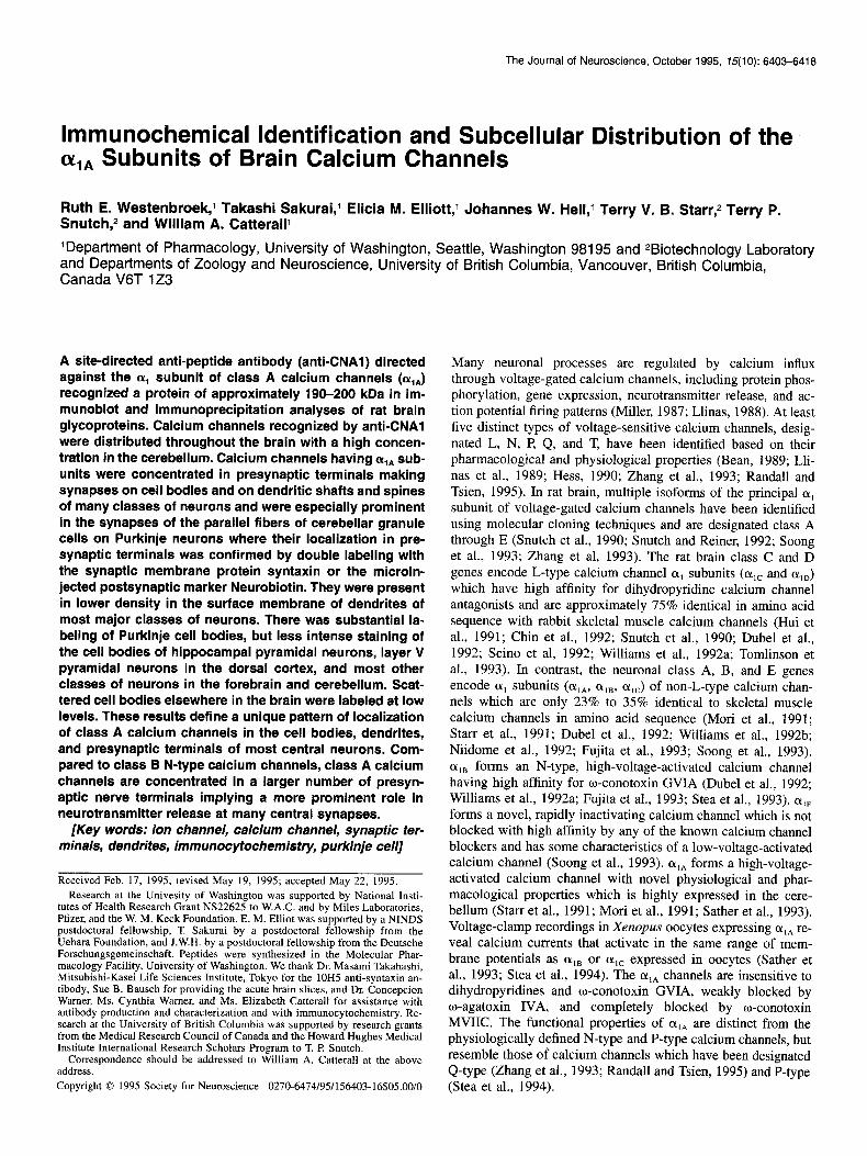
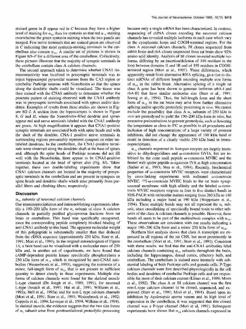
Figure 1. Immunoprecipitaion and immunoblotting with anti-CNAl antibodies detect 190 kDa olA subunits. A, Membrane glycoproteins were isolated from solubilized brain membranes by WGA affinity chromatography, biotinylated with NHS-LC-Biotin, and purified by double immuno- precipitaion with anti-CNAl. Samples were extracted with SDS sample buffer, separated by SDS-polyacrylamide gel electrophoresis, blotted, and incubated with streptavidin-biotinylated horseradish peroxidase complex as described under Materials and Methods. Calcium channels were purified by double immunoprecipitation with affinity-purified anti-CNAl antibodies (lane I), with anti-CNAl and anti-CP( 1382-1400) antibodies (lane 2), or with control antibodies (lane 5) and visualized with the ECL method. Specific block of immunoprecipitation with anti-CNAl by the peptide was tested. Anti-CNAl antibodies were preincubated over night on ice with 20 )LM CNAl peptide (lane 3) or with 20 PM CNC peptide (lane 4) as a control of the specificity of the peptide block. The sequence of CNC peptide (KYTTKINMDDLQPSENEDKS) was designed for a highly variable site in the loop domain between II and III of the class C L-type (Y, subunits. The migration positions of (Y- and B-spectrin, myosin heavy chain, a,-macroglobulin, B-galactosidase, and fructose-6-phosphate kinase are indicated at the left side of the gel together with their respective molecular masses in kilodaltons. B, Membrane glycoproteins were purified by WGA affinity chromatography and calcium channels were concentrated by adsorption to heparin-agarose. Samples were extracted, separated by SDS-PAGE, blotted, and incubated with affinity-purified anti-CNAl antibody (lane I) or nonimmune IgG (lane 2). Lane I is a representative immunoblot from four experiments with affinity-purified CNAl antibodies. Immunoreactive protein bands were visualized with the ECL method under Materials and Methods. Molecular markers are indicated as in A.
with an apparent molecular mass of 190 kDa in this glycoprotein fraction (Fig. lA, lanel). The specificity of the interaction of anti-CNAl antibodies with this band was tested with the CNAl peptide. After preincubation with this peptide at 20 FM, no sig- nal could be detected with anti-CNAl antibody, but CNCl pep- tide at the same concentration did not affect the immunoreactiv- ity with anti-CNAl antibody (Fig. lA, lanes 3 and 4). In addi- tion, neither band was recognized when immunoprecipitated with nonspecific antibodies (Fig. lA, lane 5). This specific band was only observed with affinity-purified anti-CNAl antibodies, while affinity-purified antibodies against (Y,* revealed distinct bands with apparent molecular masses of 230 kDa and 200 kDa in parallel experiments (data not shown). Calcium channels are multisubunit complexes and may interact with other cellular components such as cytoskeletal proteins and synaptic vesicle proteins. Therefore, it is possible that the proteins immunopre- cipitated by anti-CNAl antibodies under native conditions might be associated proteins of similar size to the cx, subunit rather than the (Y, subunit itself. To exclude other proteins from the immunoprecipitates, double immunoprecipitation experiments were performed under conditions which should completely dis- sociate the calcium channel subunits and associated proteins. Anti-CP( 1382-1400), which recognizes a segment of the OL, sub- unit whose sequence is conserved in calcium channels so far
characterized, was used as a probe in a second immunoprecip- itation. The CP(1382-1400) sequence is accessible to anti- CP( 1382- 1400) only after solubilization in Triton X- 100 or other detergents which cause subunit dissociation (Ahlijanian et al., 1991). Following the double immunoprecipitation with anti- CNAl and anti-CP(1382-1400), the same 190 kDa band was detected as after double immunoprecipitation with anti-CNAl antibodies and no additional bands were observed (Fig. lA, lane 2). These findings show that anti-CNAl antibodies interact spe- cifically with this 190 kDa band, indicating that it corresponds to the major form of (Y,,. In other experiments (Sakurai and Catterall, unpublished observations), we found that the antibody anti-CNA3, which is directed against an adjacent amino acid sequence m ‘Y,,, immunoprecipitated this 190-200 kDa band and also immuoprecipitated high affinity binding activity (Kd = 100 PM) for o-conotoxin MVIIC. These results provide further support for the conclusion that the 190 kDa band is the major form of olA.
Immunoblotting of a WGA fraction concentrated by adsorp- tion to heparin-agarose is shown in Figure 1B (see Materials and Methods). Affinity-purified anti-CNAl antibody revealed an im- munoreactive band with a molecular mass of 190-200 kDa un- der these experimental conditions (Fig. 1B). No specific immu- nostaining was observed with nonimmune rabbit IgG. Specific
The Journal of Neuroscience, October 1995, 75(10) 6407
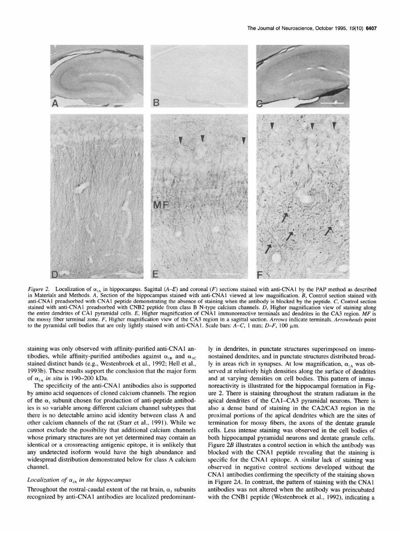
Figure 2. Localization of (Y,~ in hippocampus. Sagittal (A-E) and coronal (F) sections stained with anti-CNAl by the PAP method as described in Materials and Methods. A, Section of the hippocampus stained with anti-CNAl viewed at low magnification. B, Control section stained with anti-CNAl preadsorbed with CNAl peptide demonstrating the absence of staining when the antibody is blocked by the peptide. C, Control section stained with anti-CNAl preadsorbed with CNB2 peptide from class B N-type calcium channels. D, Higher magnification view of staining along the entire dendrites of CA1 pyramidal cells. E, Higher magnification of CNAl immunoreactive terminals and dendrites in the CA3 region. MF is the mossy fiber terminal zone. F, Higher magnification view of the CA3 region in a sagittal section. Arrows indicate terminals. Arrowheads point to the pyramidal cell bodies that are only lightly stained with anti-CNAl. Scale bars: A-C, 1 mm; D-F, 100 km.
staining was only observed with affinity-purified anti-CNAl an- tibodies, while affinity-purified antibodies against CY,~ and o,e stained distinct bands (e.g., Westenbroek et al., 1992; Hell et al., 1993b). These results support the conclusion that the major form of (Y,~ in situ is 190-200 kDa.
The specificity of the anti-CNAl antibodies also is supported by amino acid sequences of cloned calcium channels. The region of the CX, subunit chosen for production of anti-peptide antibod- ies is so variable among different calcium channel subtypes that there is no detectable amino acid identity between class A and other calcium channels of the rat (Starr et al., 1991). While we cannot exclude the possibility that additional calcium channels whose primary structures are not yet determined may contain an identical or a crossreacting antigenic epitope, it is unlikely that any undetected isoform would have the high abundance and widespread distribution demonstrated below for class A calcium channel.
Localization of (Y,* in the hippocampus
Throughout the rostral-caudal extent of the rat brain, (Y, subunits recognized by anti-CNAl antibodies are localized predominant-
ly in dendrites, in punctate structures superimposed on immu- nostained dendrites, and in punctate structures distributed broad- ly in areas rich in synapses. At low magnification, oIA was ob- served at relatively high densities along the surface of dendrites and at varying densities on cell bodies. This pattern of immu- noreactivity is illustrated for the hippocampal formation in Fig- ure 2. There is staining throughout the stratum radiatum in the apical dendrites of the CAl-CA3 pyramidal neurons. There is also a dense band of staining in the CA2KA3 region in the proximal portions of the apical dendrites which are the sites of termination for mossy fibers, the axons of the dentate granule cells. Less intense staining was observed in the cell bodies of both hippocampal pyramidal neurons and dentate granule cells. Figure 2B illustrates a control section in which the antibody was blocked with the CNAl peptide revealing that the staining is specific for the CNAl epitope. A similar lack of staining was observed in negative control sections developed without the CNAl antibodies confirming the specificty of the staining shown in Figure 2A. In contrast, the pattern of staining with the CNAl antibodies was not altered when the antibody was preincubated with the CNBl peptide (Westenbroek et al., 1992), indicating a
6406 Westenbroek et al. l Localization of Class A Calcium Channels
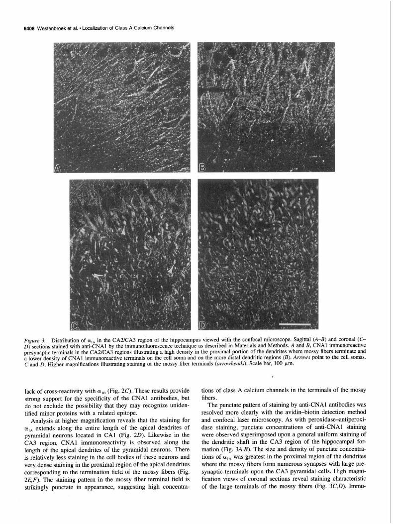
Figure 3. Distribution of oIA in the CA2KA3 region of the hippocampus viewed with the confocal microscope. Sagittal (A-B) and coronal (C- D) sections stained with anti-CNAl by the immunofluorescence technique as described in Materials and Methods. A and B, CNAl immunoreactive presynaptic terminals in the CA2/CA3 regions illustrating a high density in the proximal portion of the dendrites where mossy fibers terminate and a lower density of CNAl immunoreactive terminals on the cell soma and on the more distal dendritic regions (B). Arrows point to the cell somas. C and D, Higher magnifications illustrating staining of the mossy fiber terminals (arrowheads). Scale bar, 100 pm.
lack of cross-reactivity with on, (Fig. 2C). These results provide strong support for the specificity of the CNAl antibodies, but do not exclude the possibility that they may recognize uniden- tified minor proteins with a related epitope.
Analysis at higher magnification reveals that the staining for oIA extends along the entire length of the apical dendrites of pyramidal neurons located in CA1 (Fig. 20). Likewise in the CA3 region, CNAl immunoreactivity is observed along the length of the apical dendrites of the pyramidal neurons. There is relatively less staining in the cell bodies of these neurons and very dense staining in the proximal region of the apical dendrites corresponding to the termination field of the mossy fibers (Fig. 2E,F). The staining pattern in the mossy fiber terminal field is strikingly punctate in appearance, suggesting high concentra-
tions of class A calcium channels in the terminals of the mossy fibers.
The punctate pattern of staining by anti-CNAl antibodies was resolved more clearly with the avidin-biotin detection method and confocal laser microscopy. As with peroxidase-antiperoxi- dase staining, punctate concentrations of anti-CNAl staining were observed superimposed upon a general uniform staining of the dendritic shaft in the CA3 region of the hippocampal for- mation (Fig. 3A,B). The size and density of punctate concentra- tions of olA was greatest in the proximal region of the dendrites where the mossy fibers form numerous synapses with large pre- synaptic terminals upon the CA3 pyramidal cells. High magni- fication views of coronal sections reveal staining characteristic of the large terminals of the mossy fibers (Fig. 3C,D). Immu-
The Journal of Neuroscience, October 1995, 75(10) 6409
noreactivity in the cell bodies was relatively less than in the dendrites, although detectable staining of punctate structures was observed along the somal surface. The large size and the local- ization of these punctate concentrations of a,, immunostaining confirm identification of the presynaptic terminals of mossy fiber synapses as a major site of expression of class A calcium chan- nels.
Localization of alA in the cerebellum
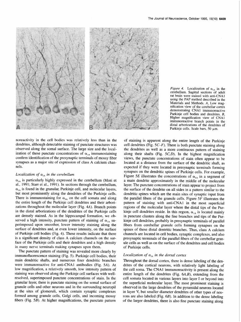
CL,* is particularly highly expressed in the cerebellum (Mori et al., 1991; Starr et al., 1991). In sections through the cerebellum, cq, is found in the granular, Purkinje cell, and molecular layers, but most prominently along the dendrites of the Purkinje cells. There is immunostaining for cqA on the cell somata and along the entire length of the Purkinje cell dendrites and their arbori- zations throughout the molecular layer (Fig. 4A). Branch points in the distal arborizations of the dendrites of the Purkinje cells are densely stained. As in the hippocampal formation, we ob- served a high intensity, punctate pattern of staining of cqa su- perimposed upon smoother, lower intensity staining along the surface of dendrites and, at even lower intensity, on the surface of Purkinje cell bodies (Fig. 4). These results indicate that there is a significant density of class A calcium channels on the sur- face of the Purkinje cells and their dendrites and a high density in many nerve terminals making synapses upon them.
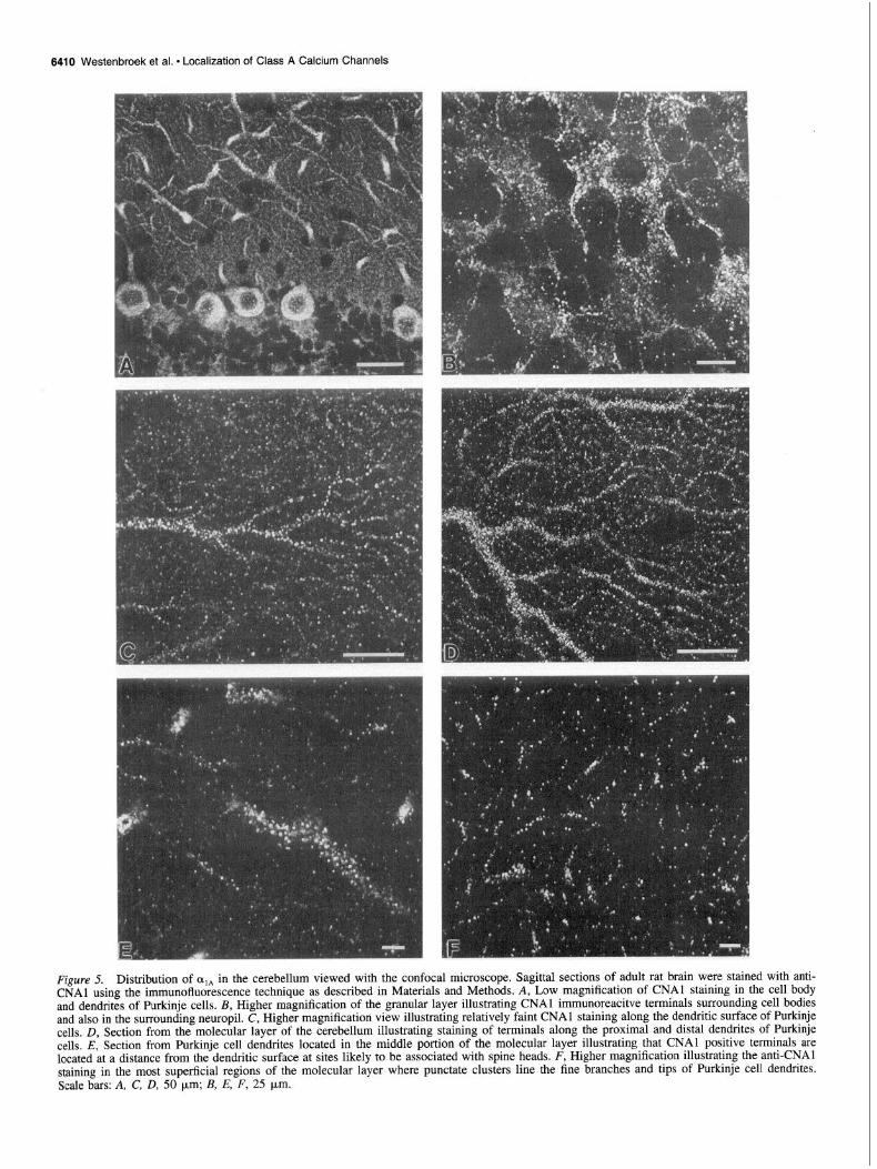
The punctate pattern of staining was revealed more clearly by immunofluorescence staining (Fig. 5). Purkinje cell bodies, their main dendritic shafts, and numerous finer dendritic branches were immunoreactive for anti-CNAl antibodies (Fig. 5A). At low magnification, a relatively smooth, low intensity pattern of staining was observed along the Purkinje cell surfaces with well- resolved, superimposed punctate concentrations of stain. In the granular layer, there is punctate staining on the somal surface of granule cells and other neurons and in the surrounding neuropil at the sites of glomeruli, which contain synaptic complexes formed among granule cells, Golgi cells, and incoming mossy fibers (Fig. 5B). At higher magnifications, the punctate pattern
Figure 4. Localization of q, in the cerebellum. Sagittal sections of adult rat brain were stained with anti-CNAl using the PAP method described in the Materials’ and Methods. A, Low mag- nification view of the cerebellar cortex demonstrating CNAl immunoreactive Purkinje cell bodies and dendrites. B, Higher magnification view of CNAl immunoreactive branch points in the distal arborizations of the dendrites of Purkinje cells. Scale bars, 50 pm.
of staining is apparent along the entire length of the Purkinje cell dendrites (Fig. 5C-F). There is both punctate staining along the dendrites as well as a more continuous pattern of staining along their shafts (Fig. 5C,D). In the highest magnification views, the punctate concentrations of stain often appear to be located at a distance from the surface of the dendritic shaft, as expected if they were located in presynaptic terminals forming synapses on the dendritic spines of Purkinje cells. For example, Figure 5E illustrates the concentrations of cxlA in a segment of a main dendrite approximately in the middle of the molecular layer. The punctate concentrations of stain appear to project from the surface of the dendrite on all sides in a pattern similar to the dendritic spines which are the main sites of synaptic input from the parallel fibers of the granule cells. Figure 5F illustrates the pattern of staining with anti-CNAl in the most superficial regions of the molecular layer where the distal tips of the Pur- kinje cell dendrites reside. In this region, cqA is located mainly in punctate clusters along the fine branches and tips of the Pur- kinje cell dendrites, probably in presynaptic terminals of parallel fibers from cerebellar granule cells forming synapses on the spines of these distal de&tic branches. Thus, class A calcium channels are located in cell bodies, synaptic complexes, and also presynaptic terminals of the parallel fibers of the cerebellar gran- ule cells as well as on the surface of the dendrites and cell bodies of Purkinje cells.
Localization of a,, in the dorsal cortex
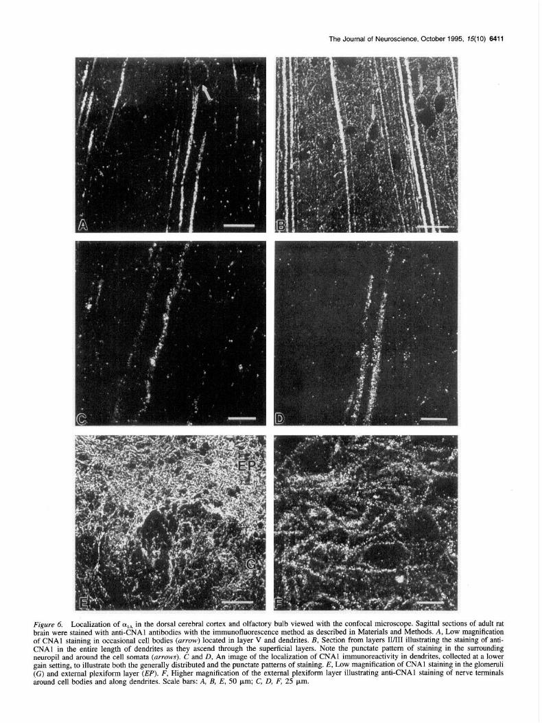
Throughout the dorsal cortex, there is dense labeling of the den- drites of the cortical neurons, with relatively light labeling of the cell soma. The CNAl immunoreactivity is present along the entire length of the dendrites (Fig. 6A,B), extending from the cell somata located in various layers into layer I or beyond into the superficial molecular layer. The most prominent staining is observed in the large dendrites of the pyramidal neurons located in layer V, but smaller diameter dendrites of other types of neu- rons are also labeled (Fig. 6B). In addition to the dense labeling of the larger dendrites, there is also fine punctate staining along
6410 Westenbroek et al. l Localization of Class A Calcium Channels
Figure 5. Distribution of olA in the cerebellum viewed with the confocal microscope. Sagittal sections of adult rat brain were stained with anti- CNAl using the immunofluorescence technique as described in Materials and Methods. A, Low magnification of CNAl staining in the cell body and dendrites of Purkinje cells. B, Higher magnification of the granular layer illustrating CNAl immunoreacitve terminals surrounding cell bodies and also in the surrounding neuropil. C, Higher magnification view illustrating relatively faint CNAl staining along the dendritic surface of Purkinje cells, D, Section from the molecular layer of the cerebellum illustrating staining of terminals along the proximal and distal dendrites of Purkinje cells. E, Section from Purkinje cell dendrites located in the middle portion of the molecular layer illustrating that CNAl positive terminals are located at a distance from the dendritic surface at sites likely to be associated with spine heads. F, Higher magnification illustrating the anti-CNAl staining in the most superficial regions of the molecular layer where punctate clusters line the fine branches and tips of Purkinje cell dendrites. Scale bars: A, C, D, 50 urn; B, E, F, 25 pm.
The Journal of Neuroscience, October 1995, 75(10) 6411
Figure 6. Localization of alA in the dorsal cerebral cortex and olfactory bulb viewed with the confocal microscope. Sagittal sections of adult rat brain were stained with anti-CNAl antibodies with the immunofluorescence method as described in Materials and Methods. A, Low magnification of CNAl staining in occasional cell bodies (arrow) located in layer V and dendrites. B, Section from layers II/III illustrating the staining of anti- CNAl in the entire length of dendrites as they ascend through the superficial layers. Note the punctate pattern of staining in the surrounding neurooil and around the cell somata (arrows). C and D, An image of the localization of CNAl immunoreactivity in dendrites, collected at a lower gain setting, to illustrate both the generally distributed and the puktate patterns of staining. E, Low magnification of CNAl staining in the glomemli (G) and external plexiform layer (EP). F, Higher magnification of the external plexiform layer illustrating anti-CNAl staining of nerve terminals around cell bodies and along dendrites. Scale bars: A, B, E, 50 km; C, D, F, 25 pm.
6412 Westenbroek et al. l Localization of Class A Calcium Channels
20 r/Y u X .- a Y- 0
ki 10
f
1
n
E
200
Fluorescence Intensity (Arbitrary Units)
$ 30 X
A III I lllll
0 100 200
Fluorescence Intensity
F (Arbitrary Units)
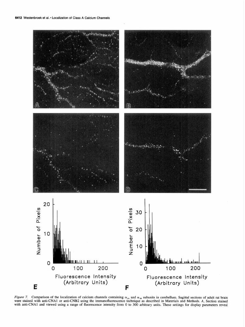
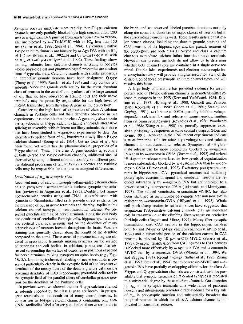
Figure 7. Comparison of the localization of calcium channels containing oIA and cqa subunits in cerebellum. Sagittal sections of adult rat brain were stained with anti-CNAl or anti-CNB2 using the immunofluorescence technique as described in Materials and Methods. A, Section stained with anti-CNAl and viewed using a range of fluorescence intensity from 0 to 300 arbitrary units. These settings for display parameters reveal
the length of smaller dendrites, on the somata of neurons, and scattered throughout the regions of the cerebral cortex that are rich in synapses (Fig. 6A,B). Punctate staining is also associated with smaller dendrites that branch off in a lateral direction. There is occasionally denser labeling of the cell soma of neurons scattered throughout the cortex, as illustrated in one example in Figure 6A (arrow). These more heavily labeled neuronal cell bodies are most often located in layer V.
In Figure 6, A and B, the gain on the confocal microscope was set at a high level in order to observe the fine punctate staining in the tissue surrrounding the more densely labeled den- drites. This results in saturation of the light emission from the more heavily stained dendrites so that their surface appears uni- formly stained. To visualize the variations in stain intensity along the surface of these heavily stained large dendrites, we examined the sections at higher magnifications and at lower gains. Under these conditions of observation, there appear to be both regions of continuous staining along the shafts of the den- drites and many punctate concentrations of stain on the dendritic surface (Fig. 6C,D). The punctate structures are observed both on the dendritic surface and at small distances from the shaft, in a position expected for presynaptic terminals making synapses on spine heads projecting from the dendrites of cortical neurons, similar to those on the dendrites of cerebellar Purkinje cells (Fig. 5E).
Localization of LY,* in the olfactory bulb The olfactory bulb is rich in synaptic plexuses segregated into discrete glomeruli. Anti-CNAl antibodies densely stain punctate structures clustered throughout all layers of this structure, as illustrated in Figure 6E where localization of class A calcium channels in the glomerular and the external plexiform layers is illustrated. A similar pattern of staining was also present in the mitral cell layer and the internal granular layer. At higher mag- nification, we observed clusters of punctate staining which ap- peared to surround the cell bodies of the neurons in this region (Fig. 6E), while the cytoplasm and nuclear regions appeared unstained. These results indicate that class A calcium channels are highly concentrated in presynaptic terminals making syn- apses on the principal cells of the olfactory bulb.
Comparison of the localization of CY,~ and (Y,~ in the cerebellum
In previous studies we found that cqB was localized in the den- dritic shafts and in presynaptic terminals making synapses on the dendrites of many central neurons (Westenbroek et al, 1992). Figure 7 compares the localization of cqA and (Y,, in the dendritic shafts and in terminals along the length of dendrites of Purkinje cells (Fig. 7A,B). cxIB is more restricted to major dendrites while ala is also found along minor dendritic branches and in synapses on them. The images in these two panels were collected using a range of fluorescence intensity of O-300 arbitrary units in the confocal microscope, the full range of intensity observed in these dendrites. In contrast, the images in Figure 7, C and D, were collected using a range of fluorescence intensity from 40
t
The Journal of Neuroscience, October 1995, 75(10) 6413
to 300 arbitrary units, blanking out regions of low intensity. In both cases, the lower level of fluorescence intensity along the dendritic shafts nearly disappears when the minimum intensity level is set at 40 arbitrary units and only the intensely staining presynaptic terminals remain. These findings indicate that the intensity of general staining along the dendritic shaft is less than approximately 13% of the staining intensity in presynaptic ter- minals. The calcium channel staining intensity in these two regions of the dendrites can also be resolved by a pixel-by-pixel analysis, as illustrated by the histograms in Figures 7, E and F. These histograms present the relative fluorescence intensity of immunostaining for oIA and qB in pixels over typical segments on two dendrites. It is evident from these histograms that very few pixels have a background level of fluorescence intensity which is set to a value of 0. Most pixels corresponding to the surface of major dendritic shafts have a fluorescence intensity of lo-50 arbitrary units. In contrast, a small fraction of pixels have fluorescence intensities ranging up to 200 units. The areas of high fluorescence intensity are likely to represent presynaptic terminals with a high concentration of calcium channels con- taining ala or cqB , while lower intensity areas likely represent dendritic areas with generally distributed calcium channels hav- ing a lower density of these two channel types. No comparisons can be made between the density of these two different classes of calcium channels using this approach due to the variability in antibody titer and unknown degree of access to the antigenic site for each of the antibodies. However, these results together with the overall pattern of staining provide direct evidence that both class A and class B calcium channels are present at low density in dendritic shafts and at a substantially (possibly eightfold) higher density in presynaptic terminals forming synapses upon them.
Localization of ffIA in presynaptic terminals
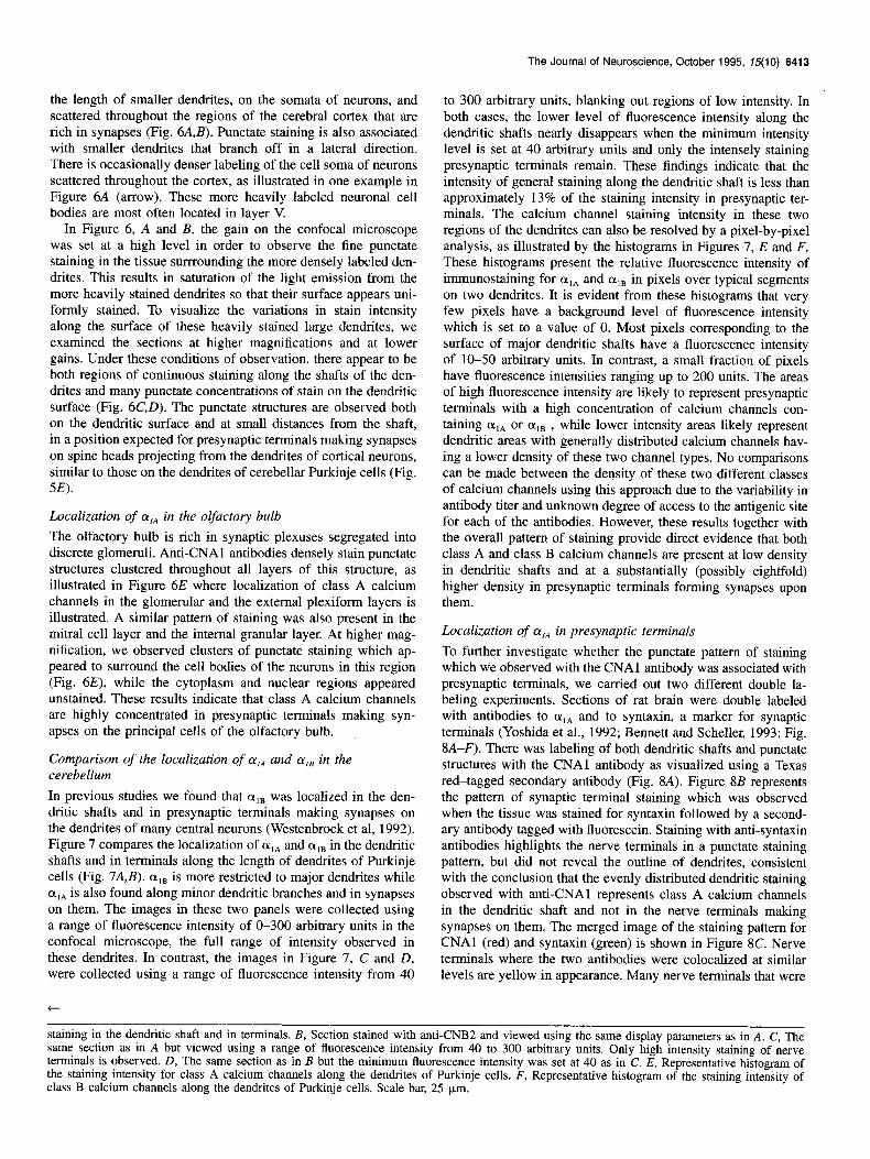
To further investigate whether the punctate pattern of staining which we observed with the CNAl antibody was associated with presynaptic terminals, we carried out two different double la- beling experiments. Sections of rat brain were double labeled with antibodies to cqA and to syntaxin, a marker for synaptic terminals (Yoshida et al., 1992; Bennett and Scheller, 1993; Fig. 8A-F). There was labeling of both dendritic shafts and punctate structures with the CNAl antibody as visualized using a Texas red-tagged secondary antibody (Fig. 8A). Figure 8B represents the pattern of synaptic terminal staining which was observed when the tissue was stained for syntaxin followed by a second- ary antibody tagged with fluorescein. Staining with anti-syntaxin antibodies highlights the nerve terminals in a punctate staining pattern, but did not reveal the outline of dendrites, consistent with the conclusion that the evenly distributed dendritic staining observed with anti-CNAl represents class A calcium channels in the dendritic shaft and not in the nerve terminals making synapses on them. The merged image of the staining pattern for CNAl (red) and syntaxin (green) is shown in Figure 8C. Nerve terminals where the two antibodies were colocalized at similar levels are yellow in appearance. Many nerve terminals that were
staining in the dendritic shaft and in terminals. B, Section stained with anti-CNB2 and viewed using the same display parameters as in A. C, The same section as in A but viewed using a range of fluorescence intensity from 40 to 300 arbitrary units. Only high intensity staining of nerve terminals is observed. D, The same section as in B but the minimum fluorescence intensity was set at 40 as in C. E, Representative histogram of the staining intensity for class A calcium channels along the dendrites of Purkinje cells. F, Representative histogram of the staining intensity of class B calcium channels along the dendrites of Purkinje cells. Scale bar, 25 pm.
6414 Westenbroek et al. * Localization of Class A Calwm Channels
Figlcl-e 8. Localization of cy14 subunits in presynaptic terminals, Sagittal sections of adult rat brain stained wtth antKNA1 and/or anti-syntaxm antibodies (A-F) as described in Materials and Methods. G-l are acute brain slices labeled with Neurobiotin and antKNA1 antibodies as described m Materials and Methods. A. Cerebellar tissue labeled with CNAl antibodies. B. Same tissue section as m A stamed wtth anti-syntaxin to localize presynaptic terminals. C. Combined image of staining shown in A and B illustrating regions of colocalization (.wllon~) between CNAl and anti- syntaxin antibodies in presynaptic terminals. D. CNAl staining in molecular layer of cerebellum. E. Syntaxm stammg m the same section as shown in D. F, Combined Image of D and E to illustrate the colocalization of CNAl and synataxm m presy naptic terminals (~e//on ) G, Acute shce from CA3 region of hippocampus m which the dendrite and spines of a single pyramidal neuron have been filled with Neurobrotrn (red) and srmulta- neously stained with anti-CNAl antibodtes (greerz). H, Higher magmfication of G illustratmg CNAl stainmg (greerr) in termmals assocrated Hith spine heads (~I.KXIT) of a Neurobiotin-filled (red) pyramidal neuron from hippocampal CA3 region. I. Purkmje cell dendrrtes and spme necks filled wth Neurobiotin (red) are assocrated M ith CNAl stained presynaptic terminals (green). CNAl positrve terminals are located at the base of spmes (nr~~~heads) and at the head of the spme (awon~). Scale bars: A-F. H. I, 10 pm: G. 25 urn.
The Journal of Neuroscience, October 1995, 75(10) 6415
stained green in B appear red in C because they have a higher level of staining for oIA than for syntaxin so that red (Y,* staining overwhelms the green syntaxin staining when the two panels are merged. Few nerve terminals that are stained green are observed in C indicating that most syntaxin-staining terminals in the cer- ebellum also contain oIA. A similar set of pictures is shown in Figure 8D-F for a different area of the cerebellum. Collectively, these pictures illustrate that the majority of synaptic terminals in the cerebellum contain class A calcium channels.
The second approach taken to determine whether CNAl im- munoreactivity was localized in presynaptic terminals was to inject hippocampal pyramidal neurons from the CA3 region or cerebellar Purkinje neurons with Neurobiotin so that the spines along the dendritic shafts could be visualized. The tissue was then stained with the CNAl antibody to determine whether the punctate pattern of staining observed with the CNAl antibody was in presynaptic terminals associated with spines and/or den- drites. Examples of results from these studies are shown in Fig- ure 8G-I. A section from the hippocampus is shown in Figure 8, G and H, where the Neurobiotin-filled dendrite and spines appear red and nerve terminals labeled with the CNAl antibody are green. At high magnification it appears that CNAl-positive synaptic terminals are associated both with spine heads and with the shaft of the dendrite. CNA-I positive nerve terminals in surrounding regions presumably represent synapses on other un- labeled dendrites. In the cerebellum, the CNAl-positive termi- nals were observed along the dendritic shaft at the base of spines and, although the spine heads of Purkinje neurons did not fill well with the Neurobiotin, there appear to be CNAl-positive terminals located at the head of spines also (Fig. 81). Taken together, these two double-labeling experiments show that CNAl calcium channels are located in the majority of presyn- aptic terminals in the cerebellum and are present in synapses on spine heads and dendritic shafts which arise primarily from par- allel fibers and climbing fibers, respectively.
Discussion CY,~ subunits of neuronal calcium channels Our immunoprecipitation and immunoblotting experiments iden- tify a 190-200 kDa form of the (Y, subunit of class A calcium channels in partially purified glycoprotein fractions from rat brain or cerebellum. This band was specifically recognized, since the corresponding peptide completely blocked binding of anti-CNAl antibody to this band. The apparent molecular weight of this polypeptide is substantially smaller than that deduced from the cDNA sequence (approximately 250 kDa; Starr et al., 1991; Mori et al., 1991). In the original autoradiogram of Figure IA, a faint band can be visualized with a molecular mass of 250 kDa and, in another set of experiments, we have found that CAMP-dependent protein kinase specifically phosphorylates a 250 kDa form of olA which is recognized by anti-CNAl anti- bodies (Westenbroek et al., 1993), suggesting the existence of a minor, full-length form of rxIA that is not present in sufficient quantity to detect clearly in these experiments. Multiple size forms of calcium channels were found for the skeletal muscle L-type channel (De Jongh et al., 1989, 1991), for neuronal L-type (Snutch et al., 1991; Hui et al., 1991; Williams et al., 1992a; Hell et al., 1993a) and for non-L-type calcium channels (Mori et al., 1991; Starr et al., 1991; Westenbroek et al., 1992; Coppola et al., 1994; Leveque et al., 1994; Willams et al., 1994). In skeletal muscle, the evidence suggests that the two size forms of IX, subunit arise from posttranslational proteolytic processing
because only a single mRNA has been characterized. In contrast, sequencing of cDNA clones encoding the neuronal calcium channels has revealed multiple isoforms in each case which vary in the cytoplasmic loops and COOH-terminal regions. For the class A neuronal calcium channels, BI clones sequenced from rabbit brain and rbA clones sequenced from rat brain show 92% amino acid identity. Analysis of BI clones revealed multiple iso- forms, differing by an insertion/deletion of 349 residues in the loop between domains II and III and of 195 residues in COOH- terminal region (Mori et al., 1991). These differences, which apparently result from alternative RNA splicing, give rise to dis- tinct mRNAs of different length encoding multiple size forms of ai.4 in the rabbit brain. Alternative splicing of a single rat class A gene has been shown to generate isoforms (rbA-I and rbA-II) that have similar molecular size (Starr et al., 1991; Soong et al., 1994). Thus, the full-length form and the short form of olA in the rat brain may arise from further alternative splicing and/or specific proteolytic processing in vivo. We cannot exclude the possibility that class A IX, subunits of 250 kDa in vivo are proteolysed to yield the 190-200 kDa form in vitro, but extensive preincubations to prevent proteolysis, such as lowering the temperature as close to 0°C as possible in all procedures and inclusion of high concentrations of a large variety of protease inhibitors, did not change the appearance of 190 kDa band or result in detection of a clearly visible 250 kDa band in immu- noprecipitates.
(Y,* channels expressed in Xenopus oocytes are largely insen- sitive to dihydropyridines and w-conotoxin GVIA, but are in- hibited by the cone snail peptide w-conotoxin MVIIC and the funnel web spider peptide w-agatoxin IVA at high concentration (Sather et al., 1993; Stea et al., 1994). Recently, biochemical properties of w-conotoxin MVIIC receptors were characterized by cross-linking experiments with iodinated w-conotoxin MVIIC. 1251-w-conotoxin MVIIC binds to the rat brain synap- tosomal membrane with high affinity and the labeled o-cono- toxin MVIIC receptors migrate as four to five distinct bands in SDS-PAGE with molecular masses ranging from 260 kDa to 140 kDa including a major band at 190 kDa (Woppmann et al., 1994). These multiple bands may not all represent the CK, sub- unit, since crosslinking of specifically bound toxin to other sub- units of the class A calcium channels is possible. However, these bands all seem to be part of the multisubunit complex with (Y,*. These observations are consistent with our results indicating a major 190-200 kDa form and a minor 250 kDa form of oIA.
Northern blot analysis shows that class A transcripts are ex- pressed in all regions of the rat CNS, but most prominently in the cerebellum (Mori et al., 1991; Starr et al., 1991). Consistent with these results, we find that the anti-CNAl antibodies label calcium channels containing oIA subunits throughout the brain, including the hippocampus, dorsal cortex, olfactory bulb, and cerebellum. The cerebellum is stained most intensely with sub- stantial labeling of both Purkinje cells and granule cells. P-Type calcium channels were first described physiologically in the cell bodies and dendrites of cerebellar Purkinje cells and are respon- sible for most of their calcium current (Llinas et al., 1989; Mintz et al., 1992). The class A or BI calcium channel was the first non-L-type calcium channel to be cloned, sequenced, and ex- pressed (Starr et al., 1991; Mori et al., 1991). Based upon its inhibition by Agelenopsis aperta venom and its high level of expression in the cerebellum, it was suggested that this cloned channel was a P-type channel (Mori et al, 1991). Subsequent experiments have shown that olA calcium channels expressed in
6416 Westenbroek et al. l Localization of Class A Calcium Channels
Xenopus oocytes inactivate more rapidly than P-type calcium channels, are only partially blocked by a high concentration (200 nM) of o-agatoxin IVA purified from Agelenopsis aperta venom, and are blocked by w-CTx-MVIIC with an IC,, less than 150 nM (Sather et al., 1993; Stea et al., 1994). By contrast, native P-type calcium channels are blocked by o-Aga-IVA with an IC,, of l-2 nM (Mintz et al., 1992a,b) and by w-CgTx-MVIIC with an IC,, of l-10 PM (Hillyard et al., 1992). These findings show that cqA subunits form calcium channels in Xenopus oocytes whose physiological and pharmacological properties are distinct from P-type channels. Calcium channels with similar properties in cerebellar granule neurons have been designated Q-type (Zhang et al., 1993; Randall et al., 1995) and may contain olA subunits. Since the granule cells are by far the most abundant class of neurons in the cerebellum, synthesis of the large amount of DllA that we have observed in granule cells and their nerve terminals may be primarily responsible for the high level of mRNA transcribed from the class A gene in the cerebellum.
Considering the high level of expression of class A calcium channels in Purkinje cells and their dendrites observed in our experiments, it is possible that the class A gene may also encode the (Y, subunits of P-type calcium channels through alternative splicing or assembly with different auxiliary subunits than those that have been studied in expression experiments to date. An alternatively spliced form of cqA inactivates slowly like a P-type calcium channel (Soong et al., 1994), but no form of (Y,* has been found yet which ‘has the pharmacological properties of a P-type channel. Thus, if the class A gene encodes CL, subunits of both Q-type and P-type calcium channels, additional sites of alternative splicing, different subunit assembly, or different post- translational processing of (Y,* in Xenopus oocytes and Purkinje cells may be responsible for the pharmacological differences.
Localization of (Y,* at synaptic sites
Localized entry of calcium through voltage-gated calcium chan- nels in presynaptic nerve terminals initiates synaptic transmis- sion (reviewed in Augustine et al., 1987). Double label immu- nocytochemical studies using anti-CNAl in combination with syntaxin or Neurobiotin-filled cells provide direct evidence for the presence of cqA in nerve terminals and thereby implicate this calcium channel subtype in neurotransmitter release. We ob- served punctate staining of nerve terminals along the cell body and dendrites of cerebellar Purkinje cells, hippocampal neurons, and cortical pyramidal neurons and on the dendrites of many other classes of neurons located throughout the brain. Punctate staining was generally denser along the length of the dendrite compared to the soma. These areas of punctate staining are sit- uated in presynaptic terminals making synapses on the surface of dendrites and cell bodies. In addition, puncta are also ob- served surrounding the surface of dendrites in positions expected for nerve terminals making synapses on spine heads (e.g., Figs. 5E, 81). Immunocytochemical labeling of nerve terminals is ob- served particularly clearly in the synaptic field of the large nerve terminals of the mossy fibers of the dentate granule cells on the proximal dendrites of CA3 hippocampal pyramidal cells and in the synaptic field of the parallel fibers of cerebellar granule neu- rons on the dendrites of the Purkinje cells.
In previous work, we showed that the N-type calcium channel (Y, subunits encoded by the class B gene are located in presyn- aptic terminals on the dendrites of many central neurons. In comparison to N-type calcium channels containing cqB, anti- CNAl antibodies label a larger population of nerve terminals in
the brain, and we observed labeled punctate structures not only along the soma and dendrites of major classes of neurons but in the surrounding neuropil as well. These results indicate that ma-. jor neuron classes, including the dentate granule neurons and CA3 neurons of the hippocampus and the granule neurons of the cerebellum, use both class B N-type and class A calcium channels to mediate calcium influx into their nerve terminals. However, our present methods do not allow us to determine whether both channel types are contained in a single nerve ter- minal. Double label experiments and electron microscopic im- munocytochemistry will provide a higher resolution view of the distribution of these presynaptic calcium channel types and will resolve this issue.
A large body of literature has provided evidence for an im- portant role of N-type calcium channels in neurotransmitter re- lease at synapses in the PNS (Kerr and Yoshikami, 1984; Koy- ano et al., 1987; Himing et al., 1988; Grinnell and Pawson, 1989; Robitaille et al., 1990; Cohen et al., 1991; Stanley and Goping, 1991). w-Conotoxin GVIA partially inhibits voltage- dependent calcium flux and release of some neurotransmitters from rat brain synaptosomes (Reynolds et al., 1986; Woodward et al., 1988; Xiang et al., 1990) and partially suppresses excit- atory postsynaptic responses in some central synapses (Horn and Kemp, 1991). However, in the CNS, recent experiments indicate a more important role for other high-voltage-activated calcium channels in neurotransmitter release. Synaptosomal 3H-gluta- mate release can be more completely blocked by w-agatoxin IVA than by w-conotoxin GVIA (Turner et al., 1992) and striatal ?H-dopamine release stimulated by low levels of depolarization is more substantially blocked by w-agatoxin IVA than by o-con- otoxin GVIA (Turner et al., 1993). Excitatory postsynaptic cur- rents in hippocampal CA1 pyramidal neurons and inhibitory postsynaptic currents in spinal and cerebellar neurons are re- duced substantially by w-agatoxin IVA but are inhibited to a lesser extent by w-conotoxin GVIA (Takahashi and Momiyama, 1993). The related conotoxin, o-conotoxin-MVIIC, has also been identified as an inhibitor of synaptic transmission that is resistant to w-conotoxin-GVIA (Hillyard et al., 1993). Whole cell patch-clamp studies in rat brain slices have suggested that o-agatoxin IVA-sensitive calcium channels play an important role in transmission at the climbing fiber synapse on cerebellar Purkinje cells (Regehr and Mintz, 1994). Mossy fiber synaptic transmission onto CA3 neurons in the hippocampus involves both N- and P-type or Q-type calcium channels (Castillo et al., 1994) and a substantial portion of the calcium current in CA3 neurons is blocked by 10 pM o-CTx-MVIIC (Swartz et al., 1993). Synaptic transmission from CA3 neurons to CA1 neurons is blocked more effectively by o-agatoxin IVA and o-conotoxin MVIIC than by u-conotoxin GVIA (Wheeler et al., 1994, Wu and Saggau, 1994). Recent findings (Sather et al., 1993; Zhang et al., 1993; Stea et al., 1994) that o-conotoxin-MVIIC and o-a- gatoxin IVA have partially overlapping affinities for the class A, P-type, and Q-type calcium channels are consistent with the pos- sibility that synaptic transmission at central synapses is mediated to a substantial degree by these calcium channels. Our detection of % in the synaptic terminals of a wide range of principal neurons and interneurons provides direct evidence for a key role of %A in presynaptic function and substantially broadens the range of neurons in which the class A calcium channel is im- plicated in transmitter release.

The Journal of Neuroscience, October 1995, 75(10) 6417
Localization of (Y,~ in dendrites
In addition to the punctate pattern of staining of class A calcium channels, we also observed immunoreactivity along the surface of dendrites. Both high-voltage-activated calcium channels and low-voltage-activated T-type calcium channels are thought to be involved in the generation of calcium-dependent action poten- tials that are conducted along the dendrite and play an important role in processing information from the multiple synaptic inputs to dendrites (Llinas and Sugimori, 1979). Earlier studies have suggested that the dendritic arbor of cerebellar Purkinje neurons is an important site of calcium-dependent electrogenesis and cal- cium entry into these neurons (Llinas and Sugimori, 1980; Tank et al., 1988). Activation of voltage-gated calcium channels was shown to be responsible for dramatic increases in cytosolic cal- cium throughout the dendritic arbor following repetitive activa- tion of synaptic input to the distal tips of hippocampal pyramidal neurons (Regehr et al., 1989). Using cultured cerebellar F’urkinje neurons it has been shown that increases in intracellular CaZ+ in dendrites evoked by brief pulses of high K+ are blocked by low concentrations of w-agatoxin IVA while w-conotoxin GVIA and o-conotoxin MVIIC block smaller components of the intracel- lular Ca*+ rise (Bindokas et al., 1993). This study suggests that P-type calcium channels highly sensitive to w-agatoxin IVA and, to a lesser extent, N-type and Q-type calcium channels are lo- cated in dendrites of Purkinje cells. These findings correlate with our previous immunocytochemical studies in which N-type cal- cium channels were localized along the entire length of dendrites of neurons using anti-peptide antibodies to (Y,~ (Westenbroek et al., 1993) and with our present identification of (Y,, localized along the length of the primary dendrites and their arborizations. This is in direct contrast to localization of L-type calcium chan- nels determined previously using antibodies to aIc and (~,o which were found to be localized predominantly in the cell soma and proximal dendrites of Purkinje cells and most other neurons (Ahlijanian et al., 1990; Westenbroek et al., 1990; Hell et al., 1993b). Collectively, these results suggest that both class A and class B N-type calcium channels are important contributors to voltage-gated calcium influx into dendrites as well as presyn- aptic terminals, compared to class C and D L-type calcium chan- nels which primarily contribute to calcium influx into cell bodies and proximal dendrites where their roles in most neurons may be to provide intracellular calcium to actiyate biochemical pro- cesses and regulate gene expression. Thus, our localization stud- ies point to important roles for class A calcium channels in ini- tiating neurotransmitter release and in integration and propaga- tion of excitatory synaptic signals in dendrites.
References Ahlijanian MK, Striessnig J, Catterall WA (1991) Phosphorylation of
an a,-like subunit of an w-conotoxin-sensitive brain calcium channels by CAMP-dependent protein kinase and protein kinase C. J Biol Chem 266:20192-20197.
Ahlijanian MK, Westenbroek RE, Catterall WA (1990) Subunit struc- ture and localization of dihydropyridine-sensitive calcium channels in mammalian brain, spinal cord, and retina. Neuron 4:819-832.
Augustine GJ, Charlton MP, Smith SJ (1987) Calcium action in syn- aptic transmitter release. Annu Rev Neurosci 10:633-693.
Bean BP (1989) Classes of calcium channels in vertebrate cells. Annu Rev Physiol 51:367-384.
Bennett MK, Scheller RH (1993) The molecular machinery for secre- tion is conserved from yeast to neurons. Proc Nat1 Acad Sci USA 90:2559-2563.
Bindokas VP, Brorson JR, Miller RJ (1993) Characteristics of voltage- sensitive calcium channels in dendrites of cultured rat cerebellar neu- rons. Neuropharmacology 11: 1213-1220.
Castillo PE, Weisskopf MG, Nicoll RA (1994) The role of CaZ+ chan- nels in hippocampal mossy fiber synaptic transmission and long-term potentiation. Neuron 12:261-269.
Chin H, Smith MA, Kim H-L, Kim H (1992) Expression of dihydro: pyridine-sensitive brain calcium channels in the rat central nervous system. FEBS Lett 299:69-74.
Cohen MW, Jones OT, Angelides KJ (1991) Distribution of Ca2+ chan- nels on frog motor nerve terminals revealed by fluorescent w-cono- toxin. J Neurosci 11: 1032-1039.
Coppola T, Waldmann R, Borsotto M, Heurteaux C, Romey G, Mattei M-G, Lazdunski M (1994) Molecular cloning of a murine N-type calcium channel (Y] subunit. Evidence for isoform, brain distribution, and chromosomal localization. FEBS Lett 338: l-5.
De Jongh KS, Merrick DK, Catterall WA (1989) Subunits of purified calcium channels: a 212~kDa form of (Y, and partial amino acid se- quence of phosphorylation site of an independent p subunit. Proc Nat1 Acad Sci USA 86:8585-8589.
De Jongh KS, Warner C, Colvin AA, Catterall WA (1991) Character- ization of the two size forms of the 01, subunit of skeletal muscle L-type calcium channels. Proc Nat1 Acad Sci USA 88:10778-10782.
Dubel SJ, Starr VB, Hell J, Ahlijanian MK, Enyeart JJ, Catterall WA, Snutch TP (1992) Molecular cloning of the (Y, subunit of an o-con- otoxin-sensitive calcium channel. Proc Nat1 Acad Sci USA 89:5058- 5062.
Fujita Y, Mynlieff M, Dirksen RT, Kim M-S, Niidome T, Nakai J, Fried- rich T, Iwabe N, Miyata T, Furuichi T, Furutama D, Mikoshiba K, Mori Y, Beam KG (1993) Primary structure and expression of the w-conotoxin-sensitive N-type calcium channel from rabbit brain. Neuron 10:585-598.
Grinnell AD, Pawson PA (1989) Dependence of spontaneous release at frog junctions on synaptic strength, external calcium and terminal length. J Phvsiol (Land) 418:397410.
Hell ;W, Westenbroek RE: Warner C, Ahlijanian MK, Prystay W, Gil- bert MM, Snutch TP, Catterall WA (1993b) Identification and dif- ferential subcellular localization of the neuronal class C and class D L-type calcium channel cx, subunits. J Cell Biol 123:949-962.
Hell JW, Yokoyama CT, Wong ST, Warner C, Snutch TP, Catterall WA (1993a) Differential phosphorylation of two size forms of the neu- ronal class C L-type calcium channel (Y, subunit. J Biol Chem 268: 19451-19457.
Hess P (1990) Calcium channels in vertebrate cells. Annu Rev Neu- rosci 13:337-356.
Hillyard DR, Monje VD, Mintz IM, Bean BP, Nadasdi L, Ramachan- dran J, Miljanich G, Azimi-Zoonooz A, McIntosh JM, Cruz LJ, Im- perial JS, Olivera BM (1992) A new Conus peptide ligand for mam- malian presynaptic Ca*+ channels. Neuron 9:69-77.
Hirning LD, Fox AP, McCleskey EW, Olivera BM, Thayer SA, Miller RJ, Tsien RW (1988) Dominant role for N-type calcium channels in evoked release of norepinephrine from synpathetic neurons. Science 239:57-61.
Horne AL, Kemp JA (1991) The effect of o-conotoxin GVIA on syn- aptic transmission within the nucleus accumbens and hippocampus of the rat in vitro. Br J Pharmacol 103: 1733-1739.
Hui A, Ellinor PT, Krizanova 0, Wang J-J, Diebold RJ, Schwartz A (1991) Molecular cloning of multiple subtypes of a novel rat brain isoform of the (Ye subunit of the voltage-dependent calcium channel. Neuron 7:35-44.
Kerr LM, Yoshikami D (1984) A venom peptide with a novel presyn- aptic blocking action. Nature 308:282-284.
Koyano K, Abe T, Nishiuchi Y, Sakakibara S (1987) Effects of syn- thetic w-conotoxin on synaptic transmission. Eur J Pharmacol 135: 337-343.
Leveque C, El Far 0, Martin-Moutot N, Sato K, Kato R, Takahashi M, Seagar MJ (1994) Purification of the N-type calcium channel asso- ciated with syntaxin and synaptotagmin. J Biol Chem 269:630& 6312.
Llinas R, Sugimori M (1979) Calcium conductances in Purkinje cell dendrites: their role in development and integration. Prog Brain Res 5 1:323-334.
Llinas R, Sugimori M (1980) Electrophysiological properties of in vitro Purkinie cell dendrites in mammalian cerebellar slices. J Phvsiol
_I d (Lond) 305:197-213.
Llinas R, Sugimori M, Lin J-W, Cherksey B (1989) Blocking and iso- lation of a calcium channel from neurons in mammals and cephalo-
6416 Westenbroek et al. - Localization of Class A Calcium Channels
pods utilizing a toxin fraction (FTX) from funnel-web spider poison. Proc Nat1 Acad Sci USA 86:1689-1693.
Llinas R (1988) The intrinsic electrophysiological properties of mam- malian neurons: insights into central nervous system function. Sci- ence 24211654-1664.
McLean IW, Nakane P (1974) Periodate-lysine-paraformaldehyde fix- ative for immunoelectron microscopy. J Histochem Cytochem 22: 1077-1083.
Merrifield RB (1963) Solid phase peptide synthesis.1. The synthesis of a tetrapeptide. J Am Chem Sot 85:2149-2154.
Miller RJ (1987) Multiple calcium channels and neuronal function. Science 235:46-52.
Mori Y, Friedrich T, Kim M-S, Mikami A, Nakai J, Ruth P Bosse E, Hofmann F, Flockerzi V, Furuichi T, Mikoshiba K, Imoto K, Tanabe T, Numa S (1991) Primary structure and functional expression from complementary DNA of a brain calcium channel. Nature 350:398- 402.
Niidome T, Kim M-S, Friedrich T, Mori Y (1992) Molecular cloning and chanracterization of a novel calcium channel from rabbit brain. FEBS Lett 308:7-13.
Orth DN (1979) Adrenocorticotrophic hormone. In: Methods of hor- mone radioimmunoassay (Jaffe BM, Behrman HR, eds), pp 245-284. New York: Academic.
Posnett DM, McGrath H, Tam JP (1988) A novel method for producing anti-peptide antibodies. Production of site-specific antibodies to the T cell antigen receptor beta-chain. J Biol Chem 263:1719-1725.
Randall A, Tsien RW (1995) Pharmacological dissection of multiple types of Ca*+ channel currents in rat cerebellar granule neurons. J Neurosci 5:350-357.
Regehr WG, Mintz IM (1994) Participation of multiple calcium chan- nel types in transmission at single climbing fiber to Purkinje cell synapses. Neuron 12:605-613.
Regehr WG, Connor JA, Tank DW (1989) Optical imaging of calcium accumulation in hippocampal pyramidal cell during synaptic activa- tion. Nature 341:533-536.
Reynolds IJ, Wagner JA, Synder SH, Thayer SA, Olivera BM, Miller RJ (1986) Brain voltage-sensitive Ca channel subtypes differentiated by w-conotoxin fraction GVIA. Proc Nat1 Acad Sci USA 83:8804- 8807.
Robitaille R, Adler EM, Charlton MP (1990) Strategic location of cal- cium channels at transmitter release sites of frog neuromuscular syn- apses. Neuron 5~773-779.
Sather WA, Tanabe T, Zhang J-E Mori Y, Adams ME, Tsien RW (1993) Distinctive biophysical and pharmacological properties of class A (BI) calcium channel oI subunit. Neuron-l 1:291-303.
Seino S. Chen L. Seino M. Blonde1 0. Takeda J. Johnson JH. Bell G (1992) Cloning of the o, subunit of a voltage-dependent ‘calcium channel expressed in pancreatic B cells. Proc Nat1 Acad Sci USA 89: 584-588.
Snutch TP, Reiner PB (1992) Calcium channels: diversity of form and function. Curr Opin Neurobiol 2:247-253.
Snutch Tp, Leonard JP, Gilbert MM, Lester HA, Davidson N (1990) Rat brain expresses a heterogeneous family of calcium channels. Proc Nat1 Acad Sci USA 87:3391-3395.
Snutch TP, Tomlinson WJ, Leonard JR Gilbert MM (1991) Distinct calcium channels are generated by alternative splicing and are dif- ferentially expressed in the mammalian CNS. Neuron 7:45-57.
Soong TW. Stea A. Hodson CD. Dubel SJ. Vincent SR. Snutch TP (1%3) Structure and functional expression of a member of the low- voltage-activated calcium channel family. Science 260: 1133-l 136.
Stanley EE Goping G (1991) Characterization of a calcium current in a vertebrate cholinergic presynaptic nerve terminal. J Neurosci 11: 985-993.
Starr TVB, Prystay W, Snutch TP (1991) Primary structure of a cal-
cium channel that is highly expressed in the rat cerebellum. Proc Nat1 Acad Sci USA 88:5621-5625:
Stea A. Dubel SJ. Praznell M. Leonard JP. Camnbell KI? Snutch TP (1993) A B subunit gormalizes the electrophysiological properties of a cloned N-type CaZ+ channel q-subunit. Neuropharmacology 32: 1103-1116.
Stea A, Tomlinson WJ, Soong TW, Bourinet E, Dubel SJ, Vincent SR, Snutch TP (1994) Localization and functional properties of a rat brain oIA calcium channel reflect similarities to neuronal Q- and P-type channels. Proc Nat1 Acad Sci USA 91:10576-10580.
Sternberger LA (1979) Immunocytochemistry, pp 104-169. New York: Wiley.
Takahashi T, Momiyama A (1993) Different types of calcium channels mediate central synaptic transmission. Nature 366: 156-158.
Tank DW, Sugimori M, Connor JA, Llinas RR (1988) Spatially re- solved calcium dynamics of mammalian Purkinje cells in cerebellar slice. Science 242~773-777.
Tomlinson WJ, Stea A, Bouinet E, Charnet P Nargoet J, Snutch TP (1993) Functional properties of a neuronal class C L-type calcium channel. Neuropharmacology 32:1112-1126.
Turner TJ, Adams ME, Dunlap K (1993) Multiple Ca*+ channel types coexist to regulate synaptosomal neurotransmitter release. Proc Nat1 Acad Sci USA 90:9518-9522.
Turner TJ, Adams ME, Dunlap K (1992) Calcium channels coupled to glutamate release identified by w-Aga-IVA. Science 258:310-313.
Westenbroek RE, Ahliianian MK, Catterall WA (1990) Clustering of L-type Caz+ channels at the base of major dendrites in hippocampal uvramidal neurons. Nature 347:281-284.
Westenbroek RE, Hell JW, Warner C, Dubel SJ, Snutch TP Catterall WA (1992) Biochemical properties and subcellular distribution of an N-tvue calcium channel o, subunit. Neuron 9:1099-l 115.
Wheeler DB, Randall A, Tsien RW (1994) Roles of N-type and Q-type Ca2+ channels in supporting hippocampal synaptic transmission. Sci- ence 264:107-111.
Williams ME, Brust BE Feldman DH, Patthi S, Smerson S, Maroufi A, McCue AE Velicelebi G. Ellis SB. Haroold MM (1992b) Structure and functional expression of an w-conoioxin-sensitive human N-type calcium channel. Science 257:389-395.
Williams ME, Feldman DH, McCue AF, Brenner R, Velicelebi G, Ellis SB, Harpold MM (1992a) Structure and functional expression of q, cl*, and B subunits of a novel human neuronal calcium channel sub- type. Neuron 8:71-84.
Williams ME, Marubio LM, Deal CR, Hans M, Brust PF, Philipson LH, Miller RJ, Johnson EC, Harpold MM, Ellis SB (1994) Structure and functional characterization of neuronal oils calcium channel subtvnes.
‘I I_
J Biol Chem 269:22347-22357. Woodward JJ. Rezazadeh SM. Leslie SW (1988) Differential sensitiv-
ity of synaptosomal calcium entry and endogenous dopamine release to o-conotoxin. Brain Res 475:141-145.
Woppmann A, Ramachandran J, Miljanich GP (1994) Calcium channel subtypes in rat brain: biochemical characterization of the high affinity receptors for o-conopeptides SNX-230 (synthetic MVIIC), SNX-183 (SVIB), and SNX-111 (MVIIA). Mol Cell Neurosci 5:350-357.
Xiang J-Z, Adamson P Brammer MJ, Campbell IC (1990) The K-o- piate agonist U50488H decreases the entry of 45Ca into rat cortical svnantosomes bv inhibiting N- but not L-tvue calcium channels. Neu- roph-armacology 29:439-4>4.
._
Yoshida A. Oho C. Omori A. Kuwahara R. Ito T Takahashi M (1992) HPC-1 is associated with synaptotagmin’and o-conotoxin receptor. J Biol Chem 267:24925-24928.
Zhang J-E Randall AD, Ellinor PT, Horne WA, Sather WA, Tanabe T, Schwartz TL, Tsien RW (1993) Distinctive pharmacology and ki- netics of cloned neuronal calcium channels and their possible coun- terparts in mammalian CNS neurons. Neuropharmacology 32:1075- 1088.