trypanosoma brucei: identification of a major repetitive ... email: [email protected]. running head:...
TRANSCRIPT

1
Isolation and characterisation of subnuclear compartments from
Trypanosoma brucei: identification of a major repetitive nuclear
lamina component
Michael P. Rout1 and Mark C. Field2
1Laboratory of Structural Cell Biology, The Rockefeller University, 1230 York
Avenue, New York, NY10021, USA, and 2Wellcome Trust Laboratories for
Molecular Parasitology, Imperial College of Science Technology & Medicine,
Department of Biology & Biochemistry, Exhibition Road, London, SW7 2AY, UK
Corresponding authors: Fractionation: M.P.Rout; tel: +1 212 327 8135, email:
[email protected]. Trypanosomatids: M.C.Field; tel: +44 020 7594
5277, email: [email protected].
Running head: Nuclear isolation from trypanosomes
Keywords: trypanosome/nucleus/proteomics/subcellular fractionation/lamina
Abbreviations: BSF; bloodstream form, NE; nuclear envelope, NPC; nuclear pore
complex, PBS; phosphate-buffered saline, PCF; procyclic culture form, PVP;
polyvinylpyrrolidone, PCLF; nuclear pore complex lamina fraction, TBST; tris-
buffered saline + 0.1% Tween20, VSG; variant surface glycoprotein.
Copyright 2001 by The American Society for Biochemistry and Molecular Biology, Inc.
JBC Papers in Press. Published on July 26, 2001 as Manuscript M104024200 by guest on June 17, 2018
http://ww
w.jbc.org/
Dow
nloaded from

2
Abstract
Protozoan parasites of the order kinetoplastida are responsible for a
significant proportion of global morbidity and economic hardship. These
organisms also represent extremely distal points within the Eukarya, and one
such organism, Trypanosoma brucei, has emerged as a major system for the
study of evolutionary cell biology. Significant technical challenges have hampered
the full exploitation of this organism, but advances in genomics, RNAi-mediated
phenocopying and proteomics provide a novel approach to acquiring rapid
functional data. However, the vast evolutionary distance between trypanosomes
and the higher eukaryotes presents significant problems with functional
assignment based on sequence similarity, and frequently homologues cannot be
identified with sufficient confidence to be informative. Direct identification of
proteins in isolated organelles has the potential of providing robust functional
insight and is a powerful approach for initial assignment. We have selected the
nucleus of T. brucei as a first target for protozoan organellar proteomics, via
isolation and protein sequencing. Trypanosomes have several unusual features to
their nuclear biology, including polycistronic transcription, closed mitosis, and
transcription of protein coding genes via RNA polymerase I. Our purification
methodology was able to reliably provide both nuclear and subnuclear fractions.
Analysis by gel electrophoresis, electron microscopy and immunoblotting against
trypanosome subcellular markers indicated that the preparations are of high yield
and purity, maintain native morphology and are well resolved from other
organelles. Minor developmental differences were observed in the nuclear
proteome for the bloodstream and procyclic stages, whilst significant
morphological alterations were visible. We demonstrate by direct sequencing that
the NUP-1 nuclear envelope antigen is a coiled coil protein, containing ~20 near
perfect copies of a 144 amino acid sequence. Immunoelectron microscopy
localised NUP-1 to the inner face of the nuclear envelope, suggesting that it is a
major filamentous component of the trypanosome nuclear lamina. These
methods provide the basis for a full analysis of the trypanosome nuclear
proteome.
by guest on June 17, 2018http://w
ww
.jbc.org/D
ownloaded from

3
Introduction
The most frequently studied eukaryote model systems are restricted to the
metazoans, nematodes and yeasts, which from an evolutionary perspective, are a
closely related subset of the kingdom (Gerhardt and Kirschner 1997, Leadbeater
and Green 2000, Coombes et al. 1998). Whilst comparisons between these
systems is a powerful strategy for the elucidation of universal and unique cellular
mechanisms, this approach is ultimately compromised by the relatively low
divergence between these organisms. As the genomes of many economically and
scientifically important divergent eukaryotes are becoming available, new
challenges in functional genomics are emerging, requiring exploitation of novel
strategies based on genome sequence and proteomic analysis.
Organisms of the order kinetoplastida, which include the parasitic
trypanosomatids Leishmania and Trypanosoma, separated from the metazoan
lineage ~3 x 10⁹ years ago (Coombes et al. 1998) and are the focus of much
interest by virtue of their impact on both human morbidity and agriculture.
Trypanosoma brucei, the causative agent of African Trypanosomiasis, is
responsible for ~100 000 deaths each year, and is considered the third most
important parasitic pathogen in overall economic impact
(www.who.int/tdr/diseases/tryp). Trypanosomatids also represent one of the most
divergent yet accessible experimental systems available. Several important
features of eukaryotes were originally described in T. brucei, mainly on account of
an extreme emphasis on these processes, for example eukaryotic polycistronic
transcription, GPI-anchoring of proteins and kinetoplastids. The presence of
specific functions or structures in either the protozoa or the higher eukaryotes has
clear potential for the identification of therapeutic targets (Ferguson 2000).
Whilst T. brucei, and related organisms, are comparatively cumbersome for
genetic analysis, recent advances in gene disruption by RNAi, genome projects
and proteomics provide potential strategies for rapid mapping of gene sequence
to function (Bastin et al. 2000, Wang et al. 2000). One strategy relies on the
comparison of gene sequences between organisms in order to identify functional
orthologues. However, a major complication is that database searches using
regular algorithms frequently fail to identify such orthologues, due to low sequence
conservation. Here we have taken a second approach, namely the direct
identification of components of organelles and complexes by proteomic analysis
by guest on June 17, 2018http://w
ww
.jbc.org/D
ownloaded from

4
of highly enriched specific subcellular fractions, which potentially avoids these
difficulties.
Unfortunately, most methodologies only apply to a particular organism or a
narrow subset of the Eukarya. We therefore attempted to modify our available
techniques such that they would function for a highly divergent eukaryote, as an
initial step towards making these methodologies more generally applicable. We
chose to focus initially on the trypanosome nucleus and substructures as this
compartment has a number of available antibody markers and several identified
protein components, in addition to representing an important system with a
number of vital biological issues that may be resolvable by proteomic analysis.
Unlike yeasts and metazoans, trypanosomatids transcribe their chromosomes in
a polycistronic manner (Pays et al. 1994). This unusual feature suggests that
these organisms use higher order chromatin structure and posttranscriptiona;
control for regulation of gene expression (Bender et al. 1992A, Navarro et al. 1999),
and hence an understanding of trypanosome nuclear structure could provide
profound insights into posttranslational control mechanisms in eukaryotes. In
addition, the chromosomes never condense, and mitosis is closed, with an intact
nuclear evelope present at all stages of the cell cycle (Ogbadoyi et al. 2000).
These organisms also appear to use unusual mechanisms for chromosome
segregation (Ersfeld and Gull 1977). Further, several T. brucei protein coding
genes, including the variant surface glycoprotein (VSG) responsible for antigenic
variation, are transcribed by RNA polymerase I (Pol I), which is restricted to
transcription of non-protein encoding genes in higher eukaryotes (Cross 1996).
Recent data suggests that a novel nucleolar-like compartment may be involved in
PolI-mediated VSG transcription (Navarro et al. 2000). Lastly, a comparison of the
composition and architecture of the trypanosome NPC and NE with those of other
eukaryotes should shed light on the core conserved mechanisms of
nucleocytoplasmic transport, NE assembly and the organization of peripheral
(silent) heterochromatin (Rout and Aitchison 2000).
We demonstrate the successful and reproducible isolation of nuclear,
nucleolar, NE and pore complex lamina fractions (PCLF) from T. brucei. The
preparations are of high purity and yield, producing sufficient protein for the
identification and characterization of specific organellar proteins. These methods
provide a basis for the exploitation of proteomics approaches in this organism.
by guest on June 17, 2018http://w
ww
.jbc.org/D
ownloaded from

5
Methods and Materials
Materials: Buffer components and other reagents were from Sigma unless
otherwise stated and of reagent grade or better. Culture media for trypanosomes
and mammalian cells were from GIBCO and were sterilised by filtration. Fetal calf
serum was heat-inactivated at 55˚C for 1 hour before use.
Trypanosomes: Procyclic culture form or bloodstream form trypomastigote (strain
427) trypanosomes were used throughout. Procyclic cells were cultured in 3.0 litre
batches of SDM79 medium (Brun and Schonenberger 1979), supplemented with
10% fetal calf serum, 10µg/ml haemin, 10U/ml penicillin, 10µg/ml streptomycin, in
sterile 6 litre glass conical flasks, with gentle shaking (100 rpm) at 27˚C. Cells
were allowed to attain a maximum density of 2-5 x 10⁷/ml before subculturing or
harvesting. Bloodstream form trypanosomes were grown by infecting mice and
rats and purified on DE-52 cellulose as described (Field and Menon 1992).
Antibodies: Mouse hybridoma cell lines secreting monoclonal antibodies against
yeast nuclear envelope antigens were developed as previously described (Rout
and Kilmartin 1990) and maintained according to Harlow and Lane 1988. Antibody
to the p67 protein and trypanosome BiP were the gift of Dr James Bangs,
University of Wisconsin, Madison, Wisconsin, USA. Antibody against trypanosome
NE (NUP-1) and nucleolar (NUMAG) proteins were the kind gift of Prof. Keith Gull
and Dr Klaus Ersfeld, University of Manchester, UK (Ogbadoyi et al. 2000).
Antibody recognising the trypanosome RNA-binding protein, RRM1, was from Prof.
John Boothroyd (Stanford, Palo Alto) (Manger and Boothroyd 1998). Polyclonal
rabbit antibodies against TbRAB2 have been described previously (Field et al.
1999, Field et al. 2000). HRP-conjugated goat anti-rabbit immunoglobulin was
from Sigma or Jackson Laboratories, HRP-conjugated donkey anti-mouse
immunoglobulin was from Jackson Laboratories. A rabbit anti-mouse IgG bridging
antibody was from ICN.
Isolation of trypanosome nuclei: Approximately 2.5 - 1010 to 7.5 x 10¹¹ procyclic cells
were used for each nuclear isolation procedure. Cellular morphology and viability
were verified beforehand by microscopy under an inverted tissue culture
microscope at 400x magnification. Cells were pelleted at 1700 gavg for 10 minutes
at 4˚C in a low speed centrifuge, resuspended in PBS (from Sigma PBS tablets)
pre-chilled on ice, and repelleted. The cell pellet was transferred to an HB-4 tube
by guest on June 17, 2018http://w
ww
.jbc.org/D
ownloaded from

6
(Sorvall) and pelleted once more in a swing out rotor benchtop centrifuge at 1800
gmax 4˚C for 15 minutes. All supernatant was carefully removed and the pellet
placed on ice. 20ml of 8% PVP (containing 0.05% TX-100 (Pierce SurfactAMPs
grade), 5mM Cleland's reagent, 100µl mammalian protease inhibitor cocktail
(Sigma) and 200µl solution P (100mg PMSF, 2mg Pepstatin A in 5ml ethanol))
was added per 2 x 10¹⁰ cells. The cell pellet was immediately processed with a
precooled Polytron PTA-10 head, setting ~6, in 1-2 minute bursts in the cold room.
The lysate was cooled on ice between each Polytron bout. Lysis was followed by
phase contrast microscopy at 400X magnification with an acceptable ~90% cell
lysis usually occurring after 5 minutes total Polytron treatment, although
proportionately longer periods were required for larger volume lysates. The lysate
derived from 2 x 10¹⁰ cells was underlaid with 10 ml of 0.3M sucrose in 8% PVP
plus 100µl solution P, 50µl 1M DTT and 50µl mammalian protease inhibitor
cocktail and then centrifuged at 11 000 gavg, in an HB-4 rotor (Sorvall) at 4˚C for 20
minutes. The top layer (containing mainly cytosol, designated SN) was carefully
decanted and stored at -80˚C. The crude nuclear pellet was immediately
resuspended with 1 minute bursts of the Polytron using setting 4.5 in a total
volume (per 2 x 10¹⁰ cell equivalents) of 8 ml of 2.1M sucrose in 8% PVP, 50µl 1M
DTT, 50µl mammalian protease inhibitor cocktail and 100µl solution P. Close to
100% cell dispersal is achieved at this point, with no detectable nuclear damage.
~12 ml of this suspension was loaded per SW28 tube (Beckman) containing a
discontinuous step gradient (from the bottom, 8ml 2.30M sucrose/PVP, 8ml 2.10M
sucrose/PVP, 8ml 2.01M sucrose/PVP, all containing protease inhibitors (Rout and
Kilmartin 1990, 1994, 1998). For the preparation to be successful, it is essential
that no more than 2.5 x 10¹⁰ cell equivalents be loaded into each SW28 tube. The
gradient was centrifuged at 100 000 gavg, in a SW28 rotor for 3 hours at 4˚C and
immediately unloaded from the top and fractions examined by phase contrast
microscopy at 1000X magnification. The nuclei were recovered at the 2.10/2.30
interface and stored at -80˚C. Nuclei, and other subfractions, are stable
morphologically at -80˚C for at least 18 months. Nuclei can be conveniently
quantitated at this stage by optical density at 260nm; for T. brucei, 1OD260nm
corresponds to ~108 nuclei, and has an OD260nm/OD280nm ratio of ~1.3.
For bloodstream form cells, nuclei were isolated in a similar manner, with
the exceptions that cells were harvested in trypanosome dilution buffer (Field and
by guest on June 17, 2018http://w
ww
.jbc.org/D
ownloaded from

7
Menon 1991), and that Tween-20 (SurfactAMPS grade, Pierce) was added to
0.05% to the lysate for Polytron resuspension of the crude nuclear pellet.
Preparation of nucleoli: ~50OD260nm of purified nuclei were dilute by the addition of
0.2 volumes of 8% PVP and pelleted at 170 000 gavg for 1 hour at 4˚C in a Type 80
rotor. The supernatant was discarded and 1ml of 10mM Bis-Tris-Cl pH6.50,
0.6mM MgCl2, 0.5mM DTT, 0.34M sucrose, 0.05% Tween-20 (SurfactAMPs),
5µl solution P and 5µl mammalian protease inhibitor cocktail (Sigma) was added.
The nuclei were then disrupted by sonication with a microprobe in 6 second bursts
with cooling of the probe and lysate on ice between bursts. Disruption of the
nuclear structure and generation of nucleoli was monitored between each
successive sonicator burst by phase contrast microscopy at 1000X magnification.
A total of six sonicator bursts was normally found to be sufficient to achieve >99%
nuclear disruption. The sonicate was then mixed 1:1 with 1.75M sucrose in 10mM
Bis-Tris-Cl pH6.50, 0.1mM MgCl2, (BT/Mg) and layered onto a step gradient in an
SW55 tube (Beckman) consisting of 1ml 2.50M sucrose, 1.5ml of 2.25M sucrose
and finally 1.5ml of 1.75M sucrose all in BT/Mg (Strambio-de-Castillia et al. 1995).
The gradient was then centrifuged at 240 000 gavg for 2 hours at 4˚C in an SW55Ti
rotor. Nucleoli were recovered at the 2.00M/2.50M interface.
Nuclear envelope fraction: 300OD260nm of purified T. brucei nuclei were diluted with
0.2 volumes of 8% PVP solution and pelleted in a Ty50.2Ti rotor (Beckman) at 140
000 gavg at 4˚C for 1 hour. The nuclear pellet was resuspended in 3 ml (per
100OD of nuclei) of nuclear lysis solution (BT/Mg, 1mM DTT, 1.0mg/ml Heparin,
20µg/ml DNase I (DN-EP, Sigma), 2µg/ml RNase A (Boehringer Mannheim), 1:100
solution P, 1:200 mammalian protease inhibitors) by vortexing vigorously at room
temperature until at least 1 minute after the last traces of the pellet had dispersed.
The suspension was incubated for a further five minutes at room temperature
before being diluted with 12 ml of 2.10M sucrose, 20% Accudenz (Accurate
Chemical Scientific Corporation), BT/Mg, and mixed thoroughly. This mixture was
placed in a SW28 tube (Beckman) and overlaid with 12 ml of 2.25M sucrose,
BT/Mg, and then 10 ml of 1.50M sucrose, BT/Mg and centrifuged at 100 000 gavg, for
4 hours at 4˚C. The NEs are recovered primarily from the 1.50M/2.25M interface,
and can be visualised as faint “C” shaped structures by phase contrast
microscopy.
by guest on June 17, 2018http://w
ww
.jbc.org/D
ownloaded from

8
Nuclear pore complex lamina fraction: One volume of the NE fraction was diluted
with 2 volumes of 1.5% TritonX-100, 1.5% sodium taurodeoxycholate, 10mM Bis-
Tris-Cl pH 6.50, 1:100 solution P and 1:500 mammalian protease inhibitors. This
mixture was vortexed for 5 minutes at room temperature and incubated for a further
25 min at room temperature. Three millilitres of the mixture was then overlaid in a
SW55 tube (Beckman) containing a step gradient of 1.0 ml 2.50 sucrose, BT/Mg
and 1.0 ml of 1.75M sucrose, BT/Mg which is then centrifuged for 30 minutes at
240 000 gavg, and 4˚C. The resulting nuclear pore complex lamina fraction is
recovered at the 1.75M/2.50M interface.
Electron microscopy: Samples were prepared for negative stain with uranyl acetate
as described (Rout and Kilmartin 1990). For thin section EM, nuclei were prepared
by dilution into an equal volume of 0.6M sucrose/8% PVP, followed by 10% EM
grade glutaraldehyde to a final concentration of 2.0%. Nucleoli and envelope
preparations were diluted into an equal volume of 1M sucrose/10mM Bis-Tris-Cl
pH6.5, 1.0mM MgCl2 and then fixed by addition of 10% glutaraldehyde to 2%.
Fixation was overnight at 4˚C, followed by pelleting in a Ti45 rotor at 40 000 rpm for
1 hour at 4˚C. Pellets were then washed in a minimum volume of sodium
phosphate/MgCl2 buffer at 4˚C, overnight prior to embedding and osmification. For
immunoelectron microscopy, NEs were processed exactly as described (Kraemer
et al., 1995; Rout et al., 2000) using a 1:5 dilution of mAb NUP-1 as the first
antibody. EM images were acquired on a JEOL 510.
SDS-PAGE: SDS-PAGE analysis was performed on methanol precipitated
proteins. Protein samples were first precipitated by addition of methanol (>5x
sample volume) and incubation at 4˚C for at least one hour, followed by washing
with 70% methanol (v/v). Proteins were solubilised by heating in reducing sample
buffer at 75˚C, and resolved by electrophoresis through precast 4-20% gradient
polyacrylamide gels (NOVEX) at 50V for 10 minutes, followed by 120V for 90
minutes. Broad range molecular weight markers were from BioRad. Proteins were
visualised by Coomassie blue staining. In some instances, proteins were
transferred to nitrocellulose by wet blot (Harlow and Lane, 1985). Gels were
documented by scanning at greater than 300dpi, and manipulated in Adobe
Photoshop v5.0 for presentation. For analysis of gradient fractions, samples were
normalised to cell equivalents by differential loading.
by guest on June 17, 2018http://w
ww
.jbc.org/D
ownloaded from

9
Extraction and analysis of DNA from nuclei and nucleoli: Material for extraction was
pelleted in a TL100 ultracentrifuge in a Ti50 rotor for one hour at 100 000 gavg, and
resuspended in 0.4 ml of 10mM Tris-HCl pH 8.0, containing 2% TX-100, 1% SDS,
100mM NaCl, 1mM EDTA. DNA was then extracted and separated on agarose
gels using standard methods. For removal of RNA, aliquots of the preparation
were digested with RNAse A at 50_g/ml at room temperature for at least one hour.
Western blot analysis: Filters were blocked in 2% freeze dried milk in Tris-buffered
saline containing 0.1% Tween 20 (TBST) (Harlow and Lane 1985). Monoclonal
antibody culture supernatants were used at 1:5 to 1:100 in 2% milk/TBST, with a
rabbit anti-mouse IgG bridging antibody followed by a goat anti-rabbit-HRP
conjugate at 1:10000, followed by luminol reagent. All antibodies for analysis of
nuclear fractionations were used in Western analysis at 1:100 to 1:1000 in 2%
milk/PBST and were incubated for one hour at room temperature. The blots were
exposed on Kodak Biomax film, and documented as above for gels.
Bioinformatics: Sequences corresponding to 26 S. cerevisiae and 15 mammalian
nucleoporins and NPC-associated proteins were used to search the T. brucei
databases at TIGR unfinished genomes (http://www.tigr.org/) and Sanger parasite
blast server (http://www.ebi.ac.uk/parasites/parasite˚blast˚server.html) on 25-26
November 2000 via an 802.11b link. Searches were performed using the full
length or partial protein sequences and tBLASTp on the remote server, using
default parameters. All putative hits were inspected by eye following retrieval.
Identification of GSS entries encoding NUP-1 were obtained by tBLASTn at the
Sanger parasite blast server and assembled using ClustalX. Coiled-coil prediction
was made using MacStripe 2.0a1 using the default settings (Lupas et al. 1991).
Quantitation: Protein concentrations were determined by the Bradford method or by
densitometry of Coomassie-stained SDS-PAGE samples. Semiquantitative data
for selected trypanosome marker proteins were obtained by high resolution
scanning at 24bit of Coomassie-stained gels or ECL -exposed films following
Western blotting.
Protein sequence determination: A protein doublet band of the nuclear pore
complex lamina fraction with a Mr of ~350,000 was selected for microsequencing
based on its abundance, separation from other proteins of the fraction and
cofractionation during the enrichment procedure. Proteins of this fraction were
separated by SDS-PAGE and transferred electrophoretically to polyvinyldiene
by guest on June 17, 2018http://w
ww
.jbc.org/D
ownloaded from

10
difluoride membrane (PVDF). The ~350 kDa protein was visualized on the
membrane with 0.1% amido black in 10% acetic acid, excised, cleaved with
endopeptidase Lys-C (Fernandez et al., 1994) and the peptides subjected to NH2-
terminal sequence analysis. High abundance proteins coenriching with the
nucleus and migrating in the histone region of the gel were also analysed.
Results and discussion
Identification of trypanosome nuclear components by comparative methods: To
identify components of the trypanosome nucleus we screened a panel of ~100
monoclonal antibodies raised against highly purified yeast NE fractions (Strambio-
de-Castillia et al. 1995). Only one hybridoma gave significant reactivity (data not
shown). We also searched the trypanosome databases against 41 yeast and
vertebrate nucleoporin protein sequences (reviewed in: Ryan and Wente 2000). In
contrast to confident identification of TbRAN (Field et al 1994), these searches
were largely unsuccessful; regions of homology were restricted to repeat-
containing domains, preventing unambiguous assignment of orthology. These
data dramatically underscore the extreme divergence between trypanosomes and
higher eukaryotes and indicate that direct analysis, by isolation and proteomics, is
likely to be a requirement for a complete characterisation of divergent eukaryote
organelles.
Isolation of enriched nuclear fractions from trypanosomes: We used as a starting
point the isolation procedure for S. cerevisiae nuclei developed previously (Rout
and Kilmartin 1990; Rout and Kilmartin 1994; Rout and Kilmartin 1998). This
method is reliable and capable of obtaining nuclei in high yield and of a quality
sufficient for subsequent subfractionation and proteome analysis (Wigge et al.
1998; Rout et al. 2000, and references therein), and in addition was potentially
adaptable to trypanosomes. We chose this method over several other protocols on
the grounds that rigorous morphological and purification data were generally
unavailable for the latter.
Our modified procedure involves a polytron lysis followed by the separation
of soluble and membranous material by centrifugation. The nuclei are then
isolated from the other membranous structures on a sucrose step gradient. The
procedure is easily completed in one day and is shown in schematic form in
Figure 1. The final nuclear fraction, recovered at the 2.10/2.30M sucrose interface,
by guest on June 17, 2018http://w
ww
.jbc.org/D
ownloaded from

11
was in very high yield and purity, and is clearly visible in the inset image of a typical
step gradient. Release of chromatin or nuclear rupture was negligible during the
isolation procedure. Also, we found that the procedure was scalable; excellent
results were obtained with starting cell numbers ranging over two orders of
magnitude, i.e. from 1010 to 1012 procyclic form parasites.
We also isolated nuclei from bloodstream form (BSF) T. brucei
trypomastigotes, with a minor additional modification of the procedure (see
Methods). Increased flocculant behaviour was observed for the bloodstream form
membranes during the initial lysis (data not shown), most probably due to
extensive stripping of the exoplasmic leaflet of VSG (below). Addition of small
quantities of the nonionic detergent, Tween-20, were sufficient to facilitate isolation
of BSF nuclei.
Analysis of isolated trypanosome nuclei: Most analysis focused on PCF nuclei as
this life stage is easier to culture in large numbers. Isolation of the nuclei results in
removal of ~90% of the total cellular protein (Table 1). SDS-PAGE analysis
demonstrated a high degree of enrichment for certain protein bands in this fraction
(Figure 2), particularly a complex of low molecular weight composed mainly of
trypanosome histones, based on mobility (Bender et al. 1992B) and peptide
sequence data (Figure 9). In BSF preparations, VSG was recovered mainly in the
first supernatant as a prominent band at ~65kDa, indicating conversion to the
sVSG form by hydrolysis of the GPI-anchor following release of cytoplasmic GPI-
phospholipase C during lysis (Bangs et al. 1986). VSG was almost completely
absent from the nuclear fraction (data not shown). Extraction of DNA from isolated
procyclic nuclei and analysis by agarose gel electrophoresis revealed that the DNA
was of very high molecular weight (Figure 2B), suggesting that the chromatin was
essentially undamaged.
We analysed the gradient fractions for the presence of a number of proteins
predicted to purify with the nucleus, as well as a group of proteins localised to
other compartments (Figure 1). The nuclear proteins examined were RRM1, an
RNA-binding protein (Manger and Boothroyd 1998), NUP-1, an NE marker, and
NUMAG, a nucleolar protein (Ogbadoyi et al. 2000). In all three cases these
proteins were strongly enriched in the nuclear fraction (Table 1), and indicating a
total nuclear recovery of nearly 80%. By contrast, no significant TbRAB2 was
detected in the nuclear fraction, consistent with this protein being associated with
by guest on June 17, 2018http://w
ww
.jbc.org/D
ownloaded from

12
the cytosolic face of the ER, and binding to the membrane in a reversible manner
(Field et al. 1999). TbBiP was mainly recovered in lighter fractions, consistent with
an ER lumenal location (Bangs et al. 1993), but a significant fraction was
associated with the nucleus, due to the presence of this protein in the perinuclear
cisternae (Field et al.1999).
We next examined the fractions by thin section EM (Figure 3). Clear flagellar
and vacuolar structures, including putative flagellar pocket structures could be
seen in the topmost fractions (data not shown), but are rare in the nuclear fraction.
In addition the nuclei are morphologically intact, each with a preserved double
membrane, extensive nucleolar and heterochromatin regions and nuclear pore
complexes (NPCs) within the NE (Ogbadoyi et al. 2000). There is also a structure
that resembles a nuclear lamina associated with the inner face of the nuclear
membrane (Fawcett 1966), consistent with the robust behaviour of the nuclei
during the isolation procedure as the trypanosome nuclei are significantly less
fragile than yeast nuclei (which lack a lamina). Together, these data demonstrate
the integrity and high degree of enrichment of the isolated trypanosome nuclei.
Strikingly, the overall shapes of the released nuclei were different for the two
stages. Isolated procyclic nuclei are close to spherical, but nuclei from
bloodstream form cells are significantly more elongated (Figure 3). Similar
developmental features have been observed previously by other workers in intact
trypanosomes. The present observations indicate that the shape of the nucleus is
due to organisation of the nuclear matrix, and not the cytoskeleton or repositioning
of the organelle during development.
Extensive electron dense regions corresponding to heterochromatin and
the nucleolus are visible in the nuclei, consistent with previous studies (Shapiro
and Doxsey 1982, Ogbadoyi et al. 2000). The most prominent feature of both BSF
and PCF nuclei is the large nucleolus. This structure is extremely electron dense
with no discernible internal organisation. In the PCF the nucleolus is compact
whilst in the BSF it is more irregular. There is clear heterochromatin-like material
associated with the periphery, but each heterochromatin region is rather more
extensive in the BSF nuclei. Compared with their bloodstream counterparts, PCF
nuclei contain larger numbers of smaller heterochromatin regions (Figure 3A and
B), consistent with a reorganisation of chromatin during differentiation from the
BSF to the PCF (Hecker et al. 1989, Bemder et al. 1992, Ersfeld et al. 1999, Belli
by guest on June 17, 2018http://w
ww
.jbc.org/D
ownloaded from

13
2000). In addition, small amounts of material with similar electron density to
heterochromatin were observed in close juxtaposition to the nucleolus. In the
procyclic nuclei the differentiation between the inner and outer NE is well defined,
making the presence of NPCs clear in transverse section (Franke 1974). For the
majority of NPCs, heterochromatin is excluded from the region immediately
adjacent to the NPC, as has been observed in many other organisms (Franke,
1974). Serial sectioning, together with negative stain EM images, allowed a
morphometric analysis of the PCF nuclei. These nuclei have overall dimensions of
1.9 x 1.5µm, and from tangential sections there are 200-300 nuclear pores per
nucleus.
We also compared the nuclear proteome of PCF and BSFs with each other,
S. cerevisiae and vertebrates (R. norvegicus) by 1D SDS-PAGE (Figure 4). The
analysis demonstrated that the protein composition of the trypanosome nuclei
does not vary greatly between life stages, with the vast majority of bands being in
common. However, a minor fraction of the bands were altered in intensity, and are
good candidates for developmentally regulated nuclear proteins. The overall
similarity in the protein profiles of BSF and PCF nuclei reflects the majority of
nuclear protein being structural and involved in chromatin/matrix assembly and
maintenance with only a very minor fraction responsible for control of differential
gene expression and other stage specific functions. By contrast a very significant
difference in the protein electrophoretograms for the three species is seen; this
may in part be due to alterations in the migration of homologous nuclear proteins,
but probably also reflects real differences in protein composition. Significantly, a
similar high abundance low molecular weight complex, consisting mainly of
histone proteins, was present in all three species.
Having obtained high quality nuclei, we sought to isolate subnuclear
fractions for further analysis. We focused on nucleoli, NEs and the pore complex
lamina fraction (PCLF). The unusual role of Pol I made production of nucleoli an
important goal, whilst the latter fractions are vital for production of NPCs and
associated structures suitable for both proteomic and morphological analysis
(Fontoura et al. 1999; Rout et al. 2000). As a starting point we used the purified
nuclei from the procyclic stage.
Isolation of nucleoli from trypanosome nuclei: Using microprobe sonication,
nucleoli were liberated from the nuclei, and recovered on a sucrose step gradient
by guest on June 17, 2018http://w
ww
.jbc.org/D
ownloaded from

14
by modifications of a procedure originally developed for isolation of nucleoli from
rat tissues (Muramatsu et al. 1978) (Figure 1). SDS-PAGE analysis of the nucleoli
preparation revealed that the nucleolar fraction, recovered at the 2.25/2.50 sucrose
interface was significantly depleted of a number of proteins, and in particular the
low molecular weight histone complex (Figure 5A). The nucleolus fraction
contained ~25% of the total nuclear protein. Western analysis, using the three
nuclear antigens described earlier, revealed that RRM1 and NUP-1 were
significantly depleted from the nucleoli, whilst by contrast NUMAG, the nucleolar
antigen, was moderately enriched, with approximately 40% of the nuclear signal
recovered in the 2.25/2.50 fraction (Table 1; Figure 5B). Light and electron
microscopy assays indicated a much higher recovery of nucleoli in the final fraction
– greater than 80% of those present in the starting nuclear material. NUMAG may
be preferentially lost in our procedure; alternatively, like many nucleolar
components, it may be dynamically localized to the nucleolus, resulting in the
presence of a significant fraction in the nucleoplasm. Furthermore, some of the
NUMAG recovered in lighter fractions may be associated with the extranucleolar
compartment recently detected in T. brucei nuclei (Navarro et al. 2000) which is
unlikely to sediment as rapidly as the larger nucleoli. The majority of nuclear matrix
was depleted, as evidenced by the loss of RRM1. EM demonstrated that the
nucleoli were comparatively homogenous (Figure 6), consisting of spherical
structures of densely packed material, similar to the appearance of nucleoli in both
intact trypanosomes and in the isolated nuclei (Figure 3).
Isolation of the nuclear envelope and a pore complex lamina fraction from
trypanosomes: Nuclear envelopes and the nuclear pore complex lamina fraction
were isolated in a sequential procedure, with the envelope fraction acting as the
starting point for the pore complex lamina preparation. The trypanosome NE
preparation methodology is based on that for yeast NEs (Strambio-de-Castillia et
al. 1995; Rout and Strambio-de-Castillia 1998). Again, SDS-PAGE analysis
indicated a significant alteration in the protein compositions of the final fractions
(Figure 5). There was a significant loss of histone complex from the envelope
preparation. In addition a high molecular weight doublet, at ~350kDa, was
enriched in this fraction. Western blot analysis indicated a depletion of RRM1 and
NUMAG, and a clear enrichment of the NUP-1 antigen. From these analyses it
became clear that the NUP-1 antigen comigrated with the 350kDa doublet,
by guest on June 17, 2018http://w
ww
.jbc.org/D
ownloaded from

15
suggesting that they may be the same molecule (Figure 5). Based on these data,
we estimate that the NE fraction recovered >70% of the NEs released from the
nuclei, whilst depletion of NUMAG, RRM1 and the histone bands from this fraction
contests to a low level of nucleoplasmic and nucleolar contamination (Table 1;
Figure 5).
Isolated NEs frequently adopted a characteristic "C" profile in EM sections
(Strambio-de-Castillia et al. 1995), and were devoid of significant amounts of
chromatin (Figure 7A). The appearance of these fractions was rather more
heterogeneous than for the isolated nuclei and nucleoli. The envelopes were
clearly quite extensive when visualised by negative stain. Both the inner and outer
nuclear membranes were resolved, and in some regions less closely juxtaposed
than the equivalent structures in the intact nuclei, possibly because of relaxation of
the envelope due to removal of the nuclear contents. The outer membrane was
studded with large numbers of particles of the correct size and morphology for
ribosomes. The envelopes exhibit a very high density of NPCs, prominently seen
as regions where the inner and outer membrane are in close juxtaposition.
Fibrous material was visible on both the cytoplasmic and the nuclear side of the
NPC; at this level of resolution the trypanosome NPC displays an organisation that
is indistinguishable from higher eukaryote NPCs (Rout and Aitchison 2001). The
NPCs themselves averaged ~110nm (dia) x 50nm thick (for the densely staining
core), with filaments extending ~ 50nm into nucleoplasm and cytoplasm. By
negative stain (Figure 7B) the high density of the NPCs is even clearer, and for a
number of the complexes, an eight-fold symmetry can be discerned, consisting of
spokes surrounding a central element (Franke 1974). Hence, the basic NPC
architecture is indeed conserved between crown eukaryotes and divergent
organisms like trypanosomes.
The pore complex lamina fraction was derived from the NEs by a detergent
extraction. The protein composition of this fraction was somewhat simplified from
the NEs and contained approximately 50% of the NE protein. This final material
contained most (~75%) of the NUP-1, consistent with the stable association of
NUP-1 with the NPC or lamina. In addition, both RRM1 and NUMAG were depleted
from the pore complex lamina, indicating further removal of trace contaminating
nucleoplasmic material. By EM the NPCs are clearly visible in the PCLF, with an
eight-fold symmetry, a central plug and peripheral filaments, indicating that the
by guest on June 17, 2018http://w
ww
.jbc.org/D
ownloaded from

16
morphology is well preserved (Figure 8A and B). Significantly, the NPCs are
interconnected by a clear, fibrous lamina a few nanometers thick, which has
strong structural similarity to the mammalian nuclear lamina (Dwyer and Blobel
1976). No obvious major lamin bands are detected in the expected 60-70 kDa
region by SDS-PAGE (Figure 5), but if present these components may have altered
molecular weights in the trypanosome or be less abundant that in vertebrates.
Overall, these data suggest a very high enrichment for the nuclear pore complex
and nuclear lamina had been achieved.
Sequence analysis of selected trypanosome nuclear proteins: We wished to
evaluate the suitability of the nuclear preparations for proteomic analysis. On
account of identification of NUP-1 as a relatively abundant Coomassie-stained
doublet band coenriching with the NE and PCLF by SDS-PAGE analysis, and the
punctate nuclear rim distribution of the antigen under immunofluorescence
microscopy (Ogbadoyi et al. 2000), we chose this protein for direct proteomic
identification of nuclear components. We also excised two of the putative histone
bands (Figure 1), predicted to be trypanosome histone H2B and H4 (Bender et al.
1992B).
We obtained high quality sequence from the H4 and H2B regions that
corresponded exactly to trypanosome histone H4 and H2B (Figure 9A). These
proteins, together with the homologues of histones H2A and H3, have previously
been characterised at the protein level (Bender et al. 1992B), and confirmed the
suitability of our nuclear preparation for proteomics. From the 350kDa band we
identified a total of five peptides. The first sequence was totally homologous to the
~320kDa microtubule-associated repetitive protein, or MARP (Affolter et al.1994),
which is localised to the subpellicular microtubule array. Detection of MARP is
probably due to its high abundance in trypanosomes, a repetitive structure
(leading to increased stoichiometric representation of the sampled sequence) and
a limited association of microtubules with the nucleus.
The remaining four peptides retrieved the same GSS from the Sanger
Centre database. Three peptides, ELHVTK, TQLEETV and LNAAGVR, precisely
matched and the fourth, TEEEELRTA, was an 89% match. Further database
interrogation allowed construction of a partial ORF of 268 amino acids (Figure 9B).
The translated sequence contained a 144 amino acid near perfect repeat (>98.5%
identical), and is strongly predicted to adopt a coiled-coil structure (Figure 9C). The
by guest on June 17, 2018http://w
ww
.jbc.org/D
ownloaded from

17
coil frame apparently breaks between each repeat, perhaps indicating an
intervening flexible region or turn. As the final assembly produced sequence from
three repeats, the incomplete homology of the TEEEELRTA peptide can be
attributed to repeat microheterogeneity as the related sequences TLEEELRTA and
TLEEELVTA were predicted in the partial ORF (Figure 9B).
To confirm that the repetitive 350kDa protein and the NUP-1 antigen are the
same molecule, we subjected isolated NEs to mild trypsin digestion and probed
them with the NUP-1 antibody. Trypsinisation resulted in cleavage of the NUP-1
antigen into a regular ladder of fragments, with an average separation between
products of ~17kDa, consistent with a single cleavage within the repeat unit of 144
amino acids and confirming the identity between NUP-1 and the sequence
retrieved from the database. At least 20 such repeats were detected in the NUP-1
protein, which accounts for the majority of the molecular weight of the 350kDa
protein (Figure 9D). It is not clear why NUP-1 runs as a doublet, but this may be
due to expansion/contraction of the number of repeats through recombination,
resulting in different sized gene products expressed either from multiple copies or
alleles; a complete explanation must await the full sequence. The assembled
portion of the NUP-1 sequence has no specific homologues in the nonredundant
database, but has low homology to a wide variety of proteins with predicted coiled
coil structure.
The structure of the NUP-1 antigen suggests that the protein is likely to be a
structural nuclear element, and could be a nucleoporin, part of the nuclear lamina
or have another role. The function of the NUP-1 antigen was further investigated by
immunoEM (Figure 10). NUP-1 localises to the interior face of the NE, ruling it out
as a component of the NPC. Rather, this location is consistent with a role for NUP-
1 in the nuclear lamina, possibly involved in the organization of the abundant
trypanosome perinuclear heterochromatin (Figure 3). As a major lamina
component, and a coiled coil protein, NUP-1 may be a trypanosome orthologue of
metazoan lamins. If so, the structure of lamins (which in metazoans generally
range in size from only 50kDa – 70kDa) is more diverse than previously believed
as trypanosomal prenylproteins with a similar molecular weight to metazoan
lamins have been detected (Field et al.1996).
The clear presence of a lamina described here has important functional
implications. In metazoans, whose nuclei disassemble at mitosis, the lamina is
by guest on June 17, 2018http://w
ww
.jbc.org/D
ownloaded from

18
believed to play a major role in reformation of the NE during late telophase (Gant
and Wilson, 1997). Thus yeast, which do not disassemble their NEs at mitosis,
lack a lamina (Strambio-de-Castillia et al. 1995, 1999). However, the presence of a
trypanosomal nuclear lamina, which like yeast maintain their nuclear envelopes
during mitosis, indicates that the lamina must fulfill a function other than
postmitotic NE reformation in these organisms.
Conclusions: Availability of sequence databases for an increasing number or
divergent eukaryotes represents an excellent opportunity for more rigorous
understanding of the repertoire of cellular processes and the degree of variation
displayed by these organisms. In particular, this concept justifies much work into
pathogenic eukaryotes as a route to development of new chemotherapeutics and
other control strategies (e.g. Ferguson 2000). This extreme divergence also
presents a major technical challenge, as identification of function based on
homology is frequently not possible. Hence there is a significant requirement for
more direct empirical evidence of function. One strategy is assignment of proteins
to specific organelles by direct identification. Our evidence, based on the failure of
both in silico and immunological similarity, indicates that such approaches are
likely to be a vital aspect of postgenomic analysis for many divergent systems.
We chose the trypanosome nucleus for initial entry into protozoan
proteomics for several reasons. Firstly, there is an advanced genome project,
facilitating identification of proteins from short peptide sequence. Secondly the
organism is amenable to study as it can be cultured and there are a number of
markers available for the trypanosome nucleus. Thirdly, trypanosomes represent a
major global health problem. In a broader context we also wished to determine if
the T. brucei nucleus could be used as a test system for establishment of
proteomic approaches for other divergent eukaryotes.
Subcellular fractionation of trypanosomatids has been performed by many
authors (e.g. Shapiro and Doxsey 1982, Grab et al. 1987, Prado-Figueroa et al.
1994, Ilgoutz et al. 1999), but full morphological and compositional
characterisation are not available. Our purified nuclear fractions meet several
important criteria for proteomic analysis. There is excellent morphological
preservation evidenced by EM. The preparations are of high purity, demonstrated
by coenrichment of nuclear proteins, EM and removal of non-nuclear proteins, and
are in good yield, as judged from quantitation of nuclear antigens in each fraction.
by guest on June 17, 2018http://w
ww
.jbc.org/D
ownloaded from

19
The procedures are scalable (1010-1012 cells may be processed), use standard
equipment facilitating exploitation by any laboratory, and can be completed within a
day. Hence, it is now possible to progress from trypanosome cultures to a highly
enriched nuclear subfraction within two days.
The fractionation procedures enabled the identification of the NUP-1 antigen
as a repetitive protein, localised to the inner face of the NE, and a probable
component of the trypanosome nuclear lamina. Further characterisation of NUP-1
function requires further analysis, but characterisation of the NUP-1 protein as a
major lamina component is an excellent example of the potential of organellar
proteomics in divergent systems. Specifically, it is unlikely that the NUP-1 protein
could have been identified by interrogation of sequence databases. Mass
spectrometric methods, in combination with 1D or 2D gel electrophoresis and
other protein seperation methodologies may now be used to identify the
components of the trypanosome nucleolus, NE and NPC, and distinguish those
proteins with expression specific to the PCF or BSF life stages. It is hoped that the
methods presented here can be extended, both to isolate other organelles of
particular interest from trypanosomes and to isolate subnuclear fractions from
other divergent eukaryotes of economic and public health importance. Such
methods provide a vital link between genome sequencing projects and functional
proteomic applications.
Acknowledgments
We wish to thank the many people who generously provided reagents and
advice for this work. Antibodies: Caterina Strambio-de-Castillia (RU) for
monoclonal antibodies against yeast nuclear proteins, Jay Bangs (Madison) for
antibodies to TbBiP and p67, Keith Gull and Klaus Ersfeld (Manchester) for
antibodies against trypanosome tubulin, NUP-1 and NUMAG. Facilities, reagents:
George Cross (RU) for bloodstream trypanosome cultures, Günter Blobel (RU) for
tissue culture facilities, Ivan Karnauchov (RU) for rat nuclei. Technical and other
assistance: Tari Suprapto for help with immunoelectron microscopy, Elena Spicas
and Helen Shio for excellent electron microscopy, Rosemary Williams for much
superb technical assistance, Joe Fernandez and Brian Imai for peptide
sequencing. Also, the members of the Rout lab for (unwittingly, or otherwise)
providing many reagents, and much other help. We also thank the following for
by guest on June 17, 2018http://w
ww
.jbc.org/D
ownloaded from

20
comments and criticism of the manuscript: Debbie Smith (IC), Helen Imogen Field
(ProteoMetrics UK) and Mark Carrington (Cambridge). We are deeply indebted to
Edward Tufte (Harvard) for insights into data presentation. The work was
supported by International Travel Fellowships and Program Grant support from the
Wellcome Trust to MCF, and the Rita Allen, Sinsheimer and Hirschl Foundations
and National Institutes of Health (RO1 grant GM62427-01) to MPR, which is
gratefully acknowledged.
References
Affolter M, Hemphill A, Roditi I, Muller N, Seebeck T. The repetitive microtubule-
associated proteins MARP-1 and MARP-2 of Trypanosoma brucei. J Struct. Biol.
1994;112:241-251
Bastin P, Ellis K, Kohl L, Gull K. Flagellum ontogeny in trypanosomes studied via
an inherited and regulated RNA interference system J Cell Sci. 2000;113:3321-8.
Bangs JD, Andrews NW, Hart GW, Englund PT. Posttranslational modification and
intracellular transport of a trypanosome variant surface glycoprotein. J Cell Biol.
1986;103:255-63.
Bangs JD, Uyetake L, Brickman MJ, Balber AE, Boothroyd JC. Molecular cloning
and cellular localization of a BiP homologue in Trypanosoma brucei. Divergent ER
retention signals in a lower eukaryote. J Cell Sci. 1993;105:1101-13.
Belli SI. Chromatin remodelling during the life cycle of trypanosomatids. Int J
Parasitol. 2000;30:679-87.
Bender K, Betschart B, Marion C, Michalon P, Hecker H. Structural differences
between the chromatin of procyclic Trypanosoma brucei brucei and of higher
eukaryotes as probed by immobilized trypsin. Acta Trop. 1992A;52:69-78.
Bender K, Betshardt B, Schaller J, Kampfer U, Hecker H. Sequence differences
between histones of procyclic Trypanosoma brucei and higher eukaryotes.
Parasitology 1992B;105:97-104.
Brun R, Schonenberger S. Cultivation and in vitro cloning or procyclic culture forms
of Trypanosoma brucei in a semi-defined medium. Acta Trop. 1979;36:289-92.
Coombes GH, Vickerman K, Sleigh MA, Warren A, (eds) evolutionary relationships
among protozoa 1998 Kluwer Academic Publishers, Dordrecht
Cross GA. Antigenic variation in trypanosomes: secrets surface slowly. Bioessays.
1996;18:283-91.
by guest on June 17, 2018http://w
ww
.jbc.org/D
ownloaded from

21
Dwyer N, Blobel G. A modified procedure for the isolation of a pore complex-
lamina fraction from rat liver nuclei. J Cell Biol 70:581-91.
Ersfeld K, Melville SE, Gull K. Nuclear and genome organization of Trypanosoma
brucei. Parasitol Today. 1999;15:58-63.
Fawcett, DW. On the occurrence of a fibrous lamina on the inner aspect of the
nuclear envelope in certain cells of vertebrates. Am J Anat 1966; 119:129-45.
Ferguson MA. Glycosylphosphatidylinositol biosynthesis validated as a drug target
for African sleeping sickness Proc Natl Acad Sci U S A. 2000;97:10673-5.
Fernandez J, Andrews L, Mische SM. An improved procedure for enzymatic
digestion of polyvinyldiene difluoride-bound proteins for internal sequence
analysis. Anal. Biochem. 1994;218:112-117.
Field H, Blench I, Croft S, Field MC. Characterisation of protein isoprenylation in
procyclic form Trypanosoma brucei. Mol Biochem Parasitol. 1996;82:67-80.
Field H, Field MC. Tandem duplication of rab genes followed by sequence
divergence and acquisition of distinct functions in Trypanosoma brucei. J Biol
Chem. 1997;272:10498-505.
Field H, Farjah M, Pal A, Gull K, Field MC. Complexity of trypanosomatid
endocytosis pathways revealed by Rab4 and Rab5 isoforms in Trypanosoma
brucei. J Biol Chem. 1998;273:32102-10.
Field H, Ali BR, Sherwin T, Gull K, Croft SL, Field MC. TbRab2p, a marker for the
endoplasmic reticulum of Trypanosoma brucei, localises to the ERGIC in
mammalian cells. J Cell Sci. 1999;112:147-56.
Field H, Sherwin T, Smith AC, Gull K, Field MC. Cell-cycle and developmental
regulation of TbRAB31 localisation, a GTP-locked rab protein from Trypanosoma
brucei. Mol Biochem Parasitol. 2000;106:21-35.
Field MC, Menon AK. Biosynthesis of glycosylphosphatidylinositol membrane
protein anchors. in 'Lipid modifications of proteins - A practical approach' (Hooper,
N., and Turner, A.J., eds), IRL Press, Oxford, UK, 1992;155-190.
Field MC, Field H, Boothroyd JC. A homologue of the nuclear GTPase ran/TC4
from Trypanosoma brucei. Mol Biochem Parasitol. 1995;69:131-4.
Franke, WW. Structure, biochemistry, and functions of the nuclear envelope. Int.
Rev. Cytol. Suppl. 1974; 4:72-236.
by guest on June 17, 2018http://w
ww
.jbc.org/D
ownloaded from

22
Gerhart J, Kirschner M Cells, Embryos, and Evolution : Toward a Cellular and
Developmental Understanding of Phenotypic Variation and Evolutionary
Adaptability 1997; Blackwell Science Inc, New York.
Grab DJ, Webster P, Ito S, Fish WR, Verjee Y, Lonsdale-Eccles JD. Subcellular
localization of a variable surface glycoprotein phosphatidylinositol-specific
phospholipase-C in African trypanosomes. J Cell Biol. 1987;105:737-46.
Harlow, E. and Lane, D., Antibodies: A Laboratory Manual, Cold Spring Harbor
Press. 1988.
Hecker H, Bender K, Betshardt B, Modespacher U. Instability of nuclear chromatin
of procyclic Trypanosoma brucei. Mol Biochem Parasitol. 1989;37:225-234.
Ilgoutz SC, Mullin KA, Southwell BR, McConville MJ. Glycosylphosphatidylinositol
biosynthetic enzymes are localized to a stable tubular subcompartment of the
endoplasmic reticulum in Leishmania mexicana. EMBO J. 1999;18:3643-54.
Kelley RJ, Alexander DL, Cowan C, Balber AE, Bangs JD. Molecular cloning of p67,
a lysosomal membrane glycoprotein from Trypanosoma brucei. Mol Biochem
Parasitol. 1999;98:17-28.
Leadbeater BSC, Green JC, (eds) The Flagellates; Unity, diversity and evolution
2000 Taylor and Francis, London
Lupas A, Van Dyke M, Stock J. (1991) Predicting Coiled Coils from Protein
Sequences, Science 252:1162-1164,
Manger ID, Boothroyd JC. Identification of a nuclear protein in Trypanosoma brucei
with homology to RNA-binding proteins from cis-splicing systems. Mol Biochem
Parasitol. 1998;97:1-11.
Marchetti MA, Tschudi C, Kwon H, Wolin SL, Ullu E. Import of proteins into the
trypanosome nucleus and their distribution at karyokinesis. J Cell Sci.
2000;113:899-906.
Muramatsu M, Onishi T. Isolation and purification of nucleoli and nucleolar
chromatin from mammalian cells. Methods Cell Biol. 1978;17:141-61.
Navarro M, Ersfeld K, Gull K. Identification of a second nucleolar-like structure in
the bloodstream form of Trypanosoma brucei. Abstract, Molecular Parasitology
Meeting, Woods Hole, 2000;18
Navarro M, Cross GA, Wirtz E. Trypanosoma brucei variant surface glycoprotein
regulation involves coupled activation/inactivation and chromatin remodelling of
expression sites. EMBO J. 1999;18:2265-2272
by guest on June 17, 2018http://w
ww
.jbc.org/D
ownloaded from

23
Ogbadoyi E, Ersfeld K, Robinson D, Sherwin T, Gull K. Architecture of the
Trypanosoma brucei nucleus during interphase and mitosis. Chromosoma.
2000;108:501-13.
Pays E, Vanhamme L, Berberof M. Genetic controls for the expression of surface
antigens in African trypanosomes. Annu Rev Microbiol. 1994;48:25-52.
Prado-Figueroa M, Raper J, Opperdoes FR. Possible localisation of dolichol-
dependent mannosyltransferase of Trypanosoma brucei to the rough
endoplasmic reticulum. Mol Biochem Parasitol. 1994;63:255-64.
Rout MP, Kilmartin JV. Components of the yeast spindle and spindle pole body. J
Cell Biol. 1990;111:1913-27.
Rout MP, Blobel G. Isolation of the yeast nuclear pore complex. J Cell Biol.
1993;123:771-83.
Rout MP, Strambio-de-Castillia C. Isolation of Yeast Nuclear Pore Complexes and
Nuclear Envelopes. In Cell Biology : A Laboratory Handbook, vol. 2, 143-152
(1998), 2nd Edition.
Rout MP, Aitchison JD, Suprapto A, Hjertaas K, Zhao Y, Chait BT. The yeast nuclear
pore complex: composition, architecture, and transport mechanism. J Cell Biol.
2000;148:635-51.
Rout MP, Aitchison JD. Pore Relations: Nuclear Pore Complexes and
Nucleocytoplasmic Exchange. Essays in Biochemistry 2000; 36:79-85.
Rout MP, Aitchison JD. The nuclear pore complex as a transport machine. J Biol
Chem 2001 in press.
Ryan, K. J. and Wente, S. R. The nuclear pore complex: a protein machine bridging
the nucleus and cytoplasm. Curr Opin Cell Biol 2000; 12:361-71.
Shapiro SZ, Doxsey SJ, Purification of nuclei from a flagellate protozoan,
Trypanosoma brucei. Anal Biochem. 1982;127:112-115.
Strambio-de-Castillia C, Blobel G, Rout MP. Isolation and characterization of
nuclear envelopes from the yeast Saccharomyces. J Cell Biol. 1995;131:19-31.
Wang Z, Morris JC, Drew ME, Englund PT. Inhibition of Trypanosoma brucei gene
expression by RNA interference using an integratable vector with opposing T7
promoters J Biol Chem. 2000;275:40174-9.
Wente SR. Gatekeepers of the nucleus. Science. 2000;288:1374-7.
by guest on June 17, 2018http://w
ww
.jbc.org/D
ownloaded from

24
Wigge PA, Jensen ON, Holmes S, Souès S, Mann M, Kilmartin JV. Analysis of the
Saccharomyces spindle pole by Matrix-assisted Laser Desorption/Ionization
(MALDI) Mass Spectrometry. J Cell Biol 141:967-77.
Yang Q, Rout MP, Akey CW. Three-dimensional architecture of the isolated yeast
nuclear pore complex: functional and evolutionary implications. Mol. Cell.
1998;1:223-34.
by guest on June 17, 2018http://w
ww
.jbc.org/D
ownloaded from

25
Table 1. Protein fractionation during nuclear and subnuclear isolation
procedures. The leftmost column (Organelle) indicates the fraction most enriched
in the named organelle or substructure generated during the procedures
(methods and figure 1), while the second column (Fraction) indicates the fractions
obtained during the isolation procedure by their position on the relevant sucrose
gradient (see figure 1). The third column (Total Protein) indicates the amount of
total protein in each fraction, expressed as a percentage (%) relative to the total
amount of protein in the starting whole cell pellet. The protein in each fraction was
quantitated by the Bradford method or by densitometry of Coomassie blue-stained
SDS gels, and the two results averaged to generate the figure given; see methods.
The rightmost six columns indicate the incidence of the indicated marker protein in
each fraction, expressed as a percentage (%) relative to the amount of that marker
present in whole cells as quantitated by densitometry of ECL signals on films; see
methods). ER; endoplasmic reticulum, Lys; lysosome, Nuc; nucleus, Nucl;
nucleolus, NE; nuclear envelope.
marker (% of total) _________________________________________Organelle Fraction Total protein TbBiP p67 TbRAB2 RRM1 NUMAG NUP-1
(%) (ER) (Lys) (ER) (Nuc) (Nucl) (NE)
S/N 46 26 27 56 12 0 0 0.3/S 19 25 23 19 12 0 4 S/2.01 19 23 27 19 13 7 8 2.01/2.10 9 12 17 2 20 9 11
Nuclei 2.10/2.30 8 14 6 4 43 84 77
S/1.50 3 24 23 44 1.50/2.25 3 12 27 20
Nucleoli 2.25/2.50 2 7 34 13
S 3 22 28 7 2.25/S 3 13 30 17
NE 1.50/2.25 2 8 26 53
S+S/1.75 1 5 17 13PCLF 1.75/2.50 1 3 9 40
by guest on June 17, 2018http://w
ww
.jbc.org/D
ownloaded from

26
Figure legends
Figure 1: Flow diagram demonstrating the relationship and origin of the various
fractions produced in this study. Icons designating the major fractions
(trypanosome, nuclei, nucleoli, NEs and PCLF are defined on this diagram and
used throughout the display items as appropriate. Sucrose concentrations of the
various gradient fractions, and the positions of the final products are indicated
(see also Methods). An example of a gradient for isolation of PCF nuclei manually
supported by Dr Field is also shown as inset. Gradient interfaces are indicated in
grey, except those containing enriched nuclear or subnuclear fractions that are
indicated in red.
Figure 2: Analysis of trypanosome nuclear isolation procedure by SDS-PAGE
and Western blot. A: Coomassie blue stained SDS-PAGE separations of the
fractions from a representative PCF nuclear isolation are shown in the top panel,
and corresponding Western blots are shown below. Note that loading volumes
have been adjusted for cell equivalents, facilitating quantitative comparison
between the lanes. Icons correspond to those used in Figure 1. Antibodies used
are indicated at the right of the blot images, and the compartments that they
predominantly recognise are given at left. The relative migration positions of the
trypanosome histones are indicated (Bender et al.1992B). Molecular weight
standards are shown at left in kDa, and sucrose gradient fraction designations
above the corresponding lanes. SN; supernatant from first centrifugation (mainly
cytosol), S; supernatant from second gradient at the loading zone. S/2.01,
2.01/2.10 and 2.10/2.30 are the retrieved fractions spanning the interfaces
between the various sucrose bands. The nuclear antigens NUP-1, NUMAG and
RRM1 are significantly enriched in the 2.10/2.30 fraction, which contains the vast
majority of nuclei, whilst the ER proteins TbBiP and TbRAB2 and the lysosomal
protein p67 are depleted from this fraction. B: DNA extracted from trypanosome
and yeast nuclei and fractionated on a 0.9% agarose gel and stained with
ethidium bromide. DNA is exclusively very high molecular weight, suggesting that
the chromatin structure is well preserved during the isolation procedure.
Figure 3: Electron microscope analysis of the nuclear fraction obtained from T.
brucei. A: Top left panel; low magnification of a typical field from a thin section of
by guest on June 17, 2018http://w
ww
.jbc.org/D
ownloaded from
27
the material from the 2.10/2.30 interface. The high abundance of intact nuclei and
the near complete absence of contaminating structures is good evidence for a
high purity isolation, and is consistent with the analysis shown in figure 2. Top
right panel shows a schematic highlighting the various nuclear features that are
easily visible in the nuclei, particularly in the PCF example (lower left). Lower
panels: procyclic culture form (left) and bloodstream form (right) nuclei at higher
magnification. Note the clear differences in the morphology of the nuclei from the
two life stages. The PCF nucleus is rounded, with a resolved NE and a large
nucleolus, whilst the nucleus from the BSF is irregular in shape, has a less
distinct NE and a smaller nucleolus. In addition, in the PCF the arrangement of the
heterochromatin is into small regions, some of which are associated with the NE;
this latter heterochromatin is excluded from the regions subtending the NPCs. In
the BSF nucleus, the heterochromatin is rather more contiguous, and
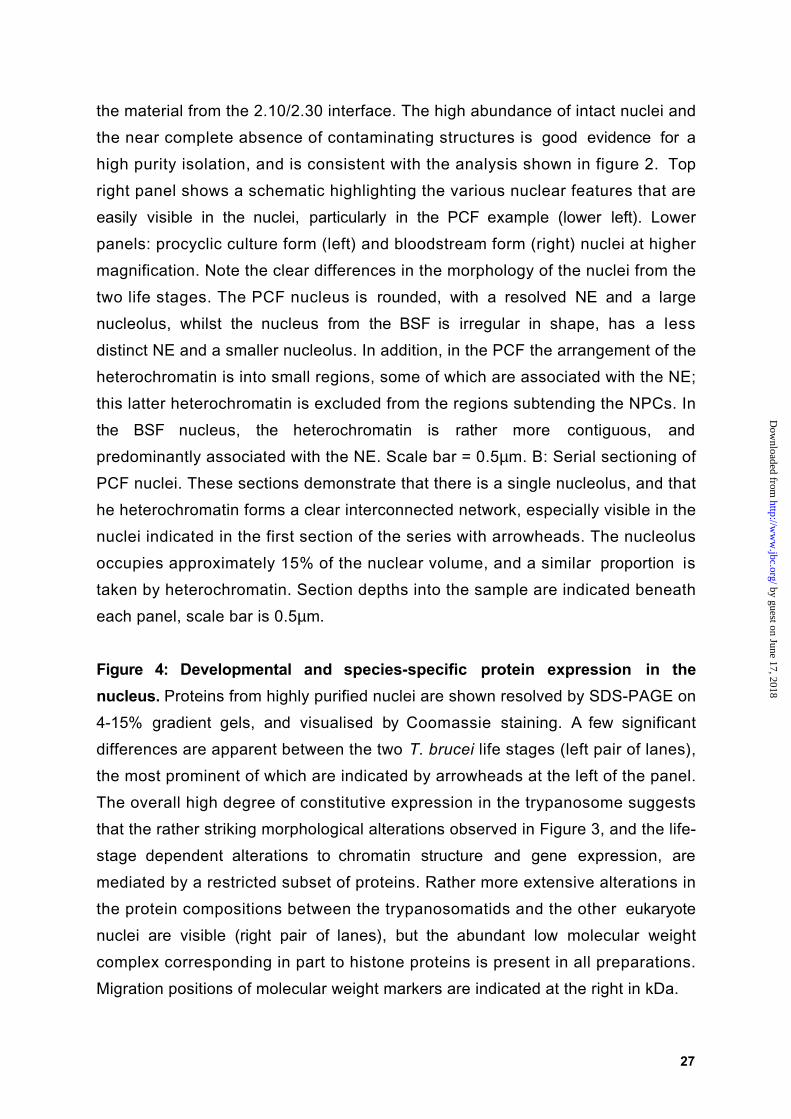
predominantly associated with the NE. Scale bar = 0.5µm. B: Serial sectioning of
PCF nuclei. These sections demonstrate that there is a single nucleolus, and that
he heterochromatin forms a clear interconnected network, especially visible in the
nuclei indicated in the first section of the series with arrowheads. The nucleolus
occupies approximately 15% of the nuclear volume, and a similar proportion is
taken by heterochromatin. Section depths into the sample are indicated beneath
each panel, scale bar is 0.5µm.
Figure 4: Developmental and species-specific protein expression in the
nucleus. Proteins from highly purified nuclei are shown resolved by SDS-PAGE on
4-15% gradient gels, and visualised by Coomassie staining. A few significant
differences are apparent between the two T. brucei life stages (left pair of lanes),
the most prominent of which are indicated by arrowheads at the left of the panel.
The overall high degree of constitutive expression in the trypanosome suggests
that the rather striking morphological alterations observed in Figure 3, and the life-
stage dependent alterations to chromatin structure and gene expression, are
mediated by a restricted subset of proteins. Rather more extensive alterations in
the protein compositions between the trypanosomatids and the other eukaryote
nuclei are visible (right pair of lanes), but the abundant low molecular weight
complex corresponding in part to histone proteins is present in all preparations.
Migration positions of molecular weight markers are indicated at the right in kDa.
by guest on June 17, 2018http://w
ww
.jbc.org/D
ownloaded from

28
Figure 5: Analysis of trypanosome subnuclear fractionation procedures by SDS-
PAGE and Western blot. A: Coomassie blue-stained 1D SDS-PAGE separations
of the fractions from representative PCF subnuclear fractionations are shown in
the top panel, and corresponding western blots are shown below. Icons
correspond to those used in Figure 1; grey arrows indicate the derivation of the
nucleolar, NE, and PCLF fractions from preceding fractions. Antibodies used are
indicated at the right of the blot images (RRM1, NUMAG and NUP-1), and the
compartments they predominantly recognise are given at left. Molecular weight
standards are shown at left in kDa, and sucrose gradient fraction designations
given above the corresponding lanes. The leftmost three lanes are the nucleolar
isolation, the centre three the NE preparation, and the rightmost two lanes are the
PCLF purification. Coenrichment of the nucleolar marker NUMAG is clear for the
nucleoli preparation, and enrichment for the NE maker NUP-1 is similarly
significant for the isolation of the NE and PCLF. Note the comparative simplicity of
the protein compositions of the final fractions, and the removal of the majority of
the histone complex (positions indicated to the right of the gel with filled circles).
The 350kDa doublet recognised by the NUP-1 antibody is clearly visible in the
1.50/2.25 and 1.75/2.50 fractions (positions indicated to the right of the gel with
open diamonds). B: Quantitation of total protein and RRM1, NUMAG and NUP-1
yields from the nuclear and subnuclear isolation procedures. Sucrose gradient
fraction designations and icons are as in Figure 1. Data are represented as a
percentage of the protein present in the relevant starting material.
Figure 6: Ultrastuctural analysis of the nucleolar fraction prepared from T.
brucei nuclei. Representative EM images of the final nucleolar preparation are
shown at low (left panel) and high (right panel) magnification. The nucleoli are
heterogeneous in size, which may reflect a real variability in the structure of this
organelle based on cell cycle position and other parameters, but this probably
also arises in part from fragmentation of the structure during isolation. Note that
nucleoli contain a highly compacted central core region, with a less dense
peripheral region; this is also seen in images of nucleoli within the intact PCF
nuclei. Some of the nucleoli are associated with peripheral material, which is most
likely chromatin and nuclear matrix remnants. Scale bars = 0.5µm.
by guest on June 17, 2018http://w
ww
.jbc.org/D
ownloaded from

29
Figure 7: Ultrastuctural analysis of the nuclear envelope fraction prepared from
T. brucei nuclei. A: Low magnification electron micrograph of isolated NEs. Note
the near absence of nucleoplasmic material (chromatin and nucleolus) from these
preparations. Clearly visible are the inner and outer nuclear membranes, and also
electron dense structures arrayed along the nuclear envelopes, which correspond
to NPCs. B: High magnification electron micrographs demonstrating detailed
ultrastructure of the trypanosome NE. Schematics showing visible features of the
NPC are shown at the left of the figure. Top panels are en face views of the NEs.
The left hand micrograph is a tangential section, clearly revealing the ring structure
of the NPCs, and in some instances a central plug and spoke elements can be
seen (arrowhead). Ribosomes studding the outer face of the NE are apparent in
the centre of the micrograph. The right hand image is a negative stain preparation;
again features of the NPC can be discerned. Note the high density of the NPCs,
especially clear in the negative stain image. Lower panels are cross sections of
NEs and again reveal details of the NPC architecture. Clearly visible are the
cytoplasmic and nuclear filaments, the central core elements and electron dense
material corresponding to the main ring structures. Good examples are indicated
in the left most two panels by arrowheads. In the right panel a clear filamentous
network is seen associated with the inner nuclear membrane and which may
constitute the trypanosome nuclear lamina (arrowhead). Insets show the same
schematics as the left but reduced to scale. Scale bar = 0.5µm.
Figure 8: Ultrastucture of the nuclear pore complex lamina fractions prepared
from T. brucei nuclei. A: Low magnification EM image of the PCLF. The majority of
the membranous material has been removed, and the NPCs are visible again as
electron dense punctata (compare this image with the one in Figure 7A), but the
overall integrity remains, indicating a robust lamina structure is present. B; High
magnification electron micrographs showing details of the NPC structures; again
a central core and peripheral filamentous structures are clearly visible. Note that
the ribosomes, visible in earlier fractions as small dense dots, have been
completely removed, consistent with an efficient extraction of membrane elements.
Insets contain schematics of the NPC to scale, en face (top) or in cross section
(lower). Scale bar = 0.5µm.
by guest on June 17, 2018http://w
ww
.jbc.org/D
ownloaded from

30
Figure 9: Identification of the 350kDa band as the NUP-1 antigen. A: Protein
sequencing data for Histone H4 and H2B. Complete sequences of trypanosome
histones H4 and H2B, with the sequences identified by peptide sequencing in
bold. The molecular weights of the bands taken for sequencing are consistent with
the migration positions of histone H4 and H2B on SDS gels (Bender et al. 1992B).
B: Partial open reading frame assembled from database retrieved GSSs using
four peptide sequences obtained from the 350kDa band from the NE preparation
predicts a coiled coil protein containing 144 amino acid repeats of unmodified
molecular weight of 15.9kDa. Peptides with an exact match are in bold, and
sequences with a partial match are underlined. Alignments of a complete repeat
with partial repeat sequences derived from the N and C-terminus of the assembly
reveals an almost prefect repeat structure. The gap in the alignment (residues 61-
75) represents missing data. C: p350 is a coiled coil protein. Secondary structure
prediction was done with the Lupas algorithm (Lupas et al. 1991) using MacStripe
2.0a1 with default settings. Two high probability peaks are contained within each
repeat. D: Mild trypsin digestion of isolated nuclei reveals that NUP-1 is a repetitive
protein. Digests were analysed by SDS-PAGE and Western blotting with the NUP-1
antibody. Trypsin treatment reveals a ladder of NUP-1 immunoreactivity, with an
approximate repeat mass of 17kDa, consistent with the repeat identified by
peptide sequencing. Increased trypsin digestion results in a greater yield of lower
molecular weight products, and indicates that there are ~20 repeats in the NUP-1
protein. Migration positions of molecular weight markers are indicated at left in
kDa.
Figure 10: Immunoelectron microscopy localises the NUP-1 antigen to the
nucleoplasmic face of the nuclear envelope. NEs were incubated with mAb NUP-
1 and the antibody visualized with gold-conjugated secondary antibodies. Top left
and top right panels, transverse sections of NEs (C; cytoplasmic side, N;
nucleoplasmic side). Bottom right panel, tangential section of NE (NPC = nuclear
pore complex). Scale bar = 0.5µm. Bottom left panel, montage of 40 aligned NPCs
showing a total of 198 gold particles. The gold particles are found an average of
57+18nm from the mid-plane of the NE. Scale bars are graduated in 10nm
intervals. Both the transverse sections and montage indicate that the NUP-1
by guest on June 17, 2018http://w
ww
.jbc.org/D
ownloaded from

31
antigen is localised to a layer beneath the inner face of the NE and that the
labelling avoids the vicinity of the NPC.
by guest on June 17, 2018http://w
ww
.jbc.org/D
ownloaded from

ttttrrrryyyyppppaaaannnnoooossssoooommmmeeeessss
SN
crude nuclearlysate
crude cytosol
0.3
2.01/2.10
2.10/2.30
2.30
S/2.01
nnnnuuuucccclllleeeeiiii
other membranous
structures
0.3/S
ppppoooollllyyyyttttrrrroooonnnn
nnnnuuuucccclllleeeeoooolllliiii
ssssoooonnnniiiiccccaaaatttteeee
S/1.50
1.50/2.25
2.25/2.50
hhhheeeeppppaaaarrrriiiinnnn////DDDDNNNNaaaasssseeeeeeeexxxxttttrrrraaaaccccttttiiiioooonnnn
S/1.75
1.75/2.50ppppccccllllffff
ddddeeeetttteeeerrrrggggeeeennnntttt eeeexxxxttttrrrraaaaccccttttiiiioooonnnn
2.25/S
1.50/2.25
S
eeeennnnvvvveeeellllooooppppeeeessss
ppppoooollllyyyyttttrrrroooonnnn
by guest on June 17, 2018http://w
ww
.jbc.org/D
ownloaded from


CALVSDVAVRNADTDLGTQLASALVALERLAEEREAALEKATEMEERVSTLEKELRTAHSTTKKMSAERELHVTKLTQLEETVSRLESYGTTPEQTVAAFTTELQHTQQRLREAEEEIIQLTNKLNAAGVRVRTSQSDKDGNARAALVSDVAVRNADTDLGTQLASALVALERLAEEREAALEKATEMEERVSTLEEELRTAHSTTKKMSAERELHVTKLTQLEETVSRLESYGTTPEQTVAAFTTELQHTQQRLREAEEEIIQLTNK
TbNUP-1 contig
144 repeatPartial repeats
144 repeatPartial repeats
144 repeatPartial repeats
11
6158
121109
E L H V T K L T Q L E E T V S R L E S Y G T T P E Q T V A A F T T E L Q H T Q Q R L R E A E E E IE L H V T K L T Q L E E T V S R L E S Y G T T P E Q T V A A F T T E L Q H T Q Q R L R E A E E E I
V R V R T S Q S D K D G N A RA A L V S D V A V R N A D T D L G T Q L A S A L V A L E R L A E E RC A L V S D V A V R N A D T D L G T Q L A S A L V A L E R L A E E R
E R V S T L E E E L R T A H S T T K K M S A E RE R V S T L E K E L R T A H S T T K K M S A E R
A
BMATPKSTPAKTRKEAKKTRRHQATWNVYVSRSLRSINSQMSMTSRTMKIVNSFVNDLFERIAAEAATIVRVNRKRTLGARELQTAVRLVLPADLAKTRWQRGTKAVSHASS
MAKGKKSGEAKGSQKRQKKVLRENVRGITRGSIRRLARRGGVKRISGVIYDEVRGVLKSFVEGVVRDATAYTEYSRKKTVTAVDVVNALRKRGKILYGYA
T. brucei H4
T. brucei H2B
CCCC
by guest on June 17, 2018http://w
ww
.jbc.org/D
ownloaded from


Michael P. Rout and Mark C. Fieldbrucei: identification of a major repetitive nuclear lamina component
Isolation and characterisation of subnuclear compartments from Trypanosoma
published online July 26, 2001J. Biol. Chem.
10.1074/jbc.M104024200Access the most updated version of this article at doi:
Alerts:
When a correction for this article is posted•
When this article is cited•
to choose from all of JBC's e-mail alertsClick here
by guest on June 17, 2018http://w
ww
.jbc.org/D
ownloaded from