liver regeneration associated protein (alr) exhibits
TRANSCRIPT

M O L M E D 1 7 ( 3 - 4 ) 2 2 1 - 2 2 8 , M A R C H - A P R I L 2 0 1 1 | D A Y O U B E T A L . | 2 2 1
INTRODUCTIONHepatocellular carcinoma (HCC) is
the most prevalent form of the primaryliver cancers, which in turn are the fifthmost common cancers worldwide— contributing substantially to cancermortality (1). HCC develops largelywithin an established background ofchronic liver diseases, caused mainly byinfections with hepatitis B and/or hepa-titis C virus or alcoholic liver disease(2), but the molecular pathogenesis isnot well understood. Although newtherapies for chronic hepatitis have beenestablished, the number of HCC patients
has not declined (1). Treatment successof HCC is dependent on the tumor stageat the time of diagnosis, with good cur-ability at early stages and poor progno-sis at advanced stages with tumor recur-rence and metastasis after surgery orablation therapy (2,3). Various factorsare involved in the regulation of metas-tasis, and epithelial-mesenchymal tran-sition (EMT) is characteristic for themost aggressive metastatic cancer cells(4,5). Therefore, biomarkers suitable fordiagnostic purposes and new targets fordeveloping therapeutic strategies areneeded.
Human augmenter of liver regenera-tion (ALR) is a member of the newly dis-covered ALR/Erv1 protein family withFAD-linked sulfhydryl oxidase activitycatalyzing disulfide bond formation (6,7).ALR is expressed ubiquitously with thehighest levels found in testis and liver(8–10). Alternative splicing generates (11)a 21/23 kDa ALR isoform predominantlylocalized to the mitochondrial inner-membrane system (IMS). In concertwith the redox-regulated receptorMia40/TOM, ALR forms a disulfiderelay system mediating the import ofproteins of mitochondria into IMS (12).Furthermore, CXXC motif of ALR is es-sential for cell survival (13) and biogene-sis of cytosolic Fe/S proteins (14). Theshort form of ALR (15 kDa) is found notonly at extracellular sites but also at nu-clear and cytosolic localizations partici-pating in intracellular redox-dependent
Liver Regeneration Associated Protein (ALR) ExhibitsAntimetastatic Potential in Hepatocellular Carcinoma
Rania Dayoub,1,5 Hannah Wagner,1,2 Frauke Bataille,3 Oliver Stöltzing,2 Thilo Spruss,4 Christa Buechler,6
Hans-Jürgen Schlitt,1,2 and Thomas S Weiss1,5
1Center for Liver Cell Research, University Medical Center Regensburg, Germany; 2Department of Surgery, University MedicalCenter Regensburg, Germany; 3Department of Pathology, University Medical Center Regensburg, Germany; 4Institute of Pharmacy,University of Regensburg, Germany; 5Department of Pediatrics, University Medical Center Regensburg, Germany; and 6Departmentof Internal Medicine, University Medical Center Regensburg, Germany
Augmenter of liver regeneration (ALR), which is critically important in liver regeneration and hepatocyte proliferation, is highlyexpressed in cirrhotic livers and hepatocellular carcinomas (HCC). In the current study, the functional role of ALR in hepato-cancerogenesis was analyzed in more detail. HepG2 cells, in which the cytosolic 15 kDa ALR isoform was reexpressed stably,(HepG2-ALR) were used in migration and invasion assays using modified Boyden chambers. Epithelial-mesenchymal transition(EMT) markers were determined in HepG2-ALR cells in vitro and in HepG2-ALR tumors grown in nude mice. ALR protein was quan-tified in HCC and nontumorous tissues by immunohistochemistry. HepG2-ALR, compared with HepG2 cells, demonstrated reducedcell motility and increased expression of the epithelial cell markers E-cadherin and Zona occludens-1 (ZO-1), whereas SNAIL, anegative regulator of E-cadherin, was diminished. Matrix metalloproteinase MMP1 and MMP3 mRNA expression and activity werereduced. HepG2-ALR cell-derived subcutaneously grown tumors displayed fewer necrotic areas, more epithelial-like cell growthand fewer polymorphisms and atypical mitotic figures than tumors derived from HepG2 cells. Analysis of tumor tissues of 53 pa-tients with HCC demonstrated an inverse correlation of ALR protein with histological angioinvasion and grading. The 15 kDa ALRisoform was found mainly in HCC tissues without histological angioinvasion 0. In summary the present data indicate that cytosolicALR reduces hepatoma cell migration, augments epithelial growth and, therefore, may act as an antimetastatic and EMT revers-ing protein.© 2011 The Feinstein Institute for Medical Research, www.feinsteininstitute.orgOnline address: http://www.molmed.orgdoi: 10.2119/molmed.2010.00117
Address correspondence and reprint requests to Thomas S Weiss, Center for Liver Cell Re-
search, University Medical Center Regensburg, F-J-S-Allee 11, 93053 Regensburg, Germany.
Phone: +49-941 9442195; Fax: +49-941 9442196; E-mail: [email protected].
Submitted July 16, 2010; Accepted for publication December 2, 2010; Epub
(www.molmed.org) ahead of print December 8, 2010.

2 2 2 | D A Y O U B E T A L . | M O L M E D 1 7 ( 3 - 4 ) 2 2 1 - 2 2 8 , M A R C H - A P R I L 2 0 1 1
A L R E X H I B I T S A N T I M E T A S T A T I C P O T E N T I A L
signaling pathways (15,16). ALR is a he-patotrophic factor stimulating prolifera-tion of hepatocytes (17,18) and augment-ing liver regeneration (15,16), therebyexerting beneficial effects in models ofhepatic failure (19) and liver fibrosis (20).
Furthermore, enhanced ALR mRNAand protein expression is found in theliver of patients with cirrhosis, cholangio-cellular and hepatocellular carcinoma.Here, ALR is localized predominantly inhepatocytes and bile duct cells (10). How-ever, it is not clear yet whether the cytoso-lic isoform of ALR plays a role in the de-velopment of HCC. The aim of this studywas to investigate the functional role andclinical importance of the short cytosolicALR isoform in hepatocancerogenesis.
MATERIALS AND METHODS
Liver SamplesTissues of 64 patients undergoing liver
surgery at the University Medical CenterRegensburg were collected, including 53patients with primary HCCs, three pa-tients with liver cirrhosis and eight pa-tients with colorectal liver metastasesserving as healthy liver tissues. Tissuesamples were fixed in formalin and paraf-fin-embedded for immunohistochemicalanalysis. Experimental procedures wereperformed according to the guidelines ofthe charitable state controlled foundationHTCR (Human Tissue and Cell Research),with the informed patients’ consent (21)approved by the local ethical committeeof the University of Regensburg.
Immunohistochemical Analysis of ALRExpression
Immunostaining was performed as de-scribed recently (10). Briefly, 2-mm sec-tions of paraffin-embedded tissue wereused, and unspecific binding was blockedby 30 min preincubation in phosphatebuffered saline (PBS) with 3% humanserum (Bio-Rad, Munich, Germany), sec-tions were subsequently incubated withanti-ALR antibody (0.26 μg/mL) at 4°Covernight followed by incubation withsecondary mouse antirabbit antibody(DAKO, Hamburg, Germany) for 1 h.
Immunostaining was developed usingAPAAP complex (DAKO) and Fast RedChromogen staining (Roche, Penzberg,Germany). Counterstaining was per-formed with hematoxylin (Vektor,Burlingame, Canada). Rabbit IgG(Sigma, Munich, Germany) was used asisotype control. To evaluate ALR im-munostaining, the percentage of positivecells and intensity of staining was ana-lyzed independently by two patholo-gists. The following scores were defined:≤10% positive cells = 0; 10% to 25 % positive cells = 1, 25% to 50 % positivecells = 2; 50% to 75 % positive cells = 3;and ε 75% positive cells = 4. Signal inten-sity was defined as 0 when no stainingwas detected, as 1 when staining wasweak, as 2 when staining was moderateand as 3 when staining was strong. Theexpression level of ALR was calculatedby multiplying the scores: 0 = negative,1–4 = positive, 6–12 = highly positive.
Cell Culture and Stable TransfectedCells
Primary human hepatocytes (PHH)were isolated and cultured as described(22). The human hepatoma cell line HepG2(clone HB-8065) was obtained from Amer-ican Type Culture Collection, (ATCC,Manassas, VA, USA) and grown at 37°C,5% CO2 in DMEM (BioWhittaker, Verviers,Belgium) supplemented with penicillin(100 units/mL), streptomycin (10 μg/mL)and 10% fetal calf serum. The human ALRcDNA encoding the short isoform was di-gested with BamHI and EcoRI and sub-cloned into the expression vectorpcDNA3.1 (Invitrogen, Karlsruhe, Ger-many). HepG2 cells were transfected usingsiPORT XP-1 (Ambion, Austin, TX, USA)regarding to the manufacturer’s instruc-tions. HepG2 cells stably expressing ALRwere selected by 1 mg/mL G-418 (GIBCOBRL, Karlsruhe, Germany) over a periodof 2 wks. Cells stably transfected withpcDNA3.1 served as controls (mock).
Immunocytochemical AnalysisCells grown on BD BioCoat Collagen I
culture slides (BD Biosciences, Heidelberg,Germanty) were fixed and incubated with
anti-ALR (1:100) (10), anti-CDH1 (1:100)(Cell Signaling, Danvers, MA, USA), oranti-ZO-1 (1:100) antibody (Invitrogen).Cryosections (2 μm) from tumors derivedfrom HepG2-ALR and HepG2 cells werestained using anti-CDH1 (1:100) or anti-ZO1 (1:100) antibody. Fluorescent signalswere visualized by an Alexa Fluor 488dye-conjugated goat antirabbit (IgG) anti-body (Invitrogen).
Migration and Invasion AssaysBriefly, 0.5 × 10⁵ cells were resus-
pended in RPMI medium containing 1%FCS and seeded on inserts with 8-mm fil-ter pores, which were either uncoated(migration assay) or matrigel-coated (in-vasion assay) (Becton Dickinson Bio-science, Heidelberg, Germany). 10% FCSserved as chemoattractant. After 48 h,cells were fixed and stained (Diff-Quickreagent, Dade Behring, Newark, NJ,USA) and migrated and invaded cellswere counted.
Scratch Assay ‘Wound-Healing Assay’Migration of cells was assayed by
scratch assays. Cells were seeded in highdensity into six-well plates and scratchedby a pipette tip in a definite array. Migra-tion into this array was documented andmeasured after 48 h. Each analysis wasperformed in triplicate.
Nude Mouse Tumor ModelA model of inoculation of tumor cells
into NMRI (nu/nu) mice to monitortumor growth in vivo was performed asdescribed previously (23). Ten-wk-oldmale athymic nude mice NMRI (nu/nu)from the universities breeding stock wereused (mean body weight was 30 g).HepG2-ALR cells or mock-transfectedHepG2 cells (1 × 10⁶ cells per mouse)were injected subcutaneously into nudemice, and six mice per clone were used.After 21 d, the formed tumors were re-moved, snap frozen and stored at –80°Cfor subsequent analysis.
RT-PCR AnalysisTotal RNA was purified using RNeasy
Mini Kit (Qiagen, Hilden, Germany). One

R E S E A R C H A R T I C L E
M O L M E D 1 7 ( 3 - 4 ) 2 2 1 - 2 2 8 , M A R C H - A P R I L 2 0 1 1 | D A Y O U B E T A L . | 2 2 3
μg of total RNA was reverse transcribedusing the Reverse-Transcription System(Promega, Madison, WI, USA). Transcriptlevels of ALR, MMP1, MMP3 and 18SrRNA as internal control were quantifiedusing real-time PCR technology (Light-Cycler, Roche, Penzberg, Germany).Primers used to amplify ALR were: for-ward 5’-CAC AAT GAA GTG AAC CGCAAG-3’, reverse 5’-CAC CCA ACT GAGACA CAA CAG-3’, MMP1: forward 5’-TCA CCA AGG TCT CTG AGG GTCAAG C-3’, reverse 5’-GGA TGC CATCAA TGT CAT CCT GAG C-3’, 18S rRNA:forward 5’-GTA ACC CGT TGA ACCCCA TT-3’, reverse 5’-CCA TCC AATCGG TAG TAG CG-3’, MMP3 primerswere obtained from TIB Molbiol (Berlin,Germany). The PCR reaction was evalu-ated by melting curve analysis. PCR wasperformed in duplicate and three inde-pendent experiments were performed.
Western Blot AnalysisProtein lysates (20 μg/lane) were sepa-
rated by SDS-PAGE under reducing con-ditions using 100 mM DTT. Proteins weretransferred to PVDF membranes and in-cubated with anti-CDH1 (1:1000), anti-ZO-1 (1:250), anti-SNAIL (1:250) (SantaCruz Biotechnology, Heidelberg, Ger-many), anti-ALR (1:440) or anti–β-actin(1:1000) (Santa Cruz Biotechnology) anti-bodies. Positive signals were developedusing enhanced chemiluminescencereagent (Pierce, Rockford, IL, USA).
Measurement of MMP1 and MMP3Activity In Vitro
HepG2 and HepG2-ALR cells were cultured for 48 h and supernatants werecollected. MMP1 and MMP3 activitieswere measured using the SensoLyte 520MMP-1 Assay Kit and the SensoLyte 520MMP-3 Assay Kit, respectively, followingmanufacturer’s instructions (AnaSpec,Fremont, CA, USA). Briefly, MMP activ-ity in culture supernatants was deter-mined using the fluorogenic MMP spe-cific substrate 5-FAM/QXL520 FRETpeptide. Assays were performed in black96-well plates with a clear flat bottom(Corning Inc, Corning, NY, USA) at 37°C.
The increase in fluorescence resultingfrom enzyme mediated cleavage of thesubstrates was measured at 490/520 nm(excitation/emission)
Statistical AnalysisSignificant differences in paired and
nonpaired samples were evaluated byusing Wilcoxon signed-rank t test andMann-Whitney U test, respectively. Clini-copathological parameters were calcu-lated by using Pearson chi-square test.
All supplementary materials are availableonline at www.molmed.org.
RESULTS
Reexpression of the Short Isoform ofALR in HepG2 Cells
To gain more insight into the func-tional role of ALR in HCC, the short iso-form of ALR (15 kDa) was reexpressedstably in the hepatoma cell line HepG2(HepG2-ALR). RT-PCR and Western blotanalysis revealed strong induction ofALR mRNA and protein in HepG2-ALRcell clones compared with mock trans-fected and nontransfected HepG2 cells,respectively (Figure 1A, B). All three iso-
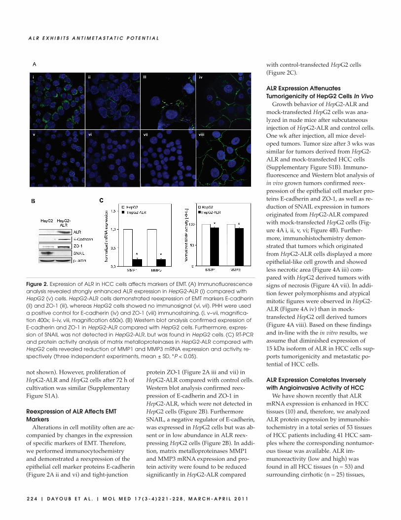
forms (23 kDa, 21 kDa, 15 kDa) were de-tected in normal liver tissue whereas the15 kDa ALR isoform was barely detectedin HepG2 cells (Figure 1B). Since HepG2-ALR2 cells showed a more intense 15 kDaALR expression than HepG2-ALR1, thisclone termed HepG2-ALR was used infurther studies. Mock transfected HepG2cells were used as control. Immunocyto-chemical analysis identified a stronglyenhanced expression of cytosolic ALR inHepG2-ALR compared with HepG2 cells(Figure 2A i and v).
Reduced Migration and Invasivenessof HCC Cells after ALR Reexpression
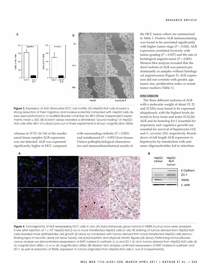
Impact of ALR expression on cellularfunction was studied by analyzing cellmotility. By performing Boyden chamberand matrigel invasion assays, a signifi-cant reduction in migration and invasionof HepG2-ALR cells compared with con-trol cells was found (Figure 3A). In addi-tion, scratch-assays (wound-healing as-says) displayed a clearly diminishedmigration capacity of ALR reexpressingHCC cells compared with control cells(Figure 3B). Furthermore, cell adherenceof HepG2-ALR after seeding was muchfaster compared with HepG2 cells (data
Figure 1. Stable reexpression of short isoform of ALR in HCC cells. HepG2 cells stably ex-pressing the 15 kDa ALR isoform (HepG2-ALR1 and HepG2-ALR2). (A) RT-PCR analysis re-vealed strong ALR mRNA expression in both ALR expressing HepG2 cells compared withnontransfected HepG2 or mock-transfected cells (three independent experiments,mean ± SD). (B) Western blot analysis demonstrated reexpression of the 15 kDa isoformALR in HepG2-ALR cell clones. Normal liver tissue served as a positive control. Expressionof β-actin was used as loading control.
2 2 4 | D A Y O U B E T A L . | M O L M E D 1 7 ( 3 - 4 ) 2 2 1 - 2 2 8 , M A R C H - A P R I L 2 0 1 1
A L R E X H I B I T S A N T I M E T A S T A T I C P O T E N T I A L
not shown). However, proliferation ofHepG2-ALR and HepG2 cells after 72 h ofcultivation was similar (SupplementaryFigure S1A).
Reexpression of ALR Affects EMTMarkers
Alterations in cell motility often are ac-companied by changes in the expressionof specific markers of EMT. Therefore,we performed immunocytochemistryand demonstrated a reexpression of theepithelial cell marker proteins E-cadherin(Figure 2A ii and vi) and tight-junction
protein ZO-1 (Figure 2A iii and vii) inHepG2-ALR compared with control cells.Western blot analysis confirmed reex-pression of E-cadherin and ZO-1 inHepG2-ALR, which were not detected inHepG2 cells (Figure 2B). FurthermoreSNAIL, a negative regulator of E-cadherin,was expressed in HepG2 cells but was ab-sent or in low abundance in ALR reex-pressing HepG2 cells (Figure 2B). In addi-tion, matrix metalloproteinases MMP1and MMP3 mRNA expression and pro-tein activity were found to be reducedsignificantly in HepG2-ALR compared
with control-transfected HepG2 cells (Figure 2C).
ALR Expression AttenuatesTumorigenicity of HepG2 Cells In Vivo
Growth behavior of HepG2-ALR andmock-transfected HepG2 cells was ana-lyzed in nude mice after subcutaneousinjection of HepG2-ALR and control cells.One wk after injection, all mice devel-oped tumors. Tumor size after 3 wks wassimilar for tumors derived from HepG2-ALR and mock-transfected HCC cells(Supplementary Figure S1B). Immuno-fluorescence and Western blot analysis ofin vivo grown tumors confirmed reex-pression of the epithelial cell marker pro-teins E-cadherin and ZO-1, as well as re-duction of SNAIL expression in tumorsoriginated from HepG2-ALR comparedwith mock-transfected HepG2 cells (Fig-ure 4A i, ii, v, vi; Figure 4B). Further-more, immunohistochemistry demon-strated that tumors which originatedfrom HepG2-ALR cells displayed a moreepithelial-like cell growth and showedless necrotic area (Figure 4A iii) com-pared with HepG2 derived tumors withsigns of necrosis (Figure 4A vii). In addi-tion fewer polymorphisms and atypicalmitotic figures were observed in HepG2-ALR (Figure 4A iv) than in mock- transfected HepG2 cell derived tumors(Figure 4A viii). Based on these findingsand in-line with the in vitro results, weassume that diminished expression of15 kDa isoform of ALR in HCC cells sup-ports tumorigenicity and metastatic po-tential of HCC cells.
ALR Expression Correlates Inverselywith Angioinvasive Activity of HCC
We have shown recently that ALRmRNA expression is enhanced in HCCtissues (10) and, therefore, we analyzedALR protein expression by immunohis-tochemistry in a total series of 53 tissuesof HCC patients including 41 HCC sam-ples where the corresponding nontumor-ous tissue was available. ALR im-munoreactivity (low and high) wasfound in all HCC tissues (n = 53) andsurrounding cirrhotic (n = 25) tissues,
Figure 2. Expression of ALR in HCC cells affects markers of EMT. (A) Immunofluorescenceanalysis revealed strongly enhanced ALR expression in HepG2-ALR (i) compared withHepG2 (v) cells. HepG2-ALR cells demonstrated reexpression of EMT markers E-cadherin(ii) and ZO-1 (iii), whereas HepG2 cells showed no immunosignal (vi, vii). PHH were useda positive control for E-cadherin (iv) and ZO-1 (viii) immunostaining. (i, v–vii, magnifica-tion 400x; ii–iv, viii, magnification 650x). (B) Western blot analysis confirmed expression ofE-cadherin and ZO-1 in HepG2-ALR compared with HepG2 cells. Furthermore, expres-sion of SNAIL was not detected in HepG2-ALR, but was found in HepG2 cells. (C) RT-PCRand protein activity analysis of matrix metalloproteinases in HepG2-ALR compared withHepG2 cells revealed reduction of MMP1 and MMP3 mRNA expression and activity, re-spectively (three independent experiments, mean ± SD, *P < 0.05).
R E S E A R C H A R T I C L E
M O L M E D 1 7 ( 3 - 4 ) 2 2 1 - 2 2 8 , M A R C H - A P R I L 2 0 1 1 | D A Y O U B E T A L . | 2 2 5
whereas in 37.5% (6/16) of the nondis-eased tissue samples ALR expressionwas not detected. ALR was expressedsignificantly higher in HCC compared
with surrounding cirrhotic (P < 0.001)and nondiseased (P < 0.001) liver tissues.Clinico-pathophysiological characteris-tics and immunohistochemical results of
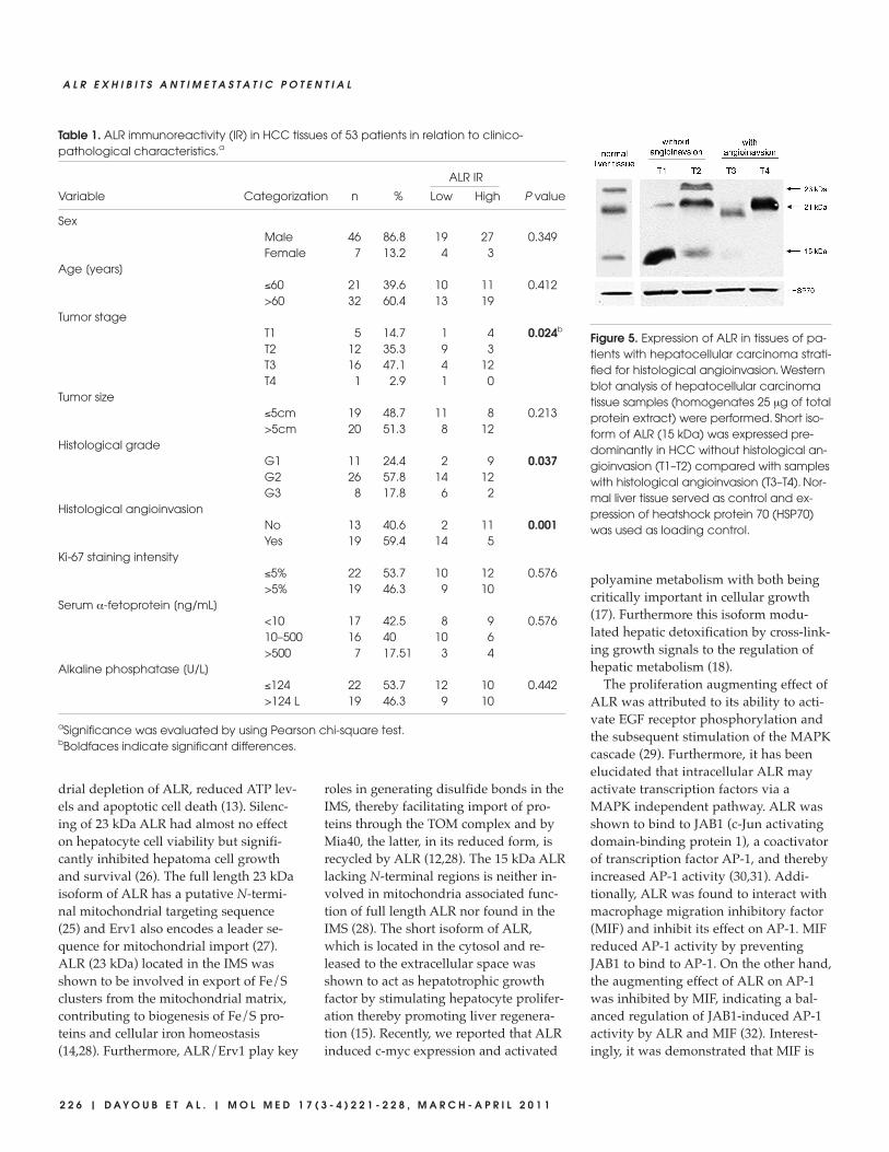
the HCC tumor cohort are summarizedin Table 1. Positive ALR immunostainingwas found to be associated significantlywith higher tumor stage (P = 0.024). ALRexpression correlated inversely withtumor grading (P = 0.037) and the rate ofhistological angioinvasion (P < 0.001).Western blot analysis revealed that theshort isoform of ALR was present pre-dominantly in samples without histologi-cal angioinvasion (Figure 5). ALR expres-sion did not correlate with gender, age,tumor size, proliferation index or serumtumor markers (Table 1).
DISCUSSIONThe three different isoforms of ALR
with a molecular weight of about 15, 21and 23 kDa were found to be expressedubiquitously with the highest levels de-tected in liver, brain and testes (9,10,24).ALR and its homolog Erv1 (essential forrespiratory and vegetative growth) areessential for survival of hepatocytes (13)and S. cerevisiae (25), respectively. Knock-down of full length ALR expression inhepatocytes by transfection with anti-sense oligonucleotides led to mitochon-
Figure 3. Expression of ALR attenuates HCC cell motility. (A) HepG2-ALR cells showed astrong reduction of their migratory and invasive potential compared with HepG2 cells. As-says were performed in a modified Boyden chamber for 48 h (three independent experi-ments, mean ± SD). (B) Scratch assays revealed a diminished “wound healing” of HepG2-ALR cells after 48 h of culture (one out of three experiments is shown, magnification 200x).
Figure 4. Tumorigenicity of ALR reexpressing HCC cells in vivo. (A) Subcutaneously grown tumors in NMRI (nu/nu) mice were analyzed 3 wks after injection of 1 × 10⁶ HepG2-ALR (i–iv) or mock transfected HepG2 cells (v–viii). HE staining of tumors derived from HepG2-ALRcells revealed more epithelial-like cell growth (iii arrow, iv) compared with tumors derived from mock transfected HepG2 cells demon-strating signs of necrotic areas (vii arrow head), cell polymorphism and atypical mitotic figures (viii arrow). Performing immunofluores-cence analysis we demonstrated reexpression of EMT markers E-cadherin (i, v) and ZO-1 (ii, vi) in tumors derived from HepG2-ALR cells. (iii,vii, magnification 400x; i, ii, iv–vi, viii, magnification 650x). (B) Western blot analysis confirmed reexpression of EMT markers E-cadherin andZO-1 as well as reduction of SNAIL expression in tumors originated from HepG2-ALR cells (1 out of 3 experiments).
2 2 6 | D A Y O U B E T A L . | M O L M E D 1 7 ( 3 - 4 ) 2 2 1 - 2 2 8 , M A R C H - A P R I L 2 0 1 1
A L R E X H I B I T S A N T I M E T A S T A T I C P O T E N T I A L
drial depletion of ALR, reduced ATP lev-els and apoptotic cell death (13). Silenc-ing of 23 kDa ALR had almost no effecton hepatocyte cell viability but signifi-cantly inhibited hepatoma cell growthand survival (26). The full length 23 kDaisoform of ALR has a putative N-termi-nal mitochondrial targeting sequence(25) and Erv1 also encodes a leader se-quence for mitochondrial import (27).ALR (23 kDa) located in the IMS wasshown to be involved in export of Fe/Sclusters from the mitochondrial matrix,contributing to biogenesis of Fe/S pro-teins and cellular iron homeostasis(14,28). Furthermore, ALR/Erv1 play key
roles in generating disulfide bonds in theIMS, thereby facilitating import of pro-teins through the TOM complex and byMia40, the latter, in its reduced form, isrecycled by ALR (12,28). The 15 kDa ALRlacking N-terminal regions is neither in-volved in mitochondria associated func-tion of full length ALR nor found in theIMS (28). The short isoform of ALR,which is located in the cytosol and re-leased to the extracellular space wasshown to act as hepatotrophic growthfactor by stimulating hepatocyte prolifer-ation thereby promoting liver regenera-tion (15). Recently, we reported that ALRinduced c-myc expression and activated
polyamine metabolism with both beingcritically important in cellular growth(17). Furthermore this isoform modu-lated hepatic detoxification by cross-link-ing growth signals to the regulation ofhepatic metabolism (18).
The proliferation augmenting effect ofALR was attributed to its ability to acti-vate EGF receptor phosphorylation andthe subsequent stimulation of the MAPKcascade (29). Furthermore, it has beenelucidated that intracellular ALR mayactivate transcription factors via aMAPK independent pathway. ALR wasshown to bind to JAB1 (c-Jun activatingdomain-binding protein 1), a coactivatorof transcription factor AP-1, and therebyincreased AP-1 activity (30,31). Addi-tionally, ALR was found to interact withmacrophage migration inhibitory factor(MIF) and inhibit its effect on AP-1. MIFreduced AP-1 activity by preventingJAB1 to bind to AP-1. On the other hand,the augmenting effect of ALR on AP-1was inhibited by MIF, indicating a bal-anced regulation of JAB1-induced AP-1activity by ALR and MIF (32). Interest-ingly, it was demonstrated that MIF is
Figure 5. Expression of ALR in tissues of pa-tients with hepatocellular carcinoma strati-fied for histological angioinvasion. Westernblot analysis of hepatocellular carcinomatissue samples (homogenates 25 μg of totalprotein extract) were performed. Short iso-form of ALR (15 kDa) was expressed pre-dominantly in HCC without histological an-gioinvasion (T1–T2) compared with sampleswith histological angioinvasion (T3–T4). Nor-mal liver tissue served as control and ex-pression of heatshock protein 70 (HSP70)was used as loading control.
Table 1. ALR immunoreactivity (IR) in HCC tissues of 53 patients in relation to clinico-pathological characteristics.a
ALR IR
Variable Categorization n % Low High P value
SexMale 46 86.8 19 27 0.349Female 7 13.2 4 3
Age [years]≤60 21 39.6 10 11 0.412>60 32 60.4 13 19
Tumor stageT1 5 14.7 1 4 0.024b
T2 12 35.3 9 3T3 16 47.1 4 12T4 1 2.9 1 0
Tumor size≤5cm 19 48.7 11 8 0.213>5cm 20 51.3 8 12
Histological gradeG1 11 24.4 2 9 0.037G2 26 57.8 14 12G3 8 17.8 6 2
Histological angioinvasionNo 13 40.6 2 11 0.001Yes 19 59.4 14 5
Ki-67 staining intensity≤5% 22 53.7 10 12 0.576>5% 19 46.3 9 10
Serum α-fetoprotein [ng/mL]<10 17 42.5 8 9 0.57610–500 16 40 10 6>500 7 17.51 3 4
Alkaline phosphatase [U/L]≤124 22 53.7 12 10 0.442>124 L 19 46.3 9 10
aSignificance was evaluated by using Pearson chi-square test.bBoldfaces indicate significant differences.

R E S E A R C H A R T I C L E
M O L M E D 1 7 ( 3 - 4 ) 2 2 1 - 2 2 8 , M A R C H - A P R I L 2 0 1 1 | D A Y O U B E T A L . | 2 2 7
expressed highly in HCC cells and stim-ulates tumor cell migration but not pro-liferation (33). Therefore, modulated cellmigration of hepatoma cells upon ALRexpression might be explained by an in-teraction of ALR and MIF as well as byaltered JAB1 binding to AP–1. AP–1 hasan indispensable role in activatingMMP3 expression in hepatoma cells (34)and, further, it was shown that MMP3activates SNAIL expression and subse-quently downregulates E-cadherin (35).Therefore, we assume that ALR regu-lates AP–1 activity, which results in re-duced MMP3 and SNAIL levels result-ing in enhanced E-cadherin expression(35). On the other hand, it was reportedthat induction of EMT associated withloss of cell polarity, and promotion of in-vasive growth by AP-1 is attributedrather to c-fos than to c-jun expression(36), and therefore, further studies haveto evaluate the role of ALR in AP-1 me-diated EMT inhibition.
MMPs and EMT related genes are reg-ulated by several important signal-trans-duction pathways such as MAPK, PI3K,Rho-GTPase as well as Smad cascades(37) and we speculate that MAPK/Erkactivation through ALR regulates cell mi-gration of HepG2. Additionally, it wasreported that tumor promoter TPA (12-tetradecanoylphorbol-13-acetate) cantrigger EMT like cell scattering and mi-gration of HepG2 cells by activation ofErk and reactive oxygen species (ROS)(38). Furthermore, recent findings impli-cate that the cellular redox state may besensed by binding of ALR to thioredoxin(TRX) based on their ability to formdisulfides, subsequently regulating AP-1and NFκ-B activity (39). Most likely,ALR, by transferring oxidizing equiva-lents and forming disulfides bonds, ei-ther modulates TRX-mediated transcrip-tion factor activity or directly acts as anantioxidative agent. A protective and antiapoptotic effect of ALR was reportedin neuroblastoma (40) and hepatomacells (26,41) after hydrogen peroxidetreatment (40,41) or irradiation-inducedoxidative stress (26). This was mainlydue to reduction of mitochondria-
mediated apoptosis characterized by in-creased mitochondrial membrane poten-tial, reduced cytochrome c release andenhanced ATP levels (26,40,41). There-fore, we hypothesize that ALR may actas an antioxidant ameliorating oxidativestress, thereby regulating cellular ROSlevels which consequently modulateredox-sensitive signaling pathways andhepatoma cell motility.
ALR expression was found to be in-creased in liver regeneration, in cirrhoticlivers and HCCs. Although reports onALR mRNA expression in HCCs vary(10,26), in the current study we con-firmed our previously reported observa-tion (10) that ALR protein expression isenhanced in HCC in a larger cohort ofpatients. Furthermore, an inverse correla-tion of ALR with tumor grading and an-gioinvasion was identified. The methodsused were not suitable to discriminate be-tween different ALR isoforms. Neverthe-less, current in vitro data provide evi-dence that the short isoform of ALRregulates cell motility, a finding in accor-dance with increased expression of ALRmost likely including higher abundanceof the 15 kDa isoform in HCC tissueswithout angioinvasion. Recently it wasreported that ALR synthesis and post-translational modification transport intoand out of mitochondria as well as extra-cellular secretion (13) is a dynamic pro-cess. Therefore, it is assumed that themetastatic potential of HCC cells with en-hanced growth activity and increased lev-els of ALR depends on the ratio of mito-chondrial- and cytosolic-localized ALR.Cytosolic ALR might not protect from ox-idative stress only, but also prevent EMTand maintain epithelial cell phenotypeunder conditions of cancerous growth aswas shown in the current study.
In this report, we demonstrate that ex-pression of 15 kDa ALR in HCC cells at-tenuates cell motility, supports and main-tains epithelial cell growth both in vitroand in vivo and, therefore, abates the pro-cess of EMT. In addition, we found thatALR expression is inversely correlatedwith tumor grading and tumor angioin-vasion. Therefore, we provide evidence
that 15 kDa ALR may be considered asan antimetastatic protein in HCC havingthe potential to become a marker in HCCdiagnosis and a therapeutic target.
ACKNOWLEDGMENTSThe authors are grateful to Friederike
Schrenk, Anja Gräbe and Birgitta Hauer,Center for Liver Research, Regensburg,Germany, for their excellent technicalassistance.
The authors also are grateful for a sup-ported grant (TSW, CB) from the MedicalFaculty of the University of Regensburg(ReForM-C).
Work was done at the Center for LiverCell Research, Department of Surgeryand Department of Pediatrics, UniversityMedical Center Regensburg, Germany.
DISCLOSURESThe authors declare that they have no
competing interests as defined by Molecu-lar Medicine, or other interests that mightbe perceived to influence the results anddiscussion reported in this paper.
REFERENCES1. El Serag HB. (2004) Hepatocellular carcinoma: re-
cent trends in the United States. Gastroenterology.127:S27–S34.
2. Farazi PA, DePinho RA. (2006) Hepatocellularcarcinoma pathogenesis: from genes to environ-ment. Nat. Rev. Cancer. 6:674–87.
3. El Serag HB, Rudolph KL. (2007) Hepatocellularcarcinoma: epidemiology and molecular carcino-genesis. Gastroenterology. 132:2557–76.
4. Thiery JP. (2002) Epithelial-mesenchymal transitionsin tumor progression. Nat. Rev. Cancer. 2:442–54.
5. Huber MA, Kraut N, Beug H. (2005) Molecularrequirements for epithelial-mesenchymal transi-tion during tumor progression. Curr. Opin. Cell.Biol. 17:548–58.
6. Senkevich TG, White CL, Koonin EV, Moss B.(2000) A viral member of the ERV1/ALR proteinfamily participates in a cytoplasmic pathway ofdisulfide bond formation. Proc. Natl. Acad. Sci.U. S. A. 97:12068–73.
7. Senkevich TG, White CL, Koonin EV, Moss B.(2002) Complete pathway for protein disulfidebond formation encoded by poxviruses. Proc.Natl. Acad. Sci. U. S. A. 99:6667–72.
8. Giorda R, et al. (1996) Analysis of the structureand expression of the augmenter of liver regener-ation (ALR) gene. Mol. Med. 2:97–108.
9. Klissenbauer M, Winters S, Heinlein UA,Lisowsky T. (2002) Accumulation of the mito-chondrial form of the sulphydryl oxidase
Erv1p/Alrp during the early stages of spermato-genesis. J. Exp. Biol. 205:1979–86.
10. Thasler WE, et al. (2005) Expression of augmenterof liver regeneration (ALR) in human liver cir-rhosis and carcinoma. Histopathology. 47:57–66.
11. Polimeno L, Lisowsky T, Francavilla A. (1999)From yeast to man—from mitochondria to liverregeneration: a new essential gene family. Ital. J.Gastroenterol. Hepatol. 31:494–500.
12. Bihlmaier K, Mesecke N, Kloeppel C, HerrmannJM. (2008) The disulfide relay of the intermem-brane space of mitochondria: an oxygen-sensingsystem? Ann. N. Y. Acad. Sci. 1147:293–302.
13. Thirunavukkarasu C, et al. (2008) Augmenter ofliver regeneration: an important intracellular sur-vival factor for hepatocytes. J. Hepatol. 48:578–88.
14. Lange H, et al. (2001) An essential function of themitochondrial sulfhydryl oxidase Erv1p/ALR inthe maturation of cytosolic Fe/S proteins. EMBORep. 2:715–20.
15. Pawlowski R, Jura J. (2006) ALR and liver regen-eration. Mol. Cell. Biochem. 288:159–69.
16. Gatzidou E, Kouraklis G, Theocharis S. (2006) In-sights on augmenter of liver regeneration cloningand function. World J. Gastroenterol. 12:4951–8.
17. Dayoub R, et al. (2006) Regulation of polyaminesynthesis in human hepatocytes by hepa-totrophic factor augmenter of liver regeneration.Biochem. Biophys. Res. Commun. 345:181–7.
18. Thasler WE, et al. (2006) Repression of cytochromep450 activity in human hepatocytes in vitro by anovel hepatotrophic factor, augmenter of liver re-generation. J. Pharmacol. Exp. Ther. 316:822–9.
19. Margeli AP, et al. (2003) Hepatic stimulator sub-stance administration ameliorates liver regenera-tion in an animal model of fulminant hepatic fail-ure and encephalopathy. Liver Int. 23:171–8.
20. Li Q, et al. (2005) Effects of augmentation of liverregeneration recombinant plasmid on rat hepaticfibrosis. World J. Gastroenterol. 11:2438–43.
21. Thasler WE, et al. (2003) Charitable state-controlledfoundation Human Tissue and Cell Research: ethicand legal aspects in the supply of surgically re-moved human tissue for research in the academicand commercial sector in Germany. Cell TissueBank. 4:49–56.
22. Weiss TS, Pahernik S, Scheruebl I, Jauch KW,Thasler WE. (2003) Cellular damage to humanhepatocytes through repeated application of 5-aminolevulinic acid. J. Hepatol. 38:476–82.
23. Rothhammer T, Bataille F, Spruss T, Eissner G,Bosserhoff AK. (2007) Functional implication ofBMP4 expression on angiogenesis in malignantmelanoma. Oncogene. 26:4158–70.
24. Tury A, Mairet-Coello G, Lisowsky T, Griffond B,Fellmann D. (2005) Expression of the sulfhydryloxidase ALR (Augmenter of Liver Regeneration)in adult rat brain. Brain Res. 1048:87–97.
25. Lisowsky T. (1996) Removal of an intron withunique 3’ branch site creates an amino-terminalprotein sequence directing the scERV1 geneproduct to mitochondria. Yeast. 12:1501–10.
26. Cao Y, et al. (2009) Human augmenter of liver re-
generation is important for hepatoma cell viabil-ity and resistance to radiation-induced oxidativestress. Free Radic. Biol. Med. 47:1057–66.
27. Lee J, Hofhaus G, Lisowsky T. (2000) Erv1p fromSaccharomyces cerevisiae is a FAD-linkedsulfhydryl oxidase. FEBS Lett. 477:62–6.
28. Daithankar VN, Farrell SR, Thorpe C. (2009)Augmenter of liver regeneration: substrate speci-ficity of a flavin-dependent oxidoreductase fromthe mitochondrial intermembrane space. Bio-chemistry. 48:4828–37.
29. Li Y, et al. (2000) Stimulation of the mitogen- activated protein kinase cascade and tyrosine phos-phorylation of the epidermal growth factor recep-tor by hepatopoietin. J. Biol. Chem. 275:37443–7.
30. Lu C, et al. (2002) Intracrine hepatopoietin poten-tiates AP-1 activity through JAB1 independent ofMAPK pathway. FASEB J. 16:90–2.
31. Wang Y, et al. (2004) Hepatopoietin interacts di-rectly with COP9 signalosome and regulates AP-1 activity. FEBS Lett. 572:85–91.
32. Li Y, Lu C, Xing G, Zhu Y, He F. (2004) Macrophagemigration inhibitory factor directly interacts withhepatopoietin and regulates the proliferation ofhepatoma cell. Exp. Cell Res. 300:379–87.
33. Ren Y, et al. (2003) Macrophage migration in-hibitory factor: roles in regulating tumor cell mi-gration and expression of angiogenic factors inhepatocellular carcinoma. Int. J. Cancer. 107:22–9.
34. Shih WL, et al. (2008) AMF/PGI transactivatesthe MMP-3 gene through the activation of Src-RhoA-phosphatidylinositol 3-kinase signaling toinduce hepatoma cell migration. Cancer Lett.270:202–17.
35. Przybylo JA, Radisky DC. (2007) Matrix metallo-proteinase-induced epithelial-mesenchymal tran-sition: tumor progression at Snail’s pace. Int. J.Biochem. Cell Biol. 39:1082–8.
36. Li S, et al. (2009) MicroRNA-101 regulates expres-sion of the v-fos FBJ murine osteosarcoma viraloncogene homolog (FOS) oncogene in human he-patocellular carcinoma. Hepatology. 49:1194–202.
37. Wu WS. (2006) The signaling mechanism of ROSin tumor progression. Cancer Metastasis Rev.25:695–705.
38. Wu WS, et al. (2006) Reactive oxygen species me-diated sustained activation of protein kinase Calpha and extracellular signal-regulated kinasefor migration of human hepatoma cell Hepg2.Mol. Cancer Res. 4:747–58.
39. Li Y, et al. (2005) Direct association of hepatopoi-etin with thioredoxin constitutes a redox signaltransduction in activation of AP-1/NF-kappaB.Cell Signal. 17:985–96.
40. Polimeno L, et al. (2009) Expression and localiza-tion of augmenter of liver regeneration in humanmuscle tissue. Int. J. Exp. Pathol. 90:423–30.
41. Wu Y, Chen L, Yu H, Liu H, An W. (2007) Trans-fection of hepatic stimulator substance gene de-sensitizes hepatoma cells to H2O2-induced cellapoptosis via preservation of mitochondria. Arch.Biochem. Biophys. 464:48–56.
A L R E X H I B I T S A N T I M E T A S T A T I C P O T E N T I A L
2 2 8 | D A Y O U B E T A L . | M O L M E D 1 7 ( 3 - 4 ) 2 2 1 - 2 2 8 , M A R C H - A P R I L 2 0 1 1