liver kinase b1 inhibits the expression of inflammation
TRANSCRIPT

Liver kinase B1 inhibits the expression of inflammation-related genespostcontraction in skeletal muscle
Ting Chen,1 Timothy M. Moore,1 Mark T. W. Ebbert,3 Natalie L. McVey,1 Steven R. Madsen,1
David M. Hallowell,1 Alexander M. Harris,1 Robin E. Char,1 Ryan P. Mackay,1 Chad R. Hancock,2
Jason M. Hansen,1 John S. Kauwe,3 and David M. Thomson1
1Department of Physiology and Developmental Biology, Brigham Young University, Provo, Utah; 2Department of Nutrition,Dietetics and Food Science, Brigham Young University, Provo, Utah; and 3Department of Biology, Brigham YoungUniversity, Provo, Utah
Submitted 26 August 2015; accepted in final form 20 January 2016
Chen T, Moore TM, Ebbert MT, McVey NL, Madsen SR,Hallowell DM, Harris AM, Char RE, Mackay RP, Hancock CR,Hansen JM, Kauwe JS, Thomson DM. Liver kinase B1 inhibits theexpression of inflammation-related genes postcontraction in skeletalmuscle. J Appl Physiol 120: 876–888, 2016. First published January21, 2016; doi:10.1152/japplphysiol.00727.2015.—Skeletal muscle-specific liver kinase B1 (LKB1) knockout mice (skmLKB1-KO)exhibit elevated mitogen-activated protein kinase (MAPK) signalingafter treadmill running. MAPK activation is also associated withinflammation-related signaling in skeletal muscle. Since exercise caninduce muscle damage, and inflammation is a response triggered bydamaged tissue, we therefore hypothesized that LKB1 plays animportant role in dampening the inflammatory response to musclecontraction, and that this may be due in part to increased susceptibilityto muscle damage with contractions in LKB1-deficient muscle. Herewe studied the inflammatory response and muscle damage with in situmuscle contraction or downhill running. After in situ muscle contrac-tions, the phosphorylation of both NF-�B and STAT3 was increasedmore in skmLKB1-KO vs. wild-type (WT) muscles. Analysis of geneexpression via microarray and RT-PCR shows that expression ofmany inflammation-related genes increased after contraction only inskmLKB1-KO muscles. This was associated with mild skeletal mus-cle fiber membrane damage in skmLKB1-KO muscles. Gene markersof oxidative stress were also elevated in skmLKB1-KO muscles aftercontraction. Using the downhill running model, we observed signifi-cantly more muscle damage after running in skmLKB1-KO mice, andthis was associated with greater phosphorylation of both Jnk andSTAT3 and increased expression of SOCS3 and Fos. In conclusion,we have shown that the lack of LKB1 in skeletal muscle leads to anincreased inflammatory state in skeletal muscle that is exacerbated bymuscle contraction. Increased susceptibility of the muscle to damagemay underlie part of this response.
LKB1; inflammation; oxidative stress; downhill running; AMPK
LIVER KINASE B1 (LKB1) is an important regulator of skeletalmuscle metabolic function. It phosphorylates and activatesmembers of the AMP-activated protein kinase (AMPK) family.The best-defined of these targets are the �1 and �2 subunits ofAMPK itself, but 11 other AMPK-related kinases are likewisephosphorylated by LKB1, although the role of these is poorlyunderstood in skeletal muscle. AMPK is activated by LKB1 inworking skeletal muscle and serves to promote catabolic pro-cesses that help maintain ATP availability, while decreasingATP consumption. AMPK is also a well-established regulator
of gene expression in skeletal muscle (33), and its activityincreases the expression of many exercise-induced genes (18).While the roles played by the other AMPK family members inmuscle have not been well studied, SNARK has been shown toaffect glucose transport (23), MARK2 is involved in utrophin-dystroglycan and dystrophin interactions (54), ARK5 maysuppress glucose uptake (15) and regulate muscle strength(53), and SIK1 appears to have prosurvival effects on musclecells (3).
We recently showed that contraction-induced expression ofseveral mitochondrial genes in skeletal muscle is dependentupon LKB1 (47). Despite the suppressed expression of thesemitochondrial genes, we found that 2 h after muscle contrac-tions, the phosphorylation of Erk was significantly elevated inmuscles from skmLKB1-KO vs. those from littermate wild-type (WT) mice. The phosphorylation of p38 mitogen-acti-vated protein kinase (MAPK) likewise tended to be elevated aswell in the skmLKB1-KO muscles, although not significantly.Because the MAPK signaling cascades are also associated withoxidative stress and inflammation-related signaling responses(4, 13, 17, 24, 32, 42, 46), we subsequently questioned whetherthe lack of LKB1 would lead to an exaggerated stress responseto contractions, which might partially explain the dysfunctionalphenotype that is associated with skmLKB1-KO muscles (22,40, 45, 47, 50).
Inflammation refers to a localized immune response to tissueinjury or infection that leads to an accumulation and activationof immune cells (neutrophils, macrophages, etc.) that coordi-nate the clearance of damaged tissue and contribute to thehealing process. Communication between the immune cellsand the other cells that make up the inflamed tissue is accom-plished through the secretion of many cytokines such as tumornecrosis factor-� (TNF-�), interleukin-1� (IL-1�), and IL-6.In skeletal muscle, the intracellular response to inflammation isstimulated by these cytokines (31) and is mediated by theactivation of several downstream signaling proteins, includingp38, Erk, jun kinase (JNK), and janus kinase (JAK). Thesepathways activate transcriptions factors including nuclear fac-tor kappa B (NF-�B), activator protein-1 (AP-1; Jun/Fos), andsignal transduction activator of transcription (STAT). Tran-scription is thus altered, leading to changes in the expression ofmany genes. Exercise or muscle contraction can lead to anacute skeletal muscle inflammatory response (9, 13, 24, 52),mediated at least in part by the generation of reactive oxygenspecies (14). In fact, NF-�B serves as a common signalingpathway in the response to both inflammation and oxidativestress, such that the inflammatory and oxidative damage re-
Address for reprint requests and other correspondence: D. M. Thomson,Dept. of Physiology and Developmental Biology, Brigham Young Univ.,Provo, UT 84660 (e-mail: [email protected]).
J Appl Physiol 120: 876–888, 2016.First published January 21, 2016; doi:10.1152/japplphysiol.00727.2015.
8750-7587/16 Copyright © 2016 the American Physiological Society http://www.jappl.org876

sponses are closely interrelated (17, 24). This response isnecessary for proper adaptation to the exercise stimulus (2, 9,24), including rapid resolution of the inflammation (34). How-ever, if inflammatory signaling or oxidative stress is excessiveor prolonged, it promotes muscle degeneration and dysfunction(6, 11, 24, 31). The role LKB1 may play in regulation of theintracellular signaling response to inflammation/oxidativestress is not currently understood.
Thus the purpose of this project was to assess the effect ofLKB1 deficiency on inflammation/oxidative stress-related sig-naling pathways, downstream transcription factors, and inflam-mation-related gene expression in muscles after contractions.Our results suggest that LKB1 plays an important role indamping the inflammatory and oxidative responses to musclecontraction, and that this may be due in part to increasedsusceptibility to contraction-induced muscle damage in LKB1-deficient muscle.
MATERIALS AND METHODS
Ethical approval. All experimental procedures involving animalswere approved by the Institutional Animal Care and Use Committeeof Brigham Young University prior to experimentation.
Animal care and generation of knockout mice. Male and femalemice were bred and housed at 21-22°C with a 12:12-h light-darkcycle, on an ad libitum diet of standard chow. Skeletal muscle-specificLKB1 knockout mice were generated by crossing LKB1 conditionalmice that express a “floxed” LKB1 gene flanked by LoxP sites(provided by R. DePinho and N. Bardeesy, Dana-Farber CancerInstitute, Boston, MA) with myf6-Cre transgenic mice (12) heterozy-gously expressing Cre recombinase specifically in skeletal muscleunder the Myf6 (MRF4) promoter (kindly provided by M. R. Capec-chi, University of Utah, Salt Lake City, UT). The skeletal muscle-specific expression of Cre in mice with homozygously “floxed” LKB1leads to recombination and deletion of the LKB1 gene specificallyfrom skeletal muscle (skmLKB1-KO), as demonstrated previously(47). Herein, LKB1 conditional mice with transgenic “floxed” LKB1,but that lack Cre expression and thus retain LKB1 gene expression,are referred to as wild-type (WT) mice. Littermate WT mice served ascontrols. Genotyping was performed via polymerase chain reactionusing primers for Cre and floxed LKB1 as described previously (51),and was verified by Western blotting for LKB1 as described below.Mice were 3–5 mo old at the time of experimentation.
Sciatic nerve stimulation. Mice were anesthetized with 2–3%isoflurane in supplemental oxygen. The sciatic nerve was accessedthrough an incision in the lateral aspect of the left hindlimb and wasstimulated at 0.5 pulses/s and 5-ms pulse duration for 15 min toproduce contractions of the lower hindlimb musculature. Unstimu-lated muscles from the unoperated right hindlimb served as restingcontrols. The resting and stimulated gastrocnemius-plantaris-soleuscomplexes were removed 0, 2, or 3 h after the cessation of stimulationand clamp-frozen at the temperature of liquid nitrogen, or, in a subsetof mice, the gastrocnemius muscle alone was frozen in isopentanechilled to the temperature of liquid nitrogen for histological exami-nation. After stimulation, the incisions on the mice in the 2- and 3-hgroups were closed with surgical staples, and the mice were main-tained under isoflurane anesthesia until the designated time of tissueharvest. RNA and protein from some of these muscle samples wereobtained and utilized for measures reported on previously (47) andhave been utilized to assess novel measures as indicated below. A newand distinct cohort of mice was injected with 0.008 ml/g body wt 1%Evan’s blue dye (EBD) in sterile PBS, pH 7.45, the evening prior toexperimentation for assessment of sarcolemmal damage.
In a separate cohort of WT and KO mice (n � 6 mice/group), thesciatic nerve was isolated and stimulated as described above, but prior
to stimulation the gastrocnemius tendon was attached to a musclelever system (Aurora Scientific, model 305C) for the measurement offorce production and fatigue during the contraction bout. The kneewas fixed and optimal voltage was determined for each mouse byassessing contraction force at varying stimulation voltages, prior toinitiation of the contraction protocol indicated above.
Downhill running. Mice were injected with EBD the evening priorto running. Mice were run on a motorized treadmill (ColumbusInstruments, Columbus, OH) with a �17° grade at 12 m/min. Miceran in bouts of 5 min separated by 2-min rest periods for a total of 61min. The rest periods were necessary because the skmLKB1-KO micefatigue very quickly (47). Immediately after the running bout, themice were anesthetized with 2–3% isoflurane in supplemental oxygenand quadriceps muscles were harvested and either frozen in liquidnitrogen for protein analysis, or in isopentane chilled to the temper-ature of liquid nitrogen for histological analysis.
Tissue homogenization. Muscles were homogenized in 19 vols ofhomogenization buffer (50 mm Tris-HCl, pH 7.4; 250 mm mannitol,50 mm NaF, 5 mm sodium pyrophosphate, 1 mm EDTA, 1 mmEGTA, 1% Triton X-100, 50 mm B-glycerophosphate, 1 mm sodiumorthovanadate, 1 mm DTT, 1 mm benzamidine, 0.1 mm phenylmeth-anesulfonyl fluoride, 5 �g ml soybean trypsin inhibitor), then frozenat �90°C and thawed 3 times to ensure disruption of intracellularmembranes. They were vortexed vigorously, centrifuged at 10,000 gfor 20 min. The supernatants were analyzed for protein content (DCProtein Assay, Bio-Rad Laboratories, Hercules, CA), then stored at�90°C for later analysis.
Western blotting. Homogenates were diluted in sample buffer (125mm Tris-HCl, pH 6.8, 20% glycerol, 4% SDS, 5% �-mercaptoetha-nol, and 0.01% bromphenol blue), then loaded on Tris-glycine gels(Bio-Rad Criterion System, Bio-Rad Laboratories). Proteins wereseparated at 200 V for 55 min. Proteins were transferred to polyvi-nylidene difluoride (PVDF) membranes which were then probed forspecific proteins via immunodetection using antibodies against thefollowing proteins: phospho-NF-�B (no. 3033), total NF-�B (no.8242), phospho-STAT3 (no. 9145), total-STAT3 (no. 9139), phos-pho-p38 MAPK (no. 4511) from Cell Signaling Technology, phos-
Table 1. RT-PCR primer sequences
Primer Direction Sequence
IL-1� forward 5=-TTCCCATTAGACAACTGC-3=reverse 5=-GGATTCTTTACTTTGAGGC-3=
IL-6 forward 5=-CCAATTTCCAATGCTCTCCT-3=reverse 5=-ACCACAGTGAGGAATGTCCA-3=
SOCS3 forward 5=-ATGGTCACCCACAGCAAGTTT-3=reverse 5=-TCCAGTAGAATCCGCTCTCCT-3=
TNF-� forward 5=-CCCACGTCGTAGCAAACCAC-3=reverse 5=-AAGGTACAACCCATCGGCTG-3=
IER3 forward 5=-GCCGAAGGGTGCTCTAC-3=reverse 5=-CATAAATGGGCTCAGGTGT-3=
BclIII forward 5=-CCGGAGGCCCTTTACTACCA-3=reverse 5=-GGAGTAGGGGTGAGTAGGCAG-3=
Fos forward 5=-CGGGTTTCAACGCCGACTA-3=reverse 5=-TTGGCACTAGAGACGGACAGA-3=
ZFP36 forward 5=-TCTCTGCCATCTACGAGAGCC-3=reverse 5=-CCAGTCAGGCGAGAGGTGA-3=
Hmox1 forward 5=-CACGCATATACCCGCTACCT-3=reverse 5=-CCAGAGTGTTATTCGAGCA-3=
Hmox2 forward 5=-TACTCAGCCCTTGAGGAGGA-3=reverse 5=-TCTGGCTCATTTTGCCCTAC-3=
NQO1 forward 5=-TTCTCTGGCCGATTCAGAGT-3=reverse 5=-GGCTGCTTGGAGCAAAATAG-3=
ALDH3a1 forward 5=-ATGGGAGGATCATCAACGAC-3=reverse 5=-TCATCCAAGCTTCGAACACA-3=
mGST3 forward 5=-CCTGAGAACGGGCATATGTT-3=reverse 5=-CTCCTCGATACCGCTTGCTA-3=
877LKB1 and Skeletal Muscle Inflammation • Chen T et al.
J Appl Physiol • doi:10.1152/japplphysiol.00727.2015 • www.jappl.org

pho-Jnk (no. 12882) from Santa Cruz Biotechnology, and LKB1 (no.07–694) from EMD Millipore.
RNA isolation. Gastrocnemius-soleus-plantaris muscles wereground to powder under liquid nitrogen. RNA was isolated from thepowdered muscle using Trizol (Life Technologies, Carlsbad, CA),then cleaned up with RNeasy columns (Qiagen) or Direct-zol RNApurification columns (Zymo Research) following the manufacturer’sdirections. RNA concentration and purity (260:280 ratio � 1.9) wasassessed by spectrophotometry.
Microarray analysis. Microarray experiments (MouseRef-8 v2.0BeadChip, Illumina) were performed by the Genome TechnologyAccess Center at Washington University, St. Louis, MO. Microarraydata were analyzed using both the limma (version 3.1) and lumi(version 3.0) R packages in R (version 3.1.2). The data were back-ground subtracted, normalized using Robust Spline Normalization(RSN), and then log2 transformed according to lumi best practices.We compared LKB1 expression values across the four groups (restingand stimulated controls, and resting and stimulated knockouts) andeliminated samples from 2 animals from further analysis because themeasured LKB1 (Stk11) expression was inconsistent with the geno-typing results by PCR.
To explore differentially expressed genes between stimulated andrest, we performed a differential expression analysis between stimu-lated and rest within wild types, and then a separate comparisonwithin knockouts. The top 30 gene probes, ranked by P value, wereretained for further analysis with a maximum P value of 0.01. We thenclustered the data by gene using all genes from both differentialexpression analyses. Samples were grouped by genotype and stimu-
lation status. Hypergeometric testing for over- and underrepresenta-tion of biological process gene ontology terms was also performedbetween resting and stimulated muscles within each genotype, as wellas between stimulated muscles from both genotypes. The significantlyaffected pathways (P � 0.01) were ranked by P value. Microarraydata have been made available at GEO (http://www.ncbi.nlm.nih.gov/geo), accession no. GSE72352.
Semiquantitative RT-PCR. Synthesis of cDNA was performed from500 ng RNA using iScript Reverse Transciption Supermix (Bio-Rad,Hercules, CA). Real-time PCR was performed using KiCqStart SYBRGreen qPCR ReadyMix (Sigma) or SsoFast EvaGreen Supermix(Bio-Rad), according to the manufacturer’s instructions using aCFX96 real-time detection system (Bio-Rad). Primer sequences weredesigned for interleukin 1� (IL-1�), interleukin 6 (IL-6), suppressorof cytokine signaling 3 (SOCS3), tumor necrosis factor � (TNF-�),immediate early response 3 (IER3), B-cell lymphoma 3 (Bcl3), FBJmurine osteosarcoma (Fos), zinc finger protein 36 (Zfp36) usingNetPrimer (Premier Biosoft). Design of oxidative stress primers[heme oxygenase 1 (Hmox1), heme oxygenase 2 (Hmox2), NADPHdehydrogenase quinone 1 (NQO1), aldehyde dehydrogenase 3A1(ALDH3a1), and microsomal glutathione S-transferase 3 (mGST3)]was performed using OligoPerfect Designer tool (ThermoFisher Sci-entific). Sequence specificity was verified via Primer Blast (http://www.ncbi.nlm.nih.gov/tools/primer-blast/). Sequences of primers areshown in Table 1. Amplification efficiency was verified prior toexperimentation and was between 90 and 105% for all primer sets.Melt curves analysis was also performed to verify the generation of a
Fig. 1. Force and fatigue characteristics of insitu skeletal muscle contraction bout. Thesciatic nerve was isolated in wild-type (WT)and littermate skeletal muscle-specificLKB1-knockout (KO) mice, and the gastroc-nemius tendon was attached to a musclelever system for force measurement. Afterthe optimal voltage for maximal force pro-duction was determined, a contraction boutwas elicited for 15 min at 0.5 pulses/s and 5ms/pulse while recording force production.Maximal force production for each mousewas recorded (A). Fatigue was measured asthe force production for the final contraction/maximal contraction (B) and for each con-traction as the percentage of initial force (C).n � 6/group.
878 LKB1 and Skeletal Muscle Inflammation • Chen T et al.
J Appl Physiol • doi:10.1152/japplphysiol.00727.2015 • www.jappl.org

single transcript. Gene expression relative to WT REST was per-formed using the 2�Ct method using beta-actin for normalization.
Hematoxylin and eosin staining. Ten-micrometer sections were cutfrom the muscle midbelly onto glass slides. Sections were clearedwith Histo-clear II, dehydrated in ethanol, and stained with hematox-ylin followed by eosin, dehydrated with ethanol, and cleared againwith Histo-clear II. Slides were sealed with Cytoseal 280 and acoverslip, then imaged via microscopy. Fibers with centralized nucleiwere counted using ImageJ, and expressed as a percentage of all fiberscounted.
Evans blue dye imaging. Ten-micrometer sections were cut fromthe muscle midbelly onto glass slides. Coverslips were mounted overthe sections using Fluormount G (Southern Biotech), then red auto-fluorescence was imaged using microscopy and the Cy3 filter. EBDpositive fibers were counted and expressed as the total number ofpositive fibers per muscle.
Statistics. Statistical comparisons were performed using NCSSstatistical software (Kaysville, UT). Data are presented as means SE. Statistical comparisons for the electrical stimulation experimentswere performed by repeated-measures analysis of variance (ANOVA)with the stimulated vs. nonstimulated muscle as the repeated measure,and for downhill running experiments using general linear model 2 �2 factorial ANOVA, with the significance level set at 0.05.
RESULTS
In situ fatigue resulting from twitch contractions is notincreased in KO muscles. Maximal specific force was notdifferent between WT and KO muscles (1.30 0.15 vs. 1.26 0.14 g/mg muscle, respectively; Fig. 1A). Fatigue was likewise notsignificantly affected by genotype as assessed by the ratio of thefinal/maximal contraction (WT � 0.80 0.03, KO � 0.72
0.07; Fig. 1B), or as percentage of initial force (Fig. 1C).Furthermore, the contraction bout itself did not produce sig-nificant fatigue in either genotype (Fig. 1, B and C), which wasnot unexpected as the contraction bout was very mild, usingonly twitch contractions.
LKB1 knock-out increases contraction-induced inflamma-tory response in skeletal muscle. STAT3 and NF-�B are bothtranscription factors that are activated by inflammatory stimuli,and are important in the inflammation response. STAT3 isactivated by phosphorylation at Tyr705. Phosphorylation of thep65 subunit of NF-�B at Ser536 by IKK results in increasedtranscriptional activity without altering recruitment to responseelements in the promoter region (41).
Immediately after 15 min of sciatic nerve stimulation, phos-pho-NF-�B increased 114% in skmLKB1-KO muscles but wasnot significantly different in WT muscles (Fig. 2A). This effectwas short-lived, however, as 2 h after contraction, NF-�Bphosphorylation had returned to resting levels in the skm-LKB1-KO muscles (Fig. 2B). However, a main effect ofgenotype was observed at this time point with phospho-NF-�Blevels being approximately twice those in skmLKB1-KO vs.WT muscles. This elevation in basal NF-�B phosphorylation inskmLKB1-KO muscles was likely due in part to elevatedNF-�B protein levels, which were significantly elevated (maineffect) in skmLKB1-KO muscles at both 0 and 2 h postcon-traction (Fig. 2, A and B).
Phosphorylation of STAT3, in contrast to NF-�B, was notimmediately affected by stimulation, but was elevated in skm-LKB1-KO muscles, regardless of contraction status (Fig. 2A).
A WT KORe St Re St
BWT KO
p-STAT3, 77,88 kDa
t-STAT3, 77,88 kDa
WT KORe St Re St
0
2000
4000
6000
8000
10000
12000
WT skmLKB1-KO
ohpsohp-
3TATS) TSE
R TW f o
%(
RESTSTIM
02000400060008000
10000120001400016000
WT skmLKB1-KO
ohpsohp-
3TATS)TSE
R TW fo
%(
RESTSTIM
0
100
200
300
400
500
600
WT skmLKB1-KO
ohpsohp-N
FκB )TSE
R TW fo
%(
RESTSTIM
0
50
100
150
200
250
300
WT skmLKB1-KO
ohpsohp-N
F κB )TSE
R TW fo
%(
RESTSTIM
0
50
100
150
200
250
WT skmLKB1-KO
NFκ
B)TSE
R TW fo
%(
p-NF-kB, 65 kDa
020406080
100120140160
WT skmLKB1-KO
NFκ
B)TSE
R TW fo
%(
t-NF-kB, 65 kDa
WT KORe St Re StRe St Re St
0
50
100
150
200
250
WT skmLKB1-KO
3TATS)TSE
R TW fo
% (
0
50
100
150
200
250
WT skmLKB1-KO
3TATS)TSE
R TW fo
%(
* #
*
aa
b
b p=0.08p=0.10
b
b
* #
p-NF-kB, 65 kDa
t-NF-kB, 65 kDa
p-STAT3, 77,88 kDa
t-STAT3, 77,88 kDa
Fig. 2. Phosphorylation of proinflammatory transcription factors is increased in LKB1-deficient muscles 0 and 2 h after in situ skeletal muscle contractions.Western blotting was performed using protein from WT and skeletal muscle-specific LKB1-knockout (skmLKB1-KO or KO) muscles immediately or 2 h aftera 15-min in situ contraction bout. A: content of phosphorylated and total NF-�B and STAT-3 protein in gastrocnemius muscles immediately after a 15-min boutof electrically stimulated contractions. B: content of phosphorylated and total NF-�B and STAT-3 protein in gastrocnemius muscles 2 h after a 15-min bout ofelectrically stimulated contractions. Re, resting muscles; St, stimulated muscles. n � 8/group. *Significant difference (P � 0.05) vs. corresponding REST group.#Significant difference vs. corresponding WT group. aMain effect of stimulation. bMain effect of genotype.
879LKB1 and Skeletal Muscle Inflammation • Chen T et al.
J Appl Physiol • doi:10.1152/japplphysiol.00727.2015 • www.jappl.org

However, 2 h after stimulation, phospho-STAT3 was againelevated in the resting skmLKB1-KO vs. WT muscles, but alsoincreased substantially in the stimulated skmLKB1-KO but notWT muscles (Fig. 2B). Similar to NF-�B, total STAT3 proteinconcentration tended to be higher in skmLKB1-KO musclesat both time points, regardless of contraction status, al-though this difference was not significant (Fig. 2, A and B).At 2 h poststimulation, stimulation induced a significantincrease in STAT3 levels in both WT and skmLKB1-KOmuscles (Fig. 2B).
LKB1 knock-out increases inflammation and oxidativestress-related gene expression after muscle contraction. Asboth NF-�B and STAT3 promote inflammation-related geneexpression, we performed a microarray analysis of RNA fromresting and stimulated muscles from skmLKB1-KO and WTmice 3 h after the 15-min sciatic nerve stimulation bout. Sinceour primary objective in this study was to determine whetherthe lack of LKB1 would exacerbate inflammatory signaling inresponse to muscle stimulation, the difference between geneexpression in STIM and REST muscles within each mouse was
Fig. 3. A cluster of inflammation/NF-�B-regulated genes is upregulated in LKB1-deficient muscles after in situ skeletal muscle contractions. Microarray analysiswas performed using RNA that was isolated from WT and skeletal muscle-specific LKB1-knockout (KO) muscles 3 h after a 15-min in situ contraction bout.Cluster analysis of the top genes differentially expressed with muscle contraction was performed. Increasing intensity of red and green colors indicates the degreeof higher and lower gene expression, respectively. n � 6/group. *Cluster of genes whose expression is elevated in KO but not WT after muscle contractions.
880 LKB1 and Skeletal Muscle Inflammation • Chen T et al.
J Appl Physiol • doi:10.1152/japplphysiol.00727.2015 • www.jappl.org

calculated and then cluster analysis was performed on the top30 gene responders within each genotype. As illustrated in Fig.3, a cluster of 14 genes was clearly induced by contraction inthe skmLKB1-KO but not WT muscles. Of these 14 genes, 12are known in the literature either to be induced by NF-�B or byother inflammatory stimuli. Comparison of GO processes instimulated muscles of WT and KO mice indicated altered geneexpression in 295 biological processes. As expected, 6 of thetop 10 processes were related to metabolism, while 3 of theother top processes that were different between genotypes wererelated to stress, and specifically, oxidative stress (see Table 2).
Contraction-induced expression of inflammation-relatedgenes is elevated in LKB1-KO muscle. To verify the results ofthe microarray analysis, we performed RT-PCR to assess geneexpression for 4 of the inflammation-related genes identified bymicroarray analysis, IER3 (Fig. 4E), Bcl3 (Fig. 4F), Fos (Fig.4G), and ZFP36 (Fig. 4H). In addition, we also used RT-PCRto analyze four classic inflammatory genes that are inducedthrough NF-�B signaling: IL-1� (Fig. 4A), IL-6 (Fig. 4B),SOCS3 (Fig. 4C), and TNF-� (Fig. 4D). IER3 is a predictivemarker for inflammation diseases (1). Fos is another well-known transcription factor that mediates many aspects of the
Table 2. GO terms overrepresented in KO vs. WT muscles following contraction, ranked by P value
Rank ID Term Pl n Genes
1 GO:0008152 Metabolic process 1.23E-07 1322 GO:0044710 Single-organism metabolic process 6.25E-07 693 GO:0006950 Response to stress 8.80E-07 474 GO:0044237 Cellular metabolic process 9.25E-07 1155 GO:0006979 Response to oxidative stress 1.11E-06 126 GO:0009987 Cellular process 4.74E-06 1677 GO:0035556 Intracellular signal transduction 7.28E-06 378 GO:0071704 Organic substance metabolic process 1.37E-05 1169 GO:0006793 Phosphorus metabolic process 1.39E-05 49
10 GO:0055114 Oxidation-reduction process 1.93E-05 2511 GO:0016265 Death 2.16E-05 3312 GO:0010941 Regulation of cell death 2.34E-05 2913 GO:0051179 Localization 2.36E-05 7114 GO:0055013 Cardiac muscle cell development 2.41E-05 515 GO:0016477 Cell migration 2.42E-05 2116 GO:0010940 Positive regulation of necrotic cell death 2.45E-05 217 GO:0044699 Single-organism process 2.56E-05 15418 GO:0040011 Locomotion 3.03E-05 2619 GO:0055002 Striated muscle cell development 3.45E-05 820 GO:0061061 Muscle structure development 3.51E-05 15
KO, skeletal muscle-specific liver kinase B1 (LKB1) knockout mice; WT, wild-type littermates.
Fig. 4. RT-PCR measurement of inflammation-related gene expression after in situ muscle contractions. A–I: RT-PCR was performed using primers for theindicated genes and RNA that was isolated from rested (REST) or stimulated (STIM) gastrocnemius muscles from WT and skeletal muscle specificLKB1-knockout (KO) mice 3 h after contractions induced by unilateral electrical stimulation of the sciatic nerve. n � 6/group. *Significant difference (P � 0.05)vs. corresponding WT group. #Significant difference vs. corresponding REST group.
881LKB1 and Skeletal Muscle Inflammation • Chen T et al.
J Appl Physiol • doi:10.1152/japplphysiol.00727.2015 • www.jappl.org

negative-feedback response to inflammation and oxidativestress and can suppress NF-�B activity (39). Bcl3 can regulateproinflammatory gene expression (7). ZFP36 also has anti-inflammatory effects (56). IL-6 can act as both a proinflam-matory cytokine and an anti-inflammatory myokine (44). NF-�B, as a transcription factor, regulates IL-6 transcription (32),and IL-6 can influence STAT3 activity (37). IL-1� is a proin-flammatory cytokine that activates NF-�B (32). Gene expres-sion of SOCS3 can be induced by IL-6, and SOCS3 can theninhibit, in part, IL-6 function (37). TNF-� is another cyto-kine that plays a key proinflammatory role via NF-�Bactivation (29).
Three hours after stimulation, IL-6, SOCS3, IER3, Fos, andZFP36 mRNA content was increased between 20- and 120-foldin stimulated skmLKB1-KO but not WT muscle. Bcl3 alsoincreased in skmLKB1-KO muscles with stimulation, but wasalso elevated basally in skmLKB1-KO muscles. IL-1� alsotended (P � 0.08) to increase in skmLKB1-KO muscles withstimulation, but this was not significant. TNF-� expression wasnot affected by genotype or treatment. Thus the lack of LKB1results in hyperexpression of inflammation-induced genes.
Contraction-induced expression of oxidative stress responsegenes is elevated in LKB1-knockout muscle. GO-term analysisindicated that oxidative-stress related processes were elevatedin skmLKB1-KO vs. WT muscles after contraction. To confirmthis, RT-PCR analysis of five oxidative stress-related genes (3from the GO-term analysis and 2 other well-characterizedoxidative stress markers) was performed. Expression ofHmox1 (Fig. 5A), Hmox2 (Fig. 5B), NQO1 (Fig. 5C),ALDH3a1 (Fig. 5D), and mGST3 (Fig. 5E) were all elevated inskmLKB1-KO vs. WT muscles after contraction. NQO1 andmGST3 were also elevated in resting skmLKB1-KO vs. WTmuscles.
Muscle damage is mildly elevated in skmLKB1-KO musclesregardless of contraction status (REST vs STIM). To determinewhether the observed hyperresponse of inflammation-relatedsignaling and gene expression in skmLKB1-KO muscles could
be due to increased muscle damage with contraction, mice wereinjected with Evans blue dye prior to the sciatic nerve stimulationto assess membrane integrity. A main effect of genotype wasobserved, with the skmLKB1-KO muscles having significantlymore EBD� fibers than WT muscles (5.2 2.2 vs. 1.9 0.6 atrest, and 11.9 3.4 vs. 3.9 1.0 after contractions, Fig. 6B).Also, contraction tended to increase the number of EBD fibers aswell, but this was not significant (P � 0.09). Therefore, skm-LKB1-KO results in mildly elevated muscle membrane damage,regardless of electrically stimulated muscle contractions.
LKB1 knockout results in muscle membrane damage afterdownhill running. To further assess the muscle damage re-sponse in a more physiologically relevant exercise model, wesubjected the mice to a downhill treadmill running protocol,again using EBD to assess muscle membrane integrity as wellas centralized nuclei to assess muscle regeneration/repair.EBD-positive fibers tended to be elevated in resting skm-LKB1-KO muscles, but this was not significant. However,downhill running strongly increased EBD-positive fibers im-mediately after exercise in skmLKB1-KO, but not WT quad-riceps muscles (Fig. 7, A and B). Five days after downhillrunning, centralized nuclei were elevated in skmLKB1-KOmuscles regardless of running status (Fig. 7, C and D).
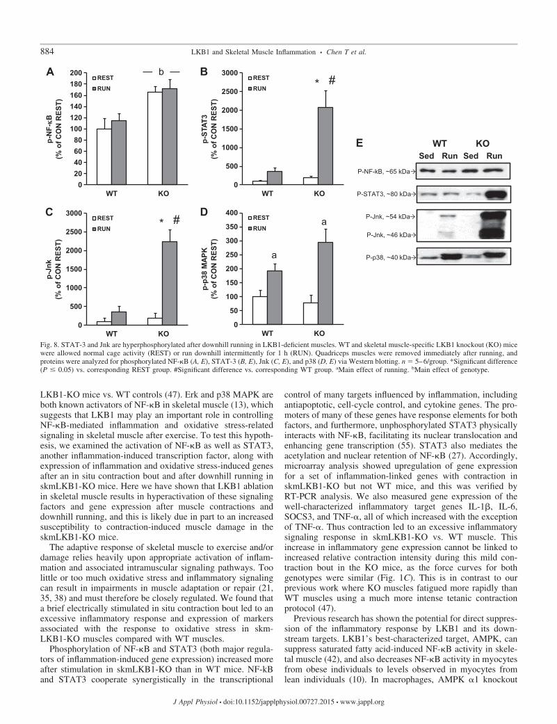
Inflammatory signaling is increased in skmLKB1-KO mus-cles after downhill running. To determine whether downhillrunning increased inflammatory signaling, we measured phos-phorylation of NF-�B, STAT3, Jnk, and p38/MAPK, all ofwhich can contribute to the inflammatory response. NF-�Bphosphorylation (Ser 536) was increased in skmLKB1-KOmuscles regardless of running status, but was unaffected byrunning in either genotype (Fig. 8, A and E). STAT3 (Tyr705)and Jnk (Thr183/Tyr185) phosphorylation, on the otherhand, was strongly induced by downhill running in skm-LKB1-KO, but not WT muscles (Fig. 8, B, C, and E).Phosphorylation of p38 (Thr180/Tyr182) was induced byrunning in both genotypes, with no genotype-dependentdifferences (Fig. 8, D and E).
Fig. 5. A–E: RT-PCR measurement ofoxidative stress-induced genes afterin situ muscle contractions. RT-PCRwas performed for the indicatedgenes using RNA that was isolatedfrom rested (REST) or stimulated(STIM) gastrocnemius muscles fromWT and skeletal muscle-specificLKB1-knockout (KO) mice 3 h aftercontractions induced by unilateralelectrical stimulation of the sciaticnerve. n � 3–4/group. *Significantdifference (P � 0.05) vs. correspond-ing WT group. #Significant differ-ence vs. corresponding REST group.
882 LKB1 and Skeletal Muscle Inflammation • Chen T et al.
J Appl Physiol • doi:10.1152/japplphysiol.00727.2015 • www.jappl.org

Inflammation-related gene expression changes in LKB1-KOmuscle after downhill running. To determine how the expres-sion of inflammation-related genes changes after downhillrunning, we performed RT-PCR for IL-6, TNF-�, SOCS3,IL-1�, Fos, and IER3 immediately after the hour-long runningbout. After running, SOCS3 expression was induced by run-ning in skmLKB1-KO, but not WT muscles (Fig. 9A). Asimilar, nonsignificant trend was observed for IL-1� (P �0.096; Fig. 9B). IL-6 expression was induced by running inboth genotypes, although this main effect seemed to be drivenprimarily by the increase in the skmLKB1-KO muscles (Fig.
9C). Fos expression was induced by running in both genotypes,but the response was about twice as great in the skmLKB1-KOmuscles compared with WT (Fig. 9D). TNF-� and IER3expression were unaffected by genotype or running (Fig. 9, Eand F).
DISCUSSION
We recently published data showing that contraction-in-duced phosphorylation of extracellular-signal regulated kinase(Erk) and possibly p38 MAPK (p38) is prolonged in skm-
Fig. 7. Downhill running leads to muscledamage in LKB1-deficient skeletal muscle.WT and skeletal muscle-specific LKB1knockout (KO) mice were injected with EBDand then allowed normal cage activity(REST) or run downhill intermittently for 1 h(RUN). Immediately (A and B; n � 8–9/group) or at 5 days (C and D; n � 5–9/group)quadriceps muscles were collected and ana-lyzed for EBD positive (EBD�) cells (A andB), or for centrally nucleated muscle fibersvia hematoxylin and eosin (H&E) staining (Cand D). A: representative images of EBD�fibers in muscles from WT and KO miceafter REST or RUN. Arrows indicate EBD�fibers. B: average EBD� fibers per quadri-ceps muscle. C: representative H&E stains ofmuscles from WT and KO mice after RESTor RUN. Arrows indicated fibers with cen-trally localized nuclei. D: average percentageof centrally nucleated muscle fibers. *Signif-icant difference (P � 0.05) vs. correspondingREST group. #Significant difference vs. cor-responding WT group. bMain effect ofgenotype.
Fig. 6. Muscle damage is mildly elevated inLKB1-deficient muscles after in situ con-tractions. Mice were injected with Evansblue dye (EBD) after which a 15-min bout ofin situ muscle contractions was performed.Gastrocnemius muscles were removed im-mediately after the contraction bout. Detec-tion of EBD-positive (EBD�) cells was per-formed via fluorescence microscopy. A: rep-resentative images of muscle sections fromWT and skeletal muscle-specific LKB1knockout (KO) muscles. Arrows indicateEBD� fibers. B: average number of EBD�fibers in whole gastrocnemius (GASTROC)muscle from WT and skmLKB1-KO mus-cles. n size � 6/group. bMain effect ofgenotype.
883LKB1 and Skeletal Muscle Inflammation • Chen T et al.
J Appl Physiol • doi:10.1152/japplphysiol.00727.2015 • www.jappl.org
LKB1-KO mice vs. WT controls (47). Erk and p38 MAPK areboth known activators of NF-�B in skeletal muscle (13), whichsuggests that LKB1 may play an important role in controllingNF-�B-mediated inflammation and oxidative stress-relatedsignaling in skeletal muscle after exercise. To test this hypoth-esis, we examined the activation of NF-�B as well as STAT3,another inflammation-induced transcription factor, along withexpression of inflammation and oxidative stress-induced genesafter an in situ contraction bout and after downhill running inskmLKB1-KO mice. Here we have shown that LKB1 ablationin skeletal muscle results in hyperactivation of these signalingfactors and gene expression after muscle contractions anddownhill running, and this is likely due in part to an increasedsusceptibility to contraction-induced muscle damage in theskmLKB1-KO mice.
The adaptive response of skeletal muscle to exercise and/ordamage relies heavily upon appropriate activation of inflam-mation and associated intramuscular signaling pathways. Toolittle or too much oxidative stress and inflammatory signalingcan result in impairments in muscle adaptation or repair (21,35, 38) and must therefore be closely regulated. We found thata brief electrically stimulated in situ contraction bout led to anexcessive inflammatory response and expression of markersassociated with the response to oxidative stress in skm-LKB1-KO muscles compared with WT muscles.
Phosphorylation of NF-�B and STAT3 (both major regula-tors of inflammation-induced gene expression) increased moreafter stimulation in skmLKB1-KO than in WT mice. NF-kBand STAT3 cooperate synergistically in the transcriptional
control of many targets influenced by inflammation, includingantiapoptotic, cell-cycle control, and cytokine genes. The pro-moters of many of these genes have response elements for bothfactors, and furthermore, unphosphorylated STAT3 physicallyinteracts with NF-�B, facilitating its nuclear translocation andenhancing gene transcription (55). STAT3 also mediates theacetylation and nuclear retention of NF-�B (27). Accordingly,microarray analysis showed upregulation of gene expressionfor a set of inflammation-linked genes with contraction inskmLKB1-KO but not WT mice, and this was verified byRT-PCR analysis. We also measured gene expression of thewell-characterized inflammatory target genes IL-1�, IL-6,SOCS3, and TNF-�, all of which increased with the exceptionof TNF-�. Thus contraction led to an excessive inflammatorysignaling response in skmLKB1-KO vs. WT muscle. Thisincrease in inflammatory gene expression cannot be linked toincreased relative contraction intensity during this mild con-traction bout in the KO mice, as the force curves for bothgenotypes were similar (Fig. 1C). This is in contrast to ourprevious work where KO muscles fatigued more rapidly thanWT muscles using a much more intense tetanic contractionprotocol (47).
Previous research has shown the potential for direct suppres-sion of the inflammatory response by LKB1 and its down-stream targets. LKB1’s best-characterized target, AMPK, cansuppress saturated fatty acid-induced NF-�B activity in skele-tal muscle (42), and also decreases NF-�B activity in myocytesfrom obese individuals to levels observed in myocytes fromlean individuals (10). In macrophages, AMPK �1 knockout
Fig. 8. STAT-3 and Jnk are hyperphosphorylated after downhill running in LKB1-deficient muscles. WT and skeletal muscle-specific LKB1 knockout (KO) micewere allowed normal cage activity (REST) or run downhill intermittently for 1 h (RUN). Quadriceps muscles were removed immediately after running, andproteins were analyzed for phosphorylated NF-�B (A, E), STAT-3 (B, E), Jnk (C, E), and p38 (D, E) via Western blotting. n � 5–6/group. *Significant difference(P � 0.05) vs. corresponding REST group. #Significant difference vs. corresponding WT group. aMain effect of running. bMain effect of genotype.
884 LKB1 and Skeletal Muscle Inflammation • Chen T et al.
J Appl Physiol • doi:10.1152/japplphysiol.00727.2015 • www.jappl.org

prevents the normal transition from proinflammatory M1 toanti-inflammatory M2 macrophages after muscle injury (36),and AMPK �1 subunit knockout decreased inflammatorymarkers in macrophages, as did inhibition of fatty acid oxida-tion (8), suggesting that AMPK may control inflammation inmacrophages by promoting fat oxidation. Since we showedthat LKB1 knockout likewise leads to decreased fatty acidoxidation in skeletal muscle (16, 49), this may likewise play arole in the increased inflammatory markers that we observedhere. Salt-inducible kinase 3 (SIK3) and sucrose non-ferment-ing-related kinase (SNRK), both additional AMPK familymembers that lie downstream of LKB1, likewise appear tohave anti-inflammatory effects on macrophages and adi-pocytes, respectively (28, 43). Interestingly, LKB1 itself canbind to and suppress inhibitor of NF-�B (I�B) kinase (IKK),leading to increased I�B stability, and therefore decreasedNF-�B nuclear translocation and transcriptional activity (30).Thus the exaggerated inflammatory signaling and gene expres-sion that we observed in skmLKB1-KO muscle after contrac-tions is likely due, at least in part, to direct effects of LKB1 andits downstream kinases.
Comparison of overrepresented gene ontology (GO) biolog-ical processes in stimulated KO vs. WT muscles showed thatmany of the most affected processes were related to metabo-lism. This was to be expected based on previous studies (16,22, 40, 47, 49–51) where mitochondrial and metabolic defectswere observed in LKB1-KO muscles. Although “inflammatoryresponse” processes were not among those most affected by the
KO, processes involved in oxidative stress response werehighly overrepresented based on the GO term analysis. Thatmetabolic and oxidative stress-related processes are both heav-ily affected in the LKB1-KO muscles is consistent with the roleof mitochondrial deficiency on reactive oxygen species gener-ation (19). This is also consistent with the elevated expressionof inflammatory genes observed in the cluster analysis. Indeed,oxidative stress activates NF-�B, and therefore the response tooxidative stress is similar in many respects to the inflammatoryresponse (24).
Nuclear factor erythroid 2-related factor 2 (Nrf2) is activatedduring periods of oxidative stress through a complex redoxshift-sensing mechanism and can upregulate many genes thathelp to manage oxidative stress and return the basal oxidativetone of the cell back to homeostatic conditions. All five of theoxidative stress response genes measured here are regulated bythe transcription factor Nrf2, and Hmox1 and NQO1 in partic-ular are hallmark genes regulated by Nrf2 activation (25).Previous work shows that loss of LKB1 results in increasedactivation of Nrf2 and would lead to an increase in Nrf2-relatedgene expression (20), including Hmox1 and NQO1. The datawe have here support these previous findings but also suggestthat during exercise, LKB1 is important in the regulation ofantioxidative pathways in skeletal muscle to abrogate exercise-induced oxidative damage.
It is also possible that part of the hyperinflammatory re-sponse in skmLKB1-KO muscles was secondary to tissue/celldamage. Shan et al. (45) previously reported substantial muscle
Fig. 9. Expression of inflammation-related genes is elevated in LKB1-deficient muscle after downhill running. WT and skeletal muscle-specific LKB1 knockout(KO) mice were left resting in their cages (REST) or run downhill intermittently for 1 h (RUN). Quadricep muscles were removed immediately after running,and gene expression was analyzed for IL-6 (A), TNF-� (B), SOCS3 (C), IL-1� (D), Fos (E), and IER3 (F) via RT-PCR. n � 5–6/group. *Significant difference(P � 0.05) vs. corresponding REST group. #Significant difference vs. corresponding WT group. aMain effect of running.
885LKB1 and Skeletal Muscle Inflammation • Chen T et al.
J Appl Physiol • doi:10.1152/japplphysiol.00727.2015 • www.jappl.org

damage and regeneration in untreated muscles from skeletalmuscle specific LKB1 knockout mice. The degree of damageand regeneration reported in that study was much more exten-sive than observed in our mice. This may be due to an earlieronset of LKB1 disruption during muscle development as theyused the MyoD promoter to drive LKB1 excision, rather thanthe Myf6 promoter that was used in our mice (45). AMPKlikely plays a role in muscle integrity since similar evidence formuscle damage and regeneration has been reported in muscle-specific AMPK knockout mice (5, 48), and potentially linkedto defects in autophagy and/or vascularity of the muscle. Here,we observed a main effect for increased EBD positive fibers inresting and stimulated skmLKB1-KO muscles, with a trend foran increased number of damaged fibers after stimulation.However, the increase in damaged fiber number in our skm-LKB1-KO mice after contraction only represented a very smallfraction of the total number of muscle fibers in the gastrocne-mius ( 10 damaged fibers of 5,000 fibers in the wholegastrocnemius). It is therefore not likely that the fiber damagealone accounts for the increase in inflammatory signaling in theKO muscle and suggests that the lack of LKB1 is likelyexacerbating inflammatory signaling independent of the mus-cle damage itself.
Nonetheless, we determined whether skmLKB1-KO muscleis more prone to contraction-induced damage using a morephysiologically relevant form of contractions by running themice downhill. We used a very mild bout of downhill runningbecause the skmLKB1-KO mice fatigue very quickly withtreadmill running (47). In preliminary experiments we foundthat the skmLKB1-KO mice were unable to sustain runningdownhill for more than 10 min even at only 12 m/min and a�17° grade. Therefore we ran the mice for 5-min bouts with2-min rest periods between bouts. The skmLKB1-KO micewere able to sustain this running protocol for a full hour,although they almost universally required substantial proddingwith a brush to do so. While this downhill running protocolproduced no significant damage in the WT mice, we observeda clear increase in EBD-positive fibers in the quadricepsmuscles immediately after running in the skmLKB1-KO mice.Centrally localized nuclei were also elevated in the skm-LKB1-KO quadriceps, although running had no significanteffect on this 5 days postexercise. STAT3 and Jnk phosphor-ylation were both increased more in the KO vs. WT muscleimmediately after the running bout, as was gene expression forSOCS3, IL-6, and Fos. It is possible that expression of theother inflammation-related genes might be affected at a latertime point. The susceptibility to damage in the skmLKB1-KOmuscles is likely mediated at least in part by AMPK sinceskeletal muscle from AMPK �1/�2 double knockout micehave an elevated number of fibers with centrally located nucleias well as increased gene expression of IL-6, suggesting thatthe muscle is prone to damage and subsequent regeneration(26). The role of other LKB1 targets in cell integrity isunknown, but MARK2, an AMPK-related kinase also phos-phorylated and activated by LKB1, has been shown to regulateinteraction between dystrophin and utrophin (54). ImpairedMARK2 activity could be an important defect contributing tothe apparent susceptibility of skmLKB1-KO muscle cells todamage. It should be kept in mind that the increased damageobserved in the KO muscles could be due in part to theincreased relative intensity of the treadmill running bout (the
running was near maximal capacity for the KO but not the WTmice). However, since the foremost reason for the downhillrunning experiment was to assess inflammatory response inrelation to susceptibility to muscle membrane damage, we keptthe overall physical strain on the whole muscle consistentbetween genotypes. Since the genotypes were of similarweight, and the distance run was identical, the strain on themuscles was likely similar, although uncharacterized biome-chanical factors could contribute as well to the strain. Ourresults suggest that while the KO mice in our study may or maynot have been limited metabolically in their running, it is likelythat the KO mice were actually limited in their running partlyby muscle damage and subsequent inflammation.
In conclusion, we have shown that the lack of LKB1 inskeletal muscle leads to an increased inflammatory and oxida-tive stress response after muscle contraction. Increased suscep-tibility of the muscle to damage may underlie at least part ofthis response, and suggests that targeting LKB1 and/or itsdownstream targets may lead to improved treatment forchronic inflammatory and degenerative skeletal muscle condi-tions.
GRANTS
This work was supported by National Institute of Arthritis and Musculo-skeletal and Skin Diseases Grant R01-AR-051928 (D. M. Thomson) andMentoring Environment Grants from Brigham Young University (D. M.Thomson).
DISCLOSURES
No conflicts of interest, financial or otherwise, are declared by the author(s).
AUTHOR CONTRIBUTIONS
Author contributions: T.C., T.M.M., M.T.E., C.R.H., J.S.K., and D.M.T.conception and design of research; T.C., T.M.M., N.L.M., S.R.M., D.M.H.,A.M.H., R.E.C., R.P.M., C.R.H., J.M.H., and D.M.T. performed experiments;T.C., T.M.M., M.T.E., N.L.M., S.R.M., D.M.H., A.M.H., R.E.C., R.P.M.,C.R.H., J.M.H., J.S.K., and D.M.T. analyzed data; T.C., T.M.M., M.T.E.,N.L.M., S.R.M., R.E.C., R.P.M., C.R.H., J.M.H., J.S.K., and D.M.T. inter-preted results of experiments; T.C. and D.M.T. drafted manuscript; T.C.,T.M.M., M.T.E., C.R.H., J.M.H., and D.M.T. edited and revised manuscript;T.C., T.M.M., M.T.E., N.L.M., S.R.M., D.M.H., A.M.H., R.E.C., R.P.M.,C.R.H., J.M.H., J.S.K., and D.M.T. approved final version of manuscript;M.T.E., N.L.M., R.P.M., J.M.H., and D.M.T. prepared figures.
REFERENCES
1. Arlt A, Schafer H. Role of the immediate early response 3 (IER3) genein cellular stress response, inflammation and tumorigenesis. Eur J CellBiol 90: 545–552, 2011.
2. Begue G, Douillard A, Galbes O, Rossano B, Vernus B, Candau R, PyG. Early activation of rat skeletal muscle IL-6/STAT1/STAT3 dependentgene expression in resistance exercise linked to hypertrophy. PLos One 8:e57141, 2013.
3. Berdeaux R, Goebel N, Banaszynski L, Takemori H, Wandless T,Shelton GD, Montminy M. SIK1 is a class II HDAC kinase that promotessurvival of skeletal myocytes. Nat Med 13: 597–603, 2007.
4. Brown AE, Palsgaard J, Borup R, Avery P, Gunn DA, De Meyts P,Yeaman SJ, Walker M. p38 MAPK activation upregulates proinflam-matory pathways in skeletal muscle cells from insulin-resistant type 2diabetic patients. Am J Physiol Endocrinol Metab 308: E63–E70, 2015.
5. Bujak AL, Crane JD, Lally JS, Ford RJ, Kang SJ, Rebalka IA, GreenAE, Kemp BE, Hawke TJ, Schertzer JD, Steinberg GR. AMPKactivation of muscle autophagy prevents fasting-induced hypoglycemiaand myopathy during aging. Cell Metab 21: 883–890, 2015.
6. Cai D, Frantz JD, Tawa NE Jr, Melendez PA, Oh BC, Lidov HG,Hasselgren PO, Frontera WR, Lee J, Glass DJ, and Shoelson SE.IKKbeta/NF-kappaB activation causes severe muscle wasting in mice.Cell 119: 285–298, 2004.
886 LKB1 and Skeletal Muscle Inflammation • Chen T et al.
J Appl Physiol • doi:10.1152/japplphysiol.00727.2015 • www.jappl.org

7. Chang TP, Vancurova I. Bcl3 regulates pro-survival and pro-inflamma-tory gene expression in cutaneous T-cell lymphoma. Biochim BiophysActa 1843: 2620–2630, 2014.
8. Galic S, Fullerton MD, Schertzer JD, Sikkema S, Marcinko K, Walk-ley CR, Izon D, Honeyman J, Chen ZP, van Denderen BJ, Kemp BE,Steinberg GR. Hematopoietic AMPK beta1 reduces mouse adipose tissuemacrophage inflammation and insulin resistance in obesity. J Clin Invest121: 4903–4915, 2011.
9. Gomez-Cabrera MC, Borras C, Pallardo FV, Sastre J, Ji LL, Vina J.Decreasing xanthine oxidase-mediated oxidative stress prevents usefulcellular adaptations to exercise in rats. J Physiol 567: 113–120, 2005.
10. Green CJ, Pedersen M, Pedersen BK, Scheele C. Elevated NF-kappaBactivation is conserved in human myocytes cultured from obese type 2diabetic patients and attenuated by AMP-activated protein kinase. Diabe-tes 60: 2810–2819, 2011.
11. Haddad F, Zaldivar F, Cooper DM, Adams GR. IL-6-induced skeletalmuscle atrophy. J Appl Physiol 98: 911–917, 2005.
12. Haldar M, Hancock JD, Coffin CM, Lessnick SL, Capecchi MR. Aconditional mouse model of synovial sarcoma: insights into a myogenicorigin. Cancer Cell 11: 375–388, 2007.
13. Ho RC, Hirshman MF, Li Y, Cai D, Farmer JR, Aschenbach WG,Witczak CA, Shoelson SE, Goodyear LJ. Regulation of IkappaB kinaseand NF-kappaB in contracting adult rat skeletal muscle. Am J Physiol CellPhysiol 289: C794–C801, 2005.
14. Hollander J, Fiebig R, Gore M, Ookawara T, Ohno H, Ji LL.Superoxide dismutase gene expression is activated by a single bout ofexercise in rat skeletal muscle. Pflügers Arch 442: 426–434, 2001.
15. Inazuka F, Sugiyama N, Tomita M, Abe T, Shioi G, Esumi H.Muscle-specific knock-out of NUAK family SNF1-like kinase 1 (NUAK1)prevents high fat diet-induced glucose intolerance. J Biol Chem 287:16379–16389, 2012.
16. Jeppesen J, Maarbjerg SJ, Jordy AB, Fritzen AM, Pehmoller C,Sylow L, Serup AK, Jessen N, Thorsen K, Prats C, Qvortrup K, DyckJR, Hunter RW, Sakamoto K, Thomson DM, Schjerling P, Wojtasze-wski JF, Richter EA, Kiens B. LKB1 regulates lipid oxidation duringexercise independently of AMPK. Diabetes 62: 1490–1499, 2013.
17. Ji LL, Gomez-Cabrera MC, Steinhafel N, Vina J. Acute exerciseactivates nuclear factor (NF)-kappaB signaling pathway in rat skeletalmuscle. FASEB J 18: 1499–1506, 2004.
18. Jorgensen SB, Jensen TE, Richter EA. Role of AMPK in skeletalmuscle gene adaptation in relation to exercise. Appl Physiol Nutr Metab32: 904–911, 2007.
19. Joseph AM, Adhihetty PJ, Leeuwenburgh C. Beneficial effects ofexercise on age-related mitochondrial dysfunction and oxidative stress inskeletal muscle. J Physiol 2015 Oct 27. doi:10.1113/JP270659. [Epubahead of print].
20. Kaufman JM, Amann JM, Park K, Arasada RR, Li H, Shyr Y,Carbone DP. LKB1 Loss induces characteristic patterns of gene expres-sion in human tumors associated with NRF2 activation and attenuation ofPI3K-AKT. J Thorac Oncol 9: 794–804, 2014.
21. Kharraz Y, Guerra J, Mann CJ, Serrano AL, Munoz-Canoves P.Macrophage plasticity and the role of inflammation in skeletal musclerepair. Mediators Inflamm 2013: 491497, 2013.
22. Koh HJ, Arnolds DE, Fujii N, Tran TT, Rogers MJ, Jessen N, Li Y,Liew CW, Ho RC, Hirshman MF, Kulkarni RN, Kahn CR, GoodyearLJ. Skeletal muscle-selective knockout of LKB1 increases insulin sensi-tivity, improves glucose homeostasis, and decreases TRB3. Mol Cell Biol26: 8217–8227, 2006.
23. Koh HJ, Toyoda T, Fujii N, Jung MM, Rathod A, Middelbeek RJ,Lessard SJ, Treebak JT, Tsuchihara K, Esumi H, Richter EA, Woj-taszewski JF, Hirshman MF, Goodyear LJ. Sucrose nonfermentingAMPK-related kinase (SNARK) mediates contraction-stimulated glucosetransport in mouse skeletal muscle. Proc Natl Acad Sci USA 107: 15541–15546, 2010.
24. Kramer HF, Goodyear LJ. Exercise, MAPK, and NF-kappaB signalingin skeletal muscle. J Appl Physiol 103: 388–395, 2007.
25. Kwak MK, Itoh K, Yamamoto M, Sutter TR, Kensler TW. Role oftranscription factor Nrf2 in the induction of hepatic phase 2 and antioxi-dative enzymes in vivo by the cancer chemoprotective agent, 3H-1,2-dimethiole-3-thione. Mol Med 7: 135–145, 2001.
26. Lantier L, Fentz J, Mounier R, Leclerc J, Treebak JT, Pehmoller C,Sanz N, Sakakibara I, Saint-Amand E, Rimbaud S, Maire P, MaretteA, Ventura-Clapier R, Ferry A, Wojtaszewski JF, Foretz M, Viollet B.
AMPK controls exercise endurance, mitochondrial oxidative capacity, andskeletal muscle integrity. FASEB J 28: 3211–3224, 2014.
27. Lee H, Herrmann A, Deng JH, Kujawski M, Niu G, Li Z, Forman S, JoveR, Pardoll DM, Yu H. Persistently activated Stat3 maintains constitutiveNF-kappaB activity in tumors. Cancer Cell 15: 283–293, 2009.
28. Li Y, Nie Y, Helou Y, Ding G, Feng B, Xu G, Salomon A, Xu H.Identification of sucrose non-fermenting-related kinase (SNRK) as asuppressor of adipocyte inflammation. Diabetes 62: 2396–2409, 2013.
29. Li YP, Reid MB. NF-kappaB mediates the protein loss induced byTNF-alpha in differentiated skeletal muscle myotubes. Am J Physiol RegulIntegr Comp Physiol 279: R1165–R1170, 2000.
30. Liu Z, Zhang W, Zhang M, Zhu H, Moriasi C, Zou MH. Liver kinaseB1 suppresses lipopolysaccharide-induced nuclear factor kappaB (NF-kappaB) activation in macrophages. J Biol Chem 290: 2312–2320, 2015.
31. Londhe P, Guttridge DC. Inflammation induced loss of skeletal muscle.Bone 80: 131–142, 2015.
32. Luo G, Hershko DD, Robb BW, Wray CJ, Hasselgren PO. IL-1betastimulates IL-6 production in cultured skeletal muscle cells throughactivation of MAP kinase signaling pathway and NF-kappa B. Am JPhysiol Regul Integr Comp Physiol 284: R1249–R1254, 2003.
33. McGee SL, Hargreaves M. AMPK-mediated regulation of transcriptionin skeletal muscle. Clin Sci 118: 507–518, 2010.
34. Minari AL, Oyama LM, Dos Santos RV. Downhill exercise-inducedchanges in gene expression related with macrophage polarization andmyogenic cells in the triceps long head of rats. Inflammation 38: 209–217,2015.
35. Mishra DK, Friden J, Schmitz MC, Lieber RL. Anti-inflammatorymedication after muscle injury. A treatment resulting in short-term im-provement but subsequent loss of muscle function. J Bone Joint Surg Am77: 1510–1519, 1995.
36. Mounier R, Theret M, Arnold L, Cuvellier S, Bultot L, Goransson O,Sanz N, Ferry A, Sakamoto K, Foretz M, Viollet B, Chazaud B.AMPKalpha1 regulates macrophage skewing at the time of resolution ofinflammation during skeletal muscle regeneration. Cell Metab 18: 251–264, 2013.
37. Niemand C, Nimmesgern A, Haan S, Fischer P, Schaper F, RossaintR, Heinrich PC, Muller-Newen G. Activation of STAT3 by IL-6 andIL-10 in primary human macrophages is differentially modulated bysuppressor of cytokine signaling 3. J Immunol 170: 3263–3272, 2003.
38. Philippou A, Maridaki M, Theos A, Koutsilieris M. Cytokines inmuscle damage. Adv Clin Chem 58: 49–87, 2012.
39. Ray N, Kuwahara M, Takada Y, Maruyama K, Kawaguchi T, Tsub-one H, Ishikawa H, Matsuo K. c-Fos suppresses systemic inflammatoryresponse to endotoxin. Int Immunol 18: 671–677, 2006.
40. Sakamoto K, McCarthy A, Smith D, Green KA, Grahame Hardie D,Ashworth A, Alessi DR. Deficiency of LKB1 in skeletal muscle preventsAMPK activation and glucose uptake during contraction. EMBO J 24:1810–1820, 2005.
41. Sakurai H, Chiba H, Miyoshi H, Sugita T, Toriumi W. IkappaB kinasesphosphorylate NF-kappaB p65 subunit on serine 536 in the transactivationdomain. J Biol Chem 274: 30353–30356, 1999.
42. Salvado L, Coll T, Gomez-Foix AM, Salmeron E, Barroso E, PalomerX, Vazquez-Carrera M. Oleate prevents saturated-fatty-acid-induced ERstress, inflammation and insulin resistance in skeletal muscle cells throughan AMPK-dependent mechanism. Diabetologia 56: 1372–1382, 2013.
43. Sanosaka M, Fujimoto M, Ohkawara T, Nagatake T, Itoh Y, KagawaM, Kumagai A, Fuchino H, Kunisawa J, Naka T, Takemori H. SIK3deficiency exacerbates LPS-induced endotoxin shock accompanied byincreased levels of proinflammatory molecules in mice. Immunology 145:268–278, 2015.
44. Scheller J, Chalaris A, Schmidt-Arras D, Rose-John S. The pro- andanti-inflammatory properties of the cytokine interleukin-6. Biochim Bio-phys Acta 1813: 878–888, 2011.
45. Shan T, Zhang P, Liang X, Bi P, Yue F, Kuang S. Lkb1 is indispensablefor skeletal muscle development, regeneration, and satellite cell homeo-stasis. Stem Cells 32: 2893–2907, 2014.
46. Szelenyi ER, Urso ML. Time-course analysis of injured skeletal musclesuggests a critical involvement of ERK1/2 signaling in the acute inflam-matory response. Muscle Nerve 45: 552–561, 2012.
47. Tanner CB, Madsen SR, Hallowell DM, Goring DM, Moore TM,Hardman SE, Heninger MR, Atwood DR, Thomson DM. Mitochon-drial and performance adaptations to exercise training in mice lackingskeletal muscle LKB1. Am J Physiol Endocrinol Metab 305: E1018–E1029, 2013.
887LKB1 and Skeletal Muscle Inflammation • Chen T et al.
J Appl Physiol • doi:10.1152/japplphysiol.00727.2015 • www.jappl.org

48. Thomas MM, Wang DC, D’Souza DM, Krause MP, Layne AS,Criswell DS, O’Neill HM, Connor MK, Anderson JE, Kemp BE,Steinberg GR, Hawke TJ. Muscle-specific AMPK beta1beta2-null micedisplay a myopathy due to loss of capillary density in nonpostural muscles.FASEB J 28: 2098–2107, 2014.
49. Thomson DM, Brown JD, Fillmore N, Condon BM, Kim HJ, BarrowJR, Winder WW. LKB1 and the regulation of malonyl-CoA and fattyacid oxidation in muscle. Am J Physiol Endocrinol Metab 293: E1572–E1579, 2007.
50. Thomson DM, Hancock CR, Evanson BG, Kenney SG, Malan BB,Mongillo AD, Brown JD, Hepworth S, Fillmore N, Parcell AC, Kooy-man DL, Winder WW. Skeletal muscle dysfunction in muscle-specificLKB1 knockout mice. J Appl Physiol 108: 1775–1785, 2010.
51. Thomson DM, Porter BB, Tall JH, Kim HJ, Barrow JR, Winder WW.Skeletal muscle and heart LKB1 deficiency causes decreased voluntaryrunning and reduced muscle mitochondrial marker enzyme expression inmice. Am J Physiol Endocrinol Metab 292: E196–E202, 2007.
52. Trenerry MK, Carey KA, Ward AC, Cameron-Smith D. STAT3signaling is activated in human skeletal muscle following acute resistanceexercise. J Appl Physiol 102: 1483–1489, 2007.
53. Windelinckx A, De Mars G, Huygens W, Peeters MW, Vincent B,Wijmenga C, Lambrechts D, Aerssens J, Vlietinck R, Beunen G,Thomis MA. Identification and prioritization of NUAK1 and PPP1CC aspositional candidate loci for skeletal muscle strength phenotypes. PhysiolGenomics 43: 981–992, 2011.
54. Yamashita K, Suzuki A, Satoh Y, Ide M, Amano Y, Masuda-HirataM, Hayashi YK, Hamada K, Ogata K, Ohno S. The 8th and 9th tandemspectrin-like repeats of utrophin cooperatively form a functional unit tointeract with polarity-regulating kinase PAR-1b. Biochem Biophys ResCommun 391: 812–817, 2010.
55. Yang J, Liao X, Agarwal MK, Barnes L, Auron PE, Stark GR.Unphosphorylated STAT3 accumulates in response to IL-6 and acti-vates transcription by binding to NFkappaB. Genes Dev 21: 1396 –1408, 2007.
56. Zhang H, Taylor WR, Joseph G, Caracciolo V, Gonzales DM,Sidell N, Seli E, Blackshear PJ, Kallen CB. mRNA-binding proteinZFP36 is expressed in atherosclerotic lesions and reduces inflammationin aortic endothelial cells. Arterioscler Thromb Vasc Biol 33: 1212–1220, 2013.
888 LKB1 and Skeletal Muscle Inflammation • Chen T et al.
J Appl Physiol • doi:10.1152/japplphysiol.00727.2015 • www.jappl.org