liver disease in children
TRANSCRIPT

Diseases of the Liverand Biliary System in ChildrenEDITED BY
DEIRDRE KELLYMD, FRCP, FRCPI, FRCPCHProfessor of Pediatric Hepatology, Liver Unit, Birmingham Children’s Hospital,Birmingham, UK
THIRD EDITION
FOREWORD BY
DAME SHEILA SHERLOCK
A John Wiley & Sons, Ltd., Publication
9781405163347_1_pre.qxd 20/06/2008 09:25 Page i

9781405163347_1_pre.qxd 20/06/2008 09:25 Page ii

Diseases of the Liverand Biliary System in ChildrenEDITED BY
DEIRDRE KELLYMD, FRCP, FRCPI, FRCPCHProfessor of Pediatric Hepatology, Liver Unit, Birmingham Children’s Hospital,Birmingham, UK
THIRD EDITION
FOREWORD BY
DAME SHEILA SHERLOCK
A John Wiley & Sons, Ltd., Publication
9781405163347_1_pre.qxd 20/06/2008 09:25 Page i

This edition first published 1999, © 1999, 2004, 2008 by Blackwell Publishing Ltd
Blackwell Publishing was acquired by John Wiley & Sons in February 2007. Blackwell’s publishingprogram has been merged with Wiley’s global Scientific, Technical and Medical business to form Wiley-Blackwell.
Registered office: John Wiley & Sons Ltd, The Atrium, Southern Gate, Chichester, West Sussex, PO198SQ, UK
Editorial offices: 9600 Garsington Road, Oxford, OX4 2DQ, UKThe Atrium, Southern Gate, Chichester, West Sussex, PO19 8SQ, UK111 River Street, Hoboken, NJ 07030-5774, USA
For details of our global editorial offices, for customer services and for information about how to applyfor permission to reuse the copyright material in this book please see our website atwww.wiley.com/wiley-blackwell
The right of the author to be identified as the author of this work has been asserted in accordance withthe Copyright, Designs and Patents Act 1988.
All rights reserved. No part of this publication may be reproduced, stored in a retrieval system, ortransmitted, in any form or by any means, electronic, mechanical, photocopying, recording orotherwise, except as permitted by the UK Copyright, Designs and Patents Act 1988, without the priorpermission of the publisher.
Wiley also publishes its books in a variety of electronic formats. Some content that appears in print maynot be available in electronic books.
Designations used by companies to distinguish their products are often claimed as trademarks. Allbrand names and product names used in this book are trade names, service marks, trademarks orregistered trademarks of their respective owners. The publisher is not associated with any product orvendor mentioned in this book. This publication is designed to provide accurate and authoritativeinformation in regard to the subject matter covered. It is sold on the understanding that the publisher isnot engaged in rendering professional services. If professional advice or other expert assistance isrequired, the services of a competent professional should be sought.
The contents of this work are intended to further general scientific research, understanding, anddiscussion only and are not intended and should not be relied upon as recommending or promoting aspecific method, diagnosis, or treatment by physicians for any particular patient. The publisher and theauthor make no representations or warranties with respect to the accuracy or completeness of thecontents of this work and specifically disclaim all warranties, including without limitation any impliedwarranties of fitness for a particular purpose. In view of ongoing research, equipment modifications,changes in governmental regulations, and the constant flow of information relating to the use ofmedicines, equipment, and devices, the reader is urged to review and evaluate the informationprovided in the package insert or instructions for each medicine, equipment, or device for, among otherthings, any changes in the instructions or indication of usage and for added warnings and precautions.Readers should consult with a specialist where appropriate. The fact that an organization or Website isreferred to in this work as a citation and/or a potential source of further information does not mean thatthe author or the publisher endorses the information the organization or Website may provide orrecommendations it may make. Further, readers should be aware that Internet Websites listed in thiswork may have changed or disappeared between when this work was written and when it is read. Nowarranty may be created or extended by any promotional statements for this work. Neither thepublisher nor the author shall be liable for any damages arising herefrom.
A catalogue record for this book is available from the British Library.
Library of Congress Cataloging-in-Publication Data
Diseases of the liver and biliary system in children / edited by Deirdre Kelly. — 3rd ed.p. ; cm.
Includes bibliographical references and index.ISBN 978-1-4051-6334-7 (alk. paper)1. Liver—Diseases. 2. Biliary tract—Diseases. 3. Pediatric gastroenterology. I. Kelly, Deirdre A.
[DNLM: 1. Liver Diseases. 2. Child. 3. Infant. WS 310 D611 2008]RJ456.L5D57 2008618.92’362—dc22
2007037665ISBN: 9781405163347
A catalogue record for this title is available from the British Library
Set in 9/12pt Meridien by Graphicraft Limited, Hong KongPrinted and bound in Singapore by COS Printers Pte Ltd
First published 1999Second edition 2004Third edition 2008
1 2008
9781405163347_1_pre.qxd 20/06/2008 09:25 Page ii
Contributors, v
Foreword to the First Edition, vii
Preface, ix
Acknowledgments, x
Section 1 Structure Function, and Repair, 1
1 Structure Function and Repair of the Liver, 3Ulrich Baumann, Alastair J.W. Millar, and Rachel M. Brown
Section 2 Investigating the Liver, 19
2 The Approach to the Child with Liver Disease:Differential Diagnosis and Useful Investigations, 21Deirdre A. Kelly
Section 3 Liver Disease in Pregnancy, 35
3 The Effects of Liver Disease on Mother and Child, 37Jane Hartley and Elwyn Elias
Section 4 Neonatal liver disease, 55
4 The Jaundiced Baby, 57Eve A. Roberts
5 The Acutely Ill Baby, 106Patrick J. McKiernan
Section 5 Acute Liver Disease, 127
6 Infective Disorders of the Liver, 129Suzanne Davison and Elizabeth H. Boxall
7 Acute Liver Failure, 169Peter F. Whitington, Estella M. Alonso, and Robert H. Squires
Section 6 Liver Disease in Older Children, 189
8 Autoimmune Liver Disease, 191Giorgina Mieli-Vergani and Diego Vergani
9 Drug-Induced Liver Disease, 207Karen F. Murray
10 Congenital and Structural Abnormalities of the Liver, 231Jaime Liou Wolfe and Kathleen B. Schwarz
11 Nonalcoholic Steatosis, 253Eve A. Roberts
12 Hepatobiliary Disease in Cystic Fibrosis, 270Carla Colombo
Section 7 Metabolic Liver Disease, 289
13 Metabolic Liver Disease in the Infant and Older Child, 291Anupam Chakrapani and Anne Green
14 Disorders of Copper Metabolism, 328Stuart Tanner
Section 8 Management of Chronic LiverDisease, 349
15 Complications and Management of Chronic LiverDisease, 351Ross Shepherd
Section 9 The Liver and Other Organs, 379
16 The Liver in Systemic Illness, 381Susan V. Beath
17 Skin Disorders in Liver Disease, 404Indra D.M. van Mourik and Michelle Thomson
iii
Contents
9781405163347_1_pre.qxd 20/06/2008 09:25 Page iii

18 Dental Care of Children with Liver Disease, 420Marie-Therese Hosey and Victoria Clark
Section 10 Surgical Management of LiverDisease, 431
19 Surgical Disorders of the Liver and Bile Ducts and PortalHypertension, 433Alastair J.W. Millar
20 Primary Hepatic Tumors, 475Bruce Morland and Jean de Ville de Goyet
Section 11 Transplantation, 501
21 Liver Transplantation, 503Deirdre A. Kelly and David Mayer
22 Small-Bowel Transplantation in Children, 531Jorge Reyes
Section 12 The Developing World, 551
23 Liver Disease in the Developing World, 553Seng-Hock Quak, Anupam Sibal, and Mei-Hwei Chang
Section 13 Supporting the Child and Family,577
24 The Role of the Multidisciplinary Team, 579Graham Gordon, Julie Reed, Jacqueline Blyth, and Carolyn J. Patchell
25 Adolescence and Transition to Adult Care, 599Janet E. McDonagh and Deirdre A. Kelly
Index, 615
Contents
iv
9781405163347_1_pre.qxd 20/06/2008 13:45 Page iv

Estella M. Alonso MDProfessor of Pediatrics, Northwestern University,
Feinberg School of Medicine; Siragusa Transplant
Center, Children’s Memorial Hospital Chicago,
IL, USA
Ulrich Baumann MDConsultant Paediatric Hepatologist, The Liver
Unit, Birmingham Children’s Hospital,
Birmingham, UK
Sue V. Beath BSc, MB BS, MRCP(UK),
DTM, FRCPCHConsultant Paediatric Hepatologist, The Liver Unit,
Birmingham Children’s Hospital, UK
Jacqueline Blyth BSc, MSc
(Health Psychology), ClinPsyD, CSci,
CPsychol, AFBPsSClinical Psychologist, Birmingham Children’s
Hospital, Birmingham, UK
Elizabeth Boxall PhD, FRCPath,
FFPHConsultant Clinical Scientist, Health Protection
Agency, West Midlands Public Health
Laboratory, Heart of England Foundation
Trust, Birmingham, UK
Rachel M. Brown BSc MBChB
FRCPathConsultant Paediatric Hepatologist, Department
of Histopathology, Birmingham Children’s
Hospital, Birmingham, UK
Anupam Chakrapani MD, DCH,
FRCPCHConsultant in Inherited Metabolic Disorders,
Department of Inherited Metabolic Disorders,
Birmingham Children’s Hospital NHS Foundation
Trust, Birmingham, UK
Mei-Hwei Chang MDProfessor, Department of Paediatrics,
National Taiwan University Hospital,
Taipei, Taiwan
Victoria Clark BChD, M Dent Sci,
FDS (paeds)Consultant in Paediatric Dentistry, Birmingham
Children’s Hospital, Birmingham, UK
Carla Colombo MDAssociate Professor of Pediatrics, University of
Milan, Fondazione IRCCS “Ospedale Maggiore
Policlinico, Mangiagalli e Regina, Elena”, Cystic
Fibrosis Centre, Milan, Italy
Suzanne Davison BSc, MRCP, MRCPCHPaediatric Hepatologist, Children’s Liver and GI
Unit, St James’s University Hospital, Leeds, UK
Elwyn Elias MDConsultant Hepatologist, Liver Unit, University
Birmingham Foundation Trust Hospital,
Birmingham, UK
Jean de Ville de Goyet FRCS,
MD, PhDProfessor of Paediatric Surgery, Tor Vergata
University, Roma, Italy; Paediatric Liver and
Transplant Surgery, Bambino Gesu Children’s
Hospital, Roma, Italy
Graham Gordon BSc, SRN, RSCNPrinciple Specialist Nurse (Hepatology and
Transplantation), The Liver Unit, Birmingham
Children’s Hospital, Birmingham, UK
Anne Green BSc, MSc, PhD, FRCPath,
FRCPCH, FIBiol, FRSCConsultant Paediatric Biochemistry and Professor
of Paediatrics and Child Health, University of
Birmingham, Birmingham, UK; Consultant
Clinical Biochemist, Research and Development,
Clinical Chemistry & Inherited Metabolic
Disorders, The Birmingham Children’s Hospital
NHS Trust, Birmingham, UK
Jane Hartley MB.ChB, MRCPCH,
M.Med.ScDepartment of Medical and Molecular Genetics,
Birmingham University and Birmingham
Children’s Hospital, Birmingham, UK
v
Contributors
Marie-Therese Hosey DDS, MSc
(Med Sci), BDS, FDS, RCPSProfessor of Paediatric Dentistry, King’s College
Dental Institute, Denmark Hill, London, UK
Deirdre A. Kelly MD, FRCP, FRCPI,
FRCPCHProfessor of Paediatric Hepatology, The Liver
Unit, Birmingham Children’s Hospital,
Birmingham, UK
A. David Mayer MS, FRCSConsultant Liver Transplant Surgeon, Queen
Elizabeth Hospital and Birmingham Children’s
Hospital, UK
Janet E. McDonagh MD, FRCP(UK)Clinical Senior Lecturer in Paediatric and
Adolescent Rheumatology, Division of
Reproductive and Child Health, University
of Birmingham, Birmingham, UK
Patrick J. McKiernan BSc, MRCP,
FRCPCHConsultant Paediatric Hepatologist, The Liver
Unit, Birmingham Children’s Hospital,
Birmingham, UK
Giorgina M. Mieli-Vergani MD, PhDDirector of Paediatric Liver Centre, Institute of
Liver Studies, King’s College London School of
Medicine at King’s College Hospital, Denmark Hill,
London, UK
Alastair J.W. Millar MBChB, FRCS(Eng
& Edin), FRACS (paed surg), FCS(SA), DCHCharles F.M. Saint Professor of Paediatric Surgery,
University of Cape Town and Red Cross War
Memorial Children’s Hospital Rondebosch, Cape
Town, South Africa
Bruce Morland MBChB, MRCP, DM,
FRCPCHConsultant Oncologist, Birmingham Children’s
Hospital, Birmingham, UK
9781405163347_1_pre.qxd 20/06/2008 09:25 Page v

Karen F. Murray MDProfessor of Pediatrics; Director, Hepatobiliary
Program, Division of Gastroenterology and
Nutrition, Seattle Children’s Hospital and Regional
Medical Center, Seattle, WA, USA
Carolyn Patchell BSc, SRDHead of Dietetic Services, Birmingham Children’s
Hospital, Birmingham, UK
Seng-Hock Quak MBBS, Mmed (Paed),
FAMS, FRCP (Glas), FRCPCH, MDDepartment of Paediatrics, National University
Hospital, Singapore
Julie Reed BSc, MPhil, ClinPsyDConsultant Clinical Psychologist, Birmingham
Children’s Hospital, Birmingham, UK
Jorge Reyes MDChief, Division of Transplant Surgery, University
of Washington Medical Center, Chief,
Transplantation Services, Children’s Hospital
and Regional Medical Center, Seattle, WA
Eve A. Roberts MD, FRCPCAdjunct Professor of Paediatrics, Medicine and
Pharmacology, University of Toronto, The Hospital
for Sick Children, Toronto, Ontario, Canada
Kathleen B. Schwarz MDDirector, Pediatric Liver Center and Professor of
Pediatrics, Johns Hopkin’s University School of
Medicine, Baltimore, MD, USA
Ross Shepherd MD, FRACP, FRCPProfessor of Pediatrics, Washington University
School of Medicine, Medical Director, Liver Care
Center, St Louis Children’s Hospital, St Louis,
MO, USA
Anupam Sibal MDGroup Medical Director, Senior Consultant
Pediatric Gastroenterologist & Hepatologist,
Indraprastha Apollo Hospitals, New Delhi, India
Robert H. Squires MDProfessor of Pediatrics, Clinical Director, Pediatric
Gastroenterology, Hepatology and Nutrition,
Children’s Hospital of Pittsburgh, Pittsburgh,
PA, USA
Stuart Tanner CBE, FRCP, FRCPHEmeritus Professor of Paediatrics, University of
Sheffield, UK
Michelle Thomson MRCPConsultant Dermatologist, The Birmingham Skin
Centre, City Hospital, Birmingham, UK
Contributors
vi
Indra D.M. van Mourik MD, MRCP,
FRCPCHConsultant Paediatric Hepatologist, The Liver Unit,
Birmingham Children’s Hospital, Birmingham, UK
Diego Vergani MD, PhDProfessor of Liver Immunopathology, Institute of
Liver Studies, King’s College London School of
Medicine at King’s College Hospital, Denmark Hill,
London, UK
Peter F. Whitington MDThe Sally Burnett Searle Professor of Pediatrics and
Transplantation, Northwestern University Feinberg
Medicine School; Chief, Division of
Gastroenterology, Hepatology and Nutrition,
Director of the Siragusa Transplantation Center,
Children’s Memorial Hospital, Chicago, IL, USA
Jaime Liou Wolfe MDDivision of Pediatric Gastroenterology and
Nutrition, Johns Hopkin’s University School of
Medicine, MD, USA
9781405163347_1_pre.qxd 20/06/2008 09:25 Page vi
Although the Ancient Egyptians believed that the liver hadmystic powers of healing, and Hippocrates gave a full descrip-tion of hepatic encephalopathy, modern hepatology has only taken off in the last 50 years. Accelerated progress hasfollowed discovery of the hepatitis viruses, now a virtualalphabet from A to E and beyond. Hepatobiliary imaging and endoscopy have added to the progress. Developmentshave depended not only on specialist hepatologists, but on developments in other related disciplines of medicinea
particularly virology, immunology, biochemistry, and now,molecular medicine. A huge literature is available describingliver disease in adults, but pediatrics has lagged behind.
This book covers all the essentials of pediatric hepatologyand is therefore particularly timely. The material covered iswide, from such aspects as the psychology of parents of chil-dren on transplant waiting lists to the genetic disturbances of
bilirubin and bile salt transport in the neonate. The chapterauthors have been well chosen. They are internationalauthorities, active both clinically and in research. They writelucidly from personal experience.
Many helpful algorithms and tables are included. The references at the end of each chapter have been carefullyselected and are up-to-date . . . This book should be availablein every pediatric department. It should be at hand at alltimes to offer practical advice on any childhood liver disease.General pediatricians will certainly benefit. It would be asuitable gift to reward a trainee.
This book fills a real gap in our knowledge of liver disease.It will be a well-deserved success.
P R O F E S S O R D A M E S H E I L A S H E R L O C K
1918–2001
vii
Foreword to the First Edition
9781405163347_1_pre.qxd 20/06/2008 09:25 Page vii

9781405163347_1_pre.qxd 20/06/2008 09:25 Page viii

ix
Pediatric liver disease is a significant cause of morbidity and mortality worldwide. Sophisticated molecular genetictechniques have not only identified new genes and catego-rized rare diseases, but have also given us an insight into pathophysiology and potential therapy. Research intogenomics and proteomics has extended our understanding ofthe mechanisms involved in cellular regulation and regener-ation. National and international collaboration through clinical databases has helped us refine our diagnosis andtreatment of diseases such as biliary atresia and acute liverfailure.
Advances in diagnosis and treatmentaparticularly the successful development of transplantationahave dramat-ically improved the outcome for infants and children withliver disease, so that many can now expect to grow into adultlife. This success brings its own challenges, as adult specialistsnow need to learn to manage young people with a lifetime ofchronic disease. They need to become familiar with pediatricdiseases new to them in a population prone to teenagebehavior and nonadherence.
The investigation and management of significant pediatricliver disease rightly remains within the remit of specialist ortransplant units. Nevertheless, the recognition of the inci-dence of liver disease, the implications of new therapies, and
the necessity for multidisciplinary working are as importantfor general pediatricians as for pediatric gastroenterologists,surgeons, and hepatologists. The gratifying survival of incre-asing numbers of young people with liver disease into adultlife means that it is essential for adult practitioners to be cognisant of pediatric liver disease.
The third edition of this book summarizes the advances of the last 4 years and provides a practical approach to thediagnosis and management of pediatric liver diseases, high-lighting the importance of multidisciplinary teamwork andholistic management of the child and family. The remit has been extended to include information on structure andfunction, with an emphasis on the basic mechanisms of dis-ease. New chapters describe the effects of liver disease frompregnancy to adolescence, to reflect the increasing survival ofyoung women with liver disease into adult reproductive life,and the importance of managing adolescent transition.
It should interest the adult gastroenterologist and hepato-logist, the general pediatrician, and the pediatric trainee, as well as providing guidance for nurses and allied healthprofessionals.
D E I R D R E A. K E L L Y
Birmingham, September 2007
Preface
9781405163347_1_pre.qxd 20/06/2008 09:25 Page ix

x
The investigation and management of pediatric liver dis-ease requires much skill and a dedicated multidisciplinaryapproach. I am indebted to my colleagues in the Liver Unit,in Birmingham Children’s Hospital, and elsewhere across theglobe for their knowledge and expertise.
I was fortunate that so many distinguished contributorsworldwide agreed to share their own areas of expertise andlearning, which have enhanced this book.
I particularly wish to thank the many colleagues who provided clinical slides and materialaparticularly Dr. HelenAlton, Dr. Kathryn Foster, and Dr. Rachel Brown.
Finally, I am grateful to Audrey Bergan for her help in editing and coordinating this book.
DedicationsFirst EditionTo my sons, Eoin and Lochlinn Parkerand my husband, Ian Byatt.
Second EditionTo the memory of my parents, Frank and Kathy Kelly, whostarted me on my medical career.
Third EditionTo all the children and families who have taught me somuch.
Acknowledgments
9781405163347_1_pre.qxd 20/06/2008 09:25 Page x

Structure Function, and Repair
1
9781405163347_4_001.qxd 20/06/2008 09:26 Page 1

9781405163347_4_001.qxd 20/06/2008 09:26 Page 2
3
The liver is an organ that has fascinated mankind ever sincemedicine existed. Ancient medicine was aware of the liver’scentral role in nutrition, and for Galen it was a “principalinstrument” of the body. In Greek mythology, Prometheusathe friend of mankind who was chained to a rock by the god Zeus as punishment for giving humans the use of firea
suffered daily as an eagle devoured his liver, only for it torestore itself overnight. This association with Prometheusand the capacity of the liver to regenerate has been quotedmany times in textbooks, editorials, and reviews.
Most lay people understand the principal roles of the heart,brain, and kidney, but are unfamiliar with the liver. Patientsand families find it difficult to understand the functions ofthe liver and the implications of liver failure, and this has tobe taken into consideration when counseling children andtheir families. In order to gain an understanding of liver disease, it is necessary to study the basics of the develop-ment, anatomy, and function of the liver and its responses toinjury.
Structure
DevelopmentOverviewThe development of the liver has been extensively studied.1,2
Human liver development begins during the third week ofgestation from the ventral foregut endoderm (the futureduodenum), which gives rise to the liver bud or hepaticdiverticulum. The liver bud grows into the septum trans-versum and the cardiac mesoderm. These structures provideconnective tissues to the developing liver and appropriategene expression, which is regulated in a time-specific man-ner by liver-enriched transcription factors such as hepatocytenuclear factor 6 (HNF6),3 required for normal developmentin the endoderm and is mesoderm. This process is termed“mesoderm inductive signaling.”4,5 In this environment, cells
from the liver bud form thick plates of hepatoblasts sur-rounding sinusoids fed from vitelline vessels derived fromthe wall of the yolk sac. Sheets of liver cells are initially many layers thick, but by 5 months after birth, the plates are two cells thick. The adult pattern of plates one cell thick(Figure 1.1) is not seen until at least 5 years of age.6 The liverreaches a peak of relative size at the ninth gestational week,accounting for 10% of fetal weight. In the healthy neonate, it represents up to about 5% of the body’s weight; duringadolescence, this decreases to the final adult proportion of2% of body weight, or a weight of 1400 g in the female and1800 g in the male.
Vascular developmentThe liver grows under the influence of its blood supply.Initially, blood is provided by the symmetrical vitelline veins,which ultimately join to form the portal vein. Later, blood issupplied by the left and right umbilical veins, rich in oxygenand nutrients, from the placenta. The right umbilical veinthen disappears, leaving the left umbilical vein as the principalsupplier. Blood in the left umbilical vein takes one of threeroutesasupplying sinusoids on the left side of liver; sup-plying sinusoids in the right half of the liver via retrogradeflow through a connection with the left branch of the portalvein; or to the inferior vena cava via the ductus venosus.Ultrasound studies in fetuses near term have shown that theleft lobe receives almost exclusively nutrient-rich umbilicalvein blood, whilst the right lobe only receives 50% of its sup-ply from the umbilical vein, with the remaining 50% comingfrom the nutrient-poor portal vein.7 The left lobe is thereforesignificantly better perfused in utero, and as such is relativelylarger than in adults and better able to withstand hypoxicinsults. At birth, the left umbilical vein becomes the ligamen-tum teres, and the ductus venosus becomes the ligamentumvenosum. Hepatic artery branches appear later in develop-ment, emerging in portal tracts (Figure 1.2) first near thehilum and then toward the periphery. This spatial and tem-poral sequence mirrors that seen in the developing bile ducts.The artery appears before the definitive bile duct and may beformed at least in part from portal constituents, specificallymyofibroblasts, rather than growing into the portal tracts fromthe hilum.8
Structure Function and Repair of the Liver
Ulrich Baumann, Alastair J.W. Millar, and Rachel M. Brown
1
Diseases of the Liver and Biliary System in Children, 3rd edition. Edited by
Deirdre Kelly. © 2008 Blackwell Publishing, ISBN: 978-1-4051-6334-7.
9781405163347_4_001.qxd 20/06/2008 09:26 Page 3

Biliary developmentThe extrahepatic and intrahepatic biliary systems developfrom the endoderm as two independent subunits, whichmerge at the end of the developmental process. The extra-hepatic bile ducts and gallbladder develop from the elongatedstalk of the hepatic diverticulum as the duodenum with-draws from the septum transversum. Formation of the intra-hepatic bile duct system begins around the eighth week of gestation. The hepatoblasts around the margins of the
mesenchyme of the portal tracts become smaller and stronglyexpress cytokeratins (intermediate cytoskeletal components,of which there are many types). This sleeve of cells surround-ing the portal vein branch, with its associated mesenchyme,is the ductal plate (Figure 1.3).
A discontinuous second layer of cells now forms aroundthe first, resulting in a double layer around variable stretchesof the portal perimeter. Within this double layer, slit-likelumens appear. The cells destined to form ducts express biliary-
SECTION 1 Structure Function, and Repair
4
Figure 1.1 Mature hepatic plates and sinusoids. A Mature hepatic plates
and sinusoids are easily identified on light microscopy. The small black arrow
shows a hepatocyte in a plate one cell thick, while the large blue arrow
shows an erythrocyte in a sinusoid. (Hematoxylin–eosin, original
magnification × 400.) B Schematic view of the cellular arrangement of
the hepatocyte in the hepatic plate, with stellate cells (yellow) located in
the space of Disse and Kupffer cells (brown) in the sinusoidal lumen.
A
B
Figure 1.2 Normal portal tract. The normal portal tract consists of the
hepatic artery (blue arrowhead), the portal vein branch (blue arrow), and bile
duct (small black arrow). (Hematoxylin–eosin, original magnification × 200.)
Figure 1.3 The ductal plate. The oval-shaped ductal plate highlighted in
this 17-week fetus on cytokeratin immunohistochemistry (AE1/AE3) is
undergoing the process of remodeling. A tubular structure has formed
within the ductal plate (arrow), which will subsequently become
incorporated into the developing portal tract to occupy a position
as seen in Figure 1.2. (Original magnification × 200.)
9781405163347_4_001.qxd 20/06/2008 09:26 Page 4

type cytokeratins, identifiable by immunohistochemistry; hepatoblasts not involved in the evolution of the ductal platedifferentiate toward mature hepatocytes and express differentcytokeratins. The early liver cells are bipotential, capable ofdifferentiating into biliary epithelial cells or mature hepato-cytes. Contact with the portal mesenchyme orchestrates thedifferentiation toward biliary epithelium; the portal myofibro-blasts have been implicated specifically in this process.8 Signalsinclude bone morphogenic protein and transforming growthfactor-β.3 The unique nature of the portal mesenchyme ininducing this differentiation is evidenced by the fact that ductal plates do not form around the central veins. From 12weeks’ gestation onward, the ductal plate is remodeled.
Both ductal plate development and its subsequent remodel-ing begin in the largest portal areas near the hilum and pro-ceed outward toward the smaller portal tracts.9 The tubularstructures that have formed in the double-layered ductalplate become surrounded by portal mesenchyme and separ-ated from the parenchyma. Connections are retained betweenthe newly forming duct in the portal tract and the ductalplate and hence to the canaliculi (canals of Hering). As only asingle duct persists, remodeling requires the disappearance ofunwanted elements of the ductal plate by apoptosis. Failureof the precise scheme of spatial and temporal remodelingleads to persistence of the ductal plate, known as “ductalplate malformation,” which can affect any caliber of portaltract.10 Periportal cells may retain the ability to differentiatetoward bile duct epithelium, seen as the ductules that appearat the portal tract margins in biliary diseases. Speculation surrounds the origin of the ductulesaeither from metaplasiaof mature hepatocytes or biliary epithelial cells, or from progenitor cells located in the canals of Hering, possibly ofbone-marrow origin.5,11 This topic is further considered inthe section on regeneration below.
At term, the remodeling process has only just reached thesmallest peripheral portal tracts, where a ductal plate maytherefore persist. Canaliculi appear as intercellular spacesbetween hepatocytes before bile secretion begins,12 fromabout 12 weeks, and the intrahepatic biliary system is inluminal continuity with the extrahepatic bile duct. However,the proliferation and development of the intrahepatic biliarysystem is not complete by 40 weeks of gestation, and bileduct genesis continues postpartum. The number of bile ductsper portal tract continues to increase and only reaches theadult 1 : 1 pairing of hepatic arteries and bile ducts per portaltract at about 15 years of age.13–15
Mature macroanatomyThe liver occupies most of the right upper quadrant of theabdomen. Physical examination demarcates the borders of anormal liver in the midclavicular line, from the fifth inter-costal space to just below the costal margin. In infants, a liverpalpable below the right costal margin is normal. A normalliver span on percussion and palpation can be estimated as:
• < 1 year: 4–5 cm• 1–5 years: 6–7 cm• 5–12 years: 8–9 cmA prominent left lobe that is palpable in the epigastrium maybe normal in infants, but in older children is suggestive ofpathology.
The macroscopic division of the liver into the right, left,quadrate, and caudate lobes does not correspond to the seg-mental organization into eight segments (Figure 1.4). Theright and left lobes of the liver are defined by the principalplane, or “Cantlie’s line,” from the gallbladder bed anteriorlyto the left side of the inferior vena cava posteriorly andbetween the right and left branch of the portal vein, with thequadrate lobe and most of the caudate lobe functionallybelonging to the left hemiliver.16 The right and left halves ofthe liver are further subdivided into two sectors by the rightand left fissures, which roughly correspond to the positions
CHAPTER 1 Structure Function and Repair of the Liver
5
Figure 1.4 Segmental anatomy of the liver. A Dorsoposterior view
of a normal adult liver. All segments can be seen only from this perspective.
B Schematic view of the anterior aspect of a normal liver. The retrograde
blood supply of segment IV is shown, which is of relevance in split-liver
techniques in liver transplantation. Segments II and III are also used for
reduction hepatectomies and living related donor transplantation.
A
B
9781405163347_4_001.qxd 20/06/2008 09:26 Page 5

of the right and left hepatic veins. The shape of the left lateralsegment (segments III and II) varies greatly between a thin,“flatfish” lobe and a short, thick lobeaparticularly segmentIIIaor “blowfish” shape. This has particular relevance inmonosegmental liver transplantation.17
More important than the topographic description ofmacroscopically visible lobes is the segmental organization of the liver, which provides the basis for all major liversurgery, including liver transplantation.18,19 The caudatelobe is segment I, and the remainder of the segments arelabeled according to their clockwise position. Each segmenthas its own independent vascular and biliary supply, which is surrounded by a fibrous sheath, the extension of Glisson’scapsule.20 Partial hepatectomies for tumor surgery or livertransplantation follow these segmental borders and are dif-ferent from the traditional lobar macroanatomy.21,22
Portal venous anatomyThe portal vein, a valveless vein, drains blood from thesplanchnic area and commences behind the neck of the pan-creas as a cranial continuation of confluence of the superiormesenteric vein and the splenic vein. It is interesting that inseveral animal species, the portal vein takes a helical or spiralshape. This has been less well documented in humans.23 Thesignificance of this helical structure and the implications for its effect on blood flow have yet to be defined, but thestructure has been documented using color duplex Dopplerultrasonography.24 The portal vein is also unique in that, in-stead of the normal pattern of solely circular smooth-musclefibers, there are two distinct muscle layers: a relatively thininner layer consisting of circular smooth-muscle cells, resem-bling the normal media of a vein, and an outer layer of lon-gitudinal muscle with abundant vasa vasorumaarchitecturethat closely resembles that of the gastrointestinal tract.25
The portal vein branches in an extrahepatic position at thehilum into a right and left portal vein; the latter supplies thecaudate and quadrate lobe before it enters the parenchyma.The venous return from the gallbladder drains into the right branch of the portal vein. Each segment of the liver issupplied by its own branch of the portal vein. Anomalies ofthe portal vein are rare, but those most frequently seen arean abnormal position anterior to the head of the pancreas,typically associated with the biliary atresia and splenic mal-formation syndrome (absent intrahepatic inferior vena cava,polysplenia, situs inversus, and malrotation) and an abnor-mal communication with the inferior vena cava, resulting ina congenital portocaval shunt (Abernethy syndrome).26
Hepatic artery anatomyThe arterial supply to the liver and biliary tree is notorious for the variation in its origin and course relative to the sur-rounding anatomy, due to the complex embryological devel-opment of the celiac and superior mesenteric arteries.27 Theusual arrangement of the hepatic artery, originating from the
celiac axis and dividing into a right and left branch above thelevel of the gastroduodenal artery, is only present in about60% of cases. In about 25% of individuals, the right hepaticartery arises from the superior mesenteric artery and may act asa fully replaced right hepatic artery or as an accessory artery.This artery runs through the head of the pancreas and liesposterior to the common bile duct and has particular relev-ance in its supply to the right liver bile ducts and gallbladder.
In a similar proportion of individuals, the left lobe of theliver may be partially or completely supplied by an arteryarising from the left gastric artery, which runs in the gastro-hepatic omentum and enters the hilar plate at the level of theumbilical fissure. Other less common anomalies are a veryshort common hepatic artery with long right and left arteries,with the gastroduodenal artery arising from the right hepaticartery or even arising separately from the celiac trunk.
The blood supply to the bile ducts is entirely arterial andmay be divided anatomically into hilar, supraduodenal, andpancreatic sections. The blood supply to the mid-portion ofthe common duct is axial, with a 3-o’clock and a 9-o’clockartery running alongside the duct, receiving an average ofeight contributions from all of the surrounding named ves-sels. There is a 60% contribution from the gastroduodenalartery and 40% from the right hepatic artery. An additionalsupply to the supraduodenal duct is a consistent retroportalartery, arising from the celiac axis or superior mesentericartery close to their origin from the aorta.28 These all form aplexus of vessels surrounding the bile ducts, which extendinto the liver. The ducts at the hilum receive blood from theright and left hepatic arteries and multiple small vessels thatenter the caudate lobe. These vessels may be arranged in anarcade pattern, suggesting good collateral supply, or in a tree-like fashion from either the left or right hepatic arteries. It isalso important to note the frequency of segment IV arterialsupply either from the right, proper, or left hepatic artery,which has important implications for split-liver transplanta-tion. From corrosion-cast studies, it is obvious that a veryimportant role for the hepatic arteries is the nourishment ofthe biliary system, and impairment of this blood supply willlead to ischemic consequences, with necrosis or stricture.28,29
Hepatic vein anatomyThe hepatic venous anatomy is relatively simple, as there arethree main hepatic veins, which lie above the portal struc-tures within the liver. They divide the liver into sectors alongan oblique plane; thus, the right hepatic vein divides the rightlobe of the liver into posterolateral and anteromedial sectors;the middle hepatic vein separates the liver into right and left,and the left hepatic vein also divides the liver into a postero-lateral sector (segment II) and an anteromedial sector (seg-ments IV and III). The caudate lobe also has bilateral drainagewith a relatively clear median plane, with direct venouschannels into the inferior vena cavaamore on the left, as this part of the caudate lobe is the larger and more consistent.
SECTION 1 Structure Function, and Repair
6
9781405163347_4_001.qxd 20/06/2008 09:26 Page 6

The right hepatic vein may not be dominant, and much ofthe right posterior sector may drain into the inferior venacava (IVC) as a large accessory, caudally placed vein. There isa short extrahepatic course, and in 60% of cases there are nobranches just before joining the IVC, which lends itself toseparate dissection and ligation.
There are multiple other “dorsal” hepatic veins that draindirectly into the IVC, which are thin-walled and fragile andrequire delicate ligation during right hepatectomy. The middle hepatic vein drains into the left hepatic vein withinthe liver substance, resulting in a common confluence inmost cases, and receives branches from the right and left liverto a variable extentamainly segments V, IVb, and VIII. Thisvenous drainage area becomes crucially important in living-donor right liver transplants, as adequate drainage must be ensured for the donor (segment IV) as well as the graft(segments V and VIII) (Figure 1.4).
Biliary anatomyThe interlobular or terminal bile ducts belong to the portaltriad and have a diameter of < 100 μm. They are accom-panied by arterial vessels, which supply oxygenated blood tothe bile ducts and also play a role in the immediate reabsorp-tion of organic compounds from primary bile into the generalcirculation. Bile is then drained into the septal, segmental,and right or left hepatic ducts. The left hepatic duct drainssegments II, III, and IV, and the right hepatic duct drains segments V, VI, VII, and VIII. Segment I, the caudate lobe,has its own biliary drainage. Variations of this are common,and in 78% of individuals the caudate lobe drains into boththe left and right hepatic duct.30 The right and left hepaticducts join to form the common hepatic duct. The left hepaticduct lies predominantly outside the liver parenchyma, andthis can be used to advantage in dealing with more distal bileduct strictures.31
An important and common anomaly is for the right sectional (sectoral) duct to cross to the left and drain into theleft hepatic duct. There is considerable variation in ductalanomalies, which are recorded in textbooks of anatomy andsurgery. In about 70% of cases, there is a clear right–leftconfluence, and in 12% there is a trifurcation of the ducts atthe porta hepatis,32 but many patterns of drainage are dis-cernible. The right hepatic posterior and anterior sectoralducts may drain separately at different levels or may join theleft duct, as mentioned. A right posterior sectoral duct mayjoin the hepatic duct as low as the insertion of the cystic ductor may even drain into the gallbladder.
The cystic duct joins the hepatic duct in most cases at anacute angle on the right side. However, the level of insertionis variable and may be anterior or on the left, with a spiral or parallel configuration around the duct. The term “hepato-cystic triangle” describes the inferolateral base, with the cysticduct and hepatic duct medially and the inferior surface of theliver superiorly. Calot’s triangle is the inferior part of this,
with the cystic artery as the base of the inverted triangle.33
The cystic duct drains the gallbladder, which lies in themedian plane between the two functioning halves of theliver on its anterior undersurface. The length and diameter ofthe cystic duct also vary greatlyafrom 4 mm to 65 mm inlength and from 3 mm to 9 mm in diameter.
The gallbladder lies wrapped in the extension of Glisson’scapsule and may be embedded within the liver substance to avariable degree, or may even have a mesentery of its ownsuspended from the undersurface of the liver.
The common bile duct, with a mean diameter of 6 mm inadults, passes distally behind the duodenum and sometimesthrough the pancreas to reach its destination in the mid-second part of the duodenum, surrounded by sphincter muscle. At its terminal portion, it is joined by the pancreaticduct, with a short common channel in most cases. However,not infrequently there may be pancreaticobiliary malunionwith a long common channel, which is associated withcholedochal dilation and cystic change due to pancreaticjuice reflux (see the section on choledochal cysts in Chapter19, pp. 441–444).
LymphaticsHepatic lymph is generated in the space of Disse, which iscontinuous with the lymph vessels. Lymphatic vessels origin-ate in the connective-tissue spaces within the portal tractsand flow toward the hepatic hilum. Lymphatics in the hep-atic capsule drain to vessels either at the hilum or around thehepatic veins and inferior vena cava and eventually into thethoracic duct.1
MicroanatomyMicroanatomy is intimately related to function and is bestconsidered by linking individual cellular constituents and theirlocal relationships with function. Blood from the hepatic arteryand portal vein needs to come into intimate contact withhepatocytes to allow the metabolism of dietary moleculesand detoxification of compounds, and to distribute the diverseproteins synthesized by the liver. In order for the liver tofulfill its exocrine function, bile secreted into intercellularcanaliculi has to find its way to the biliary duct system andultimately to the intestine. These functions require a com-plex interaction between individual cells, as well as regu-lation of blood supply and innervation. The way in whichgroups of cells are organized into “functional units” has beenthe subject of much debate and is discussed further below.
Cellular constituents of the liverThe liver parenchyma consists of a number of different celltypes. About 80% are hepatocytes; biliary epithelial cellsaccount for 1%, sinusoidal endothelium 10%, Kupffer cells(hepatic macrophages) 4%, and lymphocytes 5%.
Hepatocytes, arranged in branched and anastomosingcords, are between 30 and 40 μm in size. In keeping with
CHAPTER 1 Structure Function and Repair of the Liver
7
9781405163347_4_001.qxd 20/06/2008 09:26 Page 7
their diverse functions, they are rich in organelles, up to 1000mitochondria can be seen in a single cell, and apparatus for protein production is also abundant (e.g., endoplasmicreticulum and Golgi complex).6,32 Particulate glycogen forms much of the “background” of the cell. The hepatocyteshave different surfaces or “domains,” where they abut otherhepatocytes, with which they communicate via gap junc-tions (lateral domain). The basal domain is where the hepa-tocyte contacts blood in the sinusoid, and the apical domainforms the canaliculus. The latter two domains are coveredwith microvilli, providing an enlarged surface area. The sinusoids are lined by a specialized endothelium, which hasfenestrae (apertures) to facilitate the transfer of moleculesand particles. The sinusoidal endothelium lacks a basementmembrane, further facilitating exchange between blood andhepatocyte.
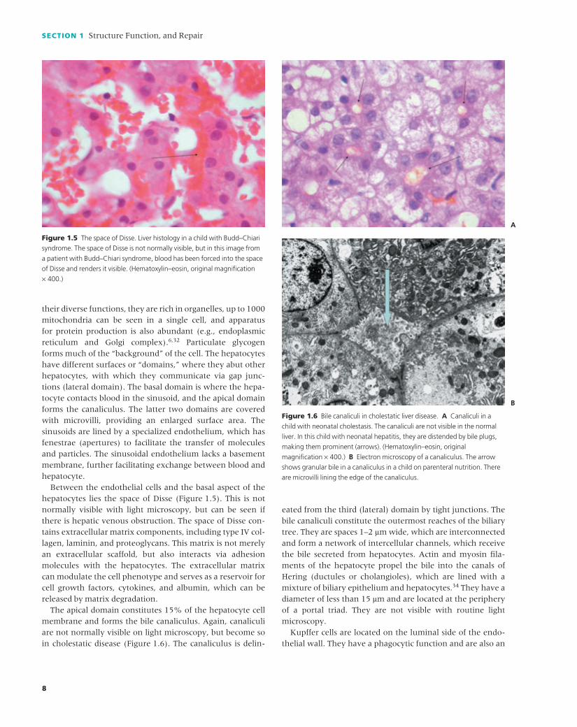
Between the endothelial cells and the basal aspect of thehepatocytes lies the space of Disse (Figure 1.5). This is notnormally visible with light microscopy, but can be seen ifthere is hepatic venous obstruction. The space of Disse con-tains extracellular matrix components, including type IV col-lagen, laminin, and proteoglycans. This matrix is not merelyan extracellular scaffold, but also interacts via adhesionmolecules with the hepatocytes. The extracellular matrix can modulate the cell phenotype and serves as a reservoir forcell growth factors, cytokines, and albumin, which can bereleased by matrix degradation.
The apical domain constitutes 15% of the hepatocyte cellmembrane and forms the bile canaliculus. Again, canaliculiare not normally visible on light microscopy, but become soin cholestatic disease (Figure 1.6). The canaliculus is delin-
eated from the third (lateral) domain by tight junctions. Thebile canaliculi constitute the outermost reaches of the biliarytree. They are spaces 1–2 μm wide, which are interconnectedand form a network of intercellular channels, which receivethe bile secreted from hepatocytes. Actin and myosin fila-ments of the hepatocyte propel the bile into the canals ofHering (ductules or cholangioles), which are lined with amixture of biliary epithelium and hepatocytes.34 They have adiameter of less than 15 μm and are located at the peripheryof a portal triad. They are not visible with routine lightmicroscopy.
Kupffer cells are located on the luminal side of the endo-thelial wall. They have a phagocytic function and are also an
SECTION 1 Structure Function, and Repair
8
Figure 1.5 The space of Disse. Liver histology in a child with Budd–Chiari
syndrome. The space of Disse is not normally visible, but in this image from
a patient with Budd–Chiari syndrome, blood has been forced into the space
of Disse and renders it visible. (Hematoxylin–eosin, original magnification
× 400.)
Figure 1.6 Bile canaliculi in cholestatic liver disease. A Canaliculi in a
child with neonatal cholestasis. The canaliculi are not visible in the normal
liver. In this child with neonatal hepatitis, they are distended by bile plugs,
making them prominent (arrows). (Hematoxylin–eosin, original
magnification × 400.) B Electron microscopy of a canaliculus. The arrow
shows granular bile in a canaliculus in a child on parenteral nutrition. There
are microvilli lining the edge of the canaliculus.
A
B
9781405163347_4_001.qxd 20/06/2008 09:26 Page 8

important source of cytokine secretion. Hepatic stellate cells(previously known as Ito cells) produce extracellular matrix,store vitamin A and lipid, and have fine extensions sur-rounding the sinusoids, possibly related to control of vasculartone. When activated, they transform into myofibroblastsand have an important role in fibrosis.
Functional anatomy/regulation of blood supplyThe dual blood supply to the liver, by the hepatic artery andportal vein, is almost unique in the body. In resting con-ditions, the liver receives about a quarter of the cardiac out-put. About 30% of this hepatic inflow is oxygen-rich bloodvia the hepatic artery; the remaining 70% is nutrient-loadedblood from the intestine and spleen, supplied by the portalvein. Arterial and portal blood mixes freely at the level of thesinusoids. Total blood flow into the liver varies considerablyand is reduced during sympathetic stimulation or sleep. Incontrast, portal blood flow increases following a meal. It ismost stimulated by a protein-rich feed and only moderatelyby carbohydrates, with little effect following lipids. The arte-rial blood supply is not determined by oxygen demand. Innormal livers, only half of the oxygen supplied is extracted,and in situations in which metabolic rates increase, oxygenextraction rises without an increase in arterial flow. Portaland arterial flow are closely related, and an experimentalreduction of portal flow in dogs resulted in arterial hyper-emia. Clinically, this phenomenon becomes apparent in livertransplantation, when thrombosis of either the hepatic arteryor the portal vein leads to compensatory flow rates in theother vessel.
About 20–25% of the normal liver consists of blood, whichis situated in the large vessels. This is about 10–15% of thebody’s total blood volume, and the liver thus serves as areservoir with capacitance function. Liver blood volume can increase by 4% for each 1-mmHg increase in hepaticvenous pressure and may be tripled to about 60% in states of severe outflow obstruction. In hemorrhagic shock, in sympathetic stimulation, and in vascular dehydration, theliver can replace systemic volume rapidly. In animal studies,it has been observed that 7% of the total blood volume can be replaced from the liver, while in dogs a 60% decrease inliver blood content can be achieved within seconds by sym-pathetic stimulation.
Portal vein perfusion pressure is approximately 6–10 mmHg.Arterial perfusion pressures depend on systemic perfusionpressures. The sinusoidal perfusion pressure is determined by a number of factors in the afferent and efferent vessels,including muscular sphincter, autonomic nervous inner-vation, and paracrine function.
In the normal liver, the sinusoids consist of a fenestratedendothelial capillary that receives blood from arterioles andvenules, with a perfusion pressure of 2–4 mmHg. The dis-tribution of blood flow in the sinusoids is determined by
variation in the size of the Kupffer and endothelial cells,which swell and shrink to control the patency of the sinu-soidal lumen. The role of stellate cells, which are thought tobe myofibroblasts, remains unclear, although their ability toinduce fibrosis appears to be the dominant effect on sinu-soidal perfusion in states of liver disease.
Functional versus anatomical unitsIn the absence of connective-tissue septa delineating struc-tural units, different models have been used to define thesmallest functional unit in the liver (Figure 1.7):• The classic lobule, hexagonal in shape, was described in1833.35 It corresponds to the unit that is outlined by connec-tive tissue in other species, such as the pig. It has a hepaticvein branch (“central vein”) at its center. Blood arriving inthe portal tracts at the periphery of the hexagon will feedsinusoids around the whole of their circumference, ratherthan all draining into the interior of the hexagon. It thereforehas limited application as a true primary unit. The primarylobule, described by Matsumoto et al.,36 uses the portal veinbranches to act as the center of the functional unit, giving riseto tortuous and branching three-dimensional units sur-rounding portal vein branches, but it does include the classiclobule as a secondary structure.6,32 This model is based onactual vascular reconstruction (rather than the gelatin infu-sions used in the acinar concept, below) and is gainingwidespread acceptance.37 Descriptive histology in the lobularmodels hence includes such terms as “centrilobular” hepato-cytes (those around the central vein).• The work of Rappaport et al. in 1954 defined the functionalunit as an acinus.38 The axis of the acinus is formed by theterminal branch of the portal vein, not visible in routinemicroscopy. The three zones of the acinar concept are illus-trated in Figure 1.7. Descriptive histology in the acinar con-cept refers to these three acinar zones, and it should be notedthat these do not equate to the regions described in the lobu-lar concept. “Acinar zone 3” is not exclusively “perivenular,”but rather extends in an arc-like fashion from one portal tractto another. The acinar concept proved popular for patho-logists from an observational point of view. In severe liverdamage, necrosis is presumed to occur in the least well oxygenated, most vulnerable hepatocytes first. In the lobularconcept, the least well oxygenated hepatocytes would becentrilobular, and necrosis would therefore be seen in theperivenular region exclusively. In practice, however, necrosisoccurs in a portal–central distribution, and this correspondsto the most peripheral acinar regions (zone 3; Figure 1.7).Many studies of functional heterogeneity within the liver donot support the acinar concept, however, and as mentionedabove, the primary lobule, based on the actual branching ofthe portal vein, is gaining in popularity.
However the functional unit is defined, the function of the hepatocytes, sinusoidal endothelium, Kupffer cells, and
CHAPTER 1 Structure Function and Repair of the Liver
9
9781405163347_4_001.qxd 20/06/2008 09:26 Page 9

extracellular matrix composition, varies between regions.“Periportal,” “perivenular,” andaalthough it does not cor-respond to a true acinar zonea“midzonal” serve as usefuldescriptors for considering functional differences or gradients.Gene expression also shows a functional gradient.32 The phenotypic variation may be determined by the declininggradient in oxygen concentration, the decreasing glucagon–insulin ratio, or other autocrine signals. Periportal hepato-cytes are responsible for oxidative energy metabolism, suchas beta-oxidation and amino acid catabolism, bile forma-tion, and cholesterol synthesis. Perivenous hepatocytes areinvolved in glucose uptake for glycogen synthesis, glycolysis,liponeogenesis, and ketogenesis.
InnervationThe liver is innervated by the autonomic nervous system,through sympathetic nerve fibers from the celiac ganglia and some parasympathetic input from the vagus nerve.Sympathetic nerves supply a dense perivascular plexus aroundthe hilar blood vessels into the sinusoids, where nervescourse in the space of Disse and surround isolated hepato-cytes and stellate cells. Parasympathetic nerve fibers accom-pany the hepatic inflow system, forming a plexus aroundhepatic artery and portal vein, but there is little cholinergicinnervation beyond the portal tract.39
It has been suggested that gap junctions may also providedirect electrical coupling between cells, bypassing the needfor nervous innervation. Cholinergic stimuli increase meta-bolic activity, whereas adrenergic stimuli increase glucosemobilization into the blood. The realization that hepaticfunction is effective even in the denervated graft followingliver transplantation has challenged long-standing views aboutthe role of the autonomic nervous system in regulatingmetabolic activity in the liver. More recent studies have sug-gested that α-adrenergic innervation is involved in hepato-cyte replication.
Function
The liver is the central organ for metabolic homeostasis. Itsmain functions are:• Regulation of uptake and processing of nutrients from theintestinal tract• Synthesis and biotransformation of proteins, carbohydrates,and lipids• Excretion of bile and elimination of hydrophobic compounds• Regulation of energy metabolism• Endocrine functions and mediation of normal growth anddevelopment• Immunological function• Drug metabolism• Regulation of fluid balance
SECTION 1 Structure Function, and Repair
10
Figure 1.7 A Light microscopy of normal liver tissue. The small arrow
points to the approximate outline of a classic hepatic lobule, centered
around a central vein. In schematic diagrams, this is often illustrated as a
regular hexagon, with portal tracts at four points and “nodal points of mall”
at the other two. This is rarely reproducible in practice, leading to the slightly
irregular hexagon shown. The elliptical structure denotes postulated acinar
zones 1, 2, and 3, centered around a terminal portal venule (not visible).
This occupies portions of two adjacent classic lobules. The dotted rectangle
shows the location of portal central bridging necrosis, which is observed
in the clinical situation and which made the acinar concept popular from
a pathological point of view. (Hematoxylin–eosin, original magnification
× 40). B Schematic view of the same anatomical and functional units
of the liver.
CV, central vein; PT, portal tract.
A
B
9781405163347_4_001.qxd 20/06/2008 09:26 Page 10

Uptake and processing (synthesis, storage anddegradation) of proteins, carbohydrates, and lipidsProteinsThe liver accounts for 15% of total body protein production,and the majority of these proteins are secreted as plasma proteins. Proteins are synthesized following the activation ofgenetic promoter sequences by transcription factors. Follow-ing translation and modification, proteins are secreted fromthe sinusoidal aspect of the hepatocytes into the circulation.Nutritional status and hormone secretion regulate the levelof protein production. There is a surge of protein productionin acute illnessesathe acute-phase response, in which C-reactive protein is the most commonly measured sign. Theliver is responsible for synthesizing many proteins, such asalbumin, transport proteins such as ceruloplasmin, coagula-tion and fibrinolytic proteins, complement, and proteaseinhibitors. Proteins are not stored in the liver, but aminoacids are recycled to synthesize new molecules. The liver alsoplays a role in protein and glycoprotein degradation. Aminoacid degradation takes place in the liver, generating thehighly toxic metabolite ammonia, which is associated withhepatic encephalopathy (see Chapters 7 and 15). The ureacycle, which is active almost exclusively in the liver, is largelyresponsible for its removal, and urea cycle defects presentwith severe encephalopathy (see Chapters 5 and 13).
CarbohydratesGlucose, fructose, and galactose are taken up by the hepato-cytes from portal blood. Glucose is converted to glucose-6-phosphate and used to replenish glycogen stores, or else usedin triglyceride production. The liver, under the influence ofhormonesaprincipally insulin (which reduces glucose output)and glucagon (which increases glucose output)ahas a majorrole in maintaining blood glucose. Glucose is either releasedfrom glycogen (glycogenolysis) or synthesized from substratessuch as lactate (gluconeogenesis). In conditions of stress orfasting, glucose uptake is reduced and glucose production isincreased from glycogenolysis. Hypoglycemia is a sensitivetest of liver function and is a sign of severe hepatic necrosis,indicating loss of liver function (see Chapter 7). For the samereason, many infants with severe liver disease are unable tomaintain their blood sugar levels during prolonged fasts.
LipidsThe liver is essential for cholesterol and lipoprotein meta-bolism. Cholesterol is a component of all cell membranes and is essential for the production of steroid hormones andbile acids. Cholesterol homeostasis is controlled by uptakefrom lipoproteins and chylomicrons, which increase hepaticcholesterol, and by the enzyme 3-hydroxy-3-methylglutarylcoenzyme A (HMG CoA), which synthesizes cholesterol de novo.The amount synthesized in the liver is twice that absorbedfrom the diet. In the liver, cholesterol is either “free” or stored
as cholesterol ester. The degradation of cholesterol takesplace through the synthesis of bile acids and biliary excretionof cholesterol (see below). A number of cholestatic liver diseases (e.g., biliary atresia or Alagille’s syndrome) lead toelevated plasma cholesterol due to deficient biliary excretionand catabolism.
Chylomicrons, which transport water-insoluble lipids,carry dietary fat from the intestine to the circulation. Theydeliver triglycerides to peripheral tissues, and the resultingcholesterol-rich chylomicron remnant is taken up by theliver. The liver also synthesizes fatty acids from glucose intimes of dietary excess, and these are subsequently stored astriglycerides, which are the principal source of energy. Fattyacids that are not converted to triglycerides or used in thesynthesis of other molecules are oxidized, following modi-fication, to ketone bodies in the mitochondria, or in the case of very-long-chain fatty acids in the peroxisomes. Micro-vesicular steatosis in hepatocytes is a sign of mitochondrial or peroxisomal disease or drug toxicity (see Chapters 5, 9,and 13).
Very-low-density lipoproteins (VLDLs) are the main lipo-proteins secreted by the liver and carry triglyceride andcholesterol to other tissues, where they are converted to low-density lipoproteins (LDLs). High-density lipoproteins(HDLs) carry cholesterol from the peripheral tissues back tothe liver. Fatty liver occurs when the synthesis of trigly-cerides exceeds the liver’s capacity for export or internalmetabolism.
Bile and bile acidsBile is produced in hepatocytes and is modified in the bileducts. In adults, about 600 mL of isotonic watery bile with apH of 7.8 is produced daily in order to facilitate the excretionof many compounds, including drugs, toxins, and waste pro-ducts, and to provide bile salts to the intestine for the emulsi-fication and absorption of dietary lipids. Bile formation is anosmotic process and is traditionally classified as “bile salt–dependent” (the relationship of canalicular bile flow to bilesalt excretion) and “bile salt–independent” (the active secre-tion of electrolytes and other solutes).
The main components of bile are bile acids (12%), phos-pholipids (4%), cholesterol (0.7%), and conjugated bilirubin(0.1%). Lecithin increases the solubility of cholesterol in bileby micelle formation exponentially to allow a 10-fold con-centration of bile acids and cholesterol in the gallbladder. Ofthe electrolytes in bile, only sodium is concentrated to about280 mmol/L; other electrolytes and bicarbonate are less con-centrated, or unchanged. The primary bile acidsacholic acidand chenodeoxycholic acidaare synthesized from choles-terol by 7α-hydroxylase and subsequently conjugated withtaurine and glycine to enhance affinity to both acids andbases (“amphophilia”).
Primary bile salts are transformed by intestinal bacteriainto secondary bile saltsacholic acid into deoxycholic acid
CHAPTER 1 Structure Function and Repair of the Liver
11
9781405163347_4_001.qxd 20/06/2008 09:26 Page 11

and chenodeoxycholic acid into lithocholic acid and sub-sequently to ursodeoxycholic acid (UDCA). They are re-absorbed in the ileum and returned to the liver via the portalvein. In normal conditions, UDCA represents only 3% of thebile salt pool. It is more hydrophilic than the other bile saltsand is used therapeutically to stimulate bile secretion; it may prevent the hepatocytes from damage caused by hydro-phobic bile salts. Only lithocholic acid is poorly reabsorbedand excreted, so that the gallbladder bile consists of the fourbile salts at a ratio of 10 : 10 : 5 : 1. In chronic liver disease,this balance is shifted to a predominant production of cheno-deoxycholic acid, which lowers the bile pH.
Hepatic bile formation and the biliary excretory functionare closely related. The rate of bile flow is determined by theenterohepatic circulation of bile salts and by the rate of secre-tion of bile salts, cholesterol, phospholipids, and glutathione,which means that if bile excretion is impaired in liver disease,there is reduced excretion of both endogenous and exoge-nous compounds. Bile salts are the main organic solutes inbile, and their active transport against a 1000-fold concentra-tion gradient into the bile canaliculus is the driving force forhepatic bile formation. In adults, the enterohepatic circula-tion of bile salts occurs more than six to eight times in 24 h,enabling the body to retain most of the 5–6 g in the body bilesalt pool. Neonates have about half the bile salt pool of anadult, and ileal bile salt reabsorption is lower. Their intestinalbile acid concentration may be low, leading to poor micelleformation and reduced uptake of fat-soluble vitamins anddietary lipid in comparison with older children and adults.Although this is rarely a cause of malnutrition and/or steat-orrhea, it needs to be considered in cholestatic conditionswhen early supplementation of fat-soluble vitamins is indi-cated. Bile acid uptake from portal blood is physiologicallylower in neonates in comparison with older children, andelevated levels of bile acids may be mistaken for cholestaticliver disease.
Intrahepatic and extrahepatic bile salt transportThe transport processes for bile salts are complex, anda
despite the recent discovery of different membrane-boundbile salt transportersathey are still not fully understood.Hepatocytes are polarized cells that absorb substrates fromthe blood in the sinusoids, such as bile salts, phospholipids,and metabolites of toxic substances, and transport them acrossthe cell to the canalicular membrane to secrete into bile. Thesinusoidal uptake of conjugated bile salts (e.g., taurocholate)by hepatocytes at the basolateral plasma membrane is medi-ated by an active transport process driven by a sodium gradi-ent via the sodium-dependent transporter for the uptake ofbile salts, sodium taurocholate cotransporting polypeptide.The uptake of unconjugated bile salts at the sinusoidal mem-brane is sodium-independent and mediated by the organicanion transporting polypeptide. This transporter also trans-ports steroids such as progesterone and cyclosporine. After
uptake into hepatocytes, the intracellular transport acrossthe cell is thought to be mediated by binding to cytosolic pro-teins, ligandins, and Y9 proteins or fatty acid–binding pro-teins. Some free intracellular bile salts reach the canalicularplasma membrane by diffusion.
Excretion of bile salts across the canalicular plasma mem-brane is the rate-limiting step in the transport of bile saltsfrom blood into bile. Canalicular secretion of monoval-ent bile salts is facilitated by the adenosine triphosphate–dependent “bile salt excreting pump” (BSEP).40 A defect in this transporter is responsible for the genetic conditionknown as progressive familial intrahepatic cholestasis (PFIC).In contrast to monoanionic bile salts, divalent sulfated andglucuronidated bile salts are excreted into bile by the multi-drug resistance–associated protein 2 (Mrp2), also known ascanalicular multispecific organic anion transporter (cMOAT).Failure to express Mrp2 at the canalicular membrane resultsin conjugated hyperbilirubinemia and forms the basis of thehereditary Dubin–Johnson syndrome. Canalicular phospho-lipid secretion is mediated by a different transporter pro-tein, multidrug resistance protein type 3 (Mdr3), which isimportant in preventing bile salt–induced toxic damage tothe biliary epithelium. Failure to express this transporterresults in progressive familial intrahepatic cholestasis type 3and biliary cirrhosis41 (see Chapter 4). FIC-1 is an amino-phospholipid translocator in the canalicular membrane ofhepatocytes that is also found on the apical membrane ofenterocytes. It is responsible for the transport of phosphati-dylserine and phosphatidylethanolamine. A genetic defect in the expression or function of this transporter causesprogressive familial intrahepatic cholestasis type 1 (PFIC-1)and benign recurrent intrahepatic cholestasis type 1 (BRIC-1;see Chapters 3 and 4).
Excretion of bilirubinAs well as its role in facilitating bile salt homeostasis, the biliarysystem also serves as the primary pathway for eliminatingbilirubin, excess cholesterol, and hydrophobic xenobiotics.About 80% of bilirubin is derived from the breakdown oferythrocytes; the remainder stems from heme-containingmyoglobin, cytochromes, and other enzymes. Mononuclearphagocytic cells oxidize heme to form biliverdin, which isthen reduced to bilirubin. This unconjugated bilirubin isalbumin-bound, transported to the hepatic sinusoids, andactively transported into the hepatocytes via the basolateralmembrane. If the unconjugated bilirubin is displaced fromalbumin, it may diffuse across the blood–brain barrier andcause kernicterus in neonates.
Bilirubin uridine diphosphate (UDP) glucuronyltrans-ferase (UGT1A1) conjugation with one or two molecules ofglucuronic acid in the endoplasmic reticulum converts biliru-bin (conjugated bilirubin), which is excreted as hydrophilicbilirubin glucuronides via the canalicular membrane.Following intestinal excretion, bacterial beta-glucuronidases
SECTION 1 Structure Function, and Repair
12
9781405163347_4_001.qxd 20/06/2008 09:26 Page 12

degrade most of these bilirubin glucuronides to colorless urobilinogen. About 20% of urobilinogen is reabsorbed inthe ileum and colon and returned to the liver via the portalvein. Some of this urobilinogen is excreted into the urinarytract.
UGT1A1 belongs to the UGT family of conjugating enzymes,which catalyze glucuronidation of various substrates, includ-ing steroid hormones, carcinogens, and drugs, and which areexpressed in a wide range of tissues. Splicing variation of the original transcripts of the UGT1A1 gene on chromosome2q37 leads to different mRNAs of the enzyme. Of several isoforms, only UGT1A1 is physiologically active. Decreasedactivity of the enzyme in the newborn period contributes tothe physiological jaundice common in the neonate. Mutationsin the UGT1A1 gene either reduce the affinity of UGT1A1toward bilirubin or reduce enzyme activity. Complete absenceof UGT1A1 activity causes Crigler–Najjar syndrome type 1,and a significant reduction of activity causes Crigler–Najjarsyndrome type 2. Only a very mild reduction of UGT1A1activity by missense mutation or reduced expression of theenzyme is present in 6% of the general population, causingGilbert’s syndrome, in which there is a mild elevation ofunconjugated bilirubin (see Chapter 4).
Regulation of energy metabolismThe energy metabolism of the body is integrated by the liverthrough glucose metabolism and fatty acid oxidation. Theliver has a central role in maintaining blood glucose homeo-stasis at constant levels between 3.3 and 6.1 mmol/L in orderto supply glucose as an energy substrate for the brain, renalmedulla, or blood cells. This glucostat function of the liver isprimarily achieved by controlling the storage and release ofglucose from glycogen, followed by glycolysis and gluconeo-genesis. The glycogen content of a liver of a 10-kg child isaround 20–25 g, increasing to about 70–80 g in an adult. As the normal resting glucose requirement is between 4 and6 mg/kg/min, the glycogen stores last for less than a day offasting, after which gluconeogenesis is activated. In pro-longed fasting, total body glucose requirements decreasefrom 160 g/glucose/day to 40 g/glucose/day after 5–6 weeksof starvation in the adult. The healthy body can tolerate this,because fatty acid oxidation becomes the main source of fuel for respiration. Soskin postulated as early as 1940 thatthe blood glucose concentration is the primary stimulus tocontrol glucose uptake or output.42 Glucose uptake into the hepatocyte is insulin-independent and has a direct regula-tory effect on glycogen synthesis. Conversion of excess glu-cose to fatty acids only takes place when hepatic glycogenstores are complete. Such fatty acids are esterified to tri-glycerides and exported from the liver as very-low-densitylipoproteins (VLDLs). Triglycerides in VLDLs from the liverand from the intestinal absorption of lipids are hydrolyzed bylipoprotein lipase and taken up in the peripheral tissues,where fatty acids are metabolized for energy or stored.
Endocrine functionThe liver plays an active role in endocrine regulation. Inresponse to growth hormone activation, the liver producesthe majority of the circulating mitosis-inducing (mitogenic)polypeptides insulin-like growth factor 1 and 2 (IGF-1 andIGF-2), which have anabolic and metabolic effects and regu-late the proliferation of various cells. The specific endocrineeffect of the IGFs and other hormones, such as steroid hormones, is modulated by different binding proteins (IGF-binding proteins 1–6, sex hormone–binding globulin, or thyroid-binding globulin) that are synthesized in the liver.These binding proteins transport the hormones, regulatetheir metabolic clearance, and directly modulate hormoneinteractions with specific receptors.43,44 Thyroxine (T4) isconverted into the metabolically active form of T3 in the liver,which accounts for the low T3 syndrome in patients withdecompensated cirrhosis. Hormonal dysfunction in liver disease may develop from reduced clearance of hormones(e.g., gynecomastia in men), from portosystemic shunting,dysregulated synthesis of binding proteins, or impaired end-organ sensitivity to the hormoneai.e., insulin resistance incirrhosis.
Immunological functionThe liver contains many lymphocytes, both of the adaptiveimmune system, which require previous exposure to antigenfor efficacy, and also cells of the innate immune systema
natural killer (NK) cells (Pit cells).45 The liver also contains a population of cells that express both T-cell and NK-cellmarkers,46 which play a role in the clearance function of theliver in filtering gut-derived endotoxins and microorganisms.Kupffer cells are macrophages that are important in thephagocytosis of particulate material and cellular debris (theyare conspicuous in acute hepatitis), but also play a role incytokine release and antigen presentation. Following livertransplantation, donor Kupffer cells are rapidly (within days)replaced by recipient Kupffer cells infiltrating the liver.
Drug metabolismThe liver is the prime site for drug metabolism in the body,which occurs in two phases. The first is an oxidation reaction,mediated by the cytochrome P450 enzymes. Reactive oxygenspecies that are toxic to the cell are generated during this process and require a range of antioxidant mechanisms(moleculesae.g., glutathione and vitamin E; and enzymesae.g., superoxide dismutase) to render them inert. The metabol-ized drug, which may itself be toxic, enters the second phaseof metabolism, which involves conjugation with hydrophiliccompoundsae.g., glucuronic acid or glutathione. Once rendered hydrophilic, the drug metabolite is excreted via the kidneys or the bile. The enzymes responsible for drugmetabolism may be either induced or inhibited by otherdrugs or chemicals, and there can also be idiosyncratic differ-ences between individuals in drug metabolism. Severe liver
CHAPTER 1 Structure Function and Repair of the Liver
13
9781405163347_4_001.qxd 20/06/2008 09:26 Page 13

failure reduces the ability to metabolize drugs, so that drugeffects are prolonged (e.g., sedatives or anesthetic agents), orthere may be an accumulation of toxic metabolites, whichcomplicates hepatic encephalopathy.
Liver function and fluid balanceAs described above (functional anatomy/regulation of bloodsupply), the liver can retain and release a significant volumeof whole blood and/or plasma and hence influence the circu-lating blood volume. Although the direct interaction betweenthe liver and kidney is not fully understood, impaired liverfunction leads to a reduced ability to excrete sodium andwater. A number of factors are involved, which include:hyperaldosteronism and/or increased tubular sensitivity toaldosteronism; increased renal sympathetic nerve activity;and reduced renal perfusion. Splanchnic vasodilation isprobably an initial adverse event that leads to renal vasocon-striction, followed by a reduction of renal blood flow and ofthe glomerular filtration rate. Sodium retention is the firstsign of renal dysfunction, followed by water retention, lead-ing to dilutional hyponatremia in plasma. Plasma volumeexpansion due to sodium and water retention, together withsinusoidal hypertension (portal pressure gradient of > 12%),is a key factor in the pathogenesis of cirrhotic ascites, whichindicates the progression from compensated to decompen-sated cirrhosis.
Growth and repair
Functional development of the liver andphysiological adaptations at birthAt birth, the change from placental to enteral nutrition stimulates bile acid secretion and the enterohepatic circu-lation. The switch from umbilical venous to portal blood supply means that new molecules and bacteria are carried tothe immature neonatal liver by the portal vein. This is bestdemonstrated by the immaturity of bile formation and thedevelopment of physiological jaundice in neonates (seeabove). The liver is vulnerable in the presence of prematur-ity, hypoxia, sepsis, drug administration, or total parenteralnutrition.47– 49 α-Fetoprotein, one of the main fetal serumproteins, is synthesized by fetal hepatocytes 25–30 days afterconception, and by the yolk sac and intestinal epithelium.Levels peak by the end of the first trimester and exponen-tially fall until normal adult levels are reached approximatelyat the end of the first year of life. Albumin levels are close to adult levels at birth, but coagulation proteins are low,increasing the risk of bleeding and hence the need for vita-min K administration at birth. Bile acids are synthesized from5 to 9 weeks’ gestation and bile secretion begins at 12 weeks,but canalicular transport mechanisms and the distal bileducts, where the bile is extensively modified, are still under
development for 4 weeks after birth.6,49 γ-Glutamyltransferase,located at the canalicular surface of the hepatocytes, is slightlyelevated in the serum in the first few months of life.
In utero, the placenta carries out most of the metabolic anddetoxifying functions that normally take place in the liver. Tocope with this change, hepatic enzymes are rapidly inducedat birth. Many conjugation reactions are mature by 2 weeks,but some UDP-glucuronyltransferase genes are not fullyexpressed for 2 years.50 The cytochrome P450 group and peroxisomal enzymes also show early functionality. The firstfeed stimulates insulin production and storage of glycogen.Term newborns have hepatic glycogen stores, but these arequickly depleted, making the infants prone to hypoglycemiaunless fed frequently. If the infant is unwell, acute-phaseproteins may have a long half-life, because the immatureliver is unable to clear them.48
At 12 weeks’ gestation, the liver is the main site ofhemopoiesis, but the bone marrow becomes active from 5months of gestation. It is normal to see evidence of residualhemopoiesis in the neonatal liver for up to 6 weeks afterbirth,48 but it is particularly prominent in neonatal hepa-titis. Hemosiderin (as hemopoiesis decreases) and copper-associated protein accumulate in the liver and are depositedin periportal hepatocytes. Both are normal constituents ofthe neonatal liver and are not indicative of disease.
Liver growth and regenerationThe expected life span of a hepatocyte is about 200–500days. In normal children and adults, hepatic regenerationoccurs by replication of mature cells. This process can be up-regulatedafor instance, following trauma or partial hep-atectomy, the liver can be reconstituted by proliferation ofmature hepatocytes within days and weeks. The liver cellmass is highly flexible and varies throughout life, dependingon metabolic demands such as disease or pregnancy. It islikely that liver is also regenerated from progenitor cells inthe liver, bone marrow–derived stem cells, and mechanismsof cell fusion, but it is still not clear how this is controlled.Initial studies were based on the search for a growth factorthat would stimulate hepatocyte regeneration and control ofhuman hepatocyte replication. Studies of rodents suggestedthe presence of alternative pathways, including a putativeprogenitor cell compartment. Animal models in which thegrowth stimulus from partial hepatectomy was combinedwith growth arrest of fast-replicating mature hepatocytesusing toxins such as 2-acetylaminofluorene or 5-aminouracilled to the proliferation of pluripotent so-called “oval cells” inthe canals of Hering.51,52 The phenotype of these putativehepatic progenitor cells, the oval cell, and the existence of astem cell compartment was accepted for animals. The pres-ence of similar oval cells in humans was more difficult todemonstrate, partly because of the inability to transfer theexperimental model into an acceptable human study and
SECTION 1 Structure Function, and Repair
14
9781405163347_4_001.qxd 20/06/2008 09:26 Page 14

partly because of phenotypic differences between humansand mice. The finding of the hemopoietic stem cell markerand proto-oncogene c-kit in certain biliary cells from diseasedpediatric liver was one of the first steps in demonstrating thepresence of this type of stem cell compartment in humans.53
An understanding of the physiology of liver regeneration was improved by animal studies conducted by Petersenet al.,54 which were confirmed in humans by Theise et al.55
They demonstrated the presence of Y chromosome–positivehepatocytes and biliary epithelium in female recipients of a therapeutic bone-marrow transplant from male donors,confirming the ability of human bone marrow–derived stemcells to differentiate into the hepatic cell lineages. However,clinically, bone marrow–derived cells are only marginallyinvolved in physiological repair, since in acute liver failureregeneration occurs via proliferation of hepatic progenitorcells, whereas following partial hepatectomy it is restored byreplication of normally quiescent hepatocytes.
Liver regeneration is now known to vary in accordancewith circadian rhythms and metabolic requirements. In-creased metabolic demands and proinflammatory cytokines are likely to be essential. Effects of cytokines (tumor necrosisfactor-α, interleukin-6) and growth factors (hepatocytegrowth factor) are probably linked by the effect of the tissue-bound metalloproteinases. Experiments with hepatocytesand cocultured biliary epithelium have shown that thedegradation of extracellular matrix by metalloproteinases to release growth factors is an essential step in hepatocyte proliferation.
The limitations of understanding of hepatic regenerationhave led to persistent problems in clinical hepatocyte trans-plantation. Therapeutic liver repopulation is an attractiveoption in a number of metabolic conditions and has beensuccessfully performed in a small number of patients. Itremains problematic to achieve persistent engraftment ofadequate numbers of cells, because it has not been possible tocreate an environment that allows preferential replication oftransplanted cells. It is possible that significant medical andsurgical hepatic preconditioning, similar to myeloablation inbone-marrow transplantation, will be necessary to providean appropriate environment for persistent engraftment.56
Clinical studies in this field are limited by the need to matchthe long-term outcome with orthotopic liver transplantation,at around 90%.
Liver fibrosisThe outcome of most disease processes in the liver is fibrosis.In hepatic fibrogenesis, stellate cells produce an excess oftype I and III collagen, which replaces the normal extracellu-lar matrix. Activation of these cells by injured hepatocytes,biliary cells, or inflammatory stimuli leads to the conversionof quiescent vitamin A–storing cells into proliferative, con-tractile, and fibrogenic myofibroblasts. The dogma that hep-
atic fibrosis is irreversible is increasingly being challenged,and an understanding of stellate-cell activation is an essentialstep forward here.57 Different metalloproteinases that cleavecollagens are mainly involved in matrix degradation of theliver, although neutrophils, macrophages, and stellate cellsalso contribute to this process. Tissue inhibitors of matrixmetalloproteinases (TIMPs) are the key regulators in deter-mining the reversal of fibrosis. Sustained TIMP-1 expressioninhibits protease activity for matrix degradation and blocksapoptosis of activated stellate cells. A number of phase I clin-ical trials are being planned to investigate the use of thesedrugs to prevent or reverse fibrosis.58
References
1 Saxena R, Zucker SD, Crawford JM. Anatomy and physiology of
the liver. In: Zakim D, Boyer TD, eds. Hepatology: a Textbook of Liver
Disease, 4th ed. Philadelphia: Saunders, 2003: 3–30.
2 Lemaigre F, Zaret KS. Liver development update: new embryo
models, cell lineage control, and morphogenesis. Curr Opin Genet
Dev 2004;14:582–90.
3 Beaudry JB, Pierreux CE, Hayhurst GP, et al. Threshold levels of
hepatocyte nuclear factor 6 (HNF-6) acting in synergy with HNF-
4 and PGC-1alpha are required for time-specific gene expression
during liver development. Mol Cell Biol 2006;26:6037–46.
4 Costa RH, Kalinichenko VV, Holterman AX, Wang X. Transcrip-
tion factors in liver development, differentiation, and regenera-
tion. Hepatology 2003;38:1331–47.
5 Crosby HA, Nijjar SS, de Ville de Goyet J, Kelly DA, Strain AJ.
Progenitor cells of the biliary epithelial cell lineage. Semin Cell Dev
Biol 2002;13:397–403.
6 Roskams T, Desmet V, Verslype C. Development, structure
and function of the liver. In: Burt AD, Portmann BC, Ferrell
LD, eds. Macsween’s Pathology of the Liver. Edinburgh: Churchill
Livingstone, 2007: 1–74.
7 Haugen G, Kiserud T, Godfrey K, Crozier S, Hanson M. Portal
and umbilical venous blood supply to the liver in the human
fetus near term. Ultrasound Obstet Gynecol 2004;24:599–605.
8 Libbrecht L, Cassiman D, Desmet V, Roskams T. The correlation
between portal myofibroblasts and development of intra-
hepatic bile ducts and arterial branches in human liver. Liver
2002;22:252–8.
9 Vijayan V, Tan CE. Developing human biliary system in three
dimensions. Anat Rec 1997;249:389–98.
10 Desmet VJ. Ludwig symposium on biliary disorders, part I.
Pathogenesis of ductal plate abnormalities. Mayo Clin Proc
1998;73:80–9.
11 Theise ND, Saxena R, Portmann BC, et al. The canals of Hering
and hepatic stem cells in humans. Hepatology 1999;30:1425–33.
12 Tan CE, Vijayan V. New clues for the developing human biliary
system at the porta hepatis. J Hepatobiliary Pancreat Surg 2001;8:
295–302.
13 Van Eyken P, Sciot R, Callea F, Van der Steen K, Moerman P,
Desmet VJ. The development of the intrahepatic bile ducts in
man: a keratin-immunohistochemical study. Hepatology 1988;8:
1586–95.
CHAPTER 1 Structure Function and Repair of the Liver
15
9781405163347_4_001.qxd 20/06/2008 09:26 Page 15

14 Nakanuma Y, Hoso M, Sanzen T, Sasaki M. Microstructure
and development of the normal and pathologic biliary tract in
humans, including blood supply. Microsc Res Tech 1997;38:552–
70.
15 Crawford JM. Development of the intrahepatic biliary tree. Semin
Liver Dis 2002;22:213–26.
16 Cantlie J. On a new arrangement of the right and left lobes of the
liver. J Anat Physiol (London) (Section Proc Anat Soc Great Britain &
Ireland) 1898;32:4–9.
17 Kasahara M, Kaihara S, Oike F, et al. Living-donor liver trans-
plantation with monosegments. Transplantation 2003;76:694–6.
18 Bismuth H, Houssin D, Castaing D. Major and minor segmentec-
tomies “réglées” in liver surgery. World J Surg 1982;6:10–24.
19 Bismuth H. Surgical anatomy and anatomical surgery of the
liver. World J Surg 1982;6:3–9.
20 Launois B, Jamieson GG. The importance of Glisson’s capsule
and its sheaths in the intrahepatic approach to resection of the
liver. Surg Gynecol Obstet 1992;174:7–10.
21 Clavien PA, Petrowsky H, DeOliveira ML, Graf R. Strategies for
safer liver surgery and partial liver transplantation. N Engl J Med
2007;356:1545–59.
22 Crawford JM. Liver and biliary tract. In: Kumar V, Abbas A,
Faustto N, eds. Robins and Cotran Pathologic Basis of Disease.
Philadelphia: Saunders, 2004: 877–939.
23 Van As AB, Hickman R, Engelbrecht GH, Makan P, Duminy F,
Kahn D. Significance of the portal vein helix. S Afr J Surg
2001;39:50–2.
24 Rosenthal SJ, Harrison LA, Baxter KG, Wetzel LH, Cox GG,
Batnitzky S. Doppler US of helical flow in the portal vein.
RadioGraphics 1995;15:1103–11.
25 Attardi G. Demonstration in vivo and in vitro of peristaltic con-
tractions in the portal vein of adult mammals (rodents). Nature
1955;176:76–7.
26 Howard ER, Davenport M. Congenital extrahepatic portocaval
shuntsathe Abernethy malformation. J Pediatr Surg 1997;32:
494–7.
27 Daly JM, Kemeny N, Oderman P, Botet J. Long-term hepatic
arterial infusion chemotherapy. Anatomic considerations, operat-
ive technique, and treatment morbidity. Arch Surg 1984;119:
936–41.
28 Northover JM, Terblanche J. A new look at the arterial supply
of the bile duct in man and its surgical implications. Br J Surg
1979;66:379–84.
29 Stapleton GN, Hickman R, Terblanche J. Blood supply of the
right and left hepatic ducts. Br J Surg 1998;85:202–7.
30 Healey JE Jr, Schroy PC. Anatomy of the biliary ducts within
the human liver; analysis of the prevailing pattern of branchings
and the major variations of the biliary ducts. AMA Arch Surg
1953;66:599–616.
31 Hepp J, Couinaud C. [Approach to and use of the left hepatic duct
in reparation of the common bile duct; in French.] Presse Med
1956;64:947–48.
32 Couinaud C. Liver anatomy: portal (and suprahepatic) or biliary
segmentation. Dig Surg 1999;16:459–67.
33 Rocko JM, Di Gioia JM. Calot’s triangle revisited. Surg Gynecol
Obstet 1981;153:410–4.
34 Roskams TA, Theise ND, Balabaud C, et al. Nomenclature of the
finer branches of the biliary tree: canals, ductules, and ductular
reactions in human livers. Hepatology 2004;39:1739–45.
35 Kiernan F. The anatomy and physiology of the Liver. Philos Trans
R Soc London 1833;123:711–70.
36 Matsumoto T, Komori R, Magara T, et al. A study on the normal
structure of human liver, with special reference to its angioarchi-
tecture. Jikeikai Med J 1979;26:1–40.
37 Malarkey DE, Johnson K, Ryan L, Boorman G, Maronpot RR.
New insights into functional aspects of liver morphology. Toxicol
Pathol 2005;33:27–34.
38 Rappaport AM, Borowy ZJ, Lougheed WM, Lotto WN.
Subdivision of hexagonal liver lobules into a structural and func-
tional unit; role in hepatic physiology and pathology. Anat Rec
1954;119:11–33.
39 McCuskey R, Robert S. Anatomy of efferent hepatic nerves. Anat
Rec 2004;280:821–6.
40 Jansen PL, Strautnieks SS, Jacquemin E, et al. Hepatocanalicular
bile salt export pump deficiency in patients with progressive
familial intrahepatic cholestasis. Gastroenterology 1999;117:1370–
9.
41 Jacquemin E. Progressive familial intrahepatic cholestasis. Genetic
basis and treatment. Clin Liver Dis 2000;4:753–63.
42 Soskin S. The liver and carbohydrate metabolism. Endocrinology
1940;26:297–308.
43 Holt RI, Crossey PA, Jones JS, Baker AJ, Portmann B, Miell
JP. Hepatic growth hormone receptor, insulin-like growth factor
I, and insulin-like growth factor-binding protein messenger
RNA expression in pediatric liver disease. Hepatology 1997;26:
1600–6.
44 Holt RI, Miell JP, Jones JS, Mieli-Vergani G, Baker AJ.
Nasogastric feeding enhances nutritional status in paediatric liver
disease but does not alter circulating levels of IGF-I and IGF bind-
ing proteins. Clin Endocrinol (Oxf) 2000;52:217–24.
45 Lang KS, Georgiev P, Recher M, et al. Immunoprivileged status of
the liver is controlled by Toll-like receptor 3 signaling. J Clin Invest
2006;116:2456–63.
46 Doherty DG, Norris S, Madrigal-Estebas L, et al. The human
liver contains multiple populations of NK cells, T cells, and
CD3+CD56+ natural T cells with distinct cytotoxic activities and
Th1, Th2, and Th0 cytokine secretion patterns. J Immunol
1999;163:2314–21.
47 Suchy F, Narkewicz MR. Development of the liver and bile ducts.
J Pediatr Gastroenterol Nutr 2002;35(Suppl 1):S4–6.
48 Beath SV. Hepatic function and physiology in the newborn.
Semin Neonatol 2003;8:337–46.
49 Knisely AS. Biliary tract malformations. Am J Med Genet A
2003;122:343–50.
50 Strassburg CP, Strassburg A, Kneip S, et al. Developmental
aspects of human hepatic drug glucuronidation in young chil-
dren and adults. Gut 2002;50:259–65.
51 Oh SH, Witek RP, Bae SH, et al. Bone marrow-derived hepatic
oval cells differentiate into hepatocytes in 2-acetylaminofluorene/
partial hepatectomy-induced liver regeneration. Gastroenterology
2007;132:1077–87.
52 Golding M, Sarraf CE, Lalani EN, et al. Oval cell differentiation
into hepatocytes in the acetylaminofluorene-treated regenerat-
ing rat liver. Hepatology 1995;22:1243–53.
53 Baumann U, Crosby HA, Ramani P, Kelly DA, Strain AJ.
Expression of the stem cell factor receptor c-kit in normal and
diseased pediatric liver: identification of a human hepatic pro-
genitor cell? Hepatology 1999;30:112–7.
SECTION 1 Structure Function, and Repair
16
9781405163347_4_001.qxd 20/06/2008 09:26 Page 16

54 Petersen BE, Bowen WC, Patrene KD, et al. Bone marrow as a
potential source of hepatic oval cells. Science 1999;284:1168–70.
55 Theise ND, Nimmakayalu M, Gardner R, et al. Liver from bone
marrow in humans. Hepatology 2000;32:11–6.
56 Grompe M. Principles of therapeutic liver repopulation. J Inherit
Metab Dis 2006;29:421–5.
57 Friedman SL, Bansal MB. Reversal of hepatic fibrosisafact or
fantasy? Hepatology 2006;43:S82–8.
58 Hemmann S, Graf J, Roderfeld M, Roeb E. Expression of MMPs
and TIMPs in liver fibrosisaa systematic review with special
emphasis on anti-fibrotic strategies. J Hepatol 2007;46:955–75.
CHAPTER 1 Structure Function and Repair of the Liver
17
9781405163347_4_001.qxd 20/06/2008 09:26 Page 17

9781405163347_4_001.qxd 20/06/2008 09:26 Page 18
Investigating the Liver2
9781405163347_4_002.qxd 20/06/2008 09:27 Page 19

9781405163347_4_002.qxd 20/06/2008 09:27 Page 20
21
The approach to the child with liver disease should be systematic and based on an accurate clinical history and athorough physical examination. This chapter will outline the main diagnostic categories and summarize key investiga-tions. Further detail will be provided in individual chapters.Investigating the liver relies on a multidisciplinary approachinvolving clinical chemistry, hematology, radiology, histo-pathology, and microbiology. It is essential to understand themany functions of the liver and to recognize the effects ofhepatic dysfunction on other body systems (Table 2.1; seealso Chapter 1).
Neonatal liver disease
Almost two-thirds of children who have liver disease presentin the neonatal period with persistent jaundice. Although phy-siologic jaundice is common in neonates, infants who developsevere or persistent jaundice should be investigated to excludehemolysis, sepsis, or underlying liver disease. Neonatal jaun-dice that persists beyond 14 or 21 days should always beinvestigated, even in breast-fed babies (see Chapter 4). It isessential to establish whether the jaundice is due to anincrease in conjugated or unconjugated hyperbilirubinemia.The common causes of unconjugated hyperbilirubinemia are rhesus incompatibility, breast-milk jaundice, and Crigler–Najjar syndrome type I or II (see Chapter 4). Liver disease isassociated with a rise in conjugated hyperbilirubinemia.
The clinical history should include:• Details about the mother’s pregnancy (drugs, alcohol, smok-ing, intercurrent illnesses, pruritus of pregnancy, hepatitisstatus, risk factorsae.g., drug abuse)• Birth weight and gestational age• Vitamin K administration• Family history
• Consanguinity• The history of the present illness should include:
aDate of jaundiceaColor of stools and urineaDrug history, particularly parenteral nutritionaBleeding, petechiae, or bruisingaFeeding history and weight gainaDiarrhea and vomitingaImmunizations, if relevant
The differential diagnosis can be guided by the birth weightand gestational age (Figure 2.1) and is between extrahepaticbiliary disease, the neonatal hepatitis syndrome, and metabolicliver disease (see Chapters 4, 5, and 13).
Clinical features suggesting liver disease include:• Family history, consanguinity, or dysmorphic featuressuggesting metabolic or inherited liver disease (Figure 2.2).• Pale stools and dark urine, suggesting cholestasis or obstruc-tion (Figure 2.3).• Bruising, petechiae, or bleeding, suggesting vitamin Kdeficiency from malabsorption or inadequate administrationat birth.• Hypoglycemia could be secondary to metabolic disease,hypopituitarism, or acute liver failure.• Hepatomegaly is a nonspecific sign, as is slow weight gainor failure to thrive.• Splenomegaly suggests an intrauterine infection, metabolicliver disease, or advanced liver disease with hepatic fibrosisand early portal hypertension.• Ascites is rare and suggests an intrauterine infection,inborn error of metabolism, rhesus hemolytic disease, oradvanced liver disease.If the infant is acutely ill, then sepsis, metabolic disease, oracute liver failure should be considered (see Chapters 5 and 7).
Liver disease in older children
Liver disease in children older than 6 months may be acute orchronic (Figure 2.4). As in infancy, there is a predominance
The Approach to the Child with LiverDisease: Differential Diagnosis and Useful Investigations
Deirdre A. Kelly
2
Diseases of the Liver and Biliary System in Children, 3rd edition. Edited by
Deirdre Kelly. © 2008 Blackwell Publishing, ISBN: 978-1-4051-6334-7.
9781405163347_4_002.qxd 20/06/2008 09:27 Page 21

SECTION 2 Investigating the Liver
22
Table 2.1 Functions of the liver.
Function Effect of dysfunction Assessment
Metabolism/storageCarbohydrate/glycogen Loss of glucose homeostasis Hypoglycemia on fasting/stressLipid Lipid accumulation in hepatocytes High/low cholesterol
↓ Oxidation of fatty acids ↑ Lactate↑ FFA : BOH ratio↑ AcylcarnitineOrganic aciduria
Protein ↑ Catabolism Low BCAA, urea↑ Ammonia↑ Tyr, –Phe, –Met
SynthesisAlbumin Loss of muscle mass Low albumin
Protein energy malnutritionFactors II, VII, IX, X Coagulopathy Prolonged PT/PTT
DegradationDrugs Prolonged drug effectae.g., sedation ClinicalEstrogens Telangiectasia Clinical
GynecomastiaToxic products Encephalopathy Abnormal EEG/clinical signs
Bile synthesis and excretion Cholestasis ↑ Conjugated bilirubinFat malabsorption ↑ GGT
↑ ALP↑ Cholesterol
Fat-soluble vitamin deficiency AnthropometryPruritusMalnutrition
ALP, alkaline phosphatase; BOH, β-hydroxybutyrate; BCAA, branched-chain amino acids; EEG, electroencephalogram; FFA, free fatty acids; GGT, γ -glutamyl transpeptidase; Met, methionine; Phe, phenylalanine; PT, prothrombin time; PTT, partial thromboplastin time; Tyr, tyrosine.
Birthweight & Gestation
Hepatomegaly
Normal with hepatomegaly
Mild/no jaundice+HypoglycemiaJaundiceHepatosplenomegaly
No JaundiceJaundice
• α1 anti-trypsin deficiency• Galactosemia• Fructosemia*• Cystic Fibrosis• Alagille Syndrome†
• Familial Intrahepatic Cholestasis• Tyrosinemia‡
• Niemann Pick A&B
• Intra-uterine Infection• Niemann Pick C
• Biliary Atresia• α1 – anti-tyrypsin deficiency• Choledocal cyst
• Endocrine hepatitis• Glycogen storage disease I, II, IV• Tyrosinaemia type I
Jaundice
abnormal/low
Figure 2.1 The approach to the diagnosis of neonatal liver disease. * Presents on weaning; † dysmorphic; ‡ jaundice may be mild.
9781405163347_4_002.qxd 20/06/2008 09:27 Page 22

CHAPTER 2 The Approach to the Child with Liver Disease
23
Figure 2.2 Dysmorphic features suggest
inherited metabolic disease. In Alagille’s
syndrome, which is an autosomal-dominant
disorder. The characteristic facies consists
of a broad forehead, deep-set eyes, mild
hypertelorism, and a pointed chin. It can be
difficult to diagnose in an infant, but the
diagnosis may be obvious in the parent.
Figure 2.3 A, B Cholestasis with pale stools and dark urine is characteristic of biliary
obstruction, usually due to biliary atresia.
A B
+/– Splenomegaly
Splenomegaly unusual
Splenomegaly
• Fibrocystic Disease• Cirrhosis* and portal hypertension from any cause• EHPVO*
• Cystic Fibrosis• Gaucher’s Disease• Glycogen Storage Disease III & IV• Niemann Pick A, B, C
• Chronic Viral Hepatitis B&C• NAFLD• Coeliac Disease• Glycogen Storage Disease I• Budd Chiari• Hepatic tumor
Mild/Absent
• Autoimmune Hepatitis• Wilson’s Disease• Drug Hepatotoxicity• a1 anti trypsin deficiency• Cirrhosis and portal hypertension from any cause
Hepatomegaly
Present +/– Splenomegaly
Jaundice
Figure 2.4 The approach to the older child
with liver disease. * Liver may be small. EHPVO,
extrahepatic vein obstruction; NAFLD,
nonalcoholic fatty liver disease.
9781405163347_4_002.qxd 20/06/2008 09:27 Page 23

SECTION 2 Investigating the Liver
24
of inherited disorders and multisystem involvement. Jaun-dice may not be a prominent feature. Acute or chronic liverdisease may be due to infection, autoimmune disease, drug-induced hepatitis, or metabolic disease, or may be secondaryto disease elsewhere (Figure 2.4; see also Sections 6 and 7,Chapters 8–14).
The clinical history for either acute or chronic liver diseaseshould include:• Previous history, particularly of neonatal jaundice (α1-antitrypsin deficiency and Niemann–Pick C disease may beassociated with transient jaundice)• Evidence of hypoglycemia• Weight gain or loss• Diarrhea or inflammatory bowel disease• Pruritus• Bleeding, bruising, epistaxis• Hematemesis or melena• Skin rashes or joint pains• Drug history, especially transfusions• Ear piercing, tattooing, previous surgery• Immunizations• Family history and consanguinity• Symptoms of extrahepatic disease/autoimmune disease• Allergies• Developmental history• Deterioration in school performance (Wilson’s disease orNiemann–Pick C)• Foreign travel• Household contacts• Local epidemics• Recreational activity• Intravenous drug abuse• Pets
Acute liver disease
The clinical presentation varies depending on the etiology,but the following clinical features are common:• A prodrome of malaise, lethargy, and anorexia• Nausea, vomiting, or diarrhea• Weight loss• Abdominal discomfort or tender hepatomegaly• Hepatomegaly• Splenomegaly• Ascites (rarely, except for acute Budd–Chiari syndrome)• Rash or joint pains• Jaundice may or may not be presentThe differential diagnosis of acute hepatitis includes:• Viral hepatitis A or B, seronegative hepatitis• Autoimmune hepatitis• Drug hepatoxicity• Metabolic liver disease, especially Wilson’s disease (seeChapters 6, 8, 9, and 14)
Acute liver failure is the development of massive hepaticnecrosis with subsequent loss of liver function, with coagu-lopathy and with or without hepatic encephalopathy. Thedisease is uncommon, but has a high mortality. The childmay present within hours or weeks with jaundice, encephalo-pathy, coagulopathy, hypoglycemia, and electrolyte disturb-ance. The main causes are infection (seronegative hepatitis),autoimmune hepatitis, drug toxicity (acetaminophen) andmetabolic disease (see Chapter 7).
Chronic liver disease
Chronic liver disease may affect every organ in the body(Figure 2.5). It may be asymptomatic, and the diagnosis maybe made:• After incidental detection of abnormal liver enzymes orhepatomegaly• After family screening for hepatitis B/C or metabolic disorders• In a transfusion recipient following diagnosis of donorinfection• In coexistent diseaseae.g., inflammatory bowel disease,celiac disease• In a recipient of a known toxic agentae.g., methotrexateThe clinical presentation varies from acute hepatitis to theinsidious development of hepatosplenomegaly, cirrhosis,and portal hypertension with lethargy and malnutrition.Common symptoms include:• Intermittent fatigue, anorexia, and weight loss• Abdominal discomfort• Variable or fluctuating jaundice with pruritus and palestools• Hematemesis or melena from variceal bleeding. This maybe the presenting symptom, especially in cirrhosis with portalhypertension or extrahepatic portal hypertension.The differential diagnosis of chronic liver disease is illustratedin Figure 2.4. Common physical features are:• Palmar erythema, or “liver palms,” is a nonspecific red dis-coloration of the palms and fingertips, indicative of a hyper-dynamic circulation, which is associated with chronic liverdisease and cirrhosis (Figure 2.6).• Spider nevi, or spider angiomas, are telangectasia consist-ing of a central arteriole with superficially radiating smallvessels, resembling spiders’ legs. The lesion will blanch whenpressure is applied to the center. Although spider nevi dooccur in healthy children at puberty, the presence of morethan five is suggestive of chronic liver disease. Superficial distended veins (telangectasia) are common on the face(Figure 2.7).• Abdominal distension may be due to hepatosplenomegalyor ascites. Distended abdominal veins are a sign of portalhypertension (Figure 2.8).• Fat malabsorption leads to malnutrition and rickets, oftenwith pathological fractures (Figure 2.9).
9781405163347_4_002.qxd 20/06/2008 09:27 Page 24
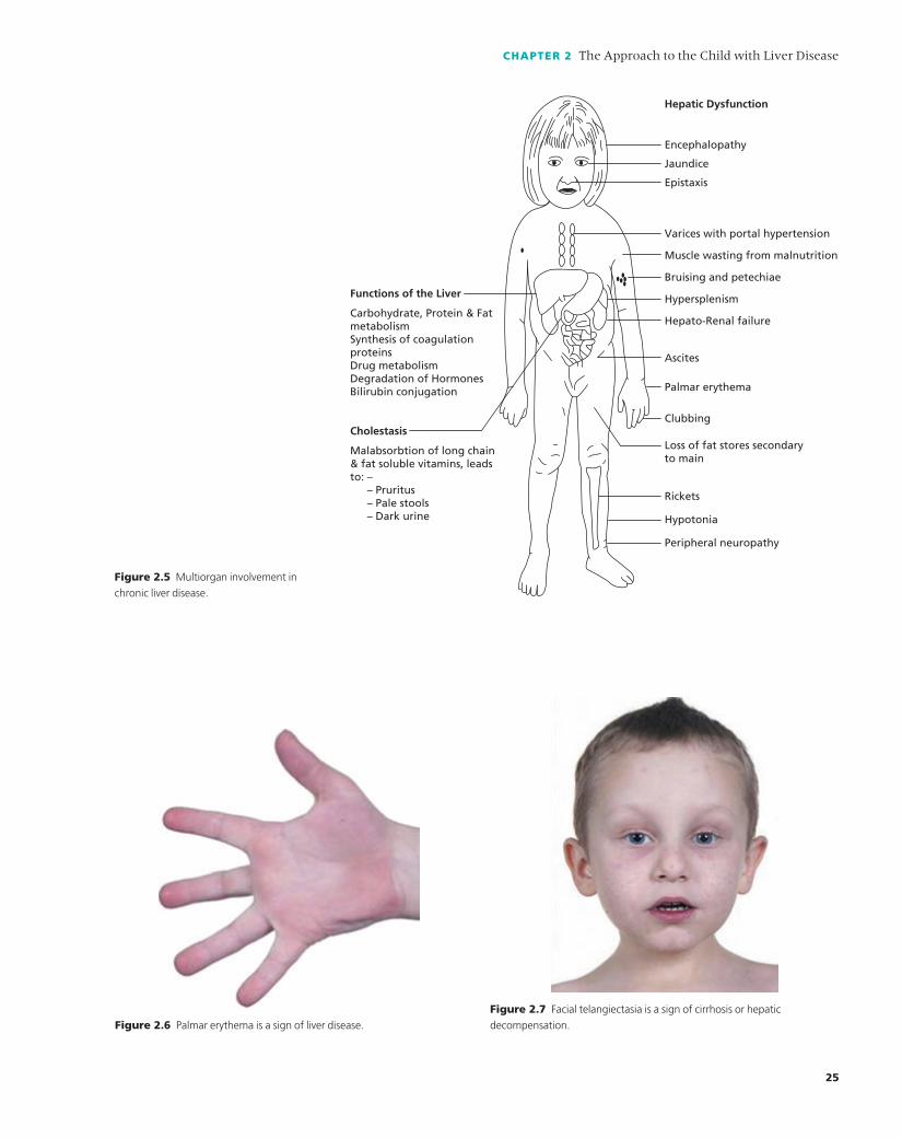
CHAPTER 2 The Approach to the Child with Liver Disease
25
Hepatic Dysfunction
Encephalopathy
Jaundice
Epistaxis
Varices with portal hypertension
Muscle wasting from malnutrition
Bruising and petechiae
Hypersplenism
Hepato-Renal failure
Ascites
Palmar erythema
Clubbing
Loss of fat stores secondaryto main
Rickets
Hypotonia
Peripheral neuropathy
Functions of the Liver
Carbohydrate, Protein & Fatmetabolism Synthesis of coagulationproteinsDrug metabolismDegradation of HormonesBilirubin conjugation
Cholestasis
Malabsorbtion of long chain& fat soluble vitamins, leadsto: – – Pruritus – Pale stools – Dark urine
Figure 2.5 Multiorgan involvement in
chronic liver disease.
Figure 2.6 Palmar erythema is a sign of liver disease.
Figure 2.7 Facial telangiectasia is a sign of cirrhosis or hepatic
decompensation.
9781405163347_4_002.qxd 20/06/2008 09:27 Page 25

SECTION 2 Investigating the Liver
26
• Malnutrition or wasting is common in advanced liver dis-ease, and jaundice may not be present.
Investigating the liver
The principles of investigation should be to:• Establish the diagnosis and exclude differential diagnoses.
• Stage disease and detect complications (Chapter 15).• Detect associated conditionsae.g., celiac disease or inflam-matory bowel disease (Chapter 16).
Biochemical liver function testsBaseline investigationsBiochemical liver function tests (Table 2.2) reflect the sever-ity of hepatic dysfunction, but rarely provide diagnostic
Figure 2.8 Abdominal distension with dilated superficial veins is
suggestive of portal hypertension.
Figure 2.9 Fat malabsorption leads to fat-soluble vitamin deficiency and
rickets. Note the splayed epiphyses and the pathological fracture.
Table 2.2 Liver function tests.
Reference range of test Abnormality
Conjugated bilirubin < 20 μmol/L Elevated: hepatocyte dysfunction or biliary obstruction
Aminotransferases Elevated: hepatocyte inflammation/damage
Aspartate (AST) < 50 U/L
Alanine (ALT) < 40 U/L
Alkaline phosphatase (ALP) < 600 U/L Elevated: biliary inflammation/obstruction
(age-dependent)
γ -Glutamyltransferase (GGT) < 30 U/L
(age-dependent)
Albumin 35–50 g/L Reduced: chronic liver disease
Prothrombin time (PT) 12–15 s Prolonged:
Partial thromboplastin time (PTT) 33–37 s (i) Vitamin K deficiency
(ii) Reduced hepatic synthesis
Ammonia < 50 μmol/L Elevated: abnormal protein catabolism/urea cycle
defect/other inherited metabolic disease
Glucose > 4 mmol/L Reduced in: acute or chronic liver failure/metabolic
disease/hypopituitarism
9781405163347_4_002.qxd 20/06/2008 09:27 Page 26
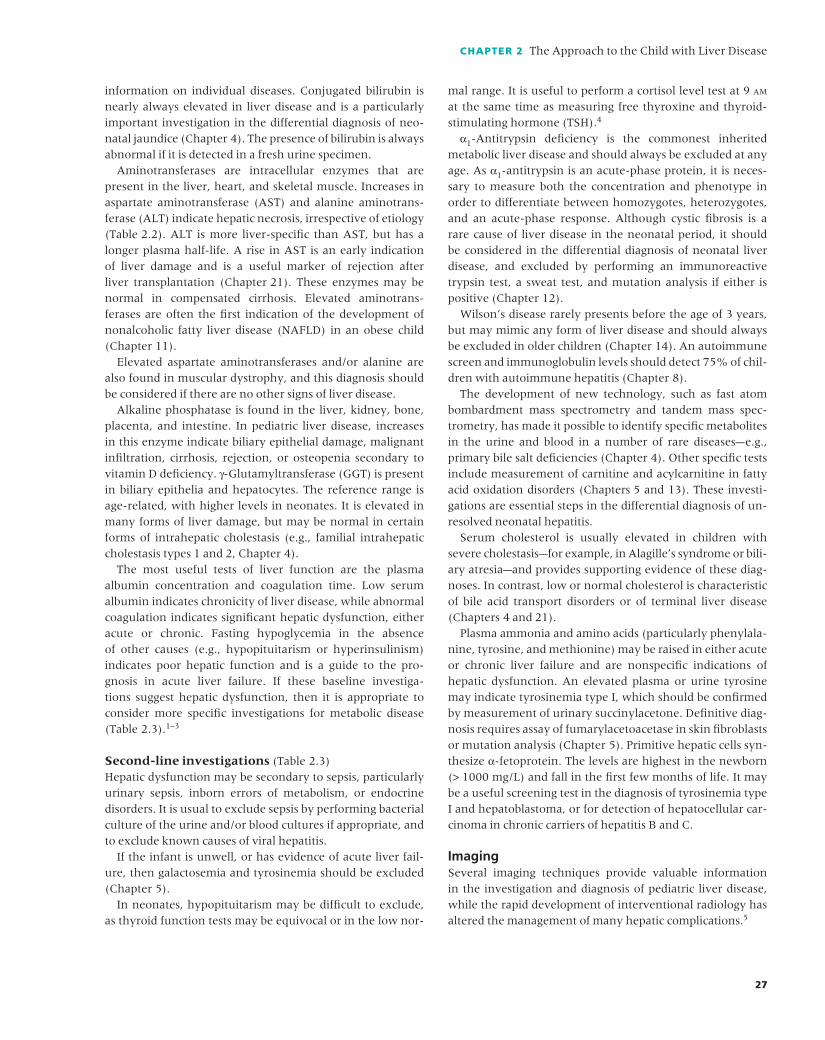
CHAPTER 2 The Approach to the Child with Liver Disease
27
information on individual diseases. Conjugated bilirubin isnearly always elevated in liver disease and is a particularlyimportant investigation in the differential diagnosis of neo-natal jaundice (Chapter 4). The presence of bilirubin is alwaysabnormal if it is detected in a fresh urine specimen.
Aminotransferases are intracellular enzymes that are present in the liver, heart, and skeletal muscle. Increases inaspartate aminotransferase (AST) and alanine aminotrans-ferase (ALT) indicate hepatic necrosis, irrespective of etiology(Table 2.2). ALT is more liver-specific than AST, but has alonger plasma half-life. A rise in AST is an early indication of liver damage and is a useful marker of rejection after liver transplantation (Chapter 21). These enzymes may benormal in compensated cirrhosis. Elevated aminotrans-ferases are often the first indication of the development ofnonalcoholic fatty liver disease (NAFLD) in an obese child(Chapter 11).
Elevated aspartate aminotransferases and/or alanine arealso found in muscular dystrophy, and this diagnosis shouldbe considered if there are no other signs of liver disease.
Alkaline phosphatase is found in the liver, kidney, bone,placenta, and intestine. In pediatric liver disease, increases in this enzyme indicate biliary epithelial damage, malignantinfiltration, cirrhosis, rejection, or osteopenia secondary tovitamin D deficiency. γ-Glutamyltransferase (GGT) is presentin biliary epithelia and hepatocytes. The reference range isage-related, with higher levels in neonates. It is elevated inmany forms of liver damage, but may be normal in certainforms of intrahepatic cholestasis (e.g., familial intrahepaticcholestasis types 1 and 2, Chapter 4).
The most useful tests of liver function are the plasma albumin concentration and coagulation time. Low serumalbumin indicates chronicity of liver disease, while abnormalcoagulation indicates significant hepatic dysfunction, eitheracute or chronic. Fasting hypoglycemia in the absence of other causes (e.g., hypopituitarism or hyperinsulinism)indicates poor hepatic function and is a guide to the pro-gnosis in acute liver failure. If these baseline investiga-tions suggest hepatic dysfunction, then it is appropriate toconsider more specific investigations for metabolic disease(Table 2.3).1–3
Second-line investigations (Table 2.3)Hepatic dysfunction may be secondary to sepsis, particularlyurinary sepsis, inborn errors of metabolism, or endocrine disorders. It is usual to exclude sepsis by performing bacterialculture of the urine and/or blood cultures if appropriate, andto exclude known causes of viral hepatitis.
If the infant is unwell, or has evidence of acute liver fail-ure, then galactosemia and tyrosinemia should be excluded(Chapter 5).
In neonates, hypopituitarism may be difficult to exclude,as thyroid function tests may be equivocal or in the low nor-
mal range. It is useful to perform a cortisol level test at 9 AM
at the same time as measuring free thyroxine and thyroid-stimulating hormone (TSH).4
α1-Antitrypsin deficiency is the commonest inheritedmetabolic liver disease and should always be excluded at anyage. As α1-antitrypsin is an acute-phase protein, it is neces-sary to measure both the concentration and phenotype inorder to differentiate between homozygotes, heterozygotes,and an acute-phase response. Although cystic fibrosis is arare cause of liver disease in the neonatal period, it should be considered in the differential diagnosis of neonatal liverdisease, and excluded by performing an immunoreactivetrypsin test, a sweat test, and mutation analysis if either ispositive (Chapter 12).
Wilson’s disease rarely presents before the age of 3 years,but may mimic any form of liver disease and should alwaysbe excluded in older children (Chapter 14). An autoimmunescreen and immunoglobulin levels should detect 75% of chil-dren with autoimmune hepatitis (Chapter 8).
The development of new technology, such as fast atombombardment mass spectrometry and tandem mass spec-trometry, has made it possible to identify specific metabolitesin the urine and blood in a number of rare diseasesae.g., primary bile salt deficiencies (Chapter 4). Other specific testsinclude measurement of carnitine and acylcarnitine in fattyacid oxidation disorders (Chapters 5 and 13). These investi-gations are essential steps in the differential diagnosis of un-resolved neonatal hepatitis.
Serum cholesterol is usually elevated in children withsevere cholestasisafor example, in Alagille’s syndrome or bili-ary atresiaaand provides supporting evidence of these diag-noses. In contrast, low or normal cholesterol is characteristicof bile acid transport disorders or of terminal liver disease(Chapters 4 and 21).
Plasma ammonia and amino acids (particularly phenylala-nine, tyrosine, and methionine) may be raised in either acuteor chronic liver failure and are nonspecific indications ofhepatic dysfunction. An elevated plasma or urine tyrosinemay indicate tyrosinemia type I, which should be confirmedby measurement of urinary succinylacetone. Definitive diag-nosis requires assay of fumarylacetoacetase in skin fibroblastsor mutation analysis (Chapter 5). Primitive hepatic cells syn-thesize α-fetoprotein. The levels are highest in the newborn(> 1000 mg/L) and fall in the first few months of life. It maybe a useful screening test in the diagnosis of tyrosinemia typeI and hepatoblastoma, or for detection of hepatocellular car-cinoma in chronic carriers of hepatitis B and C.
ImagingSeveral imaging techniques provide valuable information in the investigation and diagnosis of pediatric liver disease,while the rapid development of interventional radiology hasaltered the management of many hepatic complications.5
9781405163347_4_002.qxd 20/06/2008 09:27 Page 27

SECTION 2 Investigating the Liver
28
RadiographyPlain radiography of the abdomen will give an indication ofliver and spleen size, but is rarely of diagnostic value and is not a routine investigation. Chest radiography may showskeletal abnormalitiesafor example, butterfly vertebrae inAlagille’s syndrome, a dilated heart secondary to fluid over-load in end-stage liver disease, or evidence of congenital heartdisease. Wrist and knee radiography will demonstrate boneage and/or the development of osteopenia or rickets.
UltrasoundThe development of Doppler ultrasound has been an import-ant advance in the investigation of liver disease. Ultrasonicinvestigation of the abdomen provides information on thesize and consistency of the liver, spleen, pancreas, and kidneys,on the size of the gallbladder, and on the presence of gallstones.It may identify tumors, hemangiomas, abscesses, or cystswithin the liver, and it allows targeting of lesions for liverbiopsy. The gallbladder is best visualized after a 4–6-h fast. A
Table 2.3 Investigation of liver disease.
Baseline investigations
Bilirubin
Conjugated
Unconjugated
Aspartate aminotransferase
Alanine aminotransferase
Alkaline phosphatase
γ-Glutamyl transpeptidase
Albumin
Glucose
Full blood count and platelets
Prothrombin time
Partial thromboplastin time
Second-line investigations
Bacterial culture of blood and urine
TORCH screen
Hepatitis A, B, C
α1-Antitrypsin level and phenotype
Abdominal ultrasound
Metabolic investigations
Immunoreactive trypsin
Plasma lactate, BOH, FFA
Ammonia
Acylcarnitine
Serum iron and ferritin
Plasma amino acids
Cholesterol, triglyceride
α-Fetoprotein
Parathyroid hormone
Wrist for bone age/rickets
Urine
Reducing sugars
Organic acids
Amino acids
Succinylacetone
Bile salts
ANA, antinuclear antibodies; BOH, β-hydroxybutyrate; C3, C4, complement components 3 and 4; EBV,
Epstein–Barr virus; ERCP, endoscopic retrograde cholangiopancreatography; FFA, free fatty acids;
LKM, liver/kidney microsomal antibodies; MRI, magnetic resonance imaging; PTC, percutaneous
transhepatic cholangiography; SMA, smooth-muscle antibodies; T4, thyroxine; TORCH, toxoplasmosis,
other, rubella, cytomegalovirus, and herpes simplex; TSH, thyroid-stimulating hormone.
Neonate
Galactose 1-phosphate uridyltransferase
Free T4, TSH
9 AM cortisol
Chromosomes/DNA
Sweat test (> 4 weeks)
Older child (> 2 years)
Cu, ceruloplasmin, urinary Cu
C3, C4, ANA, SMA, LKM
Immunoglobulins
EBV
If indicated
Radioisotope scan
Liver biopsy for:
Histology
Electron microscopy
Enzyme analysis
Immunohistochemistry
Culture
Copper concentration
Skin biopsy
Ophthalmology
Cardiology
Bone-marrow aspirate
Endoscopy
ERCP
PTC
MRI
9781405163347_4_002.qxd 20/06/2008 09:27 Page 28

small or absent gallbladder after fasting suggests either severeintrahepatic cholestasis or biliary atresia in the neonate (seeChapter 4, Figure 4.4), whereas an enlarged gallbladder mayrepresent supportive evidence for primary sclerosing cholan-gitis. Extrahepatic bile ducts are usually identified, but intra-hepatic bile ducts are rarely seen unless dilated as a result ofbiliary obstruction. Dilated intrahepatic bile ducts are not afeature of extrahepatic biliary atresia in the neonate.
Color-flow Doppler techniques allow rapid evaluation of vascular patency without the use of intravenous contrastmaterial. They are particularly useful in pre- and post-transplant examinations to identify whether the portal vein,hepatic veins and artery, and splenic vessels are patent.Portal hypertension is suggested by the presence of ascites,splenomegaly, and splenic or gastric varices.
Ultrasound may be less sensitive in identifying hepaticoutflow obstruction after transplantation. This problem isrelated to “kinking” of the hepatic vein, which may not beapparent in a prone fasted child. A high index of suspicionand a low threshold for hepatic venography are required ifclinical symptoms exist.6
Radioisotope scanningSoluble radioisotopes such as technetium trimethyl 1-bromoiminodiacetic acid (TEBIDA), which are taken up wellby hepatocytes despite elevated bilirubin levels, have beenused to demonstrate either hepatic uptake or biliary excre-tion. Pretreatment with phenobarbitone (5 mg/kg) for 3–5days prior to the investigation improves hepatic uptake of theisotope. Hepatic uptake is an index of hepatic function andmay be patchy in inflammatory conditionsae.g., neonatalhepatitis (see Chapter 4, Figure 4.5).
Radioisotope scanning is most useful in the assessment of biliary excretion in the differential diagnosis of neonatalcholestasis. Under normal conditions, biliary excretion iscompleted within 4 h. Delayed excretion or no excretion after24 h suggests severe intrahepatic cholestasis or extrahepaticbiliary atresia. Delayed biliary excretion or pooling in bileducts is also a feature of cystic fibrosis liver disease (see Chap-ter 12, Figure 12.7).
In post-transplant patients, a TEBIDA scan may be of valuein identifying the degree of biliary obstruction or a biliaryleak.
Radioisotope scanning is of some value in the diagnosis ofhepatic vein obstruction (Budd–Chiari syndrome), as pooruptake of the isotope is demonstrated in most of the liverexcept for the caudate lobe, which has a separate venousdrainage. This method is rarely used now, as ultrasound orvenography is preferred.
Interventional radiologyInterventional radiology has an important role in the mod-ern management of hepatobiliary disorders and providesnew treatment options. “Keyhole” or “pinhole” techniques
using catheter-based technology are carried out under imag-ing guidance with radiography (including computed tomo-graphy), ultrasound, or magnetic resonance imaging. Usefuldiagnostic and therapeutic techniques are described below.
Computed tomographyComputed tomography (CT) of the liver is useful for iden-tifying and taking biopsies from hepatic tumors or space-occupying lesions of the liver. Intravenous contrast mediumcauses enhancement of vascular lesions and of the walls ofabscesses and may be helpful in differentiating tumors fromother solid masses. An important recent advance is the intro-duction of helical or spiral CT scanning, in which both thetable and the roentgen-ray tube move continuously to improveimaging. The use of CT angiography allows noninvasive evaluation of vascular structures. CT scans of the brain arehelpful for the detection of cerebral edema in acute liver fail-ure (Chapter 7) or for cerebral atrophy in certain metabolicconditions (Chapters 5 and 13).
Endoscopic retrograde cholangiopancreatography(ERCP)In this endoscopic technique, a fiberoptic duodenoscope ispassed into the first part of the duodenum, the ampulla of Vateris identified, the pancreatic and biliary ducts are cannulated,and radiographic contrast medium is injected. The techniquehas an 80% success rate in skilled hands and is invaluable forthe assessment of extrahepatic biliary disease in older chil-dren (e.g., choledochal cysts, primary sclerosing cholangitis)and the assessment of chronic pancreatitis. Although thistechnique should be of value in the differential diagnosis ofneonatal cholestasis, technical difficulties in cannulating thebile ducts in small infants may lead to equivocal informationbeing obtained. The development of a prototype fiberopticduodenoscope (7.5 mm in diameter) has improved the diag-nostic yield in this group of patients. In general, the diagnosticvalue of this technique has been superseded by that of mag-netic resonance imaging (MRI), which is noninvasive, butERCP retains an important role in therapy7 (see Chapter 8,Figure 8.4).
The removal of common bile duct stones, insertion of biliary and pancreatic stents, and sphincterotomy are usefultherapeutic procedures that can be performed at the sametime as the diagnostic procedure, but have limited applica-tion in children.
Endoscopic ultrasoundEndoscopic ultrasound (EUS) is a new imaging modality thatvisualizes the lower biliary tree. The technique uses mini-probes (external diameter 2.6 mm), which are small enoughto be passed through the operating channel of conventionalpediatric duodenoscopes. The technique is well established inadult practice, with a wide range of applications. It is a highlysensitive and specific method of visualizing the lower biliary
CHAPTER 2 The Approach to the Child with Liver Disease
29
9781405163347_4_002.qxd 20/06/2008 09:27 Page 29

SECTION 2 Investigating the Liver
30
tree and demonstrating pathology in this area.8,9 Althoughthe technique has not yet been evaluated in pediatric prac-tice, it may prove to be of value in the diagnosis of infantswith neonatal cholestasis. Endoscopic ultrasound has alsoproved useful in the diagnosis of submucosal esophageal andgastric varices10 (Figure 2.10).
Percutaneous transhepatic cholangiography (PTC)This technique is useful for identifying biliary disease if the intrahepatic bile ducts are dilated secondary to obstruc-tion and ERCP is impossible or unsuccessful. A thin needle(Chiba) is passed through the liver. The bile ducts or gallblad-
der are punctured and radiographic contrast medium isinjected. External drainage of the biliary tree, dilation of biliary strictures, and the insertion of biliary stents are allpossible using this technique and are useful both before andafter transplantation.
AngiographyCatheter angiography is primarily undertaken in infants andchildren with the following disorders:• Primary hepatic tumors for which complex surgical resec-tion is being consideredae.g., in auto-transplantation.• Cavernous transformation of the portal vein (also knownas extrahepatic portal vein obstruction) prior to surgicalshunting.• Portal vein thrombosis in patients being considered forliver transplantation.• If intervention is being considered for vascular stenoses orocclusion of the hepatic artery, vein, portal vein, or inferiorvena cava following liver transplantation or in the Budd–Chiari syndrome.• Hepatic vascular anomalies such as life-threatening heman-giomas, arterioportal fistulas, arteriovenous malformations,and congenital portosystemic shunts.• Refractory or life-threatening bleeding from gastrointest-inal disease or following blunt abdominal trauma or liverbiopsy.• Invasive diagnostic imaging when angiography is requiredas part of the procedure, such as transvenous (transjugular)liver biopsy, portal vein sampling, and arterially stimulatedhepatic vein sampling.• Thrombolysis to restore flow to occluded blood vessels byadministration of recombinant tissue-plasminogen activator(tPA) (0.5 mg/kg/h for 6 h, or by direct clot infusion using alow-dose regimen).Visualization of the celiac access and of the hepatic andsplenic blood vessels is achieved by femoral artery catheter-ization and injection of radiographic contrast. This techniquehas two parts:• The arterial phase, which provides information on theceliac axis, hepatic and splenic artery abnormalities, vascular-ization and anatomy of hepatic tumors, hepatic hemangiomas,or detection of hepatic artery thrombosis.• The venous phase, which provides information about thepatency of the portal, splenic, and superior mesenteric veinsand the presence of portal hypertension by identification ofmesenteric, esophageal, or gastric varices. In skilled hands,the investigation can be performed with little risk in infants.Femoral artery spasm or thrombosis is an occasional sideeffect, but rarely requires operative treatment.Hepatic artery embolization is indicated for the treatment of hepatic hemangiomas (see Chapter 10, Figure 10.7) or inthe control of liver hemorrhage from trauma or needlebiopsy. Angioplasty for portal or hepatic vein obstruction isalso feasible using angiographic techniques.
Varices
Gastric lumen
Gastric Lumen
A
B
Injectionneedlein situ
Thrombosedvarices
Figure 2.10 Endoscopic ultrasound is a useful way of detecting
submucosal varices (A) before and (B) after injection with thrombin.
9781405163347_4_002.qxd 20/06/2008 09:27 Page 30

Magnetic resonance imagingMagnetic resonance imaging (MRI) has now replaced hepaticangiography as the best way to stage or diagnose hepatictumors and identify their vascular supply. It may providevaluable information about liver or brain consistency andstorage of heavy metalsafor example, iron in hemochro-matosis, copper in Wilson’s disease, and cerebral edema inacute liver failure.11,12 The recent development of MRIcholangiography, in which both intrahepatic and extrahep-atic biliary duct abnormalities can be detected, has replacedERCP as a diagnostic investigation8 (see Chapter 8, Figure8.5). ERCP or PTC will still be required for therapeutic pro-cedures. Magnetic resonance spectroscopy is an emergingtechnique that may well be of value in the diagnosis ofmetabolic disorders, as intermediate metabolites such as lactate can be measured in the brain and other tissues.13
SplenoportographyThis technique, in which the splenic and portal veins arevisualized by the injection of radiological contrast into thespleen, has largely been replaced by hepatic angiography. It may be useful for measuring splenic pulse pressures in the evaluation of portal hypertension or if there is a post-transplant portal vein stenosis, but it carries a small risk ofsplenic rupture.
Transjugular intrahepatic portosystemic shuntLife-threatening esophageal and gastric variceal bleedingoccurs with portal hypertension secondary to liver disease. If the bleeding persists despite maximal medical and/or endoscopic therapy, a transjugular intrahepatic portosystemicshunt (TIPSS) can be carried out. TIPSS is an intrahepaticportosystemic shunt, created with radiographic assistance,between the high-pressure portal vein and the lower pressurehepatic vein. Since 1982, technological advances have madethis technique possible in pediatric practice (see Chapter 15,Figure 15.8).
Central venous access and related venous occlusive diseaseIntensive treatment regimens such as parenteral nutritionrequire long-term central venous access, particularly in patientsundergoing small-bowel or combined liver and small-boweltransplantation. Interventional radiological techniques areuseful in establishing percutaneous placement of tunneledcentral venous lines or Hickman lines, maintaining centralvenous access and managing the complications, such asthrombosis, in children of all ages.
Aspiration and drainage of fluid collections and abscessesImaging-guided (usually with ultrasound or occasionally CT)aspiration and drainage of fluid collections and abscesses inthe peritoneal cavity, liver, spleen, pancreas, and retroperi-
toneum can be performed using percutaneous fine-needleaspiration in all age groups. Complicated fluid collectionssuch as abscesses and pancreatic pseudocysts may requiredrainage over a period of time.
HistopathologyThe diagnosis of most liver diseases requires histologicalconfirmation, and liver biopsies are thus a routine proced-ure in specialist centers. An aspiration technique, using aMenghini needle (or disposable variant), has a complicationrisk of one in 1000 liver biopsies and may be performedunder sedation with local anesthesia. In fibrotic or cirrhoticlivers, a Tru-Cut needle, which removes a larger core, may benecessary. Transjugular liver biopsies, in which the liver isbiopsied through a special catheter passed from the internaljugular vein into the hepatic veins, is now possible for chil-dren as small as 6 kg, and is the only safe way to perform abiopsy if coagulation times remain abnormal despite support(prothrombin time > 5 s prolonged over control value).14 Thecomplications of this potentially dangerous procedure (seebelow) are considerably reduced if it is performed by expertsin specialized units under controlled conditions (Table 2.4).15
It is essential to have information about liver size and con-sistency and the presence of cysts or dilated bile ducts fromultrasound, and if necessary to have a “spot” marked on theabdomen to ensure an accurate biopsy. Correct informationabout coagulation parameters is vital. Prothrombin timeshould be within 3 s of control values; the platelet countshould be > 80 × 109/L. The patient’s blood group should beknown, and it is prudent to cross-match a unit of blood priorto the procedure. Biopsy specimens should be obtained forroutine histopathology, microbiology, electron microscopy,immunohistochemistry, and copper (if appropriate), andsnap-frozen in liquid nitrogen for enzymatic or metabolicinvestigations. The interpretation of the histology may bedifficult and requires considerable specialist expertise.
It is possible to carry out a liver biopsy as a day-case proced-ure in low-risk patients16 if the following criteria apply:• > 1 year old• Bilirubin < 200 μmol/L*• No other organ dysfunction• No need for coagulation or platelet support• Access to emergency facilities for 24 h after the procedureLiver biopsy is contraindicated in the following circumstances:• Abnormal coagulation parameters or thrombocytopenia(Table 2.4)• Presence of grossly dilated bile ducts or large cysts• Angiomatous malformation of the liver• Extensive ascitesIn these circumstances, image-guided or transjugular liverbiopsies should be performed if the diagnosis will change themanagement.
CHAPTER 2 The Approach to the Child with Liver Disease
31
* Bilirubin conversion factor: 1 mg/dL = 17 μmol/L
9781405163347_4_002.qxd 20/06/2008 09:27 Page 31

Complications of percutaneous liver biopsy• The main complication is bleeding, and most deaths follow-ing a liver biopsy are due to intractable bleeding. However,subclinical bleeding (as evident on ultrasound imaging) iscommon, and intrahepatic and subcapsular hematomas withno hemodynamic compromise are seen in up to 23% ofpatients.17,18 Significant nonfatal bleeding (as seen with evidence of active bleeding, shock, and a hemoglobin drop of 2.0 g/L) occurs more frequently in children than adults.
SECTION 2 Investigating the Liver
32
In adults, significant hemorrhage occurs in 0.3–0.5% ofcases, whilst bleeding requiring transfusion is seen in up to2.8% of children.15 Evidence of persistent bleeding followingliver biopsy, despite medical support and blood transfusion,warrants urgent hepatic angiography and embolization orsurgery.• Pneumothorax or hemopneumothorax, which are treatedin a standard way.• Infection (particularly if the biopsy is combined withanother procedureae.g., dental extraction).• Perforation of the gallbladder or bile ducts leading to bil-iary peritonitis and the need for emergency surgery.Adequate monitoring of vital signs after biopsy is essential inorder to detect complications such as hemorrhage or infection(Table 2.4).19
Metabolic investigationsMany inborn errors of metabolism present with hepato-megaly and/or liver disease. It is essential to screen for thesediseases as part of the investigation of liver disease inneonates (Table 2.3) and in older children (Chapters 4, 5,and 13).
Bone-marrow aspirationBone-marrow aspiration should be performed in infants withundiagnosed neonatal hepatitis with both hepatomegaly andsplenomegaly, in order to exclude Niemann–Pick type C orfamilial hemophagocytosis (Chapters 4 and 5), or at any ageif a storage disorder is suspected.
Skin biopsyThis procedure should be performed if an inborn error ofmetabolism is being considered (e.g., Niemann–Pick type A,B, or C, or tyrosinemia type I) and the specimen should bestored frozen for future culture. It may also be necessary toobtain skin biopsies from parents or siblings.
Genetic tests (chromosome and DNA)With the rapid development of molecular techniques fordiagnosis and detection of genetic diseases, samples for DNAanalysis and/or chromosomes from both child and parent areessential.
EndoscopyUpper gastrointestinal endoscopy (gastroscopy) using a flex-ible fiberoptic endoscope is now the best way to diagnosepeptic ulcer disease or esophageal and gastric varices sec-ondary to portal hypertension. The technique is normallyperformed with the patient under sedation or general anes-thetic. In children with hematemesis, gastroscopy not onlyprovides rapid diagnosis, but also makes it possible to carryout therapy with variceal banding or endoscopic sclerother-apy for bleeding varices, or injection of bleeding ulcers withepinephrine or thrombin.
Table 2.4 Liver biopsy protocol.
• Establish blood group and save serum for cross-match
• Coagulation: prothrombin (PT, normal 12–15 s), partial
thromboplastin (PTT, normal 33–37 s)
• Broad-spectrum antibiotics may be required if there are cardiac
problems or concurrent dental extraction
Coagulation support
PT: < 3 s prolonged No action
> 3–6 s prolonged 15 mL/kg FFP over 2.5 h
> 6 s prolonged Correct with FFP; recheck PT/PTT at 1 h
Reassess risk–benefit if no correction
FBC and platelets
Platelet count: > 80 x 109/L No action
40–80 x 109/L 10 mL/kg platelet infusion over 1 h
< 40 μ/L 10 mL/kg platelet infusion, recheck
Reassess risk–benefit if correction > 80
is impossible
Hemoglobin: < 8.0 g/L Transfuse to Hb > 10 g/L prebiopsy
Procedure and sedation*
• Establish size of liver: +/– mark site after ultrasound
• Apply local anesthetic cream to area of maximal dullness between 7th
and 9th intercostal spaces (or ultrasound site)
Oral midazolam 2.5 mg < 1 year old
5.0 mg > 1 year old
or
Chloral hydrate 75 mg/kg
Local anesthetic Lignocaine 1–2%
Intravenous sedation Pethidine 1–2 mg/kg
Midazolam 0.1–0.75 mg/kg
Postbiopsy observations
Blood pressure, pulse, respiration and temperature
15 min for 2 h
30 min for 2 h
Hourly for 2 h
4-hourly as required
Chest radiograph/abdominal ultrasound may be required if bleeding is
suspected. Laparotomy may be required
*N.B., most units now carry out liver biopsies in children under a general
anesthetic.
9781405163347_4_002.qxd 20/06/2008 09:27 Page 32

Capsule endoscopyThe clinical impact of wireless capsule endoscopy, which is anovel, noninvasive tool for the investigation of the smallintestine, is being evaluated in children. In this technique, a wireless capsule is swallowed and images of the small intestine are obtained. It is of most value in the diagnosis ofCrohn’s disease, obscure or occult gastrointestinal bleeding,polyposis syndromes, and protein-losing enteropathy due tolymphangiectasia. In liver disease, it may be useful for diag-nosing small-intestinal varices.20
NeurophysiologyElectroencephalography is mostly used in the assessment of hepatic encephalopathy. It will identify abnormal rhy-thms secondary to encephalopathy due to either acute orchronic liver failure or drug toxicity, such as post-transplantimmunosuppression. It may also be of value for assessingbrain death, as a flat electroencephalogram in the absence of sedation is an indication for withdrawal of therapy. CT orMRI scans of the brain (see above) may identify cerebraledema, infarction, or hemorrhage.
OphthalmologyA number of inherited conditions have associated visuallesionsafor example, Kayser–Fleischer rings in Wilson’s dis-ease, posterior embryotoxon or optic drusen in Alagille’s syndrome, and cherry-red spot in Niemann–Pick type A.Ophthalmological examination may thus provide valuablediagnostic information and should be part of the assessmentprocess. Children with Alagille’s syndrome have a higher thannormal incidence of benign intracranial hypertension, andannual fundoscopy for papilledema is therefore essential.21
Molecular biologyThe development of molecular biology has revolutionizedthe methodology for many complex diagnostic procedures,transforming many techniques into routine laboratory procedures22
aparticularly in screening for rare neonatal diseases.23
Progress in identifying specific genes and DNA sequenc-ing has made it possible to diagnose many inborn errors ofmetabolism and inherited disease (e.g., Alagille’s syndrome,Wilson’s disease, tyrosinemia type I) and has led to theidentification of mitochondrial disorders.
Advances in methodology for gene cloning and molecularcloning have been helpful in identifying viruses such as hepatitis C and G, while the polymerase chain reaction has been used to diagnose active infection and monitorpatients with many different viral diseases, such as hepatitisC, cytomegalovirus (CMV), and Epstein–Barr virus (EBV).Diagnosis for autoimmune disorders has improved, withspecific assays that use recombinant protein antigens (e.g.,antinuclear antigens and liver/kidney microsomal antibod-ies). The rapid development of molecular techniques is cer-
tain to lead to further improvements in diagnostic methodsand to a better understanding of pediatric liver disease.
Emerging techniquesThe FibroScan is a new noninvasive method for assessmentof hepatic fibrosis. This device is based on one-dimensionaltransient elastography, a technique that uses both ultra-sound (5 MHz) and low-frequency (50 Hz) elastic waves,whose propagation velocity is directly related to the elasti-city. Liver elasticity measurements are reproducible and operator-independent and correlate well with the degree of fibrosis on histology. The method has been evaluated inadults with hepatitis C,24 although not yet in children, but it has obvious advantages for the long-term follow-up ofchronic disease.
References
1 Kelly DA, Green A. Investigation of paediatric liver disease. J
Inherit Metab Dis 1991;14:531–7.
2 Kelly DA, Hull J. Investigation of prolonged neonatal jaundice.
Curr Paediatr 1991;1:228–30.
3 McKiernan PJ. The infant with prolonged jaundice: investigation
and management. Curr Paediatr 2001;11:83–9.
4 Spray CH, McKiernan P, Waldron KE, Shaw N, Kirk J, Kelly DA.
Investigation and outcome of neonatal hepatitis in infants with
hypopituitarism. Acta Paediatr 2000;89:951–4.
5 Saini S. Imaging of the hepatobiliary tract. N Engl J Med
1997;336:1889–94.
6 Lorenz JM, Van Ha T, Funaki B, et al. Percutaneous treatment of
venous outflow obstruction in pediatric liver transplants. J Vasc
Interv Radiol 2006;17:1753–61.
7 Halefoglu AM. Magnetic resonance cholangiopancreatography: a
useful tool in the evaluation of pancreatic and biliary disorders.
World J Gastroenterol 2007;13:2529–34.
8 Fernandez-Esparrach G, Gines A, Sanchez M, et al. Comparison
of endoscopic ultrasonography and magnetic resonance cholan-
giopancreatography in diagnosis of pancreatobiliary diseases: a
prospective study. Am J Gastroenterol 2007;102:1632–9.
9 Scheimann AO, Barrios JM, Al-Tawil YS, Gray KM, Gilger MA.
Percutaneous liver biopsy in children: impact of ultrasonography
and spring-loaded biopsy needles. J Pediatr Gastroenterol Nutr
2000;31:536–9.
10 Kuramochi A, Imazu H, Kakutani H, Uchiyama Y, Hino S,
Urashima M. Color Doppler endoscopic ultrasonography in iden-
tifying groups at a high-risk of recurrence of esophageal varices
after endoscopic treatment. J Gastroenterol 2007;42:219–24.
11 Wood JC. Magnetic resonance imaging measurement of iron
overload. Curr Opin Hematol 2007;14:183–90.
12 Williams H, McKiernan P, Kelly D, Baumann U. Magnetic
resonance imaging in neonatal hemochromatosisaare we there
yet? Liver Transpl 2006;12:1725.
13 Cecil KM, Kos RS. Magnetic resonance spectroscopy and metabolic
imaging in white matter diseases and pediatric disorders. Top
Magn Reson Imaging 2006;17:275–93.
14 Furuya KN, Burrows PE, Phillips MJ, Roberts EA. Transjugular
liver biopsy in children. Hepatology 1992;15:1036–42.
CHAPTER 2 The Approach to the Child with Liver Disease
33
9781405163347_4_002.qxd 20/06/2008 09:27 Page 33
20 Anato B, Bishop J, Shawis R, Thomson M. Clinical application
and diagnostic yield of wireless capsule endoscopy in children. J
Laparoendosc Adv Surg Tech A 2007;17:364–70.
21 Narula P, Gifford J, Steggall MA, et al. Visual loss and idiopathic
intracranial hypertension in children with Alagille’s Syndrome. J
Pediatr Gastroenterol Nutr 2006;43:348–52.
22 Worman HJ. Molecular biological methods in diagnosis and
treatment of liver diseases. Clin Chem 1997;43:1476–86.
23 Dhondt JL. Neonatal screening: from the “Guthrie age” to the
“genetic age.” J Inherit Metab Dis 2007;30:418–22.
24 Sandrin L, Fourquet B, Hasquenoph JM, et al. Transient elasto-
graphy: a new noninvasive method for assessment of hepatic
fibrosis. Ultrasound Med Biol 2003;29:1705–13.
SECTION 2 Investigating the Liver
34
15 Cohen MB, A-Kader HH, Lambers D, Heubi JE. Complications of
percutaneous liver biopsy in children. Gastroenterology 1992;102:
629–32.
16 Vivas S, Palacio MA, Rodriguez M, et al. Ambulatory liver biopsy:
complications and evolution in 264 cases. Rev Esp Enferm Dig
1998;90:175–82.
17 McGill DB, Rakela J, Zinsmeister AR, Ott BJ. A 21-year experi-
ence with major hemorrhage after percutaneous liver biopsy.
Gastroenterology 1990;99:1396–400.
18 Sugano S, Sumino Y, Hatori T, Mizugami H, Kawafune T, Abei T.
Incidence of ultrasound-detected intrahepatic hematomas due to
Tru-Cut needle liver biopsy. Dig Dis Sci 1991;36:1229–33.
19 Gonzalez-Vallina R, Alonso EM, Rand E, Black DD, Whitington
PJ. Outpatient percutaneous liver biopsy in children. J Pediatr
Gastroenterol 1993;17:370–5.
9781405163347_4_002.qxd 20/06/2008 09:27 Page 34

Liver Disease in Pregnancy3
9781405163347_4_003.qxd 20/06/2008 09:28 Page 35

9781405163347_4_003.qxd 20/06/2008 09:28 Page 36

37
This chapter aims to cover:• Hepatic conditions that are specifically related to pregnancy.• Neonatal liver pathology secondary to maternal disease.• Contraception and pregnancy in those with chronic liverdisease and following liver transplantation.There is an increasing trend for teenage pregnancy, one of the highest rates being in the United Kingdom, where in2002 the incidence in 15–19-year-olds reached 30 per 1000live births, in comparison with a rate of seven per 1000 livebirths in Sweden.1 It is therefore important for pediatriciansto consider the potential for conception in young womenwith chronic liver disease or those who have had a livertransplant, advise on contraception, and manage any ensu-ing pregnancy. Pediatricians should also be aware of hepaticcomplications specific to contraception and pregnancy, someof which require immediate action. Increased understandingof the pathophysiology of neonatal liver disease has led to a greater awareness of the influence of the mother on theneonatal liver, either via the fetal–placental unit or throughinheritance.
Physiological changes to the liver and biliarysystem during normal pregnancyThirty-five percent of the cardiac output normally goes to theliver; however, due to the redirection of blood to the uterusduring pregnancy, this drops to 28%, whilst the total bloodvolume increases, resulting in a relative decrease in theserum concentration of liver enzymes. Table 3.1 lists theliver-related changes seen in normal pregnancy.
Incidence of liver disease specific to pregnancyLiver disease is a rare complication of pregnancy, but when itoccurs, morbidity to both the mother and fetus can be severeand may prove fatal (Table 3.2).
Hepatic conditions specific to pregnancy
Hyperemesis gravidarumIn some pregnancies, the symptoms of nausea and vomitingin the first trimester are severe, leading to dehydration andmalnutrition and requiring hospitalization. Raised transam-inases (seldom exceeding 200 IU/dL)2 occur in up to 50% ofthe hospitalized cohort. Both conjugated and unconjugatedbilirubin may be mildly raised, as may alkaline phosphatase.Liver biopsy is not normally indicated and may be normal orhave fatty changes.3 Hypotheses to explain the transaminitisinclude a hepatic response to starvation,4 which due to the asso-ciated increased fatty acid load may unmask a heterozygousdefect of mitochondrial fatty acid oxidation in the mother.5
With hydration and nutritional therapy, the transaminasestend to settle without any further maternal pathology. Theneonate may be small for gestational age, but no long-termdetriment to the fetus has been reported.6
HELLP syndromeThis acronym was first coined in 1982 by Weinstein todescribe patients with hemolysis, elevated liver enzymes andlow platelets.7 It is associated with substantial maternal andperinatal morbidity and mortality. The diagnosis is made onclinical suspicion and supportive laboratory findings.
PathogenesisThe exact cause of the condition is unknown. Hypotheses are:• Placental ischemia, leading to the production of toxic fac-tors damaging the endothelium.8
• A thrombotic tendency, with detection of anticardiolipinantibodies being the most common finding.9
• An immunological reaction that causes cytokine-mediatedendothelial damage.10
DemographicsA pregnancy may be complicated by HELLP syndrome in a mother of any age or ethnicity. It affects primiparous ormultiparous women, with an incidence of 0.11% of liveborndeliveries.11 The majority (70%) are diagnosed in the third
The Effects of Liver Disease on Mother and Child
Jane Hartley and Elwyn Elias
3
Diseases of the Liver and Biliary System in Children, 3rd edition. Edited by
Deirdre Kelly. © 2008 Blackwell Publishing, ISBN: 978-1-4051-6334-7.
9781405163347_4_003.qxd 20/06/2008 09:28 Page 37

trimester, while the remainder are identified during the first72 h postpartum.
Maternal clinical features• Malaise.• Right upper quadrant pain or tenderness on palpation.• Blood pressure is raised above the prepregnancy baseline(19% of mothers have preexisting chronic hypertension),but there may only be mildly raised nausea and vomiting.• Features of hypertension, such as headache, edema, pro-teinuria, blurred vision, and hyperreflexia may be the pre-senting conditions in 10%.12
• Disseminated intravascular coagulation (DIC) and renalfailure occur in severe disease (7%).• Placental abruption (16%).• Pulmonary edema (6%) is more common followingabruption.• Hepatic rupture presenting as profound shock occurs in1% with HELLP syndrome.• Postpartum bleeding may be severe in the setting ofthrombocytopenia.
Laboratory findingsHemolysis. The only constant diagnostic feature is decreased
SECTION 3 Liver Disease in Pregnancy
38
Table 3.2 The incidence of liver disease specific to pregnancy and the trimesters in which each is
most likely to occur.
Disease Trimester Incidence
Hyperemesis gravidarum 1 and 2 0.3–1.0%
Acute fatty liver of pregnancy 3 0.008%
HELLP syndrome Late 2 and 3 0.1% (4–12% of those with preeclampsia)
Intrahepatic cholestasis of pregnancy Mostly 3 and 2 0.1–20% (highest in Chile and Sweden and
low in North America)
HELLP, hemolysis, elevated liver enzymes and low platelets.
Table 3.1 Physiological changes in liver investigations during normal pregnancy.
Clinical features Liver and spleen are not palpable
Palmar erythema and telangiectasia
Serum bilirubin Normal or low
Serum albumin Low
Prothrombin time Normal
ALT, AST, and GGT Normal
Alkaline phosphatase Increases steadily throughout pregnancy, with a sharp rise in the last
month. This reflects increased production by the placental
syncytiotrophoblast and skeletal maturation of the fetus. It returns to
normal within 2 weeks of delivery
Total bile acids Normal or only mildly elevated
Urea Low
Triglyceride and cholesterol Increase throughout pregnancy
Gallbladder motility Decreases
Biliary lithogenicity increases
α-Fetoprotein No changes
Liver biopsy Normal histopathology
Electron microscopy: proliferation of the smooth endoplasmic reticulum
and giant mitochondria with increased paracrystalline inclusions,
reflecting the increased protein and energy requirements
ALT, alanine aminotransferase; AST, aspartate aminotransferase; GGT, γ -glutamyltransferase.
9781405163347_4_003.qxd 20/06/2008 09:28 Page 38

serum haptoglobins. Markers of microangiopathic hemolyticanemia (burr cells and schistocytes) and reticulocytosis withpolychromasia, anisocytosis, and poikilocytosis are also fre-quently present.13
Liver transaminases. These are typically raised two to threestandard deviations above the mean.13–15
Thrombocytopenia. A platelet count of < 100 000/μL is taken todefine thrombocytopenia.13,15,16 The platelet count continuesto drop to a nadir at 24–48 h after delivery. Although theplatelet count may respond to steroid therapy, other features(elevated liver enzymes, hemolysis, hypertension, and olig-uria) are often not benefited.17
Histology of the liverLiver biopsy is not necessary if there is no diagnostic un-certainty. Liver involvement is similar to the findings inpreeclampsia. The main features are periportal neutro-philic infiltrates, hepatocyte necrosis, fibrin microthrombi in portal vessels, and fibrin deposition within the sinusoids(Figure 3.1). Fatty change in hepatocytes has also been de-scribed,18 as in acute fatty liver of pregnancy, suggesting thatthe three entities may fall within the spectrum of the samedisease.
ImagingUltrasonography and computed tomography (CT) are nor-mal in HELLP syndrome unless it is complicated by hepaticrupture or infarction (Figure 3.2). Serial CT scans are the bestmethod of monitoring the size and extent of the rupture. Thiswill also aid planning of surgery if required.
Management of the mother• The risk to the mother and the fetus increases with prolon-gation of the pregnancy, so that aggressive management anddelivery are generally advisable. Vaginal delivery is possible,but a cesarean section is often necessary.7,19 The majority ofdeliveries will be preterm via cesarean section (42–98%),16,18
with an increased risk (7–14%) of wound hematoma andinfection.20,21 Complete normalization of laboratory indiceshas been reported in some women with continuation ofpregnancy, but there is a higher risk of stillbirth.20 In a studyof 25 patients, it was found that steroids temporarily stabil-ized HELLP syndrome by increasing the platelet count,reducing liver enzymes, and increasing urine output.22
• Blood transfusions may be required. The indication forplatelet support is controversial, as there is rapid platelet consumption, with no incremental rise. When Cesarean sec-tions were studied, bleeding from the wound site was notseen to have diminished.23
• Hypertension should be controlled with standard medication.• Renal support with continuous venovenous hemofiltration.
• In those who develop a liver hematoma, no intervention isrequired if the mother is hemodynamically stable. However,rupture is a major surgical emergency requiring evacuationand drainage of the blood, packing for tamponade, or sutur-ing by experienced hepatobiliary surgeons.• Hepatic infarction may lead to hepatic insufficiency,requiring management of acute liver failure.
Seventeen liver transplants for HELLP syndrome havebeen reported, the main indication being liver necrosis andliver failure following rupture. Two patients required totalhepatectomy prior to transplantation, to control bleeding.Fourteen of the patients were alive at follow-up, suggestingthat liver transplantation is appropriate in this setting.24
CHAPTER 3 The Effects of Liver Disease on Mother and Child
39
A
B
Figure 3.1 Liver histology from the resected infarcted liver in hemolysis,
elevated liver enzymes and low platelets (HELLP) syndrome. A An area of
coagulative ischemic-type necrosis and focal infiltration by neutrophils.
(Hematoxylin–eosin, original magnification × 40). B Martius scarlet-blue
stain, showing fibrin in the periportal sinusoids (stained red) and occluding a
portal vein branch (PV).
9781405163347_4_003.qxd 20/06/2008 09:28 Page 39

Fetal presentation• One-third will have intrauterine growth restriction.• Placental abruption may lead to fetal death without ex-pedient delivery.• In those born alive, perinatal mortality has been reportedas between 5–20%; it is related to the complications of pre-term delivery,25 and is comparable with those born pretermin the absence of maternal HELLP syndrome. The mean gestational age at delivery is 32 weeks, ranging from 24 to 39weeks’ gestation.26
• The neonate may have thrombocytopenia and leukopenia,but these findings are not specific to HELLP syndrome andmay also be found in preeclampsia.26
• There is no increase in liver enzymes in neonates born tomothers with HELLP syndrome.27
Management of the neonateAs with preeclampsia, up to half of the babies born to moth-ers with HELLP syndrome will be small for gestational age.The long-term outcome for the infant depends on the extentof prematurity. The neonate should be managed initially on aneonatal intensive-care unit.
Risk in future pregnanciesThere is a risk of recurrence in subsequent pregnancies of 3–19% for HELLP syndrome and a 23% risk forpreeclampsia.28,29
Acute fatty liver of pregnancyAcute fatty liver of pregnancy (AFLP) is a consequence ofdefective mitochondrial fatty acid β-oxidation in the infant,with a high potential for both maternal and fetal morbidityand mortality.
Mitochondrial ββ-oxidationMitochondrial long-chain fatty acid β-oxidation provides themajor source of energy for cardiac and skeletal muscle andintermediary metabolism of ketone production by the liver inthe fasting state. A trifunctional protein catalyses the finalthree steps in the formation of acetyl CoA from long-chainfats. Acetyl CoA then enters the tricarboxylic acid pathway,which results in energy generation by the liver. The threeenzymatic steps for the trifunctional protein are:1 2,3-enoyl-CoA hydratase2 Long-chain 3-hydroxyacyl-CoA dehydrogenase3 Long-chain 3-keto-acyl-CoA thiolase
Two biochemical disease phenotypes have been described:• Complete deficiency of all three enzymes of the trifunc-tional protein• Isolated deficiency of long-chain 3-hydroxyacyl-CoA de-hydrogenase (LCHAD deficiency)
Genetic analysisA mutation resulting in a complete trifunctional protein ofthe fetus causes disease in the child only and has no effect onthe mother.30
• Nineteen percent of infants born to women with AFLPhave mutations leading to long-chain 3-hydroxyacyl-CoAdehydrogenase (LCHAD) deficiency in the fetus, which causesdisease in the child and may result in AFLP in the mother.31
• A fetus carrying the G1528C mutation (amino acid residueE474Q; homozygous or compound heterozygote) results indisease in the child and AFLP in the mother in 79% of cases(whether the mutation was inherited from the mother orfather).30
• A fetus with other hitherto detected mutations of long-chain 3-hydroxyacyl-CoA dehydrogenase does not causeAFLP in the mother.• A heterozygous fetus (one wild type allele) does not causeAFLP in the mother.Infants born to mothers with AFLP should always bescreened for LCHAD deficiency.32,33
Not all cases of AFLP can be attributed to fetal LCHAD defici-ency, and other causes of abnormal mitochondrial oxidationtherefore need to be considered, including drugs (aspirin andtetracyclines have been reported as causing AFLP). A casereport of carnitine palmitoyltransferase I (CPT I) deficiencyidentified two siblings with mild symptoms whose motherdeveloped AFLP whilst carrying them.34 AFLP has also beendescribed as the presenting feature of maternal previouslyundiagnosed, medium-chain acyl-CoA dehydrogenasedeficiency, with no effect on the fetus.35
Maternal presentationAFLP usually affects women in the third trimester of preg-nancy, although it has been reported exceptionally as early as 26 weeks’ gestation.36 It affects women of any ethnicgroup or age and can affect first or subsequent pregnancies.
SECTION 3 Liver Disease in Pregnancy
40
Figure 3.2 The liver in hemolysis, elevated liver enzymes and low platelets
(HELLP) syndrome. Infarction of the liver is a rare complication of HELLP
syndrome. This computed-tomographic image of the liver shows hemi-
infarction of the right lobe. The main portal vein is patent, but the right
portal vein branch has been occluded. Treatment is usually conservative.
9781405163347_4_003.qxd 20/06/2008 09:28 Page 40

The reported incidence has ranged from one in 13 000 preg-nancies37 to one in 6659,38 and in a recent prospective studyfrom Swansea, Wales, it reached one in 1000. In the latterstudy, six or more of the following features were accepted byCh’ng et al. as being diagnostic of AFLP:39
• Vomiting• Vague abdominal pain• Polydipsia/polyuria• Encephalopathy• Elevated bilirubin (bilirubin rises in severe AFLP and incontrast to HELLP syndrome is not related to hemolysis)• Hypoglycemia (profound)• Elevated urate• Leukocytosis (common)• Ascites or bright liver on ultrasound scan• Elevated transaminasesaalanine aminotransferase (ALT)and aspartate aminotransferase (AST)aare moderately raised(rarely above 10 times normal)• Elevated ammonia• Renal impairment• Coagulopathy (with DIC in 10%)• Microvesicular steatosis on liver biopsy
Histology of the liverLiver biopsy is not normally required in the acute situation,but if performed should be via the transjugular route. Biopsywithin the week following parturition will allow confirma-tion of the diagnosis.
The liver is typically small, with microvesicular steatosis in a panacinar distribution or centrilobular with minimalinflammation (Figure 3.3). Other changes are intrahepaticcholestasis and extramedullary hemopoiesis, and when thereis loss of hepatocytes, Kupffer cells aggregate.40 Followingdelivery, the lipid rapidly mobilizes to become undetectableafter a month, with normal hepatic architecture and nochronic changes.
ImagingUltrasonography. There is increased echogenicity, reflectingthe increased fat in the liver.
CT scan. There is decreased attenuation of the liver in comparison with the spleen (usually the spleen is of lowerattenuation than the liver).
Maternal managementPrompt diagnosis with expeditious delivery is recommended,as resolution of symptoms can only occur following delivery.Delivery may be vaginal, but in the face of acute liver failurerequires emergency cesarean section. Meticulous manage-ment of liver failure and encephalopathy should lead to resolution of the symptoms with no long-term sequelae.Maternal death is related to the severity of liver disease, withraised intracranial pressure and gastrointestinal bleeding
being reported as contributing to death in 12%.41 In thosewho develop fulminant hepatic failure, successful liver trans-plantation has been performed.42 Most mothers recovercompletely. Only rarely does recurrent AFLP affect sub-sequent pregnancies.43
Clinical features in the childFetal presentation. Late maternal diagnosis increases fetal mortality. There is an increased incidence of prematurity,asphyxia, intrauterine growth retardation, and intrauterinedeath, which may be related to uteroplacental insufficiency.44
Infancy. There is a genotype–phenotype correlation. Symptomsdevelop when there are periods of fasting or intercurrent illness.
Those with isolated LCHAD deficiency present with:• Hepatic failure, which may lead to death• Hepatomegaly
CHAPTER 3 The Effects of Liver Disease on Mother and Child
41
Figure 3.3 Acute fatty liver of pregnancy. A The typical distribution of
fat accumulation in zones 2 and 3 surrounding the hepatic venule (HV),
with relative sparing of zone 1 hepatocytes surrounding portal tracts (PT).
B The microvesicular nature of the fatty droplets, which accumulate
without displacing the nucleus from its central position (arrows).
HV, hepatic venule.
A
B
9781405163347_4_003.qxd 20/06/2008 09:28 Page 41

• Nonketotic hypoglycemia• Encephalopathy• Sudden death• Hypocalcemia and cholestasis45
Those with complete trifunctional protein deficiency pre-sent with:• Dilated cardiomyopathy• Peripheral neuropathy and myopathy46
• Rhabdomyolysis• Peripheral neuropathy and retinitis pigmentosa in long-term survivors
Laboratory investigations during the acute episode show:• Hypoglycemia with low ketones• Elevated transaminases• Elevated prothrombin time• Increased ammonia• Increased urate and urea• Elevated plasma acyl carnitines• Urinary organic acids show an increase in 3-hydroxy dicar-boxylic acids• Secondary carnitine deficiency
Liver histology. There is usually microvesicular fat depositionin hepatocytes, although some macrovesicular depositionmay be present.
Genetics. Mutation analysis of the trifunctional protein shouldbe carried out.
Treatment of the childAll children born to mothers with AFLP should be treated ashaving LCHAD deficiency until the molecular investigationshave been completed:• Frequent feeding, which includes overnight nasogastricfeeding to avoid prolonged fasting.• Low-fat and high-carbohydrate diet to reduce the longchain fatty acid load.• Medium-chain fats should replace most long-chain fats inthe diet.• Early intervention with intravenous fluids during inter-current illness.• Treatment of acute liver failure may be required.
Despite optimal treatment, sudden death can still occurand is most likely in the setting of an intercurrent illness,which may only be mild. Parents should be counseled as to the need for intravenous glucose even during mild infections.
ScreeningAll children born to mothers with AFLP should be screenedfor LCHAD deficiency by molecular analysis.
In mothers who’s child is a compound heterozygote orhomozygote for LCHAD deficiency, there is a 1 in 4 risk infuture pregnancies. Chorionic villus sampling in subsequent
at-risk pregnancies has been used to assess the risk of AFLP in pregnancy and LCHAD deficiency in the fetus47 (seeChapters 5 and 13).
Intrahepatic cholestasis of pregnancyIntrahepatic cholestasis of pregnancy (ICP) is characterizedby pruritus, as the initial manifestation of cholestasis causedby pregnancy, and its complete resolution following delivery.
Diagnosis• Pruritus is the main presenting symptom causing discom-fort, insomnia and fatigue.• Elevation of serum alanine aminotransferase (ALT/SGPTand serum bile acids > 10 μmol/L are the most sensitive labor-atory indicators in confirming the diagnosis of cholestasis.Conventional liver function tests are initially normal, and itis in this circumstance that serum bile acid measurementmay be needed to confirm the presence of cholestasis.Jaundice is uncommon, and γ-glutamyltransferase (GGT)remains normal in two-thirds of cases.48
• Spontaneous relief of signs and symptoms within 2–3weeks after delivery.49
The resolution of symptoms and normalization of hepaticbiochemistry after delivery help distinguish this from otherliver conditions, such as primary biliary cirrhosis, that mayfirst be unmasked in pregnancy. Interval liver function testsshould be checked for confirmation that they have returnedto normal.
DemographicsIn Europe, ICP occurs in 0.1–1.5% of pregnancies.50 The incidence is highest in Chile, the Baltic states, Scandinavia,and Bolivia, with up to 15% of pregnancies being associatedwith ICP.51 Over the past decade, a trend toward lower incid-ence has been observed in Sweden and Chile, possibly due toincreased micronutrients such as selenium in an improveddiet.52
ICP is not influenced by parity, but is five times more common in multiple pregnancies. It may be sporadic or runin families.53 There is seasonal variability, with more casesoccurring in winter.
PathogenesisEstrogens and progestagens are naturally cholestatic. It islikely that in most instances, ICP occurs in individuals whohave otherwise subclinical mutations within susceptibilitygenes for intrahepatic cholestasis, whose capacity for effi-cient biliary excretion is exceeded in the hormonal milieu ofpregnancy.
GeneticsThe increased incidence in certain populations, family clus-terings, and recurrence in subsequent pregnancies supports a genetic etiology. This led to the investigation of genes
SECTION 3 Liver Disease in Pregnancy
42
9781405163347_4_003.qxd 20/06/2008 09:28 Page 42

involved in other cholestatic disorders, progressive familialintrahepatic cholestasis (PFIC) and benign recurrent intra-hepatic cholestasis (BRIC).• Familial intrahepatic cholestasis 1 (FIC-1) is caused bymutations in the ATP8B1 gene. The gene encodes a P-typeATPase involved in bile salt transport across the canalicularmembrane. In PFIC1 and BRIC1, γ-glutamyltransferase (GGT)is normal or low. Mullenbach et al. described two ATP8B1mutations (D70N and R867C) in four women with ICP andnormal GGT. The development of ICP may be due to coinher-itance of a second gene affecting cholestasis, or externalinfluences such as changes in hormones.54
• Bile salt export pump (BSEP) deficiency is caused by muta-tions in the ABCB11 gene leading to PFIC2 and BRIC2, also with low or normal GGT. Sequence variants (single-nucleotide polymorphisms) are more common in those withICP than in the general population, suggesting increased susceptibility.55
• The multidrug resistance 3 (MDR3) or ABCB4 gene encodesthe hepatobiliary phospholipid transporter, mutations inwhich cause PFIC3, which differs from PFIC1 and 2 by hav-ing a raised GGT. In women with ICP and raised GGT,sequence variants have been identified in a small number of cases, including a frameshift deletion.56 When womenwith ICP were screened for sequence-variant polymorphismsin the ABCB4 gene in comparison with the normal popu-lation, there was a significant increase in specific variants inthose with severe disease.57 Mutations of the ABCB4 geneleading to exon-splicing abnormalities may not be associatedwith raised GGT and should be considered in women withICP and normal GGT.58
• The bile acid farnesoid X receptor (FXR) regulates bile acidhomeostasis so as to reduce hepatocyte toxicity. It binds toDNA response elements in promoter regions of target genes,including ABCB11 and ABCB4, to activate their transcription,thereby increasing bile acid secretion out of the hepatocyte.FXR also promotes genes for sulfonation and glucuronida-tion, reduces import and synthesis of bile salts in the hepato-cyte, and also reduces ileal bile salt reabsorption. Functionalvariants in the gene are associated with a predisposition to ICP.59
HormonalA hormonal role in the etiology of ICP is suggested by theobservation that symptoms are most prevalent in the thirdtrimester, when estrogen levels are highest. When the hormone levels fall after delivery, symptoms tend to im-prove rapidly. Twin and triplet pregnancies, which havehigher estrogen levels, have an increased incidence of ICP.Biliary excretion of estradiol-17β-D-glucuronide occurs via acanalicular multispecific conjugate export pump (multidrugresistance–related protein, MRP2) and exerts its cholestaticeffect via a trans-inhibition of BSEP60 from the canalicularside of the hepatocyte membrane. This causes a transient
inhibition of BSEP.61 Estradiol-17β-D-glucuronide also sup-presses the expression of MRP2.62
There is a significant alteration in the ratio of different pro-gesterone isomers in ICP in comparison with other causes ofliver disease in pregnancy, the cause and effect of which arenot known.63
Clinical presentationICP usally presents in the second or third trimester, but mayoccasonally occur as early as the first.• Pruritusaoften worst on the palms and solesais character-istic and it may be intense, leading to discomfort and fatigue.• Steatorrhea and fat-soluble vitamin malabsorption areobserved.• Excoriation is seen on clinical examination.• There are no signs of chronic liver disease, and the liver isof normal size and not tender.• Jaundice develops in 10%.
HistologyLiver biopsy is rarely undertaken unless there is diagnosticuncertainty.
Intracellular bile pigment and canalicular bile plugs areseen in the absence of any other histological abnormalities.Electron microscopy shows dilated canaliculi with loss ofmicrovilli.
Maternal managementManagement consists of:• Relief of pruritus.• A double-blind randomized controlled trial of urso-deoxycholic acid, dexamethasone, or placebo including 130women with ICP showed a significant biochemical improve-ment in the ursodeoxycholic acid group and symptomaticimprovement in those with initially high levels of bile salts (> 40 μmol/L). There were no changes in the fetal complica-tion rate.64 The meconium of neonates born to mothers withICP has an increased concentration of bile acids in compar-ison with normal. In mothers with ICP who are treated withursodeoxycholic acid, the neonatal meconium continued to have high levels of bile acids. This may explain the lack of a fetal response to ursodeoxycholic acid despite a good biochemical response in the mother. It may also suggest thatsubsequent changes in serum bile acid concentrations has little effect on those already accumulated in the meconium.65
Cholestyramine is less effective in controlling the maternalsymptoms, and no additional benefit to the fetus comparedwith ursodeoxycholic acid was observed.66
• Smaller controlled trials have suggested a benefit fromtreatment with S-adenosylmethionine and ursodeoxycholicacid, alone or in combination.67,68
• Dexamethasone and cholestyramine are not recommended.Rifampicin may be highly effective, but has not been sub-jected to a controlled trial.
CHAPTER 3 The Effects of Liver Disease on Mother and Child
43
9781405163347_4_003.qxd 20/06/2008 09:28 Page 43

• Fat-soluble vitamin supplementation should be given tothose with cholestasis, alongside nutritional support. Post-partum bleeding has been reported in this group, which maybe secondary to vitamin K deficiency.69
• Delivery of fetus. Due to the effect on the fetus, it is sug-gested that delivery should be induced at 38 weeks in thosemildly affected and at 36 weeks in severe cases.70 Prospectivestudies from Scandinavia have reported that the risk of fetalcomplications was largely confined to mothers with serumbile salt levels greater than 40 μmol/L.71
Effect on the childRisks to the child include:• Increased risk of preterm delivery (19–60%).70
• Fetal distress (22–41%), as indicated by meconium liquorand asphyxia.• Fetal loss (0.4–1.6%).70
• A neonate with reduced BSEP expression, either due tothe homozygous inheritance of SNP variants or knownPFIC2 mutations, may present in the newborn period withliver disease (including liver failure). A history of maternalcholestasis should therefore lead to the consideration ofBSEP deficiency in the differential diagnosis of liver diseaseof the neonate.
The pathogenesis of fetal complications is not fully under-stood. In stillborns, the postmortem findings are those ofacute asphyxia.71 The fetus is usually well grown, andsurveillance of the fetoplacental unit is normal, suggestingthat placental insufficiency is not the cause. Methods used to monitor the fetus, such as umbilical artery Doppler ultra-sound, are poor predictors of fetal outcome.72 Neonatalresuscitation facilities and intensive care should be availableat delivery. In those neonates who are born healthy, thereare no long-term complications.
Long-term maternal complicationsICP resolves within a few weeks of delivery, with no long-lasting effects. A retrospective study from Finland of 10 504women with ICP found an increased rate of hepatitis C infection, gallstones, and nonalcoholic pancreatitis. On follow-up, 17 were found to have cirrhosis. It is unclearwhether pregnancy enhances the symptoms of a previouslyundiagnosed chronic disorder leading to cirrhosis, orwhether, in a very small minority, ICP may precipitate achronic condition leading to fibrosis and cirrhosis.73 Womenwho have experienced ICP have an increased risk of develop-ing pruritus and jaundice whilst taking the combined oralcontraceptive pill, due to the estrogen component. For contraception, women should be advised to use the progesterone-only pill.74 Pregnancy has a natural tendencyto increase the lithogenicity of bile, and the incidence of gallstones is increased. In a prospective study, biliary sludgewas detected on ultrasound in 10.2% of women in the thirdtrimester of pregnancy.75
Neonatal liver pathology secondary tomaternal disease
Neonatal hemochromatosisThis severe disease causes liver failure with intrahepatic andextrahepatic siderosis in the newborn. The exact molecularpathogenesis is unknown. An alloimmune etiology has beensuggested, due to the inheritance pattern and the high recur-rence rate (up to 80%) in subsequent pregnancies.76 Themother is well throughout the pregnancy. Normal neonateshave been reported in between affected siblings77 (see Chap-ter 5). Maternal factors that have also been associated aresystemic lupus and anti-Ro anti-La antibodies.
The diagnosis is suspected in a neonate with liver failure thatmay have progressed to cirrhosis (confirming that the patho-logy was initiated in utero). The diagnosis is made by demon-strating siderosis in the liver and extrahepatic sites. Intracellulariron deposits are identified on biopsies of minor salivary glands.A liver biopsy demonstrates intracellular iron which sparesthe Kupffer cells.78 Abdominal magnetic resonance imagingdemonstrates loss of signal on T2-weighted images of theliver, indicating iron accumulation, whilst the spleen is nor-mal (the reticuloendothelial system is spared). The signalintensity of the pancreas has been reported as variable.78,79
Untreated, the disease is fatal in up to 60% of neonates. Ifcommenced early, an antioxidant cocktail (selenium, desfer-rioxamine, N-acetylcysteine, and prostaglandin E1) improvesoutcome in those with mild features. Those with severe liverfailure require liver transplantation for survival.80
Those who survive the neonatal period, either withantioxidants or liver transplantation, do not have long-termcomplications.
Postulation of an alloimmune etiology has led to the use of weekly intravenous infusions of immunoglobulin inthe mother from the 18th week of subsequent pregnancies.This has been reported to modify the disease process, witheither mild or no signs of neonatal hemochromatosis in thenewborn.81
Viral hepatitis in pregnancyHepatitis AAcute hepatitis A has no increased risk to the mother.Neonates born to mothers with acute hepatitis A in themonth prior to delivery may acquire self-limiting infection.82
Meconium peritonitis of the fetus has been reported follow-ing maternal infection, leading to ileal perforation andrequiring resection in the newborn period.83
Hepatitis BHepatitis B vertical transmission occurs at the time of deliveryon exposure to blood and rarely in utero.84 The transmissionrate is dependent on the mother’s infectivity at the time ofdelivery. Mothers who are HbeAg-positive have a 70–90% risk
SECTION 3 Liver Disease in Pregnancy
44
9781405163347_4_003.qxd 20/06/2008 09:28 Page 44

of infecting their infants unless the infants are vaccinated.Mothers who develop acute hepatitis B in the last trimesterhave a 70% chance of infecting their infant and need similarprophylaxis (see Chapter 6). In mothers who are HBeAg- negative with low viral load, the transmission rate is lower,but vaccination is still recommended.85 Without successfulvaccination, the majority of infected infants become “healthycarriers,” but a minorityaborn to mothers who despite beingHBeAg-negative have high hepatitis B virus DNA titers due to the precore mutant form of the virusahave a high risk offulminant hepatic failure from 6 weeks to 6 months of age.86
Vaccination and hepatitis B immunoglobulins given to theneonate provide protection against perinatal transmission inover 95% of cases87 (see Chapters 6 and 23).
Acute hepatitis in pregnancy may lead to preterm labor.88
There is no increased maternal morbidity or mortality fromhepatitis B with pregnancy.
Hepatitis CThere is a 5% transmission rate, with transmission occurringeither in utero or during delivery.89 Infectivity is related to theviral load.90 Cesarean section does not reduce transmissionrates.91 Viral genotype does not alter infectivity,92 but coin-fection with human immunodeficiency virus may increasetransmission.93 Hepatitis C does not cause acute liver failurein infancy, but is likely to progress to a chronic hepatitis Cvirus infection, with the risk of fibrosis, cirrhosis, and hepato-cellular carcinoma in adulthood.
Immunoprophylaxis for the newborn is not available.Pregnancy does not increase morbidity in the mother (seeChapter 6).
Hepatitis EHepatitis E is a self-limiting viral infection spread by thefecal–oral route, occurring sporadically or as epidemics indeveloping countries.94 In pregnant women, hepatitis E has ahigh mortality rate, which increases throughout pregnancyup to 28% in the third trimester.95 The fetal outcome is poor,with one series reporting a 70% mortality rate due to still-birth and maternal death.96
Herpes simplex virusBoth herpes simplex virus-1 (HSV-1) and -2 can cause severedisease in the neonate, either through direct shedding at thetime of delivery or in primary transplacental infection, lead-ing to miscarriage, stillbirth, and congenital malformations.97
Primary herpes infection may be more severe in pregnantwomen, with dissemination leading to hepatitis, encephal-itis, thrombocytopenia, coagulopathy, and a mortality rate ofup to 50%.98
Genital herpes has the highest risk to the neonate at thetime of delivery, due to direct viral shedding.
In the neonate, it may present as:• Localized infection of the skin, eyes and mucous membranes
• Infection of the central nervous system, which has a 5%mortality rate, with 50% of survivors having neurologicalimpairment• Disseminated infection, presenting with acute liver failure,with a high mortality rate unresponsive to antiviral therapyin the setting of multiorgan failure
Neonatal herpes preventionActive genital herpes should be treated with acyclovir orvalacyclovir in the weeks prior to delivery.99,100 Women withactive genital infection at the time of delivery should beoffered a cesarean section, as the likelihood of neonataltransmission is high.101
A high index of suspicion is needed in a septic neonate to commence acyclovir treatment early before the onset ofmultiorgan failure. Liver transplantation has been successfulin herpes simplex infection isolated to the liver.102
Contraception and pregnancy
ContraceptionHepatic complications of combined oralcontraceptivesThe combined oral contraceptive pill (COCP) has an efficacyof 99.9%. It reduces menstrual irregularities and premen-strual tension, as well as reducing the risk of fibroids, benignbreast disease, and ovarian and endometrial cancer.
No preexisting liver diseaseCholestasis. This occurs in 10 per 100 000 in the West, and aswith ICP (see above), it has a higher incidence in Chile andScandinavia (25 per 100 000). It is more common in thosewho have had ICP, or in whom there is a family history of jaundice in pregnancy or whilst taking the COCP. It tendsto occur within 3 months of commencing an estrogen-containing COCP.
Hepatic adenomas. There is an increased risk of adenoma formation, which is associated with the length of use of theCOCP and the strength of the estrogen component. With the introduction of COCPs containing lower estrogen levels,the incidence of adenoma has fallen. Adenomas may regresswhen the COCP is stopped but return again following re-introduction of the COCP or during pregnancy. Hepatic adenoma constitutes a contraindication to pregnancy, and inpatients suitable for resection should be removed before con-ception occurs. Adenomas caused by estrogens are vascularand may rupture, with hemorrhage into the adenoma orperitoneal cavity.
Focal nodular hyperplasia (FNH). 50–75% of those with FNHuse the COCP, suggesting that estrogen may be a contribut-ing factor. FNH does not increase any risks during pregnancy.
CHAPTER 3 The Effects of Liver Disease on Mother and Child
45
9781405163347_4_003.qxd 20/06/2008 09:28 Page 45

Hepatocellular carcinoma (HCC). There is an increased risk ofHCC with long-term use of COCPs containing high levels ofestrogen.103 Cofactors such as hepatitis B and alcohol mayincrease the risk. Tumors may regress following cessation ofthe COCP.
Budd–Chiari syndrome. The procoagulant effect of the COCPincreases the risk of Budd–Chiari syndrome 2.5-fold.104 Ninepercent of cases of Budd–Chiari are attributed to COCPuse.105 In a case series of 13 women taking the COCP whodeveloped hepatic vein thrombosis, 10 had an underlyingprothrombotic condition, suggesting that the development of Budd–Chiari syndrome should prompt investigation forother thrombophilic disorders.106
Women with established liver diseaseFertility may be reduced in advanced chronic liver disease,but contraception is an important issue for the majority.
Gallstones. There is an alteration in the composition of bile inthose taking the COCP, with an increase in cholic acid and adecrease in chenodeoxycholic acid, which may increase therisk of cholelithiasis.107 There is no consensus from studies asto whether this is the case, but due to the potential risk it isadvised that those with a history of biliary colic should avoidthe COCP.108
Hepatocellular carcinoma. The cirrhotic liver is at risk of devel-oping hepatocellular carcinoma. Some studies have reportedan increased incidence of hepatocellular carcinoma in chronicliver disease in those using the COCP, but this has not beenverified in subsequent studies.109–111 Due to this uncertainty,it is recommended that COCPs should be avoided in chronicliver disease.
After transplantation. Following liver transplantation, COCPshave been used safely if there have been no thrombotic complications in the peritransplantation period. An increasein cyclosporine levels has been reported in concomitant use of the COCP, and levels should therefore be monitoredclosely.112
Other forms of contraception in liver diseaseProgesterone-only oral and depot contraception is the preferred con-traceptive in all forms of liver disease, as it avoids the estro-gen component of the combined contraceptive. The pill hasto be taken within the same 3 h every day to ensure efficacy.There are no hepatic complications. The progesterone-onlypill is safe following liver transplantation and is recom-mended when there have been thrombotic complicationsafter transplantation.
Intrauterine device. The intrauterine device (IUD) is 99% effective for 1–5 years. It can be used in all forms of liver
disease except for Wilson’s disease, in which the copper coil component of the IUD may potentially increase serumcopper levels.113 In the post-transplant patient, there havebeen reported cases of IUD failure due to immunosup-pressants reducing the immunological mechanism of theIUD.114 There is also a potentially increased risk of urinarytract infections.115
Barrier contraception. In the context of hepatitis B and C infec-tion, this form of contraception should be advised in order toavoid sexual transmission of the infections. The efficacy is80–95%. There are no contraindications to the use of thisform of contraception.116
PregnancyChronic liver diseaseEvaluation before conception. Due to reduced fertility in advancedliver disease, pregnancy in the presence of cirrhosis and portal hypertension is rare. With advances in medical care,young people are surviving into adulthood with reproductivepotential, with compensated cirrhosis. In general, the moreadvanced the liver disease, the higher the risk of complica-tions to mother and child.117
Pregnancy is not contraindicated in liver disease, butshould be planned for a time when the disease is in a steadystate. If the disease is advanced, liver transplantation shouldbe considered first.
Neonatal immunization and immunoglobulin to preventtransmission of hepatitis B should be discussed with motherswho are carriers. Although there is no vaccination to preventthe transmission of hepatitis C, mothers will want to knowabout transmission rates and management for their infants(see Chapter 6).
Drug therapy should be assessed for possible teratogensa
e.g., in animal studies, spironolactone has caused genitalabnormalities in males. It is essential that therapy with peni-cillamine for Wilson’s disease and prednisolone and azathio-prine for autoimmune hepatitis should be maintained.
Cirrhosis and portal hypertension. Although studies have notshown that pregnancy increases the risk of variceal bleeding,it is advisable that esophageal varices should be eradicated in advance of any pregnancy, as bleeding during pregnancy is associated with fetal loss.118 –120 The acute management is the same as in nonpregnant variceal bleeding, with hemo-dynamic stabilization, endoscopic banding, or sclerotherapyand octreotide. Vasopressin should be avoided if possible, as it causes decreased placental perfusion and potentiatesplacental abruption. It has also been associated with fetaldigit necrosis and amputation.121
PregnancyIn the presence of decompensated liver disease with ascites,encephalopathy, or liver failure, termination of pregnancy
SECTION 3 Liver Disease in Pregnancy
46
9781405163347_4_003.qxd 20/06/2008 09:28 Page 46

should be considered, because of the difficulties in bringing a normal infant to term.122 Twenty-four percent of womenwith cirrhosis will decompensate during pregnancy,123 requir-ing intensive care and possibly transplantation. Coagulopathymay lead to massive hemorrhage at delivery.124 Splenicartery aneurysm rupture may occur in the third trimester,125
with high maternal and fetal mortality rates (70% and 80%,respectively).
Labor and deliveryDelivery should be precipitated if there is life-threateningdeterioration in the mother; otherwise, the pregnancy shouldgo to term. Cesarean section should be avoided in womenwith cirrhosis unless absolutely necessary, due to the pre-sence of intra-abdominal collaterals, leading to an increasedrisk of bleeding and infection.126 Coagulation, fibrinogen,and platelet count should be assessed and blood product support should be available as required.
Epidural anesthesia may avoid the Valsalva maneuver,which may increase variceal pressure. However, if the plateletcount is reduced, it may not be possible to provide this.
PostpartumThrombocytopenia and reduced synthetic clotting factorsincrease the risk of postpartum hemorrhage.126
Fetal complicationsPrematurity is increased in the setting of cirrhosis and requiresthe delivery to be in a setting in which neonatal intensive-care facilities are available. Beta-blockers may cause growthrestriction, and this should be monitored throughout thepregnancy. Fetal growth may also be reduced following agastrointestinal bleed.
Pregnancy issues related to specific chronic liver diseasesAutoimmune hepatitis in pregnancyPregnancy tends to induce immune tolerance in autoimmunehepatitis (AIH), with improving liver function on baselinedosages of immunosuppression. Postpartum deterioration is common and should be anticipated.127 With cirrhosis,decompensation requiring transplantation may complicatepregnancy.128
• Steroids. No teratogenic effects on the fetus have beenreported in humans. In animal studies, high doses have led tocleft lip and palate, premature rupture of membranes, andadrenal insufficiency.129
• Azathioprine. In high doses, transient abnormalities in the neonate have been reported, including thymic atrophy,leukopenia, anemia, thrombocytopenia, reduced immuno-globulins, and transient chromosomal aberrations. No struc-tural teratogenic effects have been reported. At dosages usedfor maintenance treatment in the mother, no adverse effectshave been reported in the neonate
• Tacrolimus. Transient hyperkalemia has been reported inthe neonate, but no specific teratogenicity.• Cyclosporine. No specific teratogenicity.• Mycophenolate mofetil (MMF). There are concerns regardingthe safety of MMF in the developing fetus, with an increasedrisk of spontaneous abortion and serious malformations.MMF should be avoided in pregnancy.130
Wilson’s diseasePregnancy is not contraindicated in Wilson’s disease. Fertilitymay be reduced in poorly controlled disease, with amenor-rhea and frequent miscarriages.
Penicillamine as a chelating agent is well tolerated at con-ventional doses by both mother and child, with no increasedincidence of fetal malformation.131 Unnecessarily high dosesmay lead to cutis laxa.
Zinc has also been used safely during pregnancy, with no teratogenic effect on the developing fetus.132
Pregnancy following liver transplantationBefore conceptionThe menstrual irregularities that occur in chronic liver dis-ease are reversed by liver transplantation, and fertility mayreturn within 3 weeks of transplantation.133 It is generallyadvisable that pregnancy should be avoided within the firstyear after transplantation, and contraception should there-fore be carefully addressed in those of child-bearing age134
(see above and Chapter 25).
Immunosuppressants in pregnancyThe largest experience of the use of immunosuppressiveagents in pregnancy is with azathioprine and steroids. Infor-mation on the newer immunosuppressives in pregnancy ismainly from animal studies, as there are few data in humans(Table 3.3).
PregnancyLiver and renal function, cytomegalovirus (CMV) status andhemoglobin should be closely monitored.
Pregnancy should not alter graft function, so rejection andinfection should be considered if liver function tests becomeabnormal (see Table 3.1 for the normal changes seen in liverfunction during pregnancy). Liver biopsy has no increasedrisk in comparison with the nonpregnant woman, andadjustments to immunosuppressives in the face of acuterejection should be the same as in the nonpregnant. There isan increased risk of hypertension, with the risk being greaterin those receiving cyclosporine135 and those with hyperten-sion prior to conception.136,137
Preeclampsia, premature rupture of membranes, anemia,infection, and first-trimester abortion are all increased inpregnancies following transplantation.138
CHAPTER 3 The Effects of Liver Disease on Mother and Child
47
9781405163347_4_003.qxd 20/06/2008 09:28 Page 47

DeliveryNormal vaginal delivery can occur. The risk of requiring acesarean section is related to prolonged rupture of mem-branes or preeclampsia.
Effects on the fetusCongenital malformations have not been reported. Fetalgrowth should be monitored by serial ultrasound scans, asthere is an increased risk of intrauterine growth retardationfrom preeclampsia. Congenital CMV infection is the leadcause for neonatal death, and in one series all neonataldeaths in transplanted mothers were due to acute CMVinfection.139 The risk of maternal and therefore fetal CMVinfection is greatest with high-dose immunosuppressiona
e.g., in the early post-transplantation period, when preg-nancy should be avoided.140
SECTION 3 Liver Disease in Pregnancy
48
Table 3.3 Immunosuppressant use in pregnancy and breastfeeding.
Drug
Steroids
Tacrolimus (FK506)
Cyclosporine
Mycophenolate mofetil
Azathioprine
Sirolimus
Basiliximab
Daclizumab, muromonab-CD3
(OKT3), and Thymoglobulin
Breastfeeding recommendations
Less than 10% enters breast milk,
so considered safe at maintenance
corticosteroid doses
50% enters breast milk.
Manufacturers recommend
avoiding breast feeding
Secreted into breast milk, therefore
not recommended
No information available regarding
secretion into breast milk, but the
manufacturer recommends
avoiding breast feeding
Undetectable in breast milk, but
potentially could be secreted,
therefore avoid breast feeding
No data available
No data available
Effects on fetus
In animal studies, growth retardation
and premature rupture of
membranes. At high doses, may
cause cleft palate in animal studies
Transient hyperkalemia
Growth retardation
Transient hyperkalemia
Not recommended in pregnancy.
Animal studies have shown
malformations,
intrauterine growth retardation and
intrauterine death. Limited human
experience also suggests
teratogenicity130
At high doses, growth retardation,
preterm delivery, bone-marrow
suppression if this has occurred in the
mother, and transient chromosomal
abnormalities have been reported
Delayed fetal ossification, but no
teratogenic effects
In animal studies, there were no
teratogenic effects. No human data
are available
No data available
Side effects in pregnancy
Poor wound healing, hypertension,
weight gain, gastric ulceration,
osteoporosis and glucose intolerance
Nephrotoxicity, neurotoxicity,
glucose intolerance, hyperkalemia,
diarrhea
Hypertension, nephrotoxicity,
neurotoxicity, hyperkalemia, tremor,
hirsutism, hypomagnesemia, glucose
intolerance
None known
Increased infections, alopecia,
stomatitis, hepatotoxicity, fever,
nausea and vomiting, pancreatitis,
leukopenia, thrombocytopenia, and
macrocytic anemia
None known
None known
No data available
Case study
A 28-year-old primigravida presented at 31 weeks of pregnancy
when she developed raised blood pressure, edema, and
proteinuria, which settled with bed rest. She and her husband
were fit and healthy, with no significant medical history. She then
remained well until 35 weeks, when she developed increased
blood pressure (160/95). She had proteinuria and an increase in
serum uric acid, but the full blood count and liver function tests
remained normal. Two days, later she became tachycardic,
edematous, and oliguric, and her blood results were as follows:
hemoglobin 4.3 g/dL (reference range 11.5–16.6), platelets
18 × 109/L (reference range 150–450), AST 75 U/L (reference
range 5–43). The fetus was also tachycardic, and she therefore
underwent an emergency cesarean section under general
9781405163347_4_003.qxd 20/06/2008 09:28 Page 48

References
1 Kmietowicz Z. US and UK are top in teenage pregnancy rates.
BMJ 2002;324:1354.
2 Wallstedt A, Riely CA, Shaver D, Leventhal S, Adamec TA,
Nunnally J. Prevalence and characteristics of liver dysfunction
in hyperemesis gravidarum. Clin Res 1990;38:970A.
3 Larry D, Rueff B, Feldman G, Degott C, Danan G, Benhamon J.
Recurrent jaundice caused by recurrent hyperemesis gravi-
darum. Gut 1984;25:1414–5.
4 Morali G, Braveman D. Abnormal liver function enzymes and
ketonuria in hyperemesis gravidarum. A retrospective review
of 80 patients. J Clin Gastroenterol 1990;12:303–5.
5 Outlaw W, Ibdah J. Impaired fatty oxidation as a cause of liver
disease associated with hyperemesis gravidarum. Med Hypotheses
2005;65:1150–3.
6 Conchillo J, Pijnenborg J, Peeters P, Stockbrugger R, Fevery J,
Koeg G. Liver enzyme elevation induced by hyperemesis
gravidarum: aetiology, diagnosis and treatment. Neth J Med
2002;60:374–8.
7 Weinstein L. Syndrome of hemolysis, elevated liver enzymes,
and low platelet count: a severe consequence of hypertension
in pregnancy. Am J Obstet Gynecol 1982;142:159–67.
8 Van Beek E, Peters L. Pathogenesis of preeclampsia: a compre-
hensive model. Obstet Gynecol Surv 1998;53:233–9.
9 Dekker G, de Vries J, Doelitzsch P, et al. Underlying disorders
associated with severe early-onset preeclampsia. Am J Obstet
Gynecol 1995;173:1042–8.
10 Haeger M, Undander M, Andersson B, Tarkowski A, Arnestad J,
Bengtsson A. Increased release of tumor necrosis factor-alpha
and interleukin-6 in women with the syndrome of hemolysis,
elevated liver enzymes and low platelet count. Acta Obstet
Gynecol Scand 1996;75:695–701.
11 Magann E, Perry K, Chauhan S, Graves G, Blake P, Martin J.
Neonatal salvage by week’s gestation in pregnancies com-
plicated by HELLP syndrome. J Soc Gynecol Investig 1994;1:206–
9.
12 Sibai B, Ramadan M, Usta I, Salama M, Mercer B, Friedman S.
Maternal morbidity and mortality in 442 pregnancies with
hemolysis, elevated liver enzymes, and low platelets. Am J
Obstet and Gynecol 1993;169:1000–6.
13 Wilke G, Schutz E, Armstrong V, Kuhn W. Haptoglobin as a
sensitive marker of hemolysis in HELLP-syndrome. Int J Gynecol
Obstet 1992;39:29–34.
CHAPTER 3 The Effects of Liver Disease on Mother and Child
49
anesthesia, delivering a well baby who did not require any
resuscitation and remained well.
The mother was transferred to intensive care, complaining of
right upper quadrant tenderness, and in the following 24 hours she
developed acute liver failure (hypoglycemia, encephalopathy,
acidosis, prothrombin time 27 seconds, albumin 29 g/L (reference
range 34–51), bilirubin 371 μmol/L (reference range 1–22), alkaline
phosphatase 1489 U/L (reference range 70–260), AST 4600 U/L
and D dimers 2500 (reference range < 250). She required
ventilatory support, inotropic support, (norepinephrine) and renal
support (continuous venovenous filtration). The liver failure was
treated with blood products, broad-spectrum antibiotics,
antifungals, and N-acetylcysteine.
On day 6 postpartum, she developed low-grade pyrexia and an
increasing white cell count. An ultrasound of the liver revealed
changes in the right lobe, which on CT were identified as liver
ischemia or infarction, with a possible thrombosis of the right portal
vein (Figure 3.2). This was treated conservatively. During the next
48 h, she developed symptoms of systemic sepsis with pyrexia,
rigors, tachycardia, an increased neutrophil count at 16.2 × 10⁹/L
(reference range 2.0–7.5) and increased inotropic requirements. No
organisms were identified.
To control the systemic inflammatory response, she underwent a
right hemihepatectomy. On microscopic examination, the resected
right lobe showed vascular occlusion and infarction, which was in
keeping with HELLP syndrome but more extensive than usually
occurs in this condition (Figure 3.1).
A search for additional causes of portal vein thrombosis revealed
a slightly low protein S level at 61 μ/dL (reference range 68–146)
and heterozygosity for factor V Leiden.
During the initial postoperative period, her blood pressure
was labile. Ventilatory and inotropic support was weaned over
the following 48 h. She started to pass urine, and renal
dialysis was reduced and then stopped 13 days post
hemihepatectomy.
She had striking cholestasis from the onset of the HELLP
syndrome, which peaked on day 12 postpartum, with serum
bilirubin at 970 μmol/L. This slowly resolved to 505 μmol/L at
discharge, and at 5 months postpartum was in the normal range.
She has subsequently remained well, with no long-term liver
complications.
Although she was concerned regarding bonding with her baby
after such a long period on intensive care of time, this was not a
problem when she was discharged home.
Comment. This case typifies a severe case of HELLP syndrome.
It presented in the third trimester with right upper quadrant
pain and features of preeclampsia. The patient developed renal
failure, pulmonary edema, and disseminated intravascular
coagulation. Liver failure developed following hepatic infarction,
which was initially managed conservatively until it was clear that
the ongoing systemic inflammatory response was not improving,
requiring her to undergo a right hemihepatectomy. In other
cases, complete hepatectomy has been required either in order
to control bleeding or to remove an infarcted liver, as a
temporary measure whilst awaiting organ donation and
transplantation.
The liver histological features were in keeping with HELLP
syndrome, but infarction was more extensive than is usually seen.
This may have been the result of a heterozygous state for factor V
Leiden deficiency, which may have precipitated portal vein
thrombosis, increasing the hepatic infarction.
9781405163347_4_003.qxd 20/06/2008 09:28 Page 49
14 Sibai B. The HELLP syndrome (hemolysis, elevated liver enzymes
and low platelets): much ado about nothing? Am J Obstet Gynecol
1990;162:311–6.
15 Paternoster DM, Stella A, Simioni P, Mussap M, Plebani M.
Coagulation and plasma fibronectin parameters in HELLP syn-
drome. Int J Gynecol Obstet 1995;50:263–8.
16 Harms K, Rath W, Herting E, Kuhn W. Maternal hemolysis,
elevated liver enzymes, low platelet count, and neonatal out-
come. Am J Perinatol 1995;12:1–6.
17 Vigil-De Gracia P, Garcia-Cáceres E. Dexamethasone in the
post-partum treatment of HELLP syndrome. Int J Gynaecol Obstet
1997;59:217–21.
18 Barton J, Riely C, Adamec T, Shanklin D, Khoury A, Sibai B.
Hepatic histopathologic condition does not correlate with labor-
atory abnormalities in HELLP syndrome (hemolysis, elevated
liver enzymes, and low platelet count). Am J Obstet Gynecol
1992;167:1538–43.
19 Van Pampus M, Westenberg S, van der Post J, Bonsel G, Treffers
P. Maternal and perinatal outcome after expectant manage-
ment of the HELLP syndrome compared with preeclampsia
without HELLP syndrome. Eur J Obstet Gynecol Reprod Biol
1998;76:31–6.
20 Visser W, Wallenburg H. Temporising management of severe
pre-eclampsia with and without the HELLP syndrome. Br J
Obstet Gynaecol 1995;102:111–7.
21 Audibert F, Friedman S, Frangieh A, Sibai B. Clinical utility of
strict diagnostic criteria for the HELLP syndrome (hemolysis,
elevated liver enzymes, and low platelets). Am J Obstet Gynecol
1996;175:460–4.
22 Magann E, Bass D, Chauhan S, Sullivan D, Martin R, Martin J.
Antepartum corticosteroids: disease stabilization in patients
with the syndrome of hemolysis, elevated liver enzymes, and
low platelets (HELLP). Am J Obstet Gynecol 1994;171:1148–53.
23 Roberts W, Perry K, Woods J, Files J, Blake P, Martin J. The
intrapartum platelet count in patients with HELLP (hemolysis,
elevated liver enzymes, and low platelets) syndrome: is it pre-
dictive of later hemorrhagic complications? Am J Obstet Gynecol
1994;171:799–804.
24 Shames B, Fernandez L, Sollinger H, et al. Liver transplantation
for HELLP syndrome. Liver Transpl 2005;11:224–8.
25 Ganzevoort W, Rep A, de Vries JI, Bonsel GJ, Wolf H. Prediction
of maternal complications and adverse infant outcome at
admission for temporizing management of early-onset severe
hypertensive disorders of pregnancy. Am J Obstet Gynecol 2006;
195:495–503.
26 Raval D, Co S, Reid M, Pildes R. Maternal and neonatal outcome
of pregnancies complicated with maternal HELLP syndrome.
J Perinatol 1997;17:266–9.
27 Knapen M, van Schaiijk F, Mulder T, Peters W, Steegers E.
Marker for liver damage in neonates born to mothers with
HELLP syndrome. Lancet 1997;349:1519–20.
28 Sibai B, Ramadan M, Chari R, Friedman S. Pregnancies compli-
cated by HELLP syndrome (hemolysis, elevated liver enzymes
and low platelets): subsequent pregnancy outcome and long-
term prognosis. Am J Obstet Gynecol 1995;172:125–9.
29 Sullivan C, Magann E, Perry K, Roberts W, Blake P, Martin J.
The recurrence risk of the syndrome of hemolysis, elevated liver
enzymes, and low platelets (HELLP) in subsequent gestations.
Am J Obstet Gynecol 1994;171:940–3.
30 Ibdah J, Bennett M, Rinaldo P, et al. A fetal fatty acid oxidation
disorder as a cause of liver disease in pregnant women. N Engl J
Med 1999;340:1723–31.
31 Yang Z, Yamanda J, Zhao Y, Strauss A, Ibdah J. Prospective
screening for pediatric mitochondrial trifunctional protein defects
in pregnancy complicated by liver disease. JAMA 2002;288:
2163–6.
32 Treem W, Rinaldo P, Hale D, et al. Acute fatty liver of pregnancy
and long-chain 3-hydroxyacyl-coenzyme A dehydrogenase
deficiency. Hepatology 1994;19:339–45.
33 Ibdah JA. Acute fatty liver of pregnancy: an update on patho-
genesis and clinical implications. World J Gastroenterol 2006;
12:7397–404.
34 Innes A, Seargeant L, Balachandra K, et al. Hepatic carnitine
palmitoyltransferase I deficiency presenting as maternal illness
in pregnancy. Pediatr Res 2000;47:43–5.
35 Santos L, Patterson A, Moreea SM, Lippiatt CM, Walter J,
Henderson M. Acute liver failure in pregnancy associated with
maternal MCAD deficiency. J Inherit Metab Dis 2007;30:103.
36 Buytaert I, Elewaut A, van Kets H. Early occurrence of acute
fatty liver in pregnancy. Am J Gastroenterol 1996;91:603–4.
37 Pockros P, Reynolds T. Acute fatty liver of pregnancy. Dig Dis Sci
1985;30:601–2.
38 Castro M, Fassett M, Reynolds T, Shaw K, Goodwin T.
Reversible peripartum liver failure: a new perspective on the
diagnosis, treatment, and cause of acute fatty liver of preg-
nancy, based on 28 consecutive cases. Am J Obstet Gynecol
1999;181:389–95.
39 Ch’ng C, Morgan M, Hainsworth I, Kingham J. Prospective
study of liver dysfunction in pregnancy in Southwest Wales.
Gut 2002;51:876–80.
40 Rolfes D, Ishak K. Liver disease in pregnancy. Histopathology
1986;10:555–570.
41 Rolfes D, Ishak K. Acute fatty liver of pregnancy: a clinicopatho-
logical study of 35 cases. Hepatology 1985;5:1149–58.
42 Ockner S, Brunt E, Cohn S, Krul E, Hanto D, Peters M.
Fulminant hepatic failure caused by acute fatty liver of preg-
nancy treated by orthotopic liver transplantation. Hepatology
1990;11:59–64.
43 Sims H, Brackett J, Powell C, et al. The molecular basis of pedi-
atric long chain 3-hydroxyacyl-CoA dehydrogenase deficiency
associated with maternal acute fatty liver of pregnancy. Proc
Natl Acad Sci 1995;92:841–5.
44 Moise K, Shah D. Acute fatty liver of pregnancy: etiology of
fetal distress and fetal wastage. Obstet Gynecol 1987;69:482–5.
45 Ibdah J, Dasouki M, Strauss A. Long-chain 3-hydroxyacyl-CoA
dehydrogenase deficiency: variable expressivity of maternal
illness during pregnancy and unusual presentation with infan-
tile cholestasis and hypocalcaemia. J Inher Metab Dis 1999;22:
811–4.
46 Ibdah J, Tein I, Dionisi-Vici C, et al. Mild trifunctional pro-
tein deficiency is associated with progressive neuropathy and
myopathy and suggests a novel genotype-phenotype correla-
tion. J Clin Invest 1998;102:1193–9.
47 Ibdah J, Yang Z, Bennett M. Liver disease in pregnancy and
fetal fatty acid oxidation defects. Mol Genet Metab 2000;71:
182–9.
48 Milkiewicz P, Gallagher R, Chambers J, Eggington E, Weaver J,
Elias E. Obstetric cholestasis with elevated gamma glutamyl
SECTION 3 Liver Disease in Pregnancy
50
9781405163347_4_003.qxd 20/06/2008 09:28 Page 50

transpeptidase: incidence, presentation and treatment. J Gastro-
enterol Hepatol 2003;18:1283–6.
49 Beuers U, Pusl T. Intrahepatic cholestasis of pregnancyaa het-
erogeneous group of pregnancy-related disorders? Hepatology
2006;43:647–9.
50 Glanz A, Marschall H, Mattsson L. Intrahepatic cholestasis of
pregnancy: relationship between bile acid levels and fetal com-
plication rates. Hepatology 2004;40:467–74.
51 Lammert F, Marschall H, Glantz A, Matern S. Intrahepatic
cholestasis of pregnancy: molecular pathogenesis, diagnosis and
management. J Hepatol 2000;33:1012–21.
52 Reyes H, Baez M, Ganzalez M, Hernandez I, Palma J, Ribalta J.
Selenium, zinc and copper plasma levels in intrahepatic
cholestasis of pregnancy, in normal pregnancies and in healthy
individuals. J Hepatol 2000;32:542–9.
53 Reyes H, Simon F. Intrahepatic cholestasis of pregnancy: an
estrogen related disease. Semin Liver Dis 1993;13:289–301.
54 Mullenbach R, Bennett A, Tetlow N, et al. ATP8B1 mutations in
British cases with intrahepatic cholestasis of pregnancy. Gut
2005;54:829–34.
55 Eloranta M, Hakli T, Hiltunen M, Helisalmi S, Punnonen K,
Heinonen S. Association of single nucleotide polymorphisms of
the bile salt export protein gene with intrahepatic cholestasis of
pregnancy. Scand J Gastroenterol 2003;38:648–52.
56 Jaquemin E, Cresteil D, Manouvrien S, Boute O, Hadchouel M.
Heterozygous non-sense mutation of the MDR3 gene in familial
intrahepatic cholestasis of pregnancy. Lancet 1999;353:210–1.
57 Wasmuth H, Glantz A, Keppeler H, et al. Intrahepatic cholestasis
of pregnancy: the severe form is associated with common vari-
ants of the hepatobiliary phospholipids transporter ABCB4 gene.
Gut 2007;56:265–70.
58 Schneider G, Paus T, Kullak-Ublick G, et al. Linkage between
a new slicing site mutation in the MDR3 alias ABCB4 gene
and intrahepatic cholestasis of pregnancy. Hepatology 2007;45:
150–8.
59 van Mil S, Milona A, Dixon P, et al. Functional variants of the
central bile acid sensor FXR identified in intrahepatic cholestasis
of pregnancy. Gastroenterology 2007;133:507–16.
60 Steiger B, Fattinger K, Madon J, Kullak-Ublick G, Meier P.
Drug- and estrogen-induced cholestasis through inhibition of
the hepatocellular bile salt export pump (Bsep) of a rat liver.
Gastroenterology 2000;118:422–30.
61 Trauner M, Arrese M, Soroka C, Ananthanarayanan M,
Koeppel T, Schlosser S. The rat canalicular conjugate export
pump Mrp2 is down regulated in intrahepatic and obstructive
cholestasis. Gastroenterology 1997;113:255–64.
62 Bacq Y. Intrahepatic cholestasis of pregnancy. Clin Liver Dis
1999;3:1–13.
63 Milkiewicz P, Elias E, Williamson C, Weaver J. Obstetric
cholestasis. BMJ 2002;324:123–4.
64 Glanz A, Marschall H, Lammert F, Mattsson L. Intrahepatic
cholestasis of pregnancy: a randomized controlled trial com-
paring dexamethasone and ursodeoxycholic acid. Hepatology
2005;42:1399–405.
65 Rodrigues C, Martin J, Brites D. Bile acid patterns in meconium
are influenced by cholestasis of pregnancy and not altered by
ursodeoxycholic acid treatment. Gut 1999;45:446–52.
66 Kondrackiene J, Beuers U, Kupcinskas L. Efficacy and safety
of ursodeoxycholic acid versus cholestyramine in intrahe-
patic cholestasis of pregnancy. Gastroenterology 2005;129:894–
901.
67 Nicastri P, Diaferia A, Tartagni M, Loizzi P, Fanelli M. A ran-
domised placebo-controlled trial of ursodeoxycholic acid and
S-adenosylmethionine in the treatment of intrahepatic cholest-
asis of pregnancy. Br J Obstet Gynaecol 1998;105:1205–7.
68 Roncaglia N, Locatelli A, Arreghini A, et al. A randomised
controlled trial of ursodeoxycholic acid and S-adenosyl-l-
methionine in the treatment of gestational diabetes. BJOG
2004;111:17–21.
69 Shaw D, Frohlich J, Wittman B, Wilms M. A prospective study
of 18 patients with cholestasis of pregnancy. Am J Obstet Gynecol
1982;142:621–4.
70 Rioseco A, Ivankovic M, Manzur A, Hamed F, Kato S, Parer J.
Intrahepatic cholestasis of pregnancy: a retrospective case-
control study of perinatal outcome. Am J Obstet Gynecol
1994;170:890–5.
71 Fisk, N, Storey G. Fetal outcome in obstetric cholestasis. Br J
Obstet Gynaecol 1988;95:1137–43.
72 Zimmermann P, Koskien J, Vaalamo P, Ranta T. Doppler umbil-
ical artery velocimetry in pregnancies complicated by intrahep-
atic cholestasis. J Perinat Med 1991;19:351–5.
73 Ropponen A, Sund R, Riikonen S, Ylikorkala O, Aittomaki K.
Intrahepatic cholestasis of pregnancy as an indicator of liver and
biliary disease: a population-based study. Hepatology 2006;43:
723–8.
74 Connolly T, Zuckerman A. Contraception in the patient with
liver disease. Seminars in Perinatology 1998;22:178–82.
75 Ko C, Beresford S, Schulte S, Matsumoto A, Lee S. Incidence,
natural history, and risk factors for biliary sludge and stones
during pregnancy. Hepatology 2005;41:359–65.
76 Whitington P, Malladi P. Neonatal hemochromatosis: is it an
alloimmune disease? J Pediatr Gastroenterol Nut 2005;40:544–9.
77 Knisely A, Mieli-Vargani G, Whitington P. Neonatal hemochro-
matosis. Gastroenterol Clin North Am 2003;32:877–89.
78 Udell I, Barshes N, Voloyiannis T, et al. Neonatal hemochro-
matosis: radiological and histological signs. Liver Transpl 2005;
11:998–1000.
79 Williams H, McKiernan P, Kelly D, Baumann U. Magnetic
resonance imaging in neonatal hemochromatosisaare we there
yet? Liver Transpl 2006;12:1725.
80 Flynn D, Mohan N, McKiernan P, et al. Progress in treatment
and outcome for children with neonatal haemochromatosis.
Arch Dis Child Fetal Neonatal Ed 2003;88:F124–7.
81 Whitington P, Hibbard J. High-dose immunoglobulins during
pregnancy for recurrent neonatal haemochromatosis. Lancet
2004;364:1690–8.
82 Renge R, Dani V, Chitamber S, Arankalle V. Vertical transmis-
sion of hepatitis A. Indian J Pediatr 2002;69:535–6.
83 McDuffie R, Bader T. Fetal meconium peritonitis after maternal
hepatitis A. Am J Obstet Gynecol 1999;180:1031–2.
84 Yao J. Perinatal transmission of hepatitis B virus infection and
vaccination in China. Gut 1996;38:S37–8.
85 Aggarwal R, Ranjin P. Preventing and treating hepatitis B infec-
tion. BMJ 2004;329:1080–6.
86 Lee G, Gong Y, Brok J, Boxall E, Gluud C. Effects of hepatitis B
immunisation in newborn infants of mothers positive for hep-
atitis B surface antigen: systematic review and meta-analysis.
BMJ 2006;332:328–36.
CHAPTER 3 The Effects of Liver Disease on Mother and Child
51
9781405163347_4_003.qxd 20/06/2008 09:28 Page 51
87 Heiber J, Dalton D, Shorey J, Combes B. Hepatitis and preg-
nancy. J Pediatr 1977;91:545–9.
88 Chang M, Lee C, Chen D, Hsu H, Lai M. Fulminant hepatitis in
children in Taiwan: the important role of hepatitis B virus. J
Pediatr 1987;111:34–9.
89 Scwimmer J, Balistreri W. Transmission, natural history, and
treatment of hepatitis C virus infection in the pediatric popula-
tion. Semin Liver Dis 2000;20:37–46.
90 Ceci O, Margiotta M, Marello F, et al. Vertical transmission of
hepatitis C virus in a cohort of 2447 HIV-seronegative pregnant
women: a 24-month prospective study. J Pediatr Gastroenterol
Nutr 2001;33:570–5.
91 McIntyre PG, Tosh K, McGuire W. Caesarean section versus
vaginal delivery for preventing mother to infant hepatitis C
virus transmission. Cochrane Database Syst Rev 2006;(4):CD005546.
92 Resti M, Jara P, Hierro L, Azzari C, Giachino R, Zuin G. Clinical
features and progression of perinatally acquired hepatitis C
virus infection. J Med Virol 2003;70:373–7.
93 Tovo P, Palomba E, Ferraris G, Principi N, Ruga E, Dallacasa P.
Increased risk of maternal–infant hepatitis C virus transmission
for women coinfected with human immunodeficiency virus
type 1. Italian study group for HCV infection in children. Clin
Infect Dis 1997;25:1121–4.
94 Reyes GR. Overview of the epidemiology and biology of the
hepatitis E virus. In: Willson RA, ed. Viral Hepatitis: Diagnosis,
Treatment, Prevention. New York: Decker, 1997: 239–58.
95 Kumar A, Beniwal M, Kar P, Sharma J, Murthy N. Hepatitis E in
pregnancy. Int Gynecol Obstet 2004;85:240–4.
96 Banait V, Sandur V, Parikh F, et al. Outcome of acute liver
failure due to acute hepatitis E in pregnant women. Indian J
Gastroenterol 2007;26:6–10.
97 Duin L, Willekes C, Baldewijns M, Robben S, Offermans J,
Vles J. Major brain lesions by intrauterine herpes simplex
virus infection: MRI contribution. Prenatal Diagnosis 2007;27:
81–4.
98 Young E, Chafizadeh E, Oliveira V, Genta R. Disseminated
herpes virus infection during pregnancy. Clin Infect Dis 1996;
22:51–8.
99 Sheffield J, Hollier L, Hill J, Stuart G, Wendel G. Acyclovir pro-
phylaxis to prevent herpes simplex virus recurrence at delivery:
a systematic review. Obstet Gynecol 2007;196:89–94.
100 Sauerbrei A, Wutzler P. Herpes simplex and varicella-zoster
infection during pregnancy: current concepts of prevention,
diagnosis and therapy. Part 1: herpes simplex infection. Med
Microbiol Immunol 2007;196:89–94.
101 Brown Z, Selke S, Zeh J, et al. The acquisition of herpes simplex
during pregnancy. N Engl J Med 1997;337:509–15.
102 Verma A, Dhawan A, Zuzkerman M, Hadzic N, Baker A, Mieli-
Vergani G. Neonatal herpes simplex virus infection presenting
as acute liver failure: prevalent role of herpes simplex virus type
1. J Pediatr Gastroenterol Nutr 2006;42:282–6.
103 Giannitrapani L, Soresi M, La Spada E, Cervello M, D’Alessandro
N, Mantalto G. Sex hormones and risk of liver tumor. Ann N Y
Acad Sci 2006;1089:228–36.
104 O’Grady JG, Lake JR, Howdle PD. Comprehensive Clinical Hepato-
logy. London: Mosby, 2000.
105 Mitchell MC, Boitnott JK, Kaufman S, Cameron JL, Maddrey
WC. Budd–Chiari syndrome: etiology, diagnosis and manage-
ment. Medicine (Baltimore) 1982;61:199–218.
106 Denninger M, Chait Y, Casadevall N, et al. Cause of portal or
hepatic venous thrombosis in adults: the role of multiple con-
current factors. Hepatology 2000;31:587–91.
107 Bennion L, Ginsburg R, Gernick M, Bennett P. Effects of oral
contraceptives on the gallbladder or normal women. N Engl J
Med 1976;294:189–92.
108 Lindberg M. Hepatobiliary complications of oral contraceptives.
J Gen Intern Med 1992;7:199–209.
109 Forman D, Doll R, Peto R. Trends in mortality from carcinoma
of the liver and the use of oral contraceptives. Br J Cancer
1983;48:349–54.
110 Henderson B, Preston-Martin, Edmonson H, Peters R, Pike M.
Hepatocellular carcinoma and oral contraceptives. Br J Cancer
1983;48:437–440.
111 [No authors listed.] Combined oral contraceptives and liver
cancer. The WHO Collaborative Study of Neoplasia and Steroid
Contraceptives. Int J Cancer 1989;43:254–9.
112 Deray G, Le Hoang P, Cacoub P, Assogba U, Grippon P,
Baumelou A. Oral contraceptive interaction with cyclosporin
[letter]. Lancet 1987:i:158–9.
113 Speroff L, Darney PD. A Clinical Guide for Contraception.
Baltimore: Williams and Wilkins, 1992:171.
114 Zerner J, Doil KL, Drewry J, Leeber DA. Intrauterine contracept-
ive device failures in renal transplant patients. J Reprod Med
1981;26:99–102.
115 Laifer S, Guido R. Reproductive function and outcome of
pregnancy after liver transplantation in women. Mayo Clin Proc
1995;70:388–94.
116 Alter M, Mast E. The epidemiology of viral hepatitis in the
United States. Gastroenterol Clin North Am 1994;23:437–52.
117 Lee W. Pregnancy in patients with chronic liver disease.
Gastroenterol Clin North Am 1992;21:889–903.
118 Pajor A, Lehoczky D. Pregnancy in liver cirrhosis. Gynecol Obstet
Invest 1994;38:45–50.
119 Britton R. Pregnancy and esophageal varices. Am J Surg
1982;143:421–5.
120 Yip DM, Baker AL. Liver diseases in pregnancy. Clin Perinatol
1985;12:683–94.
121 Davies J, Robson JM. The effects of vasopressin, adrenaline
and noradrenaline on the mouse foetus. Br J Pharmacol
1970;38:446P.
122 Fine R, Webber S, Harmon W, Kelly D, Olthoff K. Pediatric Solid
Organ Transplantation, 2nd ed. Oxford: Blackwell, 2007.
123 Varma RR, Michelsohn NH, Borkowf HI, Lewis JD. Pregnancy
in cirrhotic and noncirrhotic portal hypertension. Obstet Gynecol
1977;50:217–22.
124 Laifer SA, Darby MJ, Scantlebury VP, Harger JH, Caritis SN. Preg-
nancy and liver transplantation. Obstet Gynecol 1990;76:1083–8.
125 O’Grady JP, Day EJ, Toole AL, Paust JC. Splenic artery
aneurysm rupture in pregnancy. A review and case report.
Obstet Gynecol 1977;50:627–30.
126 Heriot JA, Steven CM, Sattin RS. Elective forceps delivery and
extradural anaesthesia in a primigravida with portal hyperten-
sion and oesophageal varices. Br J Anaesth 1996;76:325–7.
127 Buchel E, Van Steenbergen W, Nevens F, Fevery J. Improve-
ment of autoimmune hepatitis during pregnancy followed by a
flare-up after delivery. Am J Gastroenterol 2002;97:3160–5.
128 Laifer S, Abu-Elmagd K, Fung J. Hepatic transplantation during
pregnancy and the puerperium. J Matern Fetal Med 1997;6:40–4.
SECTION 3 Liver Disease in Pregnancy
52
9781405163347_4_003.qxd 20/06/2008 09:28 Page 52
129 Fraser FC, Fainstat TD. Production of congenital defects in the
off-spring of pregnant mice treated with cortisone; progress
report. Pediatrics 1951;8:527–33.
130 Sifontis N, Coscia L, Constantinescu S, Lavelanet A, Moritz M,
Armenti V. Pregnancy outcomes in solid organ transplant recipi-
ents with exposure to mycophenolate mofetil and sirolimus.
Transplantation 2006;82:1698–702.
131 Sternlieb I. Wilson’s disease and pregnancy. Hepatology 2000;
31:531–2.
132 Brewer G, Johnson V, Dick R, Fink J, Kluin K. Treatment of
Wilson’s disease with zinc. XVII: treatment during pregnancy.
Hepatology 2000;31:364–70.
133 Laifer S, Guido R. Reproductive function and outcome of
pregnancy after liver transplantation in women. Mayo Clin Proc
1995;70:388–94.
134 Laifer S, Darby M, Scantlebury V, Harger J, Caritis S. Pregnancy
and liver transplantation. Obstet Gynecol 1990;76:1083–8.
135 Pruvot F, Declerk N, Valat-Rigot A. Pregnancy after liver trans-
plantation: focusing on risks to the mother. Transpl Proc
1997;29:2470–1.
136 Scantlebury V, Gordon R, Tzakis A. Childbearing after liver
transplantation. Transplantation 1990;49:317–21.
137 Paternoster D, Floreani A, Burra P. Liver transplantation and
pregnancy. Int J Gynecol Obstet 1995;50:199–200.
138 Ville Y, Fernandez, Samuel D, Bismuth H, Frydman R.
Pregnancy in liver transplant recipients: course and outcome in
19 cases. Am J Obstet Gynecol 1993;168:896–902.
139 Cundy T, O’Grady J, Williams R. Recovery of menstruation and
pregnancy after liver transplantation. Gut 1990;31:337–8.
140 Laifer S, Ehrlich G, Huff D, Balsan M, Scantlebury V. Congenital
cytomegalovirus infection in offspring of liver transplant re-
cipients. Clin Infect Dis 1995;20:52–5.
CHAPTER 3 The Effects of Liver Disease on Mother and Child
53
9781405163347_4_003.qxd 20/06/2008 09:28 Page 53