ligand-induced internalization of the p75 neurotrophin - journal of
TRANSCRIPT

Ligand-Induced Internalization of the p75 NeurotrophinReceptor: A Slow Route to the Signaling Endosome
Francisca C. Bronfman,1 Marianna Tcherpakov,1 Thomas M. Jovin,2 and Mike Fainzilber1
1Molecular Neurobiology Group, Department of Biological Chemistry, Weizmann Institute of Science, 76100 Rehovot, Israel, and 2Department of MolecularBiology, Max Planck Institute for Biophysical Chemistry, D-37077 Goettingen, Germany
The nerve growth factor (NGF) family of neurotrophins binds two classes of cell-surface receptors, trk receptor tyrosine kinases and theshared p75 receptor. Rapid internalization and retrograde trafficking of neurotrophin-trk complexes have been demonstrated in anumber of systems and are thought to transmit trophic signals from terminals to neuronal cell bodies. In contrast, the internalization andtrafficking of neurotrophin-p75 complexes are not well understood. In this study, we used biotinylated NGF and a fluorescent-labeledanti-p75 antibody to follow the kinetics and route of ligand-induced internalization of the p75 receptor in cycling and differentiated PC12cells. Binding of neurotrophins to p75 induced internalization at a rate approximately three times slower than that of transferrin andNGF-TrkA complexes in the same cells. The ligand-p75 complex was internalized via clathrin-coated pits into early endosomes andeventually accumulated in recycling endosomes in the cell body and vesicles colabeled by the cholera toxin B-subunit in the growth cones.Both internalized ligand and p75 were protected from proteolytic degradation and accumulated in vesicles that did not undergo acidifi-cation. Finally, NGF induced endosomal association of p75 and its MAGE interactors, necdin and NRAGE. These data suggest thatsignaling endosomes containing activated p75 are involved in neurotrophin signaling, and that such endosomes may be temporally andspatially distinct from those containing trk receptors.
Key words: neurotrophin; p75 receptor; retrograde transport; internalization; recycling endosome; signaling endosome
IntroductionThe nerve growth factor (NGF) family of neurotrophins interactswith specific members of the trk family of receptor tyrosine ki-nases to signal differentiation or survival of neuronal cells (Pata-poutian and Reichardt, 2001). In addition, all neurotrophinsbind to a shared p75 receptor (Hutson and Bothwell, 2001) thatalso binds other ligands (Fainzilber et al., 1996; Tuffereau et al.,1998; Della-Bianca et al., 2001; Lee et al., 2001). p75 has diverseroles in the nervous system, ranging from enhancement of axonaloutgrowth through modulation of cell survival or cell death (Leeet al., 2001; Hempstead, 2002).
Neurotrophin signaling via trk receptors ultimately leads toactivation of transcription factors, most notably cAMP responseelement-binding protein (CREB) (Riccio et al., 1997, 1999; Lonzeet al., 2002). This nuclear response requires transduction of thetarget-derived neurotrophin signal along the full length of theaxon (for review, see Miller and Kaplan, 2001; Neet and Campe-not, 2001; Ginty and Segal, 2002; Heerssen and Segal, 2002). Pro-posed mechanisms for such retrograde transduction include cal-cium/phosphorylation “waves” progressing along the axon(Senger and Campenot, 1997; MacInnis and Campenot, 2002),
axonal transport of activated signaling molecules (Kuruvilla etal., 2000; Watson et al., 2001), or retrograde transport of activatedneurotrophin-trk complexes (Bhattacharyya et al., 1997; Tsui-Pierchala and Ginty, 1999; Watson et al., 1999). The latter possi-bility has been elaborated as the “signaling endosome” hypothesis(Di Fiore and De Camilli, 2001). Potential TrkA-signaling endo-somes in PC12 cells contain NGF and activated TrkA, are inter-nalized via clathrin-coated pits, and are associated with activatedsignaling proteins of the Ras-MAP kinase pathway (Grimes et al.,1997; Howe et al., 2001). A direct interaction between trk recep-tors and dynein has been reported previously (Yano et al., 2001),providing a possible mechanism for trafficking of trk-signalingendosomes.
In contrast to the detailed studies on trk retrograde signaling,evidence for ligand-induced internalization and trafficking of thep75 receptor is rather weak. Anti-p75 monoclonal antibodieshave been used to target toxins into cholinergic neurons (Heckerset al., 1994; Berger-Sweeney et al., 2001), and p75 may be in-volved in neurotrophin trafficking in certain neuronal subtypes(Curtis et al., 1995; von Bartheld et al., 1996; Kramer et al., 1999;Butowt and von Bartheld, 2001; Gatzinsky et al., 2001). However,a number of studies have failed to establish a role for p75 in NGFinternalization (Eveleth and Bradshaw, 1992; Kahle et al., 1994;Gargano et al., 1997). Recent studies have emphasized indepen-dent signaling roles for p75 in both the nervous and vascularsystems (Kaplan and Miller, 2000; von Schack et al., 2001; Hemp-stead, 2002). These findings, and the fact that p75 lacks intrinsiccatalytic activities, rekindles the question as to how ligand bind-ing to p75 might transmit a retrograde signal. We have addressedthis issue by directly examining ligand-induced internalization ofp75. Neurotrophin-p75 complexes internalize slowly and accu-
Received Dec. 12, 2002; revised Jan. 23, 2003; accepted Jan. 28, 2003.This work was supported by Grant GR-1802 from the German and Israeli Ministries of Science (M.F., T.M.J.), Grant
647/01 from the Israel Science Foundation (M.F.), and an EMBO short-term fellowship and Feinberg postdoctoralfellowship (F.C.B.) M.F. is the incumbent of the Daniel Koshland Sr. Career Development Chair at the WeizmannInstitute. We thank Dr. Phil Barker, Dr. Louis Reichardt, and Dr. Kazuaki Yoshikawa for generous provision of anti-bodies. We are grateful to Dr. Dmitry Charny (Goettingen, Germany) for advice on NGF labeling, Dr. Vladimir Kiss(Rehovot, Israel) for assistance with confocal microscopy, and Zehava Levy for excellent technical assistance.
Correspondence should be addressed to Mike Fainzilber, Department of Biological Chemistry, Weizmann Insti-tute of Science, Herzl Street 1, 76100 Rehovot, Israel. E-mail: [email protected] © 2003 Society for Neuroscience 0270-6474/03/233209-12$15.00/0
The Journal of Neuroscience, April 15, 2003 • 23(8):3209 –3220 • 3209

mulate in nonacidified vesicles in PC12 cells. NGF enhances as-sociation of p75 with intracellular interactors on these vesicles,thus defining signaling endosomes that may be temporally andspatially distinct from those containing trk receptors.
Materials and MethodsReagents. EZ-link biotin-PEO-amine (amino-biotin), 1-ethyl-3-(3-dimethyl aminopropyl)carbodiimide (EDC), and 2(4�-hydroxyazo-benzene) benzoic acid (HABA) were from Pierce (Rockford, IL). MouseNGF and the MC192 anti-p75 antibody conjugated to FITC were fromAlomone Labs (Jerusalem, Israel). Recombinant human BDNF was agenerous gift from Dr. Carlos Ibanez (Karolinska Institute, Stockholm,Sweden). Transferrin-Alexa-594, transferrin-Alexa-547, streptavidin-Alexa-594, streptavidin-Alexa-647, B-subunit of cholera toxin-Alexa-594, and LysoTracker Red DND-99 were from Molecular Probes (Eu-gene, OR). Rhodamine Red-X (RRX)-conjugated donkey-anti-goat,RRX-anti-mouse, goat RRX-anti-horseradish peroxidase (HRP), Cy5-conjugated donkey anti-rabbit, anti-goat and anti-mouse, IgG rat trans-ferrin-HRP, and protease-free bovine serum albumin (BSA) were fromJackson ImmunoResearch (West Grove, PA). Rab7, Rab5, cathepsin D,hemagglutinin (HA), pan trk (C14), tyrosine-phospho-specific rabbitpolyclonals, Clathrin heavy chain (Clathrin HC) goat polyclonal, andpan trk monoclonal (B3) were from Santa Cruz Biotechnology (SantaCruz, CA). Early endosomal autoantigen1 (EEA1) rabbit polyclonal wasfrom Affinity BioReagents (Golden, CO). Rabbit anti-p75 intracellulardomain and neurotrophin receptor-interacting MAGE homolog(NRAGE) polyclonals were from Dr. Philip Barker (Montreal Neurolog-ical Institute, Montreal, Canada), rabbit anti-necdin was from Dr. Ka-zuaki Yoshikawa (Osaka University, Osaka, Japan), and rabbit r-TrkApolyclonal was from Dr. Louis Reichardt (University of California, SanFrancisco, San Francisco, CA). Magnetic particles were from RocheProducts (Hertforshire, UK). The ABC/peroxidase kit was from Dako(Copenhagen, Denmark). All other reagents were from Sigma (St. Louis,MO).
Cell culture. PC12 cells were grown in DMEM containing glutamineand high glucose and supplemented with 6% horse serum, 6% fetal calfserum, and 100 U/ml penicillin–streptomycin. For differentiation exper-iments, cells were seeded at a concentration of 10 –15,000 cells per squarecentimeter on poly-D-lysine-coated 16 mm coverslips and maintainedfor 4 –5 d in the presence of 4 nM NGF in complete media. Differentiationwas scored by morphological criteria and AChE activity determination(Bronfman et al., 2000). PC12–nnr5 (nnr5) cells were grown in collagen-coated dishes in RMPI-1640 media containing 4 mM glutamine, supple-mented with 5% horse serum, 10% fetal calf serum and 100 U/ml peni-cillin–streptomycin. PC12 cells stably transfected with necdin-HA wereestablished as described previously (Tcherpakov et al., 2002). Transienttransfections of COS cells were with DEAE-dextran.
NGF iodination. NGF was iodinated by the lactoperoxidase method asdescribed previously (Ryden et al., 1997). Steady-state competition-binding assays were performed on cells suspended in PBS supplementedwith 1 mg/ml BSA, 1 mM MgCl2, and 0.5 mM CaCl2 in the presence orabsence of native or biotinylated NGF. Binding was allowed to occur withgentle rocking at 4°C for 90 min, and the bound ligand was separatedfrom free ligand by rapid pelleting of the cells in an Eppendorf microfuge(Eppendorf Scientific, Westbury, NY) at 4°C.
NGF biotinylation. The water-soluble carbodiimide EDC can mediatecoupling between free amine and carboxyl groups to form an amidelinkage. We took advantage of this property to selectively biotinylateNGF on carboxyl groups, by reacting EDC with NGF in the presence ofEZ-link biotin-PEO-amine. NGF (0.5 mg) was dissolved together with 20mg of amino-biotin in 710 �l of MES-NaOH buffer, pH 5.0. Forty mi-croliters of EDC (0.5 M) were added to this solution, and the reaction wasallowed to proceed for 2 hr with vigorous shaking at room temperature(RT). Biotinylated NGF (NGFb) was then purified from the reactionmixture by reverse-phase HPLC over a C4 column (Vydac) and quanti-fied by spectroscopy. The degree of NGF biotinylation was then mea-sured by competitive displacement of HABA dye from avidin, yielding anaverage 9.2 mol biotin per mole NGF.
NGFb binding and internalization in PC12 and nnr5 cells. Cells weregrown in monolayers and harvested in PBS with 2 mM EDTA, pelleted,and resuspended in ice-cold DMEM HEPES with 1 mg/ml BSA and 4 – 6nM NGFb. Cells from one confluent plate (6 –9 � 10 6 cells) were used foreach experimental point. NGFb was allowed to bind for 90 min at 4°Cwith gentle rotation, followed by four washes in PBS. The final cell pelletwas resuspended in lysis buffer, composed of 20 mM Tris-HCl, pH 8.0,150 mM NaCl, 0.1% Igepal, 10% glycerol, 2 mM EDTA, and proteaseinhibitors mix (Roche Products). After clearing by 15 min centrifugationat 18,000 � g, the supernatant was adsorbed with streptavidin magneticparticles (Roche Products) overnight at 4°C. The particles were washedthree times in lysis buffer for 10 min at 4°C, followed by one wash inwater. Bound NGFb was then eluted by 10 min of boiling in 100 �l ofSDS-PAGE sample buffer, followed by separation on 17.5% SDS-PAGEand blotting to nitrocellulose. NGFb was visualized with ABC/peroxidaseusing enhanced chemiluminescence (ECL) detection (Pierce). For inter-nalization experiments, cells were incubated for the desired times (2–180min) at 37°C, followed by rapid chilling on ice and centrifugation for 2min at 1200 � g. Cell pellets were then acid washed by resuspension in 0.2M acetic acid and 0.5 M NaCl for 7 min on ice, followed by three PBSwashes. Internalized NGFb was then identified by lysis of the cell pelletand streptavidin pull-downs as described above. The band correspond-ing to NGFb was quantified with Image Gauge V3.41 software (Fuji,Tokyo, Japan) and regression analysis of the curves obtained was per-formed as described by Shogomori and Futerman (2001).
Tyrosine phosphorylation and Western blot analysis for TrkA. PC12 cellswere treated with NGF at the indicated concentrations for 15 min at37°C, washed with ice-cold PBS, and lysed with 10 mM Tris, pH 8.0, 150mM NaCl, 10% glycerol, 1 mM orthovanadate, and proteinase inhibitors(Roche Products) containing 0.1% Igepal. Lysates were spun at 10,000 �g for 5 min and then precleared with 100 �l of a 50% suspension ofprotein-A agarose beads (Roche Products) for 1 hr at 4°C. The beads wereremoved and the supernatant (1 ml) was incubated with 6 �g of C14primary antibody overnight at 4°C, followed by 100 �l of protein-Aagarose for 1 hr at 4°C. Beads were precipitated and washed three times inlysis buffer, and proteins were eluted by 10 min of boiling in 60 �l ofSDS-PAGE sample buffer, followed by separation on 10% SDS-PAGEand blotting to nitrocellulose. Tyrosine phosphorylation of immunopre-cipitated TrkA was determined with a specific phosphotyrosine antibodyand a secondary antibody conjugated to HRP and developed with ECL.The presence of TrkA in transfected COS cells was determined by immu-noprecipitation as described above and development of the nitrocellu-lose membrane with a pan trk B3 monoclonal.
Immuno-endocytosis assay. MC192-FITC was used in an immuno-endocytosis assay to visualize ligand-induced internalization of p75.PC12 or nnr5 cells were seeded onto poly-D-lysine-coated coverslips athigh density and used for endocytosis assays after 24 – 48 hr. Cells werecultured as described above with the exception that the DMEM mediumdid not contain phenol red. After preincubation for 1 hr at 37°C withincubation buffer (DMEM HEPES, 1 mg/ml BSA) to deplete endogenoustransferrin and growth factors, cells were incubated with MC192-FITC(3 �g/ml), NGF (6 –20 nM), and transferrin-Alexa-647 (60 �g/ml) inincubation buffer for 15–180 min at 37°C, followed by three washes inice-cold PBS and fixation in 3% paraformaldehyde for 20 min. The fix-ation reaction was quenched in 0.1 mM glycine for 10 min. Cells werethen washed in PBS and incubated for 60 min at RT with Cy5 or RRXanti-mouse (1:500; to label cell surface-associated MC192), washed, andmounted in Moviol. For cointernalization studies of p75 and NGF, dif-ferentiated PC12 cells were incubated with MC192-FITC (3 �g/ml) andNGFb (20 nM) for 90 min at 4°C, followed by 30 min incubation withstreptavidin-Alexa-647 (1:100) in the presence of NGF (6 nM). Cells werethen washed three times with PBS, and the coverslip was inverted in 100�l ice-cold incubation buffer in a silicon rubber chamber on a micro-scope slide. Internalization was then induced by warming the slide for10 – 60 min at 37°C, followed by in vivo visualization of the cells.
Fluorescence microscopy and quantitative analysis. Labeled cells wereexamined under an Olympus (Olympus Optical, Tokyo, Japan) laser-scanning microscope FV500 equipped with an Olympus IX70 invertedmicroscope (Olympus Optical) and Argon ion 488 nm, He-Ne 543 nm,
3210 • J. Neurosci., April 15, 2003 • 23(8):3209 –3220 Bronfman et al. • Internalization of p75 to Signaling Endosomes

and He-Ne 633 nm lasers. For fluorescein, Rhodamine Red-X and Alexa-594, Alexa-647, and Cy5 visualization, BA505IF, BA610If, and BA610IFfilters were used.
Digital images of cell-associated fluorescence were acquired with ahigh-aperture objective (PLAPO 60�, numerical aperture 1.4). To quan-tify the fluorescence intensity associated with cells after MC192-FITC,transferrin-Alexa-647, and HRP internalization, six to eight digital im-ages were taken from at least two different coverslips at 6� magnificationfor each time point. Cells presenting good morphology in phase imageswere chosen without bias as to their level of fluorescence and opticallysectioned at 1 �m resolution. A total of 12–15 cells were imaged perexperimental point. Cell-associated fluorescence was quantified withFluoview Olympus (Olympus Optical) software station, after cell borderswere defined with the help of the contrast phase image. Total cellularfluorescence was determined after subtracting nonspecific fluorescenceof images obtained with the same illumination and exposure conditionsin untreated cells.
Internalized transferrin and HRP were expressed as total cellular flu-orescence (arbitrary units). Internalized MC192-FITC was expressed as aratio of the internalized receptor (intracellular fluorescence) versus thecell surface-associated receptor (cell surface-associated fluorescence).Cell surface-associated MC192 was labeled by incubation with Cy5 orRRX-conjugated anti-mouse. Absence of internalization was defined asthe fluorescence value obtained with cells treated with MC192-FITC for90 min at 4°C. This value was subtracted from the different time points at37°C. The regression analysis of the curves obtained was performed ac-cording to Shogomori and Futerman (2001). Colocalization of MC192-FITC and transferrin-Alexa-647 was quantified with ImagePro Plus soft-ware (Media Cybernetics, Silver Spring, MD).
Cointernalization of TrkA and p75. Differentiated PC12 cells weretreated with MC192-FITC (3 �g/ml, green), r-TrkA antibody (1:500)(Clary et al., 1994), and BDNF (4 nM) for 60 min at 4°C; washed andincubated for 30 min at 4°C with anti-rabbit-Cy5 (1:250, red) and BDNF(4 nM); and then washed and incubated for different times (15– 60 min)at 37°C in the presence of BDNF (4 nM). Cells were fixed and mounted asdescribed above. The presence of vesicles positive for p75 or TrkA, orboth, was quantified by measuring the fluorescence intensity levels foreach dye (FITC for p75, Cy5 for TrkA) along a line through the respectivevesicles. Results are presented as a percentage of total vesicles containingthe receptors per cell; 12–15 different cells were analyzed per time pointin each experiment.
Inhibition of receptor-mediated endocytosis via clathrin-coated pits. In-ternalization via the clathrin route was assessed by two separate methods:potassium depletion (Larkin et al., 1983) and cytosol acidification (Sand-vig et al., 1987). All manipulations were performed at 37°C. Potassiumdepletion was performed by passing cells through hypotonic medium(DMEM/water, 1:1) for 5 min, followed by 30 min in potassium deple-tion buffer (PDF) consisting of 50 mM HEPES and 100 mM NaCl, pH 7.4.This was followed by 90 min of incubation in PDF containing 1 mg/mlBSA, 6 nM NGF, and 3 �g/�l MC192-FITC, or 15 min in PDF with 60�g/ml transferrin-Alexa-594 or 2 mg/ml HRP. Controls underwent thesame procedure in PDF supplemented with 10 mM KCl. For cytosolacidification, cells were incubated for 30 min in DMEM HEPES, pH 7.0,with 40 mM NH4Cl, followed by 5 min in cytosol acidification buffer(CAB) composed of 20 mM HEPES, 140 mM KCl, 2 mM CaCl2, 1 mM
MgCl2, pH 7.0. p75 internalization was assessed after 90 min of incuba-tion in CAB with 1 mg/ml BSA, 6 nM NGF, and 3 �g/ml MC192-FITC,whereas transferrin receptor and HRP internalization were visualizedafter 15 min of incubation in CAB containing 60 �g/ml tranferrin-Alexa-594 and 2 mg/ml HRP, respectively. Controls were incubated with theligands in DMEM HEPES. After the treatments, cells were washed andfixed as described above, followed by 30 min of blocking with 5% donkeyserum in PBS (blocking buffer). Cell surface-associated MC192 was thenlabeled by 60 min of incubation with Cy5-conjugated donkey anti-mouse (1:500), and the cells were then washed and mounted in Moviolfor confocal analysis. To visualize HRP, cells were treated as describedbelow for double labeling. Quantification of receptor internalization wasperformed as described above.
Double labeling. Colocalization of p75 and markers for fluid phase(HRP), early endosomes (EEA1), lysosomes (cathepsin D), late endo-somes (rab7), or acid organelles (LysoTracker Red DND-99) was as-sessed in differentiated PC12 cells. To label early endosomes, cells wereincubated for 30 min at 37°C with MC192-FITC (3 �g/ml), NGF (6 nM),and HRP (2 mg/ml). For EEA1, cells were incubated for 60 min at 37°Cwith MC192-FITC (3 �g/ml) and NGF (6 nM). To label the late endo-some and lysosome compartments, cells were first incubated for 15 minwith HRP (2 mg/ml), washed, and then incubated with MC192-FITC (3�g/ml) and NGF (6 nM) for 3 hr at 37°C. For cathepsin D or rab7, cellswere incubated with MC192-FITC (3 �g/ml) and NGF (6 nM) for 3 hr at37°C, followed by fixing and immunostaining with the relevant markerantibody. Cells were blocked for 30 min in 10% goat or donkey serum, 1mg/ml BSA, and 0.2% saponin, followed by 2 hr in 0.05% saponin andprimary antibodies: Rhodamine Red-X-Goat anti-HRP (1/100) or rabbitpolyclonals for EEA1 (1/500) cathepsin D and rab7 (1/100). The latterwere visualized with Cy5-donkey anti-rabbit (1:100). Loading of acidicorganelles with LysoTracker Red DND-99 was performed according tothe manufacturer’s instructions, followed by wash, fixation, and directmounting in Moviol. The association of cell-surface p75 after ligandstimulation with clathrin matrices was assessed by patching MC192-FITC on the cell surface with rabbit anti-mouse antibody (1:25; Dako),followed by washes and 15 min of incubation at 37°C and fixation andimmunostaining with goat anti-Clathrin HC (1:250) as described above.
Subcellular fractionation. PC12 or PC12-necdin cells (15–20 confluent10 cm plates) were preincubated for 1 hr at 37°C with incubation buffer(DMEM HEPES, 1 mg/ml BSA) to deplete endogenous transferrin andgrowth factors, followed by 2 hr at 37°C with NGF (4 nM) and 15 min at37°C with HRP (1 mg/ml) in the presence of NGF, or 1 hr at 37°C withiron-loaded rat transferrin-HRP (8 �g/ml). After labeling, cells werewashed extensively with ice-cold PBS and, after a final wash with 320 mM
sucrose and 20 mM HEPES buffer (HB), pH 7.4, were scraped and sedi-mented at 800 � g for 5 min. The pellet (�0.5 ml) was resuspended in 1.2ml of HB and homogenized with a ball-bearing homogenizer (cell crack-er; European Molecular Biology Laboratory, Heidelberg, Germany) with12 �m clearance. A postnuclear supernatant was prepared by centrifu-gation for 10 min at 1000 � g and then additionally centrifuged for 1 hrat 105,000 � g in a TLS 55 rotor (Beckman, Coulter, CA) over 1% Ficollin HB to obtain a P2 pellet. The �1 subunit of Na �/K �-ATPase was usedas a marker for plasma membrane contamination during the subcellularfractionation procedure. P2 was resuspended in 1 ml HB and loaded inan 11 ml linear Ficoll gradient prepared in HB (1–16%), the gradient wascentrifuged for 3 hr at 210,000 � g in a SW41 rotor (Beckman), and 1 mlfractions were manually collected from the top. HRP or transferrin-HRPwas used to define the early/recycling compartment. The three HRP ortransferrin-HRP peak fractions from gradient 1 were pooled and centri-fuged over a 9 ml linear Ficoll gradient prepared in HB (3–16%) for 3 hrat 35,000 rpm in an SW41 rotor, and 1 ml fractions were manuallycollected from the top (gradient 2). Gradient 2 was free of plasma mem-brane marker. From cells treated with NGF for 2 hr at 37°C, 70 �l of eachfraction of gradient 2 were boiled for 10 min in SDS-PAGE sample bufferand loaded in a 12% PAGE gel, blotted to nitrocellulose, and probed forrab5 (1:500), p75 (1:1000), NRAGE (1:1000), and necdin (1:5000) orHA- (1:3000) specific antibodies. For coimmunoprecipitation, 5–10fractions of gradient 2 were pooled and diluted eight times with HB andthen centrifuged for 10 –12 hr at 35,000 rpm in an SW41 rotor. Thesedimented endosomes were additionally centrifuged for 1 hr at 55,000rpm in a TLS 55 rotor and lysed with 10 mM Tris, pH 8.0, 150 mM NaCl,10% glycerol, and proteinase inhibitors containing 0.1% Igepal. Lysateswere spun at 10,000 � g for 5 min and precleared with 30 �l of a 50%suspension of protein-G Sepharose beads (Amersham Biosciences, Ar-lington Heights, IL) for 30 min at 4°C. The beads were removed, and thesupernatant (1 ml) was incubated with 5 �g of MC192 for 2 hr at 4°C,followed by 30 �l of protein-G Sepharose for 1 hr at 4°C. Beads wereprecipitated and washed three times in lysis buffer, and proteins wereeluted by 10 min of boiling with SDS-PAGE sample buffer. Eluted pro-teins were loaded in a 12% SDS-PAGE gel, blotted to nitrocellulose, anddeveloped with rabbit anti-NRAGE (1:1000) or anti-HA (1:3000)antibodies.
Bronfman et al. • Internalization of p75 to Signaling Endosomes J. Neurosci., April 15, 2003 • 23(8):3209 –3220 • 3211

HRP activity assay. Each gradient fraction (15– 40 �l) was brought to75 ml with lysis buffer and assayed for HRP activity in a 96-well tissue-culture plate by the addition of 115 �l of citrate buffer (50 mM citrate/100mM PO4, pH 5.0), O-phenylenediamine (Sigma), and 0.03% H2O2. Theassay was stopped with the addition of 60 �l of 2N H2SO4 to each well.Absorbance was followed at 492 nm (Lichtenstein et al., 1998).
ResultsBiotinylated and fluorescent probes for p75Biotinylation of NGF provides a convenient tag for monitoringNGF trafficking with a wide range of different probes. Most bi-otinylation protocols are targeted to amino groups in proteins,but unfortunately the amino side chains in NGF are crucial forbinding to the p75 receptor (Ibanez, 1995). We therefore devel-oped a protocol to biotinylate carboxyl side groups in NGF, usingamino-biotin and EDC to activate and derivatize carboxylgroups. The modified NGF was purified by reverse-phase HPLC,and biotinylation was confirmed by Western blot analysis withavidin–peroxidase (Fig. 1A).
NGFb was able to compete with the binding of 125I-NGF toPC12 cells in the same dose range as native NGF (Fig. 1B), andnative NGF competed with binding of NGFb to nnr5 cells thatexpress p75 but not TrkA (Fig. 1C). NGFb bound TrkA tran-siently expressed in COS cells (Fig. 1D) and activated TrkA in thesame effective dose range as native NGF (Fig. 1E). Finally, NGFbinduced differentiation of PC12 cells in a similar manner as na-tive NGF (Fig. 1F). Thus, the biotinylated protein was shown tofaithfully mimic native NGF with respect to receptor binding andcellular responses, confirming its use for our purposes.
A complementary tool to NGFb was provided by afluorescein-labeled preparation of the MC192 monoclonal anti-body (Chandler et al., 1984). MC192 binds to an extracellularepitope on rat p75, which is distinct from the binding sitefor NGF. Moreover, MC192 binding does not deleteriouslyaffect NGF signaling in PC12 cells (Chandler et al., 1984;Heckers et al., 1994; Maliartchouk and Saragovi, 1997) or neu-rons (Brann et al., 1999).
P75 internalizes to a transferrin-positive endosomeAlthough internalization of the p75 receptor has been demon-strated in glial cells (Kahle and Hertel, 1992), to date there is noconvincing evidence for ligand-induced p75 internalization inneurons or neuronal cell lines. We addressed this question byincubating PC12 cells with MC192-FITC (3 �g/ml) in the pres-ence of NGF (20 nM) and transferrin-Alexa-647 (60 �g/ml. Thehigh NGF concentration was chosen to ensure maximal internal-ization of neurotrophin receptors. After 15 min at 37°C, MC192-FITC was associated primarily with the cell surface (Fig. 2A, toppanel), similar to the distribution observed in cells treated for 90min at 4°C (data not shown). Tranferrin-Alexa-647 internalizedwithin 15 min at 37°C. After 60 –90 min at 37°C, internalizationof MC192-FITC was evident (Fig. 2A, middle panel), and thelabel colocalized with transferrin-Alexa-647. At steady state(120 –180 min at 37°C), MC192-FITC and transferrin-Alexa-647extensively colocalized (Fig. 2A, bottom panel), and this was alsoconfirmed by a confocal three-dimensional reconstruction of se-rial z-planes (Fig. 2D). Quantification revealed up to 80% colo-calization at the 90 –180 min time points (Fig. 2C). We calculatedthe initial rate of internalization by regression analysis from theinternalization curves shown in Figure 2E (Shogomori and Fu-terman, 2001). The internalization rate thus calculated for thep75 receptor (t1/2 �46 min) proved to be approximately threetimes that of transferrin (t1/2 �17 min). The kinetics of internal-
Figure 1. Generation of a stably labeled bioactive derivative of NGF. A, NGF was labeledwith biotin through the COOH groups using as reactants amino-biotin and EDC; the bio-tinylated NGF was separated by HPLC on a Vydac C4 reverse phase column. Inset, NGFb wasrun in an SDS-PAGE gel and silver stained or blotted and detected with avidin peroxidase.B, NGF and NGFb displacement of 125I-NGF bound to PC12 cells (EC50 NGF, 2.8 � 0.4 nM;EC50 NGFb, 2.2 � 0.6 nM). C, nnr5 cells were incubated at 4°C with 6 nM NGFb and differentratios of NGF, as shown. NGFb was precipitated from cell homogenates on streptavidinmagnetic beads and visualized on Western blots with avidin peroxidase. D, Left panel,Western blot to show TrkA expression in transfected COS cells. Right panel, Control orTrkA-transfected COS cells were incubated with 4 nM NGFb at 4°C, with or without 100-foldexcess NGF, and binding was visualized as described above. E, PC12 cells were incubatedwith different concentrations of NGF or NGFb for 15 min, and TrkA tyrosine phosphoryla-tion was determined. F, Morphological differentiation of PC12 cells after 3 d of treatmentwith 2 nM NGF or NGFb concomitant with increased acetyl cholinesterase activity as acholinergic differentiation marker.
3212 • J. Neurosci., April 15, 2003 • 23(8):3209 –3220 Bronfman et al. • Internalization of p75 to Signaling Endosomes

ization of MC192-FITC in the presence of 6 nM NGF in differen-tiated versus cycling PC12 was similar (t1/2 �46 min in cyclingPC12 vs t1/2 �50 min in differentiated PC12) (Fig. 2E,F).
p75 internalization is ligand dependent andTrkA independentMC192-FITC internalization in PC12 cells in the absence of li-gand was very slow and equally potentiated by NGF or BDNF(Fig. 2F). NGF-induced internalization of MC192-FITC was alsoobserved in nnr5 cells, in which the label accumulated intransferrin-positive vesicles after 2 hr at 37°C (Fig. 3A). Thesedata indicate that ligand-induced p75 internalization does notrequire trk activation.
To obtain an independent measurement of p75 internaliza-tion kinetics, we used NGFb to monitor internalization in PC12and nnr5 cells. Consistent with the MC192-FITC data in PC12,NGFb was internalized less rapidly by nnr5 cells (that express p75only) than by PC12 cells that express both trk and p75 (t1/2 �18min for PC12 vs t1/2 �48 min for nnr5) (Fig. 3C). The totalcellular levels of p75 after NGF treatment remained constant afterup to 3 hr of incubation (Fig. 3B), suggesting that p75 does notundergo degradation.
The p75 colocalization with transferrin at incubation timesabove 30 min suggests that p75 is a recycling receptor. We there-fore investigated whether NGFb recycles back to the cell surfacein both PC12 and nnr5 cells by allowing NGFb to internalize for
Figure 2. p75 is internalized to a transferrin-positive endosome in a ligand-dependent manner. A, Visualization of the internalization of p75 (labeled with MC192-FITC; 3 �g/ml, green) in thepresence of NGF (20 nM) and transferrin-Alexa-647 (red, 60 �g/ml) by confocal microscopy on PC12 cells. After incubation, cells were fixed and incubated with anti-mouse-RRX (1:250, blue) to labelcell-surface p75. B, Binding of MC192 to PC12 cells at 4°C in the presence or absence of NGF (20 nM). C, Degree of colocalization of p75 and transferrin in the recycling endosome; 12–15 cells wereanalyzed for time point. D, Confocal three-dimensional reconstruction of serial z-planes of a PC12 cell incubated for 120 min at 37°C with MC192-FITC, NGF, and transferrin-Alexa-647. Differentrotation angles are shown. Scale bar, 10 �m. E, Internalization kinetics of p75 labeled with MC192-FITC as shown in A (relative fluorescence, normalized to cell surface p75), and transferrin-Alexa-647 (arbitrary fluorescence units) in the presence of NGF (20 nM); 12–15 cells were analyzed for each time point. F, p75 internalization kinetics in differentiated PC12 treated with MC192-FITC (3�g/ml) and 6 nM of the indicated neurotrophin. The plots show average � SEM for 12–15 cells at each time point.
Bronfman et al. • Internalization of p75 to Signaling Endosomes J. Neurosci., April 15, 2003 • 23(8):3209 –3220 • 3213

2 hr at 37°C, followed by acid wash and incubation of the cells infresh medium in the presence of 100-fold excess of NGF. Rela-tively little NGFb (�15%) was recovered in the incubation me-dia, and a substantial amount was retained intracellularly. Thiscontrasted with transferrin recycling, in which 90% was recov-ered in the cell media (Fig. 3D). These results suggest that therecycling kinetics of NGF is inherently slow. Slow recycling kinet-ics has been described previously for other receptors that alsoaccumulate in transferrin-positive endosomes (Innamorati et al.,1998; Chatterjee et al., 2001; Innamorati et al., 2001).
Internalization kinetics and vesicle accumulation of TrkAversus p75The kinetics of NGFb internalization in PC12 versus nnr5 sug-gested that TrkA receptors internalize faster than p75 receptors.We assessed this possibility by performing a cointernalizationexperiment of TrkA and p75 in differentiated PC12. TrkA inter-nalization was induced and visualized with an NGF-mimickinganti-TrkA antibody (Clary et al., 1994), whereas p75 internaliza-
Figure 3. Slow internalization and limited recycling of p75-bound NGF. A, Confocal micros-copy of nnr5 cells treated for 120 min at 37°C with MC192-FITC (green, 3 �g/ml) andtransferrin-Alexa-594 (red, 60 �g/ml), in the presence of 6 nM NGF. Colocalized ligands appearin yellow. Scale bar, 10 �m. B, Top panels, PC12 and nnr5 cells incubated with 4 or 6 nM,respectively, of NGFb, at different internalization times before acid wash. NGFb was precipitatedfrom cell homogenates on streptavidin magnetic beads and visualized on Western blot withavidin peroxidase. Bottom panel, Corresponding total p75 levels in cell lysates. C, Internalizationkinetics of NGFb in PC12 versus nnr5 (average � SEM; n � 3). D, Recycling of NGFb in PC12 andnnr5. The cells were incubated with 4 – 6 nM NGFb for 60 min at 4°C, followed by 2 hr at 37°C.Cells were then acid washed and incubated for 1 hr at 37°C with 100-fold excess of NGF. Ap-proximately 15% of the NGFb was recovered in the incubation media in both cases. Approxi-mately 90% of transferrin-HRP recycled under similar conditions in PC12 cells.
Figure 4. Distinct p75 and TrkA internalized vesicles in differentiated PC12 cells. A, Confocalmicroscopy of differentiated PC12 cells treated with MC192-FITC (3 �g/ml, green), the ligand-mimicking r-TrkA antibody (1:500), and BDNF (4 nM). Cells were incubated with primary anti-bodies for 60 min at 4°C, anti-rabbit-Cy5 (1:250, red) and BDNF (4 nM) for 30 min at 4°C, and forthe indicated times at 37°C in the presence of BDNF (4 nM). B, Visualization of p75 and TrkA-positive vesicles in the growth cone of differentiated PC12 treated as in A (60 min internaliza-tion). Scale bar, 10 �m. Bottom panel, Confocal three-dimensional reconstruction of serialz-planes of the indicated region. The graph on the right shows fluorescence intensity levelsalong the line drawn through the three vesicles present in the boxed region of the growth cone.C, Quantification of the experiment shown in A. The presence of p75 or TrkA, or both, wasquantified for individual vesicles by measuring the fluorescence intensity of each dye (FITC forp75 and Cy5 for TrkA), summing �15 vesicles per cell and 12–15 cells for each time point.
3214 • J. Neurosci., April 15, 2003 • 23(8):3209 –3220 Bronfman et al. • Internalization of p75 to Signaling Endosomes
tion was induced by BDNF and visualized with MC192-FITC.After 15 min at 37°C, �80% of the labeled vesicles were TrkApositive and did not contain p75 (Fig. 4A,C). At longer incuba-tion times (60 min), three vesicle populations were observed:TrkA only, p75 only, or mixed (Fig. 4). Interestingly, all threepopulations could also be discerned in PC12 growth cones(Fig. 4 B).
Internalized p75 is sorted to early endosomes viaclathrin-coated pitsIn the well characterized clathrin-mediated endocytic pathway,endocytosed molecules are delivered to sorting or early endo-somes and then trafficked to the recycled endosomal compart-ment or to late endosomes and lysosomes for degradation. Thefact that internalized p75 colocalized with transferrin at steadystate strongly suggests that p75 avoids the degradation route and
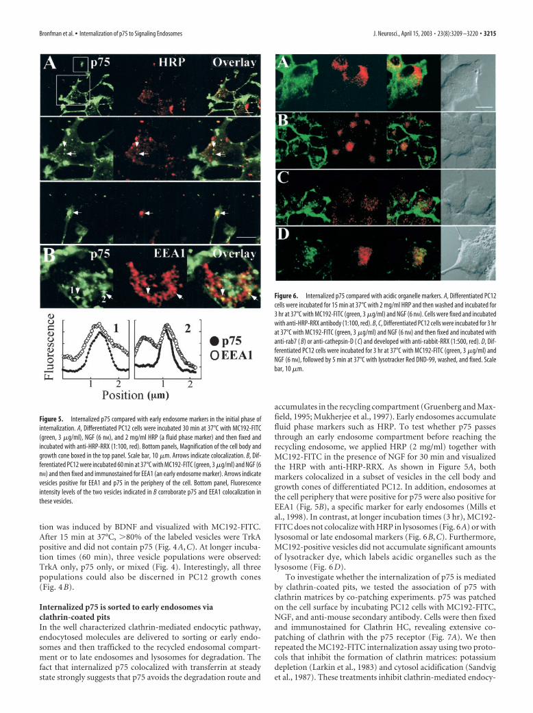
accumulates in the recycling compartment (Gruenberg and Max-field, 1995; Mukherjee et al., 1997). Early endosomes accumulatefluid phase markers such as HRP. To test whether p75 passesthrough an early endosome compartment before reaching therecycling endosome, we applied HRP (2 mg/ml) together withMC192-FITC in the presence of NGF for 30 min and visualizedthe HRP with anti-HRP-RRX. As shown in Figure 5A, bothmarkers colocalized in a subset of vesicles in the cell body andgrowth cones of differentiated PC12. In addition, endosomes atthe cell periphery that were positive for p75 were also positive forEEA1 (Fig. 5B), a specific marker for early endosomes (Mills etal., 1998). In contrast, at longer incubation times (3 hr), MC192-FITC does not colocalize with HRP in lysosomes (Fig. 6A) or withlysosomal or late endosomal markers (Fig. 6B,C). Furthermore,MC192-positive vesicles did not accumulate significant amountsof lysotracker dye, which labels acidic organelles such as thelysosome (Fig. 6 D).
To investigate whether the internalization of p75 is mediatedby clathrin-coated pits, we tested the association of p75 withclathrin matrices by co-patching experiments. p75 was patchedon the cell surface by incubating PC12 cells with MC192-FITC,NGF, and anti-mouse secondary antibody. Cells were then fixedand immunostained for Clathrin HC, revealing extensive co-patching of clathrin with the p75 receptor (Fig. 7A). We thenrepeated the MC192-FITC internalization assay using two proto-cols that inhibit the formation of clathrin matrices: potassiumdepletion (Larkin et al., 1983) and cytosol acidification (Sandviget al., 1987). These treatments inhibit clathrin-mediated endocy-
Figure 5. Internalized p75 compared with early endosome markers in the initial phase ofinternalization. A, Differentiated PC12 cells were incubated 30 min at 37°C with MC192-FITC(green, 3 �g/ml), NGF (6 nM), and 2 mg/ml HRP (a fluid phase marker) and then fixed andincubated with anti-HRP-RRX (1:100, red). Bottom panels, Magnification of the cell body andgrowth cone boxed in the top panel. Scale bar, 10 �m. Arrows indicate colocalization. B, Dif-ferentiated PC12 were incubated 60 min at 37°C with MC192-FITC (green, 3 �g/ml) and NGF (6nM) and then fixed and immunostained for EEA1 (an early endosome marker). Arrows indicatevesicles positive for EEA1 and p75 in the periphery of the cell. Bottom panel, Fluorescenceintensity levels of the two vesicles indicated in B corroborate p75 and EEA1 colocalization inthese vesicles.
Figure 6. Internalized p75 compared with acidic organelle markers. A, Differentiated PC12cells were incubated for 15 min at 37°C with 2 mg/ml HRP and then washed and incubated for3 hr at 37°C with MC192-FITC (green, 3 �g/ml) and NGF (6 nM). Cells were fixed and incubatedwith anti-HRP-RRX antibody (1:100, red). B, C, Differentiated PC12 cells were incubated for 3 hrat 37°C with MC192-FITC (green, 3 �g/ml) and NGF (6 nM) and then fixed and incubated withanti-rab7 ( B) or anti-cathepsin-D ( C) and developed with anti-rabbit-RRX (1:500, red). D, Dif-ferentiated PC12 cells were incubated for 3 hr at 37°C with MC192-FITC (green, 3 �g/ml) andNGF (6 nM), followed by 5 min at 37°C with lysotracker Red DND-99, washed, and fixed. Scalebar, 10 �m.
Bronfman et al. • Internalization of p75 to Signaling Endosomes J. Neurosci., April 15, 2003 • 23(8):3209 –3220 • 3215
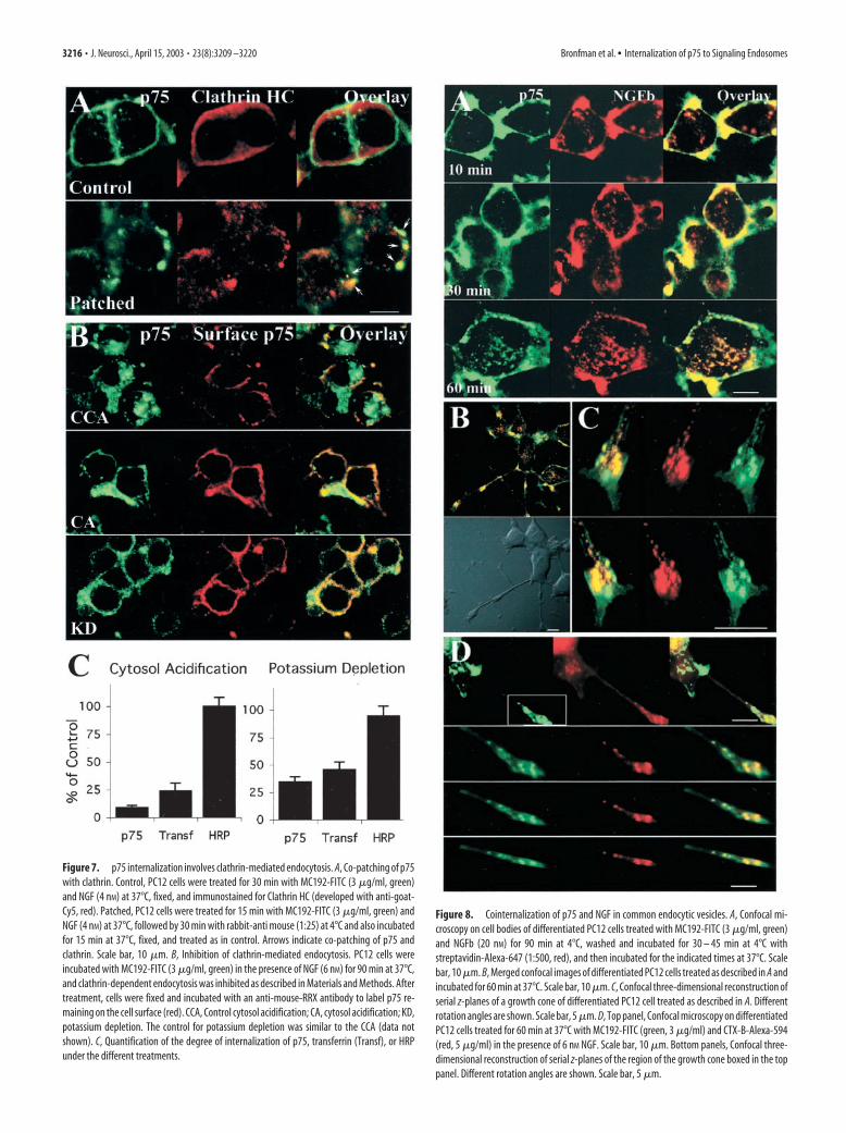
Figure 7. p75 internalization involves clathrin-mediated endocytosis. A, Co-patching of p75with clathrin. Control, PC12 cells were treated for 30 min with MC192-FITC (3 �g/ml, green)and NGF (4 nM) at 37°C, fixed, and immunostained for Clathrin HC (developed with anti-goat-Cy5, red). Patched, PC12 cells were treated for 15 min with MC192-FITC (3 �g/ml, green) andNGF (4 nM) at 37°C, followed by 30 min with rabbit-anti mouse (1:25) at 4°C and also incubatedfor 15 min at 37°C, fixed, and treated as in control. Arrows indicate co-patching of p75 andclathrin. Scale bar, 10 �m. B, Inhibition of clathrin-mediated endocytosis. PC12 cells wereincubated with MC192-FITC (3 �g/ml, green) in the presence of NGF (6 nM) for 90 min at 37°C,and clathrin-dependent endocytosis was inhibited as described in Materials and Methods. Aftertreatment, cells were fixed and incubated with an anti-mouse-RRX antibody to label p75 re-maining on the cell surface (red). CCA, Control cytosol acidification; CA, cytosol acidification; KD,potassium depletion. The control for potassium depletion was similar to the CCA (data notshown). C, Quantification of the degree of internalization of p75, transferrin (Transf), or HRPunder the different treatments.
Figure 8. Cointernalization of p75 and NGF in common endocytic vesicles. A, Confocal mi-croscopy on cell bodies of differentiated PC12 cells treated with MC192-FITC (3 �g/ml, green)and NGFb (20 nM) for 90 min at 4°C, washed and incubated for 30 – 45 min at 4°C withstreptavidin-Alexa-647 (1:500, red), and then incubated for the indicated times at 37°C. Scalebar, 10 �m. B, Merged confocal images of differentiated PC12 cells treated as described in A andincubated for 60 min at 37°C. Scale bar, 10 �m. C, Confocal three-dimensional reconstruction ofserial z-planes of a growth cone of differentiated PC12 cell treated as described in A. Differentrotation angles are shown. Scale bar, 5 �m. D, Top panel, Confocal microscopy on differentiatedPC12 cells treated for 60 min at 37°C with MC192-FITC (green, 3 �g/ml) and CTX-B-Alexa-594(red, 5 �g/ml) in the presence of 6 nM NGF. Scale bar, 10 �m. Bottom panels, Confocal three-dimensional reconstruction of serial z-planes of the region of the growth cone boxed in the toppanel. Different rotation angles are shown. Scale bar, 5 �m.
3216 • J. Neurosci., April 15, 2003 • 23(8):3209 –3220 Bronfman et al. • Internalization of p75 to Signaling Endosomes
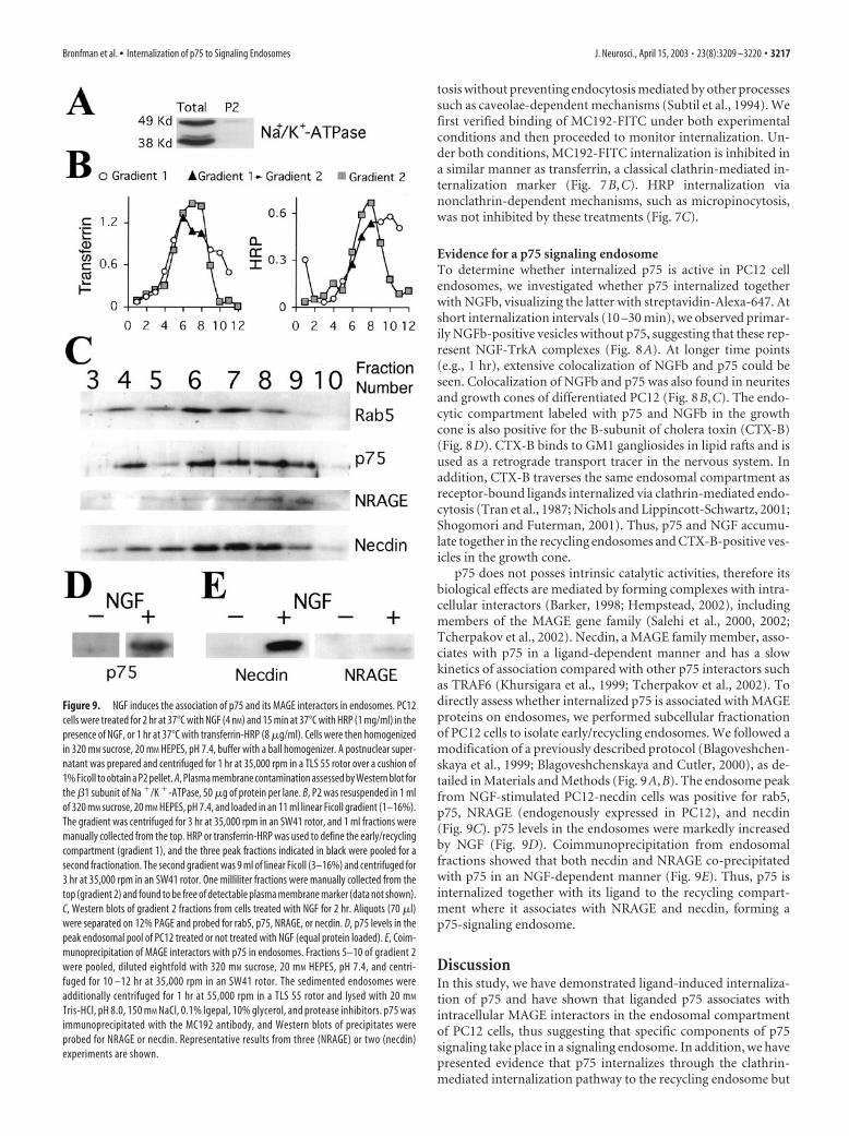
tosis without preventing endocytosis mediated by other processessuch as caveolae-dependent mechanisms (Subtil et al., 1994). Wefirst verified binding of MC192-FITC under both experimentalconditions and then proceeded to monitor internalization. Un-der both conditions, MC192-FITC internalization is inhibited ina similar manner as transferrin, a classical clathrin-mediated in-ternalization marker (Fig. 7B,C). HRP internalization vianonclathrin-dependent mechanisms, such as micropinocytosis,was not inhibited by these treatments (Fig. 7C).
Evidence for a p75 signaling endosomeTo determine whether internalized p75 is active in PC12 cellendosomes, we investigated whether p75 internalized togetherwith NGFb, visualizing the latter with streptavidin-Alexa-647. Atshort internalization intervals (10 –30 min), we observed primar-ily NGFb-positive vesicles without p75, suggesting that these rep-resent NGF-TrkA complexes (Fig. 8A). At longer time points(e.g., 1 hr), extensive colocalization of NGFb and p75 could beseen. Colocalization of NGFb and p75 was also found in neuritesand growth cones of differentiated PC12 (Fig. 8B,C). The endo-cytic compartment labeled with p75 and NGFb in the growthcone is also positive for the B-subunit of cholera toxin (CTX-B)(Fig. 8D). CTX-B binds to GM1 gangliosides in lipid rafts and isused as a retrograde transport tracer in the nervous system. Inaddition, CTX-B traverses the same endosomal compartment asreceptor-bound ligands internalized via clathrin-mediated endo-cytosis (Tran et al., 1987; Nichols and Lippincott-Schwartz, 2001;Shogomori and Futerman, 2001). Thus, p75 and NGF accumu-late together in the recycling endosomes and CTX-B-positive ves-icles in the growth cone.
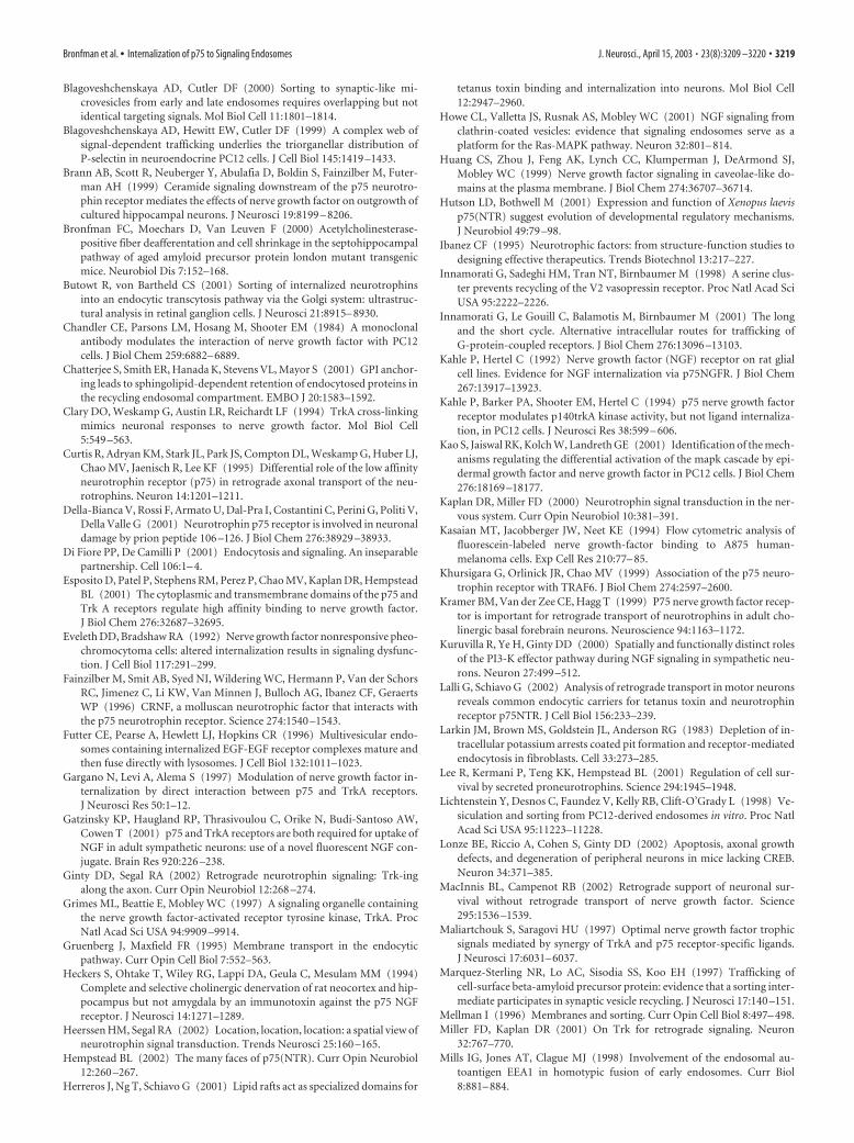
p75 does not posses intrinsic catalytic activities, therefore itsbiological effects are mediated by forming complexes with intra-cellular interactors (Barker, 1998; Hempstead, 2002), includingmembers of the MAGE gene family (Salehi et al., 2000, 2002;Tcherpakov et al., 2002). Necdin, a MAGE family member, asso-ciates with p75 in a ligand-dependent manner and has a slowkinetics of association compared with other p75 interactors suchas TRAF6 (Khursigara et al., 1999; Tcherpakov et al., 2002). Todirectly assess whether internalized p75 is associated with MAGEproteins on endosomes, we performed subcellular fractionationof PC12 cells to isolate early/recycling endosomes. We followed amodification of a previously described protocol (Blagoveshchen-skaya et al., 1999; Blagoveshchenskaya and Cutler, 2000), as de-tailed in Materials and Methods (Fig. 9A,B). The endosome peakfrom NGF-stimulated PC12-necdin cells was positive for rab5,p75, NRAGE (endogenously expressed in PC12), and necdin(Fig. 9C). p75 levels in the endosomes were markedly increasedby NGF (Fig. 9D). Coimmunoprecipitation from endosomalfractions showed that both necdin and NRAGE co-precipitatedwith p75 in an NGF-dependent manner (Fig. 9E). Thus, p75 isinternalized together with its ligand to the recycling compart-ment where it associates with NRAGE and necdin, forming ap75-signaling endosome.
DiscussionIn this study, we have demonstrated ligand-induced internaliza-tion of p75 and have shown that liganded p75 associates withintracellular MAGE interactors in the endosomal compartmentof PC12 cells, thus suggesting that specific components of p75signaling take place in a signaling endosome. In addition, we havepresented evidence that p75 internalizes through the clathrin-mediated internalization pathway to the recycling endosome but
Figure 9. NGF induces the association of p75 and its MAGE interactors in endosomes. PC12cells were treated for 2 hr at 37°C with NGF (4 nM) and 15 min at 37°C with HRP (1 mg/ml) in thepresence of NGF, or 1 hr at 37°C with transferrin-HRP (8 �g/ml). Cells were then homogenizedin 320 mM sucrose, 20 mM HEPES, pH 7.4, buffer with a ball homogenizer. A postnuclear super-natant was prepared and centrifuged for 1 hr at 35,000 rpm in a TLS 55 rotor over a cushion of1% Ficoll to obtain a P2 pellet. A, Plasma membrane contamination assessed by Western blot forthe �1 subunit of Na �/K �-ATPase, 50 �g of protein per lane. B, P2 was resuspended in 1 mlof 320 mM sucrose, 20 mM HEPES, pH 7.4, and loaded in an 11 ml linear Ficoll gradient (1–16%).The gradient was centrifuged for 3 hr at 35,000 rpm in an SW41 rotor, and 1 ml fractions weremanually collected from the top. HRP or transferrin-HRP was used to define the early/recyclingcompartment (gradient 1), and the three peak fractions indicated in black were pooled for asecond fractionation. The second gradient was 9 ml of linear Ficoll (3–16%) and centrifuged for3 hr at 35,000 rpm in an SW41 rotor. One milliliter fractions were manually collected from thetop (gradient 2) and found to be free of detectable plasma membrane marker (data not shown).C, Western blots of gradient 2 fractions from cells treated with NGF for 2 hr. Aliquots (70 �l)were separated on 12% PAGE and probed for rab5, p75, NRAGE, or necdin. D, p75 levels in thepeak endosomal pool of PC12 treated or not treated with NGF (equal protein loaded). E, Coim-munoprecipitation of MAGE interactors with p75 in endosomes. Fractions 5–10 of gradient 2were pooled, diluted eightfold with 320 mM sucrose, 20 mM HEPES, pH 7.4, and centri-fuged for 10 –12 hr at 35,000 rpm in an SW41 rotor. The sedimented endosomes wereadditionally centrifuged for 1 hr at 55,000 rpm in a TLS 55 rotor and lysed with 20 mM
Tris-HCl, pH 8.0, 150 mM NaCl, 0.1% Igepal, 10% glycerol, and protease inhibitors. p75 wasimmunoprecipitated with the MC192 antibody, and Western blots of precipitates wereprobed for NRAGE or necdin. Representative results from three (NRAGE) or two (necdin)experiments are shown.
Bronfman et al. • Internalization of p75 to Signaling Endosomes J. Neurosci., April 15, 2003 • 23(8):3209 –3220 • 3217

at distinct kinetics from TrkA, thus indicating that p75-signalingendosomes may be distinct from trk-signaling endosomes.
Monitoring the internalization and trafficking of p75-ligandcomplexes requires the use of selectively tagged ligands or probesfor the complex. Most of the previous studies of neurotrophin-receptor internalization have relied primarily on preparations ofneurotrophins labeled with radioactive iodine (Eveleth and Brad-shaw, 1992; Kahle et al., 1994; Gargano et al., 1997). Althoughiodinated neurotrophins provide a highly sensitive probe causedby the high-specific activities obtainable in labeling and the rela-tive ease of detection of 125I, there are inherent limitations to thisapproach (i.e., the short half-life of iodinated proteins and theirreduced stability). Nonradioactive labeling of neurotrophins hasbeen attempted previously by a number of groups with varyingdegrees of success. Neet and colleagues described a detailedprotocol for fluorescein labeling of NGF; however, an unex-pected quenching limited the use of this preparation (Kasaian etal., 1994). A recent preliminary report described the use ofrhodamine-NGF to follow neuronal retrograde transport in vivo(Weible et al., 2001). Unfortunately, the labeling protocol ofWeible et al. (2001) was targeted to amine groups that are crucialfor NGF binding to p75 (Ibanez, 1995). To obtain a useful andstably labeled NGF that retains the capacity to bind to p75, wedevised a biotinylation strategy targeted to carboxyl groups onthe protein. Carboxyl groups in NGF have been successfully la-beled previously with fluorescent high molecular weight dextrans(Gatzinsky et al., 2001). Our protocol leads to incorporation of anaverage of approximately nine biotins on carboxyl groups in eachNGF dimer, whereas it retains both TrkA- and p75-bindingproperties of the modified NGF. The labeled NGF was stable andcould be visualized or pulled down with various avidin- orstreptavidin-based reagents, thus providing a highly versatile toolfor studies of internalization and trafficking. This was comple-mented by the use of MC192-FITC as a selective fluorescentprobe for p75, thus providing an additional and independentpossibility for monitoring p75 internalization.
p75 internalization proceeds via clathrin-coated pits to earlyendosomes, with subsequent transfer to the recycling compart-ment. Internalization occurs in both cycling and postmitotic dif-ferentiated cells, and in the latter can also be observed in theprocesses and terminals. p75 and NGF colocalize in endosomesin the cell body and terminals, suggesting that internalized p75may be activated. Intriguingly, the kinetics of internalization ofligand-bound p75 is slower than that for classical rates ofclathrin-mediated endocytosis, such as those measured for thetransferrin receptor. Moreover, p75-NGF internalization pro-ceeds at a rate approximately one-third (t1/2 �42–50 min) thanthat of NGF-TrkA. The different rates of internalization of p75and TrkA define different populations of endocytic vesicles in thecell body and growth cone of differentiated PC12 cells, whichcontain TrkA only, p75 only, or a mixed population of bothreceptors.
In the clathrin-mediated endocytic pathway, endocytosedmolecules are delivered to the sorting endosome and then traf-ficked to the recycling endosome or the late endosome–lysosomepathway (Gruenberg and Maxfield, 1995; Mellman, 1996). If ac-tivated p75 transports an NGF-mediated retrograde signal, itmust avoid degradation in the axon. Our data support this affir-mation. Indeed, p75 and NGF avoid degradation and remain inthe recycling endosome for at least up to 3 hr after internalization.Likewise, NGF-TrkA internalized via clathrin (Howe et al., 2001)has been shown to avoid degradation (Zapf-Colby and Olefsky,1998). In contrast to the neurotrophin receptors, the EGF recep-
tor is not retrogradely transported and is efficiently degraded inthe lysosomal pathway after ligand stimulation (Futter et al.,1996; Kao et al., 2001; Waterman et al., 2002). Thus, receptorsthat are retrogradely transported must first evade rapid degrada-tion in the synaptic terminal. The recycling organelle in PC12 hasbeen reported to contain synaptic vesicle components that recy-cle or sort to synaptic microvesicles (Lichtenstein et al., 1998).This raises the possibility that p75 normally recycles in the syn-aptic terminal, sharing a similar endosome as components of thesynaptic vesicles. Ligand association with p75 might regulate in-teraction with specific docking proteins, which might then sortthe receptor to the retrograde transport pathway. Indeed, theassociation of endosomal p75 with MAGE proteins supports thisinteresting possibility and suggests that internalized p75 mightcarry a signaling complex from the synaptic terminal to the cellbody. Such a concept has been exemplified by the amyloid pre-cursor protein that internalizes to recycling synaptic vesicles inprimary cerebellar neurons and subsequently traffics retro-gradely to the soma (Marquez-Sterling et al., 1997).
P75 and TrkA may form a high-affinity joint-binding complexfor NGF (Esposito et al., 2001), raising the question of how thesereceptors internalize with different kinetics. Although we cannotrule out a fast-internalizing high-affinity complex in other celltypes, the high excess of p75 over TrkA in PC12 cells (Urdiales etal., 1998) suggests that the majority of p75 receptors should beexcluded from such a complex. Alternatively, the high-affinitycomplex may dissociate after ligand binding, allowing each re-ceptor to follow separate kinetics of internalization. Nonetheless,because both p75-NGF and TrkA-NGF enter the cell via clathrin-mediated endocytosis, their differential internalization kinetics isintriguing. Slow internalization kinetics has been observed in thepast for GPi-linked receptors associated with lipid rafts (Chatter-jee et al., 2001). Both p75 and TrkA have been found in thesespecialized microdomains (Huang et al., 1999), and internalizedp75 colocalizes with the lipid raft marker CTX-B in differentiatedPC12. Thus, lipid rafts may regulate differential internalization ofp75 versus TrkA (for example, via different affinities of the recep-tors for rafts). Notably, tetanus toxin-labeled retrograde traffick-ing vesicles in motor neurons have been reported to contain p75(Lalli and Schiavo, 2002), and tetanus toxin is also associated withlipid rafts in the cell membrane (Herreros et al., 2001). Together,these studies might suggest that lipid rafts in the synaptic termi-nal regulate the sorting of retrograde-transported vesicles.
To summarize, our observations support the possibility thatinternalized neurotrophin-p75 complexes can act as signalingplatforms in neuronal cells and may be incorporated to signalingendosomes. The differential kinetics of internalization of p75complexes versus trk complexes may provide a mechanism fortemporal separation between signaling endosomes containingeach of these receptors. Such endosomes could thereby beshunted into a retrograde transport pathway of a neuron in aspatially separated manner, thus allowing differential signaltransmission to the cell body.
ReferencesBarker PA (1998) p75NTR: a study in contrasts. Cell Death Differ
5:346 –356.Berger-Sweeney J, Stearns NA, Murg SL, Floerke-Nashner LR, Lappi DA,
Baxter MG (2001) Selective immunolesions of cholinergic neurons inmice: effects on neuroanatomy, neurochemistry, and behavior. J Neurosci21:8164 – 8173.
Bhattacharyya A, Watson FL, Bradlee TA, Pomeroy SL, Stiles CD, Segal RA(1997) Trk receptors function as rapid retrograde signal carriers in theadult nervous system. J Neurosci 17:7007–7016.
3218 • J. Neurosci., April 15, 2003 • 23(8):3209 –3220 Bronfman et al. • Internalization of p75 to Signaling Endosomes
Blagoveshchenskaya AD, Cutler DF (2000) Sorting to synaptic-like mi-crovesicles from early and late endosomes requires overlapping but notidentical targeting signals. Mol Biol Cell 11:1801–1814.
Blagoveshchenskaya AD, Hewitt EW, Cutler DF (1999) A complex web ofsignal-dependent trafficking underlies the triorganellar distribution ofP-selectin in neuroendocrine PC12 cells. J Cell Biol 145:1419 –1433.
Brann AB, Scott R, Neuberger Y, Abulafia D, Boldin S, Fainzilber M, Futer-man AH (1999) Ceramide signaling downstream of the p75 neurotro-phin receptor mediates the effects of nerve growth factor on outgrowth ofcultured hippocampal neurons. J Neurosci 19:8199 – 8206.
Bronfman FC, Moechars D, Van Leuven F (2000) Acetylcholinesterase-positive fiber deafferentation and cell shrinkage in the septohippocampalpathway of aged amyloid precursor protein london mutant transgenicmice. Neurobiol Dis 7:152–168.
Butowt R, von Bartheld CS (2001) Sorting of internalized neurotrophinsinto an endocytic transcytosis pathway via the Golgi system: ultrastruc-tural analysis in retinal ganglion cells. J Neurosci 21:8915– 8930.
Chandler CE, Parsons LM, Hosang M, Shooter EM (1984) A monoclonalantibody modulates the interaction of nerve growth factor with PC12cells. J Biol Chem 259:6882– 6889.
Chatterjee S, Smith ER, Hanada K, Stevens VL, Mayor S (2001) GPI anchor-ing leads to sphingolipid-dependent retention of endocytosed proteins inthe recycling endosomal compartment. EMBO J 20:1583–1592.
Clary DO, Weskamp G, Austin LR, Reichardt LF (1994) TrkA cross-linkingmimics neuronal responses to nerve growth factor. Mol Biol Cell5:549 –563.
Curtis R, Adryan KM, Stark JL, Park JS, Compton DL, Weskamp G, Huber LJ,Chao MV, Jaenisch R, Lee KF (1995) Differential role of the low affinityneurotrophin receptor (p75) in retrograde axonal transport of the neu-rotrophins. Neuron 14:1201–1211.
Della-Bianca V, Rossi F, Armato U, Dal-Pra I, Costantini C, Perini G, Politi V,Della Valle G (2001) Neurotrophin p75 receptor is involved in neuronaldamage by prion peptide 106 –126. J Biol Chem 276:38929 –38933.
Di Fiore PP, De Camilli P (2001) Endocytosis and signaling. An inseparablepartnership. Cell 106:1– 4.
Esposito D, Patel P, Stephens RM, Perez P, Chao MV, Kaplan DR, HempsteadBL (2001) The cytoplasmic and transmembrane domains of the p75 andTrk A receptors regulate high affinity binding to nerve growth factor.J Biol Chem 276:32687–32695.
Eveleth DD, Bradshaw RA (1992) Nerve growth factor nonresponsive pheo-chromocytoma cells: altered internalization results in signaling dysfunc-tion. J Cell Biol 117:291–299.
Fainzilber M, Smit AB, Syed NI, Wildering WC, Hermann P, Van der SchorsRC, Jimenez C, Li KW, Van Minnen J, Bulloch AG, Ibanez CF, GeraertsWP (1996) CRNF, a molluscan neurotrophic factor that interacts withthe p75 neurotrophin receptor. Science 274:1540 –1543.
Futter CE, Pearse A, Hewlett LJ, Hopkins CR (1996) Multivesicular endo-somes containing internalized EGF-EGF receptor complexes mature andthen fuse directly with lysosomes. J Cell Biol 132:1011–1023.
Gargano N, Levi A, Alema S (1997) Modulation of nerve growth factor in-ternalization by direct interaction between p75 and TrkA receptors.J Neurosci Res 50:1–12.
Gatzinsky KP, Haugland RP, Thrasivoulou C, Orike N, Budi-Santoso AW,Cowen T (2001) p75 and TrkA receptors are both required for uptake ofNGF in adult sympathetic neurons: use of a novel fluorescent NGF con-jugate. Brain Res 920:226 –238.
Ginty DD, Segal RA (2002) Retrograde neurotrophin signaling: Trk-ingalong the axon. Curr Opin Neurobiol 12:268 –274.
Grimes ML, Beattie E, Mobley WC (1997) A signaling organelle containingthe nerve growth factor-activated receptor tyrosine kinase, TrkA. ProcNatl Acad Sci USA 94:9909 –9914.
Gruenberg J, Maxfield FR (1995) Membrane transport in the endocyticpathway. Curr Opin Cell Biol 7:552–563.
Heckers S, Ohtake T, Wiley RG, Lappi DA, Geula C, Mesulam MM (1994)Complete and selective cholinergic denervation of rat neocortex and hip-pocampus but not amygdala by an immunotoxin against the p75 NGFreceptor. J Neurosci 14:1271–1289.
Heerssen HM, Segal RA (2002) Location, location, location: a spatial view ofneurotrophin signal transduction. Trends Neurosci 25:160 –165.
Hempstead BL (2002) The many faces of p75(NTR). Curr Opin Neurobiol12:260 –267.
Herreros J, Ng T, Schiavo G (2001) Lipid rafts act as specialized domains for
tetanus toxin binding and internalization into neurons. Mol Biol Cell12:2947–2960.
Howe CL, Valletta JS, Rusnak AS, Mobley WC (2001) NGF signaling fromclathrin-coated vesicles: evidence that signaling endosomes serve as aplatform for the Ras-MAPK pathway. Neuron 32:801– 814.
Huang CS, Zhou J, Feng AK, Lynch CC, Klumperman J, DeArmond SJ,Mobley WC (1999) Nerve growth factor signaling in caveolae-like do-mains at the plasma membrane. J Biol Chem 274:36707–36714.
Hutson LD, Bothwell M (2001) Expression and function of Xenopus laevisp75(NTR) suggest evolution of developmental regulatory mechanisms.J Neurobiol 49:79 –98.
Ibanez CF (1995) Neurotrophic factors: from structure-function studies todesigning effective therapeutics. Trends Biotechnol 13:217–227.
Innamorati G, Sadeghi HM, Tran NT, Birnbaumer M (1998) A serine clus-ter prevents recycling of the V2 vasopressin receptor. Proc Natl Acad SciUSA 95:2222–2226.
Innamorati G, Le Gouill C, Balamotis M, Birnbaumer M (2001) The longand the short cycle. Alternative intracellular routes for trafficking ofG-protein-coupled receptors. J Biol Chem 276:13096 –13103.
Kahle P, Hertel C (1992) Nerve growth factor (NGF) receptor on rat glialcell lines. Evidence for NGF internalization via p75NGFR. J Biol Chem267:13917–13923.
Kahle P, Barker PA, Shooter EM, Hertel C (1994) p75 nerve growth factorreceptor modulates p140trkA kinase activity, but not ligand internaliza-tion, in PC12 cells. J Neurosci Res 38:599 – 606.
Kao S, Jaiswal RK, Kolch W, Landreth GE (2001) Identification of the mech-anisms regulating the differential activation of the mapk cascade by epi-dermal growth factor and nerve growth factor in PC12 cells. J Biol Chem276:18169 –18177.
Kaplan DR, Miller FD (2000) Neurotrophin signal transduction in the ner-vous system. Curr Opin Neurobiol 10:381–391.
Kasaian MT, Jacobberger JW, Neet KE (1994) Flow cytometric analysis offluorescein-labeled nerve growth-factor binding to A875 human-melanoma cells. Exp Cell Res 210:77– 85.
Khursigara G, Orlinick JR, Chao MV (1999) Association of the p75 neuro-trophin receptor with TRAF6. J Biol Chem 274:2597–2600.
Kramer BM, Van der Zee CE, Hagg T (1999) P75 nerve growth factor recep-tor is important for retrograde transport of neurotrophins in adult cho-linergic basal forebrain neurons. Neuroscience 94:1163–1172.
Kuruvilla R, Ye H, Ginty DD (2000) Spatially and functionally distinct rolesof the PI3-K effector pathway during NGF signaling in sympathetic neu-rons. Neuron 27:499 –512.
Lalli G, Schiavo G (2002) Analysis of retrograde transport in motor neuronsreveals common endocytic carriers for tetanus toxin and neurotrophinreceptor p75NTR. J Cell Biol 156:233–239.
Larkin JM, Brown MS, Goldstein JL, Anderson RG (1983) Depletion of in-tracellular potassium arrests coated pit formation and receptor-mediatedendocytosis in fibroblasts. Cell 33:273–285.
Lee R, Kermani P, Teng KK, Hempstead BL (2001) Regulation of cell sur-vival by secreted proneurotrophins. Science 294:1945–1948.
Lichtenstein Y, Desnos C, Faundez V, Kelly RB, Clift-O’Grady L (1998) Ve-siculation and sorting from PC12-derived endosomes in vitro. Proc NatlAcad Sci USA 95:11223–11228.
Lonze BE, Riccio A, Cohen S, Ginty DD (2002) Apoptosis, axonal growthdefects, and degeneration of peripheral neurons in mice lacking CREB.Neuron 34:371–385.
MacInnis BL, Campenot RB (2002) Retrograde support of neuronal sur-vival without retrograde transport of nerve growth factor. Science295:1536 –1539.
Maliartchouk S, Saragovi HU (1997) Optimal nerve growth factor trophicsignals mediated by synergy of TrkA and p75 receptor-specific ligands.J Neurosci 17:6031– 6037.
Marquez-Sterling NR, Lo AC, Sisodia SS, Koo EH (1997) Trafficking ofcell-surface beta-amyloid precursor protein: evidence that a sorting inter-mediate participates in synaptic vesicle recycling. J Neurosci 17:140 –151.
Mellman I (1996) Membranes and sorting. Curr Opin Cell Biol 8:497– 498.Miller FD, Kaplan DR (2001) On Trk for retrograde signaling. Neuron
32:767–770.Mills IG, Jones AT, Clague MJ (1998) Involvement of the endosomal au-
toantigen EEA1 in homotypic fusion of early endosomes. Curr Biol8:881– 884.
Bronfman et al. • Internalization of p75 to Signaling Endosomes J. Neurosci., April 15, 2003 • 23(8):3209 –3220 • 3219

Mukherjee S, Ghosh RN, Maxfield FR (1997) Endocytosis. Physiol Rev77:759 – 803.
Neet KE, Campenot RB (2001) Receptor binding, internalization, and ret-rograde transport of neurotrophic factors. Cell Mol Life Sci58:1021–1035.
Nichols BJ, Lippincott-Schwartz J (2001) Endocytosis without clathrincoats. Trends Cell Biol 11:406 – 412.
Patapoutian A, Reichardt LF (2001) Trk receptors: mediators of neurotro-phin action. Curr Opin Neurobiol 11:272–280.
Riccio A, Pierchala BA, Ciarallo CL, Ginty DD (1997) An NGF-TrkA-mediated retrograde signal to transcription factor CREB in sympatheticneurons. Science 277:1097–1100.
Riccio A, Ahn S, Davenport CM, Blendy JA, Ginty DD (1999) Mediation bya CREB family transcription factor of NGF-dependent survival of sympa-thetic neurons. Science 286:2358 –2361.
Ryden M, Hempstead B, Ibanez CF (1997) Differential modulation of neu-ron survival during development by nerve growth factor binding to thep75 neurotrophin receptor. J Biol Chem 272:16322–16328.
Salehi AH, Roux PP, Kubu CJ, Zeindler C, Bhakar A, Tannis LL, Verdi JM,Barker PA (2000) NRAGE, a novel MAGE protein, interacts with thep75 neurotrophin receptor and facilitates nerve growth factor-dependentapoptosis. Neuron 27:279 –288.
Salehi AH, Xanthoudakis S, Barker PA (2002) NRAGE, a p75 neurotrophinreceptor interacting protein, induces caspase activation and cell deaththrough a JNK-dependent mitochondrial pathway. J Biol Chem277:48043– 48050.
Sandvig K, Olsnes S, Petersen OW, van Deurs B (1987) Acidification of thecytosol inhibits endocytosis from coated pits. J Cell Biol 105:679 – 689.
Senger DL, Campenot RB (1997) Rapid retrograde tyrosine phosphoryla-tion of trkA and other proteins in rat sympathetic neurons in compart-mented cultures. J Cell Biol 138:411– 421.
Shogomori H, Futerman AH (2001) Cholera toxin is found in detergent-insoluble rafts/domains at the cell surface of hippocampal neurons but isinternalized via a raft-independent mechanism. J Biol Chem276:9182–9188.
Subtil A, Hemar A, Dautry-Varsat A (1994) Rapid endocytosis of interleu-kin 2 receptors when clathrin-coated pit endocytosis is inhibited. J Cell Sci107:3461–3468.
Tcherpakov M, Bronfman FC, Conticello SG, Vaskovsky A, Levy Z, NiinobeM, Yoshikawa K, Arenas E, Fainzilber M (2002) The p75 neurotrophinreceptor interacts with multiple MAGE proteins. J Biol Chem277:49101– 49104.
Tran D, Carpentier JL, Sawano F, Gorden P, Orci L (1987) Ligands internal-ized through coated or noncoated invaginations follow a common intra-cellular pathway. Proc Natl Acad Sci USA 84:7957–7961.
Tsui-Pierchala BA, Ginty DD (1999) Characterization of an NGF-P-TrkAretrograde-signaling complex and age-dependent regulation of TrkAphosphorylation in sympathetic neurons. J Neurosci 19:8207– 8218.
Tuffereau C, Benejean J, Blondel D, Kieffer B, Flamand A (1998) Low-affinity nerve-growth factor receptor (P75NTR) can serve as a receptorfor rabies virus. EMBO J 17:7250 –7259.
Urdiales JL, Becker E, Andrieu M, Thomas A, Jullien J, van Grunsven LA,Menut S, Evan GI, Martin-Zanca D, Rudkin BB (1998) Cell cycle phase-specific surface expression of nerve growth factor receptors TrkA andp75(NTR). J Neurosci 18:6767– 6775.
von Bartheld CS, Williams R, Lefcort F, Clary DO, Reichardt LF, Bothwell M(1996) Retrograde transport of neurotrophins from the eye to the brainin chick embryos: roles of the p75NTR and trkB receptors. J Neurosci16:2995–3008.
von Schack D, Casademunt E, Schweigreiter R, Meyer M, Bibel M, Dechant G(2001) Complete ablation of the neurotrophin receptor p75NTR causesdefects both in the nervous and the vascular system. Nat Neurosci4:977–978.
Waterman H, Katz M, Rubin C, Shtiegman K, Lavi S, Elson A, Jovin T,Yarden Y (2002) A mutant EGF-receptor defective in ubiquitylationand endocytosis unveils a role for Grb2 in negative signaling. EMBO J21:303–313.
Watson FL, Heerssen HM, Moheban DB, Lin MZ, Sauvageot CM, Bhatta-charyya A, Pomeroy SL, Segal RA (1999) Rapid nuclear responses totarget-derived neurotrophins require retrograde transport of ligand-receptor complex. J Neurosci 19:7889 –7900.
Watson FL, Heerssen HM, Bhattacharyya A, Klesse L, Lin MZ, Segal RA(2001) Neurotrophins use the Erk5 pathway to mediate a retrograde sur-vival response. Nat Neurosci 4:981–988.
Weible Jr MW, Bartlett SE, Reynolds AJ, Hendry IA (2001) Prolonged recy-cling of internalized neurotrophins in the nerve terminal. Cytometry43:182–188.
Yano H, Lee FS, Kong H, Chuang J, Arevalo J, Perez P, Sung C, Chao MV(2001) Association of Trk neurotrophin receptors with components ofthe cytoplasmic dynein motor. J Neurosci 21:RC125(1–7).
Zapf-Colby A, Olefsky JM (1998) Nerve growth factor processing and traf-ficking events following TrkA-mediated endocytosis. Endocrinology 139:3232–3240.
3220 • J. Neurosci., April 15, 2003 • 23(8):3209 –3220 Bronfman et al. • Internalization of p75 to Signaling Endosomes