identification of the neurotrophin receptors p75 and trk in a series of
TRANSCRIPT

American Journal of Pathology, Vol. 145, No. 4, October 1994Copyight © American Society for Investigative Pathology
Identification of the Neurotrophin Receptors p75and trk in a Series of Wilms' Tumors
Michael J. Donovan,* B. Hempstead,ttL. Julie Huber,A D. Kaplan,§ Pantelis Tsoulfas,§M. Chao,* Luis Parada,§ andDeborah Schofield*From the Department ofPathology,* Children's Hospital,Boston, Massachusetts; Departments ofHematologylOncologyt and Cell Biology,* Cornell University MedicalCenter, New York, New York; and Molecular EmbryologySection,j NCI-Fredenick Cancer Research and DevelopmentCenter, ABL-Basic Research Program, Frederick, Maryland
The molecular mechanisms underlying thepatho-genesis of Wilms' tumor (WTJ are poorly under-stood, although a variety ofgrowth factors in-cluding platelet-derived growth factor andinsulin-like growthfactor are expressed and arethought to contribute to tumor development. Inearlier studies, WT ceUs in culture werefound toexpress the low affinity nerve growth factor re-ceptor, p75. These WT ceUs were capable of re-sponding to the neurotrophin (NT) NGF, suggest-ing that NT may be involved in WTfpathogenesis.We have examined a group of WT immunohisto-chemically with antibodies recognizing known trkreceptorproteins, thep75 receptor, and the NTs,NGF and NT-3. Confirmatory immunoprecipita-tion and Western blots were then performed onrepresentative WTsamplesfrom the studygroup.Thep75 receptor wasfoundpredominantly in theepithelial and blastemal components where highlevels ofNT were also identifled The trk A and Breceptors were primarily within stromal compo-nents, whereas the trk C and C' receptors werepresent within epithelial structures. Western blotanalyses confirmed thepresence ofthe respectivereceptor proteins with variations correlating insome cases with histological type. The selectivepresence ofNT receptors and growth factors inthis series of WT implies autocrine/paracrinemechanisms for tumor development. (Am JPathol 1994, 145:792-801)
There is convincing evidence for the implication ofgrowth factors as regulators of cell growth and dif-
ferentiation in specific tumors. A variety of peptidegrowth factors such as epidermal growth factor(EGF), platelet-derived growth factor (PDGF), trans-forming growth factors (TGF-a and -43) and insulin-likegrowth factors (IGF-1 and -2) are reportedly mito-genic in vitro12 and under some conditions promoteexpression of phenotypic and molecular features oftransformed cells.3A tumor that has been investigated rather exten-
sively in this regard is Wilms' tumor (WT), a pediatrickidney neoplasm that arises from multipotential stemcells of the metanephric blastema.4 Because WT re-capitulates some aspects of normal kidney develop-ment, the expression of a number of fetal growth fac-tors has been examined, specifically PDGF andIGF.56 Early studies have identified the expression ofboth PDGF and IGF-2 transcripts in a small series ofWT and implicated these fetal growth factors in theetiology of this tumor. More recently, experiments withWT tissue samples reported increased expression ofIGF-2 mRNA, increased IGF-2 protein, and increasedIGF-1 receptor binding activity.7.8 It has been sug-gested that IGF production may at least be partiallyresponsible for increased proliferation and inhibitionof terminal differentiation of this neoplasm. In addi-tion, a role of the WT-1 gene product in the develop-ment of these tumors has been postulated given theproteins ability to regulate the expression of the PDGFa-chain and IGF-2 genes.9We would like to suggest that an additional set of
growth factors, the neurotrophins (NT) and their re-ceptors, are also involved in the pathogenesis ofsome WT. We are aware of only one published studythat identified the low affinity nerve growth factor(LANGF) receptor, p75, on epithelial WT cells in cul-ture.10 The authors demonstrated that these cells re-
BLH was supported by grants from the National Institutes ofHealth, ACS, and the March of Dimes. MVC was supported bygrants the the National Institutes of Health, Zenith Award from Alz-heimer's Association, and the Dorothy Radbell Cohen Foundation.DK, PL, and LP are supported by the National Institutes of Health.
Accepted for publication July 7, 1994.
Address reprint requests to Dr. Michael J. Donovan, BostonChildren's Hospital, Department of Pathology, 300 Longwood Av-enue, Boston, MA 02115.
792

Neurotrophin Receptors in Wilms' Tumors 793AJP October 1994, Vol. 145, No. 4
sponded to the NT NGF with an induction in c-fos RNAexpression. These results, although limited, suggestthat WT cells express one class of intact NT receptorscapable of responding to a specific ligand. In addi-tion, recent studies have implicated the p75 receptorin normal recent kidney development11 and othershave found the p75 receptor in mesangial cells of thedeveloping human kidney.12'13 The function of thisreceptor during the development of the kidney is stillelusive. The status of another set of receptors, thehigh affinity NT receptors or trk receptor tyrosine ki-nases, on WT cells is currently unknown, however, thetrk receptors have been identified at distinct sites dur-ing the development of the rodent kidney.14 Theirfunction in kidney development is unknown.Members of the trk tyrosine kinase family have re-
cently been identified as functional receptors of theNGF family of NTs. The interactions between NTs andtheir respective receptors stimulates receptor ty-rosine kinase activity and elicits biological responsesincluding differentiation and proliferation, dependingon the cellular environment in which the receptor isexpressed. 15 Recent evidence has suggested that trkA expression is required for differentiation in somepediatric small round blue cell tumors, such as neu-roblastoma.16'17 Additional studies have shown highlevels of trk protein in terminally differentiated gan-glion cells of neuroblastomas and in differentiatingrhabdomyoblasts in rhabdomyosarcoma.18 These re-sults expand the known association between NTs, trkreceptors, and differentiation.The trk gene family encodes three receptor ty-
rosine kinases, trk A, trk B, and trk C, which selectivelyinteract with the NTs, NGF, BDNF, and NT-3, respec-tively. 15 In addition to the full-length trk A, B, or C tran-scripts, each trk gene is subject to alternative splicingevents, leading to the generation of numerous iso-forms for each trk species. The functional signifi-cance of these splice variants with regards to cell sig-naling is not completely clear. The truncated forms oftrk B and trk C have been postulated to inhibit sig-naling when co-expressed with full-length trk B andtrk C receptors.15'19 Whether this inhibition of NT sig-naling reflects a sequestration of ligand by the trun-cated receptors, or direct inhibition of dimerizationand autophosphorylation of full-length trk isoforms isnot clear. The trk C gene also encodes additional iso-forms that contain 14 or 25 amino acid inserts withinthe tyrosine kinase domain. The isoforms with kinaseinserts display an attenuated tyrosine phosphoryla-tion response to ligand and fail to promote cell dif-ferentiation when expressed in a neural crest cell linecapable of neuronal differentiation. This system rep-resents the first receptor tyrosine kinase system ex-
pressing variable isoforms that retain both ligandbinding and ligand-inducible kinase activation, yetdiffer in biological capabilities.20The developmental expression of trk B and trk C is
significantly more widespread than that described fortrk A. Although most investigations of trk B and trk Cexpression have focused on the central and periph-eral nervous systems, it is now known that all the trksare expressed in extraneural tissues, including themouse and rat kidney.15'19 Based on these studiesand by analogy with results derived from neuronal celllines, the interactions between the NTs and their cog-nate receptors stimulate receptor tyrosine kinase ac-tivity thereby eliciting different biological responsesfor survival and differentiation.20'21 However, the spe-cific roles of the NT receptors in the development ofnonneuronal organs is not yet known.
Using antibodies that recognize the NT receptorsp75 and trk, including trk A, B, and C, we assessedthe expression of these receptors in a series of WTsusing immunohistochemistry and Western blot analy-ses. We also evaluated NT expression in these sameWT samples. The localization and involvement ofthese factors in the pathogenesis of WT is discussed.
Materials and MethodsTen cases of WT diagnosed at the Children's Hospitalof Boston between 1980 and 1993 were identified.After examination of a large series of cases, we se-lected a cross-sectional study group that consisted ofsix females and four males, ranging in age from 11weeks to 8 years of age. There were three stage 1, fivestage II, and two stage IV categorized as follows:4 twoanaplastic, two blastemal predominant, two epithelialpredominant, two classic triphasic, and two rhab-domyoblastic. After review of the hematoxylin and eo-sin (H&E)-stained slides on the study group of cases,a representative block was selected for analysis and4 p-thick sections were cut. All tissues were pro-cessed as previously described using an avidin-biotin-based alkaline phosphatase detection kit (Bio-Genex Labs, San Ramon, CA).18 Each of theantibodies were individually titrated first with selectivecell lines overexpressing the particular receptor pro-tein. 18 These antibody results served as negative andpositive controls and for optimal use on paraffin sec-tions with dilutions ranging from 1:400 to 1:600. Sec-tions were routinely incubated for 1 to 2 hours at roomtemperature. Additional rinses and preincubationwith goat serum (5%) were incorporated as neededto reduce background. Normal goat serum and non-immune serum were routinely used as negative con-

794 Donovan et alAJP October 1994, Vol. 145, No. 4
trols. In addition, the peptide immunogen for the trk C'antibody was used as an additional negative control.
Frozen tissue from each category of WT was ana-lyzed using tissue lysates (100 pg/lane) equallyloaded and immunoprecipitations with the pan-trk an-tibody and subsequent Western immunoblotting withtrk A, trk B, trk C, and the anti-trk C' antibodies. De-tergent (RIPA) extracts of fibroblasts and insect cellsstably expressing trk A, B, C, and C' were normalizedfor protein content and subjected to immunoprecipi-tation using the pan-trk antisera, followed by Westernanalyses using the antisera specific for each trk re-ceptor.
Immunocomplexes to protein were detected by in-cubation with 1251-labeled protein A. As shown in Fig-ure 1, no cross-reactivity could be detected. Lysatesfrom insect (SF9) cells (2 x 10⁶) expressing rat trk Bwere electrophoresed and Westerns performed withanti-trk B and reprobed with pan-trk antibody for con-firmation. A mouse melanoma cell line overexpress-ing p75 was also used as a control cell line. The meth-ods for Western blot analysis and cell cultureutilization have been previously described.23 The ex-posure times for the culture utilization have been pre-viously described.23 The exposure times for the au-toradiographs were 18 and 24 hours, as indicated.For the insect cell blots, the exposure times were 7seconds (chemiluminescence).
Results
Specificity of Antibodies
Antibody specificity for p75 and trk protein was con-firmed by Western blot analyses. All antibodies wereevaluated immunohistochemically using selective
A
200
110
BA B C A B C
200
110
cell lines overexpressing the individual receptorsserving as positive and negative controls (data notshown). The trk and p75 positivity was seen as red,coarsely granular to globular staining within cell cy-toplasm. The anti-p75 rabbit polyclonal antibody wasgenerated against the intracellular rat p75 protein andthe specificity of this antibody is demonstrated usinga human melanoma cell line A875 expressing only thep75 receptor as a methodological control for theWestern blots (Figure 3).18.22 The complete charac-terization and utilization of the polyclonal pan-trk an-tibody has been previously described. 18'23 Theaffinity-purified rabbit polyclonal trk A antibody wasgenerated against a unique extracellular sequence ofhuman trk A and identifies a distinct band at 140 kdby Western blot (Figure 1, A). The affinity-purified rab-bit polyclonal trk B antibody was generated against aunique juxtamembrane sequences of rat trk B andidentifies the variably glycosylated forms (110 and145 kd; Figure 1, B). The affinity-purified rabbit poly-clonal antisera specific for extracellular rat trk C rec-ognizes all trk C isoforms (140 kd; faint band at 100kd; Figure 1, C) and affinity-purified antisera specificfor the unique amino acids within the cytoplasmic do-main of rat truncated trk C (C') lacking the catalyticdomain are demonstrated (100 kd; Figure 1, D). Theantinerve growth factor antibody is a polyclonal an-tibody that recognizes both nerve growth factor andNt-3 (BLH, unpublished observations) and was ob-tained from Collaborative Research, New York.
The p75 Receptor in WT
Previous studies have used a cultured WT cell line todetect p75 receptor expression.10 However, no pub-lished studies have examined the expression of the
CA B C
200
11070-
DA B C
200
11070
anti-trk A anti-trk B anti-trk C anti-trk C truncFigure 1. A: Specificity of antisera to trk receptors as detected by one-dimensional We-stern blot. Cells expressing individual trk ftmnill memberswere cextracted in detergent anid eqnivalent amounts ofprotein u'ere immnnoprecipitated with the pan-trk antisera. Immune comlplexe-s were re-solved on sodinim dodecyl sulfate-poltvacrnlamide gel electrophoresis and Westerni blots were developed iasinig antisera specificfor each trkfamily. A,B, C and D blots probed with aniti-trk A, anti-trk B, anti-trk C, and anti-trncated trk C antisera, respectively. A anld C: lane a, 3T3-trk A cells:lane b, 3T3-trk B cells; lane c. 3T3-trk C cells. B: lanes a-c, SF9 cells expressing trk A, trk B, or trk CG respectively. D, lanes a-c, 3T3 cells expressingfidll-length trk CG trk C = 14 aminlo acid kinase insert. trk C-trtnncated. A, C, azd D nsed 125JI-labeleld protein A for detection. nith 24-hboir anito-radiography eAposure time. B ntsed ECL, 7-second exposure.

Neurotrophin Receptors in Wilms' Tumors 795AJP October 1994, Vol. 145, No. 4
low affinity nerve growth factor receptor, p75, inpathological specimens of WT. We report a high levelof immunoreactivity for NTs and p75 diffusely in theblastemal component and focally within mature tu-bules (Figure 2A, 2B, respectively) with a nervebundle composed of Schwann cells and axons serv-ing as an internal control for p75 (Figure 2, B, inset).The level of NT immunoreactivity was present in alltubules, glomerular structures, and blastemal com-ponents identified, independent of the tumor type ex-amined. The level of p75 immunoreactivity was great-est in pure blastemal areas of all tumors andappeared to be somewhat decreased in regionswhere epithelial differentiation, ie, tubule formation,had occurred. The tubules thought to be the mosthistologically immature contained the highest levelsof p75 antigen. The p75 was not identified in S bodiesor glomerular structures. The components of the pe-ripheral nervous system, ie, nerve fibers and perineu-ral cells, as well as Schwann cells and axons havepreviously been shown by immunohistochemistry tocontain the p75 low affinity nerve growth factor re-ceptor.24 The diffusely anaplastic WT exhibited thehighest levels of p75 within anaplastic blastemal ar-eas, whereas the nonanaplastic appearing cells wereless immunoreactive (data not shown).We verified that p75 was present in the WT study
group by Western blot analysis of individual tumorsamples. As shown in Figure 3, the expression of p75protein varied among individual WT cases examinedbut correlated for the most part with the immuno-histochemistry results. Indeed, the WT sample withthe highest level of anti-p75 immunoreactivity (Figure
1 2 3 4 5200-
110-
70-
anti-p75Figure 3. Autoradiograph of3T3 trk A (lane 1), A875 (lane 2) p75conitrol cell linle, blastemal WT(lane 3), epithelial WT(lane 4), andclassical WT (lane 5) after immunoprecipitation and probing withthe antibody to p75 (exposure 24 hours).
2, B) contained primarily blastemal components thatdemonstrated the greatest amount of p75 receptorprotein on an equal protein-loaded Western immu-noblot (Figure 3, Lane 3). Both of the anaplastic WTsby Western immunoblotting of tissue lysates had highlevels of p75 protein identified with no evidence fornonspecific binding (data not shown).
Figure 2. A: Immunostaining of classical WT with polyclonal antinerve growth factor. Note intense epithelial and blastemal staining, (magnifica-tion X 250). B: Immunostaining of blastemal component of WTwith anti-p75. Note blastemal staining with absence ofstromal immunoreactivity.Nerve bundle stains with anti-p 75 (inset) (magnification X 250).

796 Donovan et alAIP October 1994, Vol. 145, No. 4
The trk A and B Receptors in WT
Using polyclonal antisera specific for trk A or trk B,expression could be demonstrated immunohisto-chemically within the stromal component of each WT.Interestingly, in all WT cases examined, the strongestimmunoreactivity for trk A and B was seen within in-dividual cells at the stromal-blastemal interface.Whether the identical cells express both trk A and Breceptors has yet to be entirely confirmed. Suchquestions are currently being investigated using insitu hybridization with double labeling. The observedstaining pattern was true for all the WT types exam-ined with the exception of the anaplastic variety, inwhich trk A and B were present only within the ana-plastic blastemal component and not within thestroma. Both trk A and B were focally present withinmore histologically mature tubules but not S bodies orglomerular structures.On examination of the two rhabdomyoblastic WT
samples that contained focal collections of stromalrhabdomyoblasts, we found additional evidence fordifferential receptor localization. The trk B reactionproduct was identified within isolated blastemal cellsand stromal cells (Figure 4, A). The immunoreactive
cells are distinguished by their more abundant cyto-plasm compared with their negative counterparts.Using antibodies that recognize desmin (BiogenexLabs) and smooth muscle actin (Biogenex Labs) onthese two WT cases, additional positive cells werefound within the blastema and the stroma, suggestinga skeletal muscle phenotype (data not shown). Ad-ditional studies are needed to further characterize thisobservation.
In the anaplastic WT examined, focal trk A- andB-positive blastemal cells were also noted. Thesecells were not morphologically similar to those in therhabdomyoblastic WT in that they were desmin andactin negative. However, special stains for the lowmolecular weight cytokeratins, ie, CAM 5.2 (BiogenexLabs) antibody, were focally positive for these cells,suggesting epithelial and mesenchymal differentia-tion (data not shown).
The trk C and Truncated trk C
We found receptor proteins trk C and trk C' (trun-cated) primarily within mature epithelial tubular ele-ments of the classical triphasic WT (Figure 4, B). The
Figure 4. A: Immunostaining qfrhabdomyoblastic type WT with anti-trk B. Note staining of differentiating rhabdornvoblasts in blastemaand within the stromal component (magnification X250). B: Im-munostaining of classical WT witb anti-trk C. Note specific staining ofdifferentiated epithelial tubule (magnification X250). C: Immuno-staining of anaplastic WT with anti-trk B. Note focal intense stainingof anaplastic blastemal cells (magnification x 250).
Neurotrophin Receptors in Wilms' Tumors 797AJP October 1994, Vol. 145, No. 4
protein was localized to the membrane and cyto-plasm of epithelial cells but was not present within theblastema or stromal components. Trk C was also seenfocally within the blastema cells of both anaplasticWTs.We did not find trk receptor protein within the non-
rhabdomyoblastic blastemal component of any WTexamined except for some focal intense immunore-activity of trk A, B, and C/C' in the focal and diffuselyanaplastic WT samples, as alluded to earlier (Figure4, C). In these two cases the trk-positive cells weremoderately pleomorphic and contained more cyto-plasm than the nonanaplastic blastemal cells. Theydid not have rhabdomyoblastic features in that theywere negative for desmin and other skeletal musclemarkers.
Western Analysis/lmmunohistochemistryCorrelation
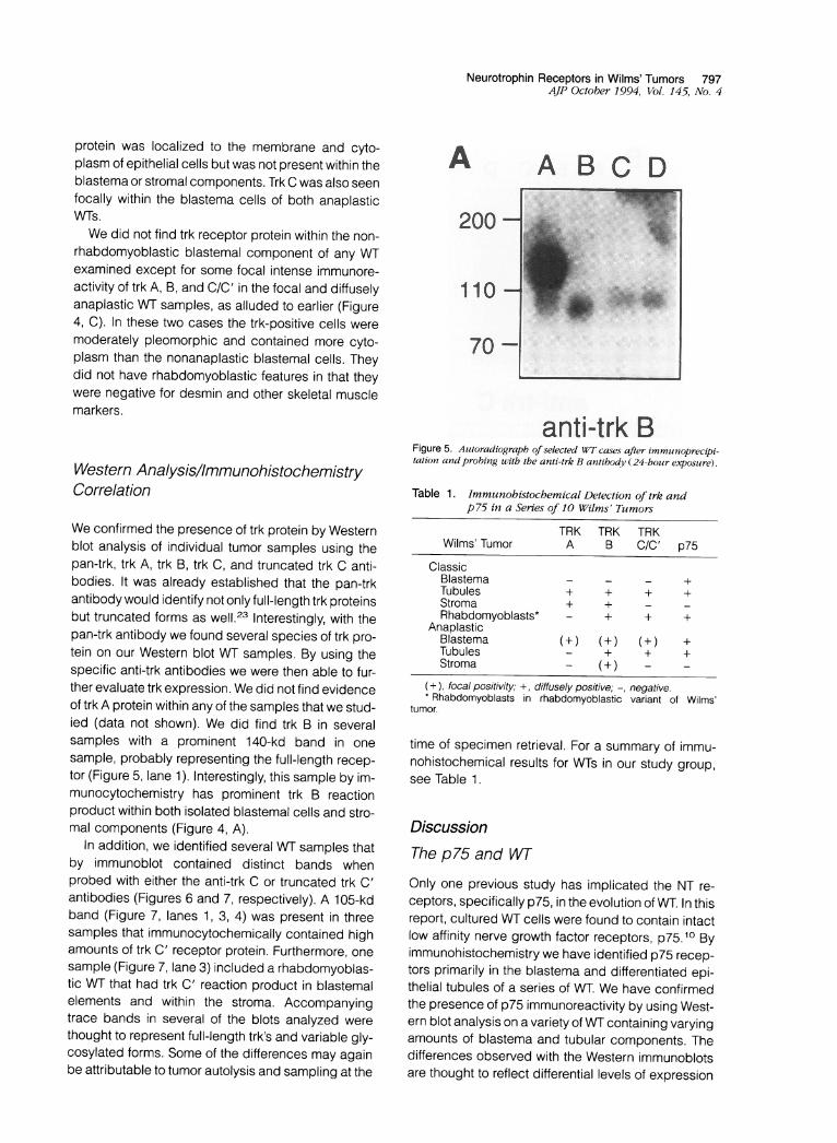
We confirmed the presence of trk protein by Westernblot analysis of individual tumor samples using thepan-trk, trk A, trk B, trk C, and truncated trk C anti-bodies. It was already established that the pan-trkantibody would identify not only full-length trk proteinsbut truncated forms as well.23 Interestingly, with thepan-trk antibody we found several species of trk pro-tein on our Western blot WT samples. By using thespecific anti-trk antibodies we were then able to fur-ther evaluate trk expression. We did not find evidenceof trk A protein within any of the samples that we stud-ied (data not shown). We did find trk B in severalsamples with a prominent 140-kd band in onesample, probably representing the full-length recep-tor (Figure 5, lane 1). Interestingly, this sample by im-munocytochemistry has prominent trk B reactionproduct within both isolated blastemal cells and stro-mal components (Figure 4, A).
In addition, we identified several WT samples thatby immunoblot contained distinct bands whenprobed with either the anti-trk C or truncated trk C'antibodies (Figures 6 and 7, respectively). A 105-kdband (Figure 7, lanes 1, 3, 4) was present in threesamples that immunocytochemically contained highamounts of trk C' receptor protein. Furthermore, onesample (Figure 7, lane 3) included a rhabdomyoblas-tic WT that had trk C' reaction product in blastemalelements and within the stroma. Accompanyingtrace bands in several of the blots analyzed werethought to represent full-length trk's and variable gly-cosylated forms. Some of the differences may againbe attributable to tumor autolysis and sampling at the
A A BC D
200 -
110 -
70-.... ....2.. ~ ~~~~~~-.1111.... .... ....
anti-trk BFigure 5. Autoradiograph of selected WT cases after immunoprecipi-tation and probing uwith the anti-trk B antibody (24-hour exposure).
Table 1. Immunohistochemical Detection of trk andp75 in a Series of 10 Wilms' Tumors
TRK TRK TRKWilms' Tumor A B C/C' p75
ClassicBlastema - - - +Tubules + + + +Stroma + +Rhabdomyoblasts* - + + +
AnaplasticBlastema (±) (+) (+) +Tubules - + + +Stroma - (+) - -
(+), focal positivity; +, diffusely positive; -, negative.*Rhabdomyoblasts in rhabdomyoblastic variant of Wilms'
tumor.
time of specimen retrieval. For a summary of immu-nohistochemical results for WTs in our study group,see Table 1.
DiscussionThe p75 and WT
Only one previous study has implicated the NT re-ceptors, specifically p75, in the evolution of WT. In thisreport, cultured WT cells were found to contain intactlow affinity nerve growth factor receptors, p75.10 Byimmunohistochemistry we have identified p75 recep-tors primarily in the blastema and differentiated epi-thelial tubules of a series of WT. We have confirmedthe presence of p75 immunoreactivity by using West-ern blot analysis on a variety ofWT containing varyingamounts of blastema and tubular components. Thedifferences observed with the Western immunoblotsare thought to reflect differential levels of expression

798 Donovan et alAJP October 1994, Vol. 145, No. 4
B A B C D
200-
110-
70-
anti-trk CFigure 6. Auitoradiograph of selected WT cases after immunoprecipi-tation anid probing with the anti-trk C antibody (18-hour exposuire).
11o0
7O0
Figure 7. Autoradiograph of selected WT cases after immunoprecipi-tation and probing with the anti-trk C (truncated trk C) antibody(24-hour exposure).
by different tumor cell types (blastemal predominantversus epithelial), although we cannot rule out sec-
ondary effects from tumor autolysis. Indeed, the WTsamples that were blastemal predominant containedthe greatest amount of p75 protein.
Previous studies have proposed that during tumorprogression the blastema retains the capacity to dif-ferentiate into the various components of the nephronsimilar to processes occurring during normal kidneymorphogenesis.25 26 The differentiative pathway thatresults in mature kidney is one in which pleuripotentialstem cells progressively lose the ability to divide as
they become further committed toward their final phe-notypes. The factors responsible forthe induction andpromotion of this differentiative response are likely toreflect the actions of a variety of growth factors and
the regulation in expression of these factors is likelyto be a direct consequence of the genetic rearrange-ments characteristic of WT.
Indeed, the identification of a WT gene at 11 p15 inclose proximity to the IGF-2 gene27'28 and the abilityto inhibit WT growth both in vitro and in vivo with an-tibodies against the IGF-1 receptor2 help support thishypothesis. Previously, it was shown that IGF levelsare elevated not only in WT samples examined butalso in normal fetal kidney.8 In situ hybridization ofIGF-2 in normal kidney identified expression primarilyin blastemal cells, whereas the differentiated glomer-ular and tubular elements had reduced levels of ex-pression.29 Interestingly, the same authors found el-evated IGF-2 expression in differentiated epithelialcomponents of a monomorphous WT. They con-cluded that IGF-2 is a marker for differentiation and itsidentification in WT tubules could be a manifestationof unregulated expression.
Recently, both the p75 receptor and the WT-1 genehave been implicated in the early stages of normalkidney development.11,30 The p75 has been identi-fied in S bodies of early condensed mesenchyme(blastema) but was lacking on more differentiated tu-bules.11 Our finding of p75 on mature tubules in WTmay in some way reflect a disregulation of receptorexpression as suggested for IGF-2. In the earlierstudy by Sariola et al, 11 the function of p75 in epithelialdifferentiation and tubular branching was analyzed inorganotypic cultures of embryonic rat kidneys. Theutilization of antisense oligonucleotides to the p75 re-ceptor (thus inhibiting p75 expression) in kidney cul-tures resulted in the inhibition of tubule formation withonly rudimentary branching of the ureteric bud. Theauthors concluded that depletion of p75 specificallyperturbed epithelial differentiation of the nephrons,although this experimental result has not been dupli-cated using similar conditions.14 Additional work30implicated the WT-1 gene in the promotion of blast-emal differentiation because WT-1 transcripts can befound in fetal kidney metanephric blastemal cells andlevels of WT-1 increase as cells divide and differen-tiate. The most recent findings indicate that the WT-1gene is required for the survival and early differen-tiation of the metanephric blastema.31
Because the WT-1 protein has been implicated inthe regulation of the IFG-1 receptor by binding to itspromoter,32 so may the WT-1 protein bind to the pro-moter for p75 and quite possibly the promoter regionsfor the NT and trk receptors as well. Our immunohis-tochemical and Western blot results suggest high ex-pression and misregulation of the p75 receptor. Bycomparison, immunoblots of normal human fetal kid-ney demonstrate only low levels of p75 expression12

Neurotrophin Receptors in Wilms' Tumors 799AJP October 1994, Vol. 145, No. 4
(L. Julie Huber, personal communication). We hypoth-esize that the functional WT-1 protein may serve in asimilar regulatory capacity for the p75 receptor andthus a functionally inactive protein would allow for up-regulation of p75. If our preliminary NT studies areaccurate, then an autocrine loop involving both p75and NT may account for some aspects of tumor de-velopment. We have recently begun studies to verifythe expression of the individual NTs in WT by in situhybridization. Furthermore, the presence of the p75receptor (and trk receptors) on differentiated tubulesallows for competent receptor response to growthfactors in a disregulated setting. We are currently inthe process of testing the association between theWT-1 protein and various NT and receptor promoters.
The mechanism by which the low affinity nervegrowth factor p75 receptor induces cellular changesremains uncertain. There is evidence that p75 con-tributes to the formation of high affinity nerve growthfactor binding sites33 and enhances the specificity ofthe trk family of NT receptors.34 Recent experimentshave suggested that this receptor retains some func-tional similarities to other members of the super familyof receptors that include tumor necrosis factor, Fas(Apo-1), and CD40. In this regard p75 may be in-volved as a constitutive cell death-promoting mole-cule that is inhibited by nerve growth factor bindingwhen p75 is expressed in a central nervous system-derived cell line.35 These results have been inter-preted as implying that some cells become depend-ent for their survival on the binding of nerve growthfactor and this response coincides with an increasedexpression of p75. Our observations of high expres-sion levels for both p75 and NT in WT blastemal cellsand differentiating tubules suggest a similar mecha-nism for tumor progression.
The trk A and B Receptors
By immunohistochemistry we have identified both trkA and B receptor protein localized to cells that makeup the stroma of all WT examined (except the ana-plastic variety). In many cases it appeared that thehighest level of immunoreactivity was present at thestromal-blastemal interface. The functional signifi-cance of this immunoreactivity is unclear. One hy-pothesis is that these focally positive cells within thestroma may be involved in some inductive pathwayinvolving the proliferating/differentiating blastemalcells and the adjacent stroma. Recent studies usingNorthern analyses on embryonic rodent kidney iden-tified only the truncated form of the trk B receptor.14In addition, by in situ hybridization, the authors dem-
onstrated that the transcripts for the trk B receptorswere localized to the more uninduced mesenchymein the upper cortex, which is destined to become thetrue stromal component of the adult kidney. Immu-nohistochemical results may again be supporting thetheory that during the development of a WT there iscontinual recapitulation of normal kidney morphology.Furthermore, our Western immunoblots also demon-strate that of the four samples analyzed, three con-tained truncated trk B receptors and one containedthe full-length receptor for trk B. Again, the samplewith the full-length receptor was a WT that containedboth stromal and isolated blastemal positivity. The full-length receptors are those that contain the tyrosinekinase moiety and are thought capable of respondingto signals eliciting proliferation and/or differentiation.
Evidence suggests that the stromal portion of theWT is derived from mesenchymal stem cells that havethe capacity to differentiate into a variety of cell andtissue types including skeletal muscle, cartilage, andbone.36 The identification of the trk receptors in thismultipotential stroma implies a possible role for thesereceptors in the subsequent differentiation of this tis-sue. Indeed, it has already been shown that trk re-ceptors identified with a pan-trk antibody are presentin differentiating rhabdomyoblasts of rhabdomyosar-comas.18 Furthermore, NT mRNA expression hasbeen identified in muscle cells in the major elasticarteries of the developing rat cardiovascular sys-tem.37 Intriguing to our study, the only examples ofblastemal trk expression are with the anaplastic WTand the two rhabdomyoblastic variants (diagnosedas such because of the large amounts of stromalmuscle differentiation).
Skeletal muscle differentiation within a WT is a well-documented histopathological entity. The skeletalmuscle is believed to arise from the differentiation ofthe mesenchymal or stromal component of the tu-mor.38 On examination of the two rhabdomyoblasticWT samples we found preliminary evidence for dif-ferential receptor localization. The trks B, C, and trun-cated trk C' were found within isolated blastemal cellsand stromal cells (Figure 4, A). These preliminary re-sults suggest that these immunoreactive cells (pos-sibly primitive skeletal muscle cells) may arise fromthe mesenchymally derived blastemal cells. Interest-ingly, we found trk B receptor expression in severalmuscle types including vascular tissues (unpub-lished data) and the p75 receptor was found in fetalskeletal muscle.24 Further studies using in situ hy-bridization and double labeling experiments are re-quired to elucidate the origin of muscle in WT. Thesestudies are currently underway. The anaplastic WT

800 Donovan et alA/P October 1994, Vol. 145, No. 4
also contained trk B receptor protein, and this will bediscussed later.
The trk C and Truncated trk C (C')
Our identification of trk C and C' receptors in maturetubular structures implies a potential role for thesereceptors in epithelial differentiation. Previous experi-ments have already demonstrated preferentially highlevels of trk C and C' mRNA in more differentiatedstructures.14,15 It may be that trk C is necessary forearly differentiation to occur and trk C' may serve toexert a dominant negative effect on the signal trans-duction mechanism. This would be similar to the in-hibition of the wild-type PDGF receptor by the co-expression of a truncated receptor.39 The trk C genewas already shown to be highly expressed in adultstructures of both the brain and extraneural tissues.15Recently, full-length trk C transcripts were identifiedover regions of fully formed collecting ducts in fetalrodent kidney. 14 Our Western immunoblots identifiedboth full-length and truncated receptor proteins onselected WT, including a rhabdomyoblastic type.We identified trk A, B, and C/C' receptor protein
within the nonrhabdomyoblastic blastemal compo-nents in the focal and diffusely anaplastic WTsamples. We also found high levels of p75 receptorprotein present in similar anaplastic blastemal cells.By exhibiting a multireceptor phenotype, we hypoth-esize that these cells are capable of responding to avariety of NT growth factors, giving these cells a dis-tinct advantage in terms of tumor growth. In addition,by containing so many different receptor types, thereis evidence for receptor disregulation in that the cellswill not respond appropriately to differentiative sig-nals, thus allowing for continued tumor progression.We did find evidence of p75 receptor protein on West-ern immunoblots and trk B protein, further supportingour immunohistochemical evidence. Interestingly,many of the anaplastic blastemal cells also containepithelial antigens, suggesting a commitment ofthese cells toward more epithelial structures (tubularcomponents) as opposed to mesenchymal tissues.
In these studies, we have attempted to add the NTand their receptors to the growing list of factors as-sociated with inappropriate cell regulation, differen-tiation, and tumor formation. The immunoreactive pat-terns for trk and p75 imply involvement of thesefactors in the pathobiology of the WT. Furthermore,the presence of the truncated trk C' species in severalof the WT examined may have relevance in the overallfavorable histology and behavior of this tumor. Theevidence of high levels of both p75 and NT in undif-
ferentiated blastemal components lends further sup-port for a misguided autocrine/paracrine signal re-ceptor mechanism in the progression of this tumor.Further studies are underway to characterize both NTand trk receptor function in the pathogenesis of WT.
AcknowledgmentsWe thank Steve Borack for photographic reproduc-tion and Louis Whitney for illustration layout.
References1. Deuel TF: Polypeptide growth factors: roles in normal
and abnormal cell growth. Ann Rev Cell Biol 1987,3:443-492
2. Gansler T, Furlanetto R, Gramling T, Robinson K,Blocker N, Buse M, Sens, Garvin J: Antibody to typeinsulin-like growth factor receptor inhibits growth ofWilms' tumor in culture and athymic mice. Am J Pathol1989, 135:961-966
3. Goustain AS, Leof EB, Shipley GD, Moses HL: Growthfactors and cancer. Cancer Res 1986, 46:1015-1029
4. Beckwith JB, Palmer NF: Histopathology and progno-sis of Wilms tumor. Cancer 1978, 41:1937-1948
5. Frazier G, Bowen-Pope D, Vogel A: Production ofplatelet-derived growth factor by cultured Wilms' tumorcells and fetal kidney cells. J Cell Physiol 1987, 133:169-174
6. Reeve AE, Eccles MR, Wilins RJ, Bell GI, Millow LJ:Expression of insulin-like growth factor-lI transcripts inWilms' tumor. Nature 1985, 317:258-260
7. Haselbacher GK, Irminger J-C, Aapf J, Ziegler WH,Humbel RE: Insulin-like growth factor-lI in human adre-nal pheochromocytomas and Wilms tumors: expres-sion at the mRNA and protein level. Proc Nat Acad SciUSA 1987, 84:1104-1106
8. Werner H, Re G, Drummond I, Sukhatme V, RauscherF, Sens D, Garvin J, LeRoith D, Roberts C: Increasedexpression of the insulin-like growth factor receptorgene, IGF1R, in Wilms' tumor is correlated with modu-lation of IGF1 R promoter activity by the WT1 Wilms tu-mor gene product. Proc Nat Acad Sci USA 1993, 90:5828-5832
9. Wang Z, Madden S, Deuel T, Rauscher F: The Wilms'tumor gene product, WT-1, represses transcription ofthe platelet-derived growth factor A-chain gene. J BiolChem, 1992, 267:21999-22002
10. Thomson T, Pellicer A, Greene L: Functional receptorsfor nerve growth factor on Ewing's sarcoma and Wilmstumor cells. J Cell Physiol 1989, 141:60-64
11. Sariola H, Saarma M, Saino K, Arumae U, Palgi J,Vaahtokari A, Thesleff I, Karavanov A: Dependence ofkidney morphogenesis on the expression of nervegrowth factor receptor. Science 1992, 254:571-573

Neurotrophin Receptors in Wilms' Tumors 801AJP October 1994, Vol. 145, No. 4
12. Alpers C, Hudkins K, Ferguson M, Johnson R, Schat-teman G, Bothwell M: Nerve growth factor receptorexpression in fetal mature and diseased human kid-neys. Lab Invest 1993, 69:703-713
13. Ernfors P, Wetmore C, Erkidsdotter-Nilsson M, Bygde-mann M, Stromberg I, Olson L, Persson H.: The nervegrowth factor receptor gene is expressed in both neu-ronal and non-neuronal tissues in the human fetus. IntJ Dev Neurosci 1991, 9:57-66
14. Durbeej M, Soderstrom S, Ebendal T, Birchmeier, Ek-blom P: Differential expression of neurotrophin recep-tors during renal development. Development 1993,119:977-989
15. Tessarollo L, Tsoulfas P, Martin-Zanca D, Gilbert D,Jenkins N, Copeland N, Parada L: trk C, a receptor forneurotrophin-3, is widely expressed in the developingnervous system and in non-neuronal tissues. Develop-ment 1993, 118:463-475
16. Nakagawara A, Arima M, Azar C, Scavarda NJ, Bro-deur GM: Inverse relationship between trk expressionand n-myc amplification in human neuroblastomas.Cancer Res 1992, 52:1394-1368
17. Nakagawara A, Arima-Nakagawara M, Scavarda NJ,Azar C, Cantor A, Brodeur GM: Association betweenhigh levels of expression of the trk gene and favorableoutcome in human neuroblastoma. N Engl J Med1993, 328:847-854
18. Donovan M, Hempstead B, Horvath C, Chao M,Schofield D: Immunohistochemical localization of trkreceptor protein in pediatric small round blue cell tu-mors. Am J Pathol 1993, 143:1560-1567
19. Klein R, Conway D, Parada LF, Barbacid M: The trk Btyrosine kinase gene codes for a second neurogenicreceptor that lacks the catalytic kinase domain. Cell1990, 61:647-656
20. Tsoulfas P, Soppet D, Escandon E, Tessarollo L,Mendoza-Ramirez J-L, Rosenthal A, Nikolics K, ParadaL: The rat trk C locus encodes multiple neurogenic re-ceptors that exhibit differential response toneurotrophin-3 in PC12 cells. Neuron 1993, 10:975-990
21. Ibanez CF, Ebendal T, Barbany G, Murray-Rust J,Blundell TL, Persson H: Disruption of the low affinityreceptor binding site in NGF allows neuronal survivaland differentiation by binding to the trk gene product.Cell 1992, 69:329-341
22. Huber LJ, Chao MV: Mesenchymal and neuronal cellexpression of the p75 neurotrophin receptor are distin-guished during morphogenesis of transgenic animals.Dev Biol (in press)
23. Horvath C, Wolfen A, Machadeo D, Huber J, Boter L,Benedetti M, Hempstead B, Chao M: Analysis of thetrk NGF receptor tyrosine kinase using recombinantfusion proteins. J Cell Sci 1993, S17:223-228
24. Garin-Chesa P, Rettig W, Thomson T, Old L, MelamedM: Immunohistochemical analysis of nerve growth fac-tor receptor expression normal and malignant humantissues. J Histochem Cytochem 1988, 36:383-389
25. Albeda FW, Molenaar WM, de Leij L, Thijs-lpema AH:Heterogeneity of Wilms' tumour blastema: an immuno-histological study. Virchows Arch 1989, 414:263-271
26. Van Heyningen V, Hastie N: Wilms' tumour: reconcilinggenetics and biology. Trends Genet 1992, 8:16-21
27. Brissenden JE, Ullrich A, Francke U: Human chromo-somal mapping of genes for insulin-like growth factorsand 11 and epidermal growth factor. Nature 1984,
310:781-78428. Tricoli J, Rall LB, Scott J, Bell GI, Shows TB: Localiza-
tion of insulin-like growth factor genes to human chro-mosomes 11 and 12. Nature 1984, 310:784-786
29. Paik S, Rosen N, Jung W, You J, Lippman M, PerdueJ, Yee D: Expression of insulin-like growth factor 11mRNA in fetal kidney and Wilms' tumor. Lab Invest1989, 61 :522-526
30. Prichard-Jones K, Fleming S, Davidson D, BickmoreW, Porteous D, Gosden C, Bard J, Houseman D, vanHeyningen V, Hastie N: The candidate Wilms' tumorgene is involved in genitourinary development. Nature1990, 346:194-197
31. Kreidberg J, Sariola H, Loring J, Maeda M, Pelletier J,Housman D, Jaenisch R: WT-1 is required for earlykidney development. Cell 1993, 74:679-691
32. Werner H, Re G, Drummond I, Sukhatme V, RauscherF, Sens D, Garvin J, LeRoith D, Roberts C: Increasedexpression of the insulin-like growth factor receptorgene, IGF1R, in Wilms tumor is correlated with modu-lation of IGF1 R promoter activity by the WT1 Wilms tu-mor gene product. Proc Natl Acad Sci USA 1993, 90:5828-5832
33. Hempstead BL, Martin-Aanca D, Kaplan D, Parada L,Chao M: High-affinity NGF binding requires coexpres-sion of the trk proto-oncogene and the low-affinityNGF receptor. Nature 1991, 350:678-683
34. lp NY, Stitt TN, Tapley P, Klein R, Glass DJ, Fandl J,Greene LA: Similarities and differences in the wayneurotrophins interact with the trk receptors in neuro-nal and nonneuronal cells. Neuron 1993, 10:137-149
35. Rabizadeh S, Oh J, Li-Tao Z, Yang J, Bitler C, ButcherL, Bredesen D: Induction of apoptosis by the low-affinity NGF receptor. Science 1993, 261:345-348
36. Hazen-Martin D, Garvin J, Gansler T, Tarnowski B,Sens D: Morphology and growth characteristics ofepithelial cells from classic Wilms' tumors. Am JPathol 1993, 142:893-905
37. Scarisbrick IA, Jones EG, Isackson P: Coexpressionof mRNAs for NGF, BDNF, and NT-3 in the cardiovas-cular system of the pre- and postnatal rat. J Neurosci1993, 13:875-893
38. Garvin J, Surrette F, Hintz D, Rudisill M, Sens MA,Sens D: The in vitro growth and characterization of theskeletal muscle component of wilms' tumor. Am JPathol 1985, 121:298-310
39. Ueno H, Colbert H, Escobedo J, Williams L: Inhibitionof PDGF B signal transduction by coexpression of atruncated receptor. Science 1991, 252:844-848