life-threatening infections in children in europe a
TRANSCRIPT

1
Life-threatening infections in children in Europe – a prospective cohort study (The
EUCLIDS Project)
Prof. F. Martinon-Torres1,2*, Prof. A. Salas2,3*, M.D. I. Rivero1,2*, PhD. M. Cebey-
López2*, PhD. J. Pardo-Seco2*, PhD. J. Herberg4*, M.D. N.P. Boeddha5, M.D. D.S.
Klobassa6, MMED. F. Secka7, M.D. S. Paulus8, Prof. R. de Groot9, M.D. L.J.
Schlapbach10,11,12, 13, PhD. G.J. Driessen5, PhD. S.T. Anderson7, PhD. M. Emonts14,15,
Prof. W. Zenz6, Prof. E.D. Carrol8+, PhD. M. Van der Flier 9, Prof. M. Levin4+ on behalf
of EUCLIDS consortium&.
* contributed equally, + contributed equally & EUCLIDS consortium members are detailed in Appendix, page 35
Affiliations: 1. Translational Pediatrics and Infectious Diseases Section- Pediatrics Department,
Santiago de Compostela, Spain.
2. Instituto de Investigación Sanitaria de Santiago (IDIS), Genetics- Vaccines-
Infectious Diseases and Pediatrics research group GENVIP, Santiago de Compostela,
Spain.
3. Unidade de Xenética, Departamento de Anatomía Patolóxica e Ciencias Forenses,
Instituto de Ciencias Forenses, Facultade de Medicina, Universidade de Santiago de
Compostela, and GenPoB Research Group, Instituto de Investigaciones Sanitarias
(IDIS), Hospital Clínico Universitario de Santiago (SERGAS), Galicia, Spain
4. Section of Paediatrics Imperial College London, London, United Kingdom.
5. Erasmus MC-Sophia Children’s Hospital, University Medical Center Rotterdam,
Intensive Care and Department of Pediatric Surgery, Rotterdam, The Netherlands.
6. Medical University of Graz, Department of General Pediatrics, Graz, Austria.
7. Medical Research Council Unit The Gambia, Fajara, The Gambia.
8. University of Liverpool Institute of Infection and Global Health, Department of
Clinical Infection Microbiology and Immunology, Liverpool, United Kingdom.
9. Department of Pediatrics, division of Pediatric Infectious Diseases and Immunology
and Laboratory of Infectious Diseases, Radboud Institute of Molecular Life Sciences,
Radboudumc Nijmegen, the Netherlands
10. Faculty of Medicine, The University of Queensland, Brisbane, Australia
11. Department of Pediatrics, Inselspital, Bern University Hospital, University of Bern,
Switzerland
12. Paediatric Intensive Care Unit, Lady Cilento Children`s Hospital, Brisbane, Australia
13. Paediatric Critical Care Research Group, Mater Research, University of Queensland,
Brisbane, Australia
14. Institute of Cellular Medicine, Newcastle University, Newcastle upon Tyne, United
Kingdom
15. Paediatric Infectious Diseases and Immunology Department, Newcastle upon Tyne
Hospitals Foundation Trust, Great North Children's Hospital, Newcastle upon Tyne,
United Kingdom
Author for correspondence: Federico Martinón-Torres, Hospital Clínico Universitario
de Santiago de Compostela, Choupana s/n, 15706, Santiago de Compostela, A Coruña,
Spain. E-mail: [email protected], tel. +34981955093, fax
+34981950596.

2
Research in context
Evidence before this study
The burden of life-threatening infections on childhood morbidity and mortality persists
in spite of the substantial reduction in vaccine-preventable invasive bacterial infections
since the introduction of conjugate vaccines in childhood, and the availability of
antimicrobial agents.
We carried out comprehensive and focused reviews of the scientific literature published
between 2000 and July 2017, on severe childhood infections. For this purpose, we
searched the PubMed and Medline databases for articles published in English, Spanish
and French up to July 31, 2017. Our search terms included a combination of the following
terms “meningococcal disease”, “bacterial infection”, “sepsis”, “septic shock”,
“children”, “severe focal infection”, “paediatric intensive care”, “microorganism” and
“diagnosis”.
We found that information on the global epidemiology of severe infections in the
paediatric population is scarce and most published studies on sepsis and severe focal
infection are biased towards the paediatric intensive care population (Schlapbach LJ et al.
and Weiss et al).
Added value of this study
Our study highlights the burden of severe childhood infections, drawing on detailed
clinical information from the largest prospective cohort of children with severe infection
in Europe published to date. We demonstrate the continued impact of severe bacterial
infection and mortality caused by vaccine-preventable infections (N. meningitidis and S.
pneumoniae), and by pathogens for which vaccines are urgently required (S. aureus and
Group A streptococcus).
Implications of all the available evidence
Data collection was made possible by a diverse and widely representative EU funded
European network (EUCLIDS Project GA: 279185): 194 hospitals in 9 European
countries, with information on 98 hospitals of 6 of these countries included in this report.
Conclusions from our data are likely to reflect generalised patterns of illness and to be
widely relevant across Europe. Our findings emphasize the current burden of infection
and the need for on going studies of the prevalence and characteristics of serious
infections in childhood, to guide priorization of therapeutic, diagnostic and preventive
measures.
This project has received funding from the European Union’s seventh Framework
program under EC-GA no. 279185 (EUCLIDS).

3
Abstract
Background: Sepsis and severe focal infections (SFI) represent a significant burden of
disease in hospitalized children. To understand the burden of disease and outcome of
childhood infection in Europe, children with life-threatening bacterial infections were
studied in a multi-centre study in six countries in Europe.
Methods: Children aged 1 month-to-18 years old with sepsis or SFI, admitted to 98
European EUCLIDS network hospitals were prospectively recruited during July 2012-
December 2016. Demographic, clinical, microbiological data and outcomes were
collected.
Findings: A total of 2,844 patients were included (53.2% male; median age: 39.1 months).
43.2% of patients (n=1229) had sepsis and 56.8% (n=1615) SFI. Sepsis was diagnosed
predominantly in younger children and SFI in older ones (P-value<0.0001). Main SFI
were pneumonia (n=511, 18%), central nervous system infection (n=469, 16.5%) and
skin and soft tissue infection (n=247, 8.7%). Causal microorganism was identified in
47.8% of children (n=1,359). Most prevalent causative agent was Neisseria meningitidis
(9.1%, n=259) followed by Staphylococcus aureus (7.8%, n=222), Streptococcus
pneumoniae (7.7%, n=219) and Group A streptococcus (5.7%, n=162). Mortality rate was
2.2% (n=57); and 37.6% of patients (n=1,070) required intensive care.
Interpretation: Mortality rate in European children hospitalised due to sepsis or SFI is
low. Burden of disease lies predominantly in children under 5 years and is largely due to
vaccine-preventable infections by meningococcus and pneumococcus. More than a third
of children required intensive care. Despite availability and application of current clinical

4
methods for microbiological diagnosis, the causative organism remained unidentified in
approximately 50% of the patients.
This project has received funding from the European Union’s seventh Framework
program under EC-GA no. 279185 (EUCLIDS).

5
Introduction
The Confidential Enquiry into Maternal and Child Health (CEMACH) report ‘Why
Children Die’ demonstrated that infectious illness was ‘the single largest cause of death
in children dying of an acute physical illness’, constituting ‘20% of the deaths overall’
with the 1-4 year old group the most affected [1]. Amongst all the infectious agents,
bacteria represent the principal cause of death in young children, accounting for over a
third of all child deaths globally [2].
The World Health Organization (WHO) recently issued a resolution on sepsis in all age
groups, recognizing deaths by severe infection as a main target for global and national
prioritization in healthcare delivery [3]. This burden on childhood morbidity and
mortality persists despite of the substantial reduction in vaccine-preventable invasive
bacterial infections after the introduction of conjugate vaccines in childhood and the
availability of antimicrobial agents [4-6], highlighting the need for a better understanding
of the host response to infection, novel treatments of acute infection, new methods to
identify those at risk, and better preventative strategies.
Currently, information regarding the global epidemiology of severe infections in the
paediatric population is scarce. Most published studies on sepsis and severe focal
infection (SFI) are biased towards a predominantly paediatric intensive care unit (PICU)
population. Reported mortality and morbidity from recent large paediatric sepsis and
septic shock studies ranged from 17% to 25% [7, 8].
In this paper, we present data from the European Union Childhood Life-threatening
Infectious Disease Study (EUCLIDS), which aimed to describe the current burden of
severe paediatric infectious diseases, with respect to demographic, clinical,
microbiological data and outcomes, across Europe.

6
Materials and methods
Study design and recruitment criteria
This prospective, multicenter, observational study of children with life-threatening
bacterial infection presenting to hospital was conducted between July 2012-December
2016 by the EUCLIDS Consortium (http://www.euclids-project.eu/). This network
included 194 hospitals in Europe (in 9 countries) and one hospital in Africa (The Gambia).
Data from Switzerland were not included in the analysis because they used different
inclusion criteria. The African partner was also excluded because the present study
focuses on the European burden of disease.
Eligible participants were children from 1 month to 18 years of age admitted to hospital
with sepsis (or suspected sepsis) and/or severe focal infection including but not limited
to pneumonia, soft tissue infection, meningitis, encephalitis, osteomyelitis, and septic
arthritis (Appendix: Full definitions document, page 43). In order to enrol children as
early as possible during the infection, potential recruits were identified from their clinical
characteristics on presentation often before the results from confirmatory microbiology
tests were available. Additionally, children admitted with proven infections due to N.
meningitidis, S. pneumoniae, S. aureus and Group A streptococcus (GAS) who had not
been included in the study on initial presentation to hospital were specifically targeted for
recruitment. For this reason our findings cannot be used to accurately establish the relative
prevalence of other potentially causative pathogens. although recruitment mostly took
place before any causal pathogen was identified. Patients with hospital-acquired
infections were not included.

7
The study used harmonised procedures for patient recruitment, sample processing and
sample storage. A common clinical protocol agreed by EUCLIDS Clinical Network and
approved by the Ethics Comitte was implemented at all hospitals. All clinical staff were
trained in the projects proceedures, and specified criteria were used for clinical definitions
and assignment of patients to diagnostic categories. Written informed consent was
obtained from a parent or legal guardian for each subject before study inclusion.
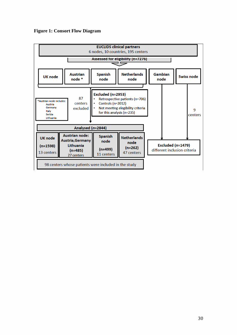
Among 7,276 eligible patients included in the EUCLIDS database, we excluded 2,012
patients labelled as controls, 706 patients recruited retrospecively, 1,479 patients from
the Swiss and Gambian Cohorts, and 235 that did not meet eligibility criteria or were
incomplete (Figure 1). Analysis was limited to the remaining 2,844 subjects with a
complete minimal dataset including patient age and discharge diagnosis.
Clinical data collection
The clinical information for each patient was collected using a secured web-based
platform, including data on demographics, comorbidity, immunisation status, selected
laboratory results, and past medical and family history of severe infectious diseases
defined as: (a) any infection requiring hospitalization, if outpatient at onset; (b) any
infection requiring oxygen, pressors or fluids to support blood pressure, or intubation; or
(c) deep tissue (invasive) infection requiring intravenous or oral antibiotics to treat
infection. Discharge diagnosis, clinical course, treatments and specific procedures during
admission and outcomes (such as death or sequelae) were recorded.
Patients were categorised into two main groups according to the clinical characteristics
during the hospital admission: sepsis or SFI. Sepsis was defined as suspected or
confirmed infection (infectious organisms or toxins) plus systemic inflammatory
response syndrome (SIRS) [9], and SFI included those illnesses with a suspected or

8
confirmed infection but without SIRS. Patients were assigned one or more pre-defined
clinical syndromes. (Appendix: Full definitions document, page 43).
Laboratory methods
Microbiological diagnosis was undertaken as part of clinical care using locally available
clinical diagnostic procedures, including, as appropriate, bacterial culture from normally
sterile sites (blood, cerebrospinal fluid, urine and invasive diagnostic samples), and from
non-sterile sites (throat and wound swabs); bacterial and viral molecular diagnostics were
applied to blood, cerebrospinal fluid and respiratory secretions, according to local
availability.
In order to assign microbiological aetiology of infection in prospective patients recruited
to the study, each patient was phenotyped according to their likelihood of bacterial
infection, using an agreed algorithm, when all the results of investigations were available
(Figure 2).
Specific inflammatory parameters: maximum levels of serum C-reactive protein (CRP)
and neutrophil counts were compared to further assess their utility and sensitivity in
discriminating focal vs. sepsis, PICU vs. non-PICU admission, and prognosis (survivors
vs. death). For CRP values, all cohort values were used; while for neutrophil counts only
UK values were available. Sensitivity and specificity was assessed using pre-agreed cut
offs and numeric values were used to obtain receiver operating characteristic curves
(ROC) Figure 2 [10].
Statistical analysis
General data are presented as percentages and odds ratios (OR) computed from
contigency tables, and medians and interquartile ranges (IQR). Analysis was performed

9
using R version 3.3.1 (www.r-project.org). The level of statistical significance was set at
0.05. Bonferroni correction was used in order to reduce the likelihood of false positive
results caused by multiple testing. Associations were assessed using non-parametric tests:
Fisher’s exact test for discrete variables and Wilcoxon test for continuous variables
(package stats). ROC curves and areas under curve (AUC) were calculated with P-values
to test the null hypothesis that the AUC equals 0.50 (package pROC).
Role of the funding source
This project has received funding from the European Union’s seventh Framework
program under EC-GA no.279185 (EUCLIDS). The sponsor of the study had no role in
study design, data collection, data analysis, data interpretation, or writing of the report.
The corresponding author had full access to all the data in the study and had final
responsibility for the decision to submit for publication.
Results
Characteristics of the EUCLIDS cohort
A total of 2,844 subjects were analysed. 53.2% (1512/2841) were male and the median
age was 39.1 months (IQR=12.4-93.9). Characteristics of the patients are summarised in
Table 1.
A history of previous severe infection was found in 432 (16.9%) cases, whilst 240 cases
had 1st or 2nd order family members with a history of serious infection (11.0%, 240/2174).
Previous infections included meningitis (32.9%, 79/240), pneumonia (20.4%, 49/240),
severe sepsis (11.3%, 27/240) and meningococcemia (7.5%, 18/240). 2.4% of cases
(51/2127) had parental consanguinity and 2.1% (45/2150) had first- or second-degree

10
relatives with an immunodeficiency. Prematurity was present in 9.8% (230/2343) of the
cases. 30.1% (497/1652) of the patients lived with smokers at home (Table 1).
Immunisations were up-to-date according to the local schedules in 93.0% (2240/2409) of
the patients. Nevertheless, we found that 89.5% (204/228) of the meningococcus isolated
and serotyped could be eventually covered by vaccines that were not available or not
included in the immunization calendars implemented in Europe at that time.
Sepsis was diagnosed predominantly in younger children and SFI in older ones (Figure
3A), with significant statistical differences in the age distribution between those in whom
a causative organism was identified and those with no organism identified (Figure 3B,
Table 1).
Most of patients (93.4%, 2282/2444) had a favourable clinical course (no death, skin
grafts, amputations, hearing loss >40dB) with complete recovery from the illness. The
mortality rate was 2.2% (57/2569) in the entire cohort, 0.5% (7/1549) in SFI vs. 4.9%
(50/1020) for sepsis. The cause of death for patients included in the SFI sub-cohort is
specified in Appendix: Cause of death for patients with SFI, page 63.
A total of 37.6% (1070/2844) patients were admitted to PICU of which 62.1% (763/1229)
admissions presented with sepsis.
Microbiological and clinical diagnosis
A total of 44.8% of children (1155/2581) had definite bacterial infection; 5.9%
(152/2581) had definite viral; and 47.9% (1202/2509) suffered from uncertain type of
infection (454 probable bacterial, 65 probable viral and 683 unknown) (Figure 2).

11
A causative microorganism was identified in 47.8% (1359/2844) of the cases. The most
prevalent bacterial causative agent was Neisseria meningitidis in 9.1% (259/2844)
followed by Staphylococcus aureus (7.8%, 222/2844), Streptococcus pneumoniae (7.7%,
219/2844) and GAS (5.7%, 162/2844) (Figure 4). Viruses were identified as causative
agents in 6.5% (185/2844) of the patients with the most common ones being: enterovirus,
rhinovirus and respiratory syncytial virus.
In patients admitted to PICU, the main identified bacteria were: N. meningitidis (16.5%,
162/981), S. pneumoniae, (9.9%, 97/981), GAS (8.1%, 79/981) and S. aureus (5.5%,
54/981). Viruses were the causative pathogen in the 8.1% (79/981) of the cases, and there
was no organism identified in 41.6% (408/981) of the patients. Ward and PICU clinical
syndromes, and causal agents are shown in Appendix Figure 1, page 64.
Significant differences were found in N. meningitidis rates in patients with a family
history of severe bacterial infection [OR: 2.02 (95%CI: 1.31-3.04), P-value=0.0011], and
in patients exposed to tobacco [OR: 3.21 (95%CI: 2.19-4.74), P-value<0.0001]. In
premature patients there is a significant difference for viral infection rates [OR: 2.13
(95%CI: 1.38-3.22), P-value=0.0005].
Those patients in whom a causative organism was identified were more likely to have
severe disease: a higher proportion was admitted to PICU (P-value<0.0001) and had a
prolonged hospital length of stay (LOS) (P-value<0.0001), furthermore, they required
more respiratory support (P-value<0.0001), and supplemental oxygen (P-value<0.0001).
Additionally, inotropes (P-value=0.0122) and mortality were higher in patients with an
identified causative organism (P-value=0.0045) although this was not statistically
significant after Bonferroni adjustment (Table 1A).

12
Among patients with bacterial SFI, the most prevalent clinical syndromes were
pneumonia (20.4%, 329/1615), central nervous system (CNS) infection (12.1%,
196/1615), skin and soft tissue infection (11.5%, 185/1615) and osteomyelitis (9.6%,
155/1615).
No correlation was found between administration of antimicrobial agents before cultures
and organism identification (P-value=0.7813).
Children whose immunisations were not up to date (7.0%, 169/2409) were admitted
mainly due to pneumonia (18.9%, 32/169), CNS infections (15.4%, 26/169) and urinary
tract infections–pyelonephritis (11.8%, 20/169); with S. pneumoniae and Escherichia coli
being the main causative microorganisms (6.5%, 11/169; and 5.9%, 10/169, respectively).
We further analysed the main presenting clinical syndromes according to the presence of
a microorganism. For the main pathologies studied we found that CNS infections were
caused mainly by N. meningitidis (29.9%, 140/469) and S. pneumoniae (19.0%, 89/469);
soft tissue infection, osteomyelitis, toxic shock syndrome and septic arthritis by S. aureus
and GAS, and abdominal conditions and urinary tract infections-pyelonephritis by E. coli.
(Figure 4A)
Infection with N. meningitidis (22.8%, 13/57) was the most prevalent among the fatal
cases, mainly associated with severe sepsis, followed by S. pneumoniae (19.3%, 11/57)
and S. aureus (10.5%, 6/57). In 33.3% (19/57) of the non-survivors no causative pathogen
was identified (Figure 4B).
Sepsis vs. SFI
The main differences observed between patients with sepsis or SFI were that septic
patients had a more severe course, with significant differences for all parameters

13
including full recovery at discharge (P-value<0.0001), need for supplemental oxygen (P-
value<0.0001), respiratory support requirement (P-value<0.0001), inotropes (P-
value<0.0001), PICU admission (P-value<0.0091) and death outcome (P-value<0.0001)
(Table 1B).
Antibiotics had been administrated before blood cultures were taken in 40.0% (355/887)
of septic patients and in 29.8% (359/1204) patients with SFI (P-value<0.0001).
Utility of inflammatory markers
We compared maximum CRP and neutrophil counts levels between different groups
(Table 2). Patients with sepsis and those requiring intensive care, had an increased serum
CRP (≥60 mg/L) compared to those with focal infection and non-PICU admission (P-
value<0.0001). (Appendix Figure 2, page 65). No differences were found when
comparing survivors vs. non-survivors.
ROC analysis for CRP to discriminate sepsis vs. SFI showed an AUC of 0.655 (95%CI
0.616-0.694, P-value<0.0001) and 0.661 (95%CI 0.621-0.701, P-value<0.0001) for
distinguishing between PICU vs. non-PICU admission. The CRP AUC for discriminating
between survivors and death was also significant (0.655, 95%CI 0.535-0.776, P-
value=0.0153) (Appendix Figure 3, page 66).
ROC analysis for neutrophil count to discriminate sepsis vs. SFI showed an AUC of 0.553
(95%CI 0.523-0.583, P-value<0.0001) and 0.550 (95%CI 0.518-0.582, P-value=0.0015)
for discriminating between PICU vs. non-PICU admission. The neutrophil AUC for
discriminating between survivors and death was not significant (0.522, 95%CI 0.390-
0.655, P-value=0.7158) (Appendix Figure 3, page 66).
Discussion

14
Our study highlights the burden of severe childhood infections, drawing on detailed
clinical information from the largest prospective cohort of children with severe infection
in Europe, recruited at 98 hospitals in 6 European countries. We demonstrate the
continued importance of severe illness and mortality caused by vaccine-preventable
infections (N. meningitidis and S. pneumoniae), and by pathogens for which vaccines are
urgently required (S. aureus and GAS).
Laboratory tests failed to identify a causative pathogen in over half of children with severe
illness, in line with data from the previous two decades [8, 11], despite the introduction
of more sensitive and precise techniques in diagnostics in recent years. In over 50% of
paediatric patients admitted with suspected life-threatening infections, decisions on need,
type and duration of antimicrobial therapy thus have to be made with no clear guidance
from the microbiological findings, indicating an urgent need for improved diagnostics.
Patients with an identified microorganism suffered from more severe disease, which may
suggest a higher pathogen load and more successful detection in these patients, but may
be associated as well to increased diagnostic effort in the sickest patients.
Mortality
In our study, the case fatality ratio was 2.2%, significantly lower than that recently
reported by two recent large studies [7, 8], although it should be noted that these studies
were restricted to PICU patients with a more severe population (sepsis/septic shock).
Mortality was highest in children with sepsis as defined by the International Paediatric
Sepsis consensus conference [9]. The new sepsis definitions from 2016 [12] were not
established for children, hence were not used in our study. Delay in timely treatment has
been considered to increase the mortality risk in sepsis [6, 13]. Esteban et al. [14] reported
a trend towards reduction in mortality after implementing an educational intervention for

15
appropriate empiric antibiotic administration within the first hour of admission in children
with sepsis. However, we were not able to assess this in our data. Our results are
consistent with the reported mortality rates of patients with sepsis after the introduction
of adequate treatment guidelines (hospital mortality 1%–3% in previously healthy, and
7%–10% in chronically ill children) [15], and with a recent population-based study on
blood culture-proven bacterial sepsis [16]. As previously described [15], we found that
mortality in community-acquired severe infections [6] was associated with the
identification of the causative organism, the presence of sepsis, higher PICU admission
rates, oxygen and/or respiratory support requirement, inotrope administration and
prolonged LOS.
Severity and pathogen type
Though our study was not designed to reliably establish the relative prevalence of
potentially causative pathogens; our results show the relative frequency of N. meningitidis,
S. pneumoniae, S. aureus and GAS are roughly equal. Overall, the most frequent clinical
syndromes were meningitis and pneumonia. Almost half of the patients admitted to
hospital with a bacterial infection required intensive care admission. These findings are
consistent with the reported leading causes of morbidity and mortality in children
worldwide [1, 2]. The causative pathogens in our study differed from findings in Asia:
were Salmonella enterica serotype Typhi was the most common bacterial pathogen,
followed by S. pneumoniae and Haemophilus influenzae [17] and Africa: were S.
pneumoniae is the most common isolate in children, followed by S.aureus and E. coli
[18]. We also observed differences form studies in the United States were S. aureus,
Pseudomonas species and Enterobacteriacae (mainly E.coli) were the main pathogens
isolated. [19]

16
Vaccinations are an essential tool in our fight against infectious disease [4, 20, 21], and
they have greatly reduced the global burden of infectious disease [21]. Although most
patients were up-to-date according to their local immunisation schedule, we found that
there was a considerable burden of mortality and morbidity caused by vaccine preventable
infections, particularly meningococcal and pneumococcal disease. Vaccines for
meningococcal serogroup B, Y, W and for a major proportion of pneumococcal serotypes
are not available or implemented in Europe. Thus, improved vaccines and implementation
of current vaccines may yield further health gain. Wider implementation of existing
vaccines and development of vaccines for S. aureus or GAS could contribute to a further
decline in the burden of paediatric infectious diseases.
Tobacco smoke exposure
We found an increased risk of meningococcal infections in children exposed to tobacco
(P-value<0.0001). In previous studies tobacco smoke exposure was associated with
increased susceptibility to infections including tuberculosis, pneumonia, meningitis or
otitis media. This could be explained by increased nasopharyngeal colonization with
pathogens including S. pneumoniae, H. influenzae (non-type b), M. catarrhalis, GAS and
S. aureus [22]. Furthermore, increased infection risk can also be explained by the reported
interference of tobacco smoke with the antibacterial function of leukocytes (e.g.
neutrophils, monocytes, T cells and B cells) [23].
Family history
The huge variation in clinical response to identical infecting pathogens is most likely the
result of the combined effects of genetic variation in both the infecting pathogen and the
infected host [24]. There is now strong evidence that host genetic factors influence
occurrence of meningococcal disease, and a number of genes controlling susceptibility

17
and severity of meningococcal disease have been identified in candidate gene association
studies [25-28]. We found a significant association between family history of severe
infectious diseases and meningococcal infection (P-value=0.0011),
Inflammatory biomarkers
In our study, higher CRP levels were associated with an increased risk of severe outcomes.
Biomarkers may contribute to outcome prediction in life threatening infections [29].
However, there is still a need for improved host biomarker and pathogen diagnostics that
can establish the clinical diagnosis, direct appropriate therapy and enable prediction of
outcome [10]. Improved diagnostic discrimination in this group could have major
implications for tackling rising antimicrobial resistance.
Sepsis outcomes for children in high-income countries have not changed dramatically
over the past decade [6-8, 13]. Additional diagnostic approaches may help to establish
the clinical diagnosis, direct early and adequate therapy and enable a more reliable
outcome prognosis. It has been proposed that an approach combining sensitive pathogen
diagnostics and novel host response biomarkers may improve treatment and clinical
outcomes for children with serious infection [10]
PIRO concept
We identified specific variables associated with each of the components of PIRO concept
[30] (predisposition, infection characteristics, host response and organ dysfunction):
including age, gender, family history of severe infection, tabacco exposure, type of
microorganism, infection focus, inflammatory biomarkers and a dynamic view of the
patient’s clinical course and outcomes. All of these variables described, contribute as a

18
proof of concept of this novel approach and as a predictor of mortality for patients with
community-acquired sepsis.
Limitations
Although children were recruited early in their clinical course, before a pathogen
diagnosis was known, a limitation of our study is that children with known infection due
to N. meningitidis, S. pneumoniae, S. aureus, and GAS were targeted for recruitment. The
reason behind this targeted recruitment was to study the genetic basis of these pathogens
as one of the main objectives of EUCLIDS Project (GA 279185). Overall the majority of
patients were recruited unbiased, providing a cross section of different etiologies in
children presenting with severe infection. But specific targeting of the four core
pathogens will have caused bias towards these infections. The burden of disease from
these selectively-target pathogens is within this study cohort but our study design limits
the ability to generalize this to the broader population. Our findings thus cannot be used
to establish population prevalence of each organism. Of note, one of EUCLIDS centres
(Switzerland), where the recruitment strategy was to enroll children at a later time point,
solely after confirmation of positive blood culture, were not included in this paper. In
order to determine the disease burden and to elucidate the contributing factors to severe
infection outcome, large epidemiological studies are needed. Recruitment was restricted
to the hospital setting and did not capture out-of-hospital deaths, or severe focal infections
managed as outpatients; our data therefore under-represent less severe infections.
Eventhough the study used harmonised procedures for patient recruitment, sample
processing, and sample storage, microbiological diagnosis was undertaken as part of
clinical care using locally available clinical diagnostic procedures which could have
limited in some way the assignment of patients as having viral infection, bacterial

19
infection or co infection. We will report separately on additional viral studies undertaken
as part of EUCLIDS using molecular diagnosis for a wide range of viruses.
Conclusions
This is the largest reported prospective study of severe childhood infections in Europe.
Data collection was made possible by a diverse and widely representative European
network. Recommendations or interventions based on our data are likely to reflect
generalised patterns of illness and to be widely relevant across Europe. Although the
mortality rate due to sepsis or SFI was low, we found considerable morbidity associated
with severe childhood infection and more than a third of children required PICU
admission. The burden of disease lies predominantly in children under 5 years and was
predominantly caused by infections where vaccines are available: pneumococcus and
meningococcus. We found that children had infections by common pathogens such as S.
aureus (7.8%) and GAS (5.7%) for which there are no effective vaccines, and that 11.0%
of the bacterial microorganisms were Gram-negative bacilli. Both of which, should have
important implications for vaccine development and for empirical antimicrobial strategies
implementation in Europe.
Contributions
FMT, JH, EDC, MVdF, ME, RdG, WZ and ML designed the study.

20
IRC, MCL, JH, NPB, DK, FS, SP, MVdF, LJS, GJD, STA, ME, EDC assisted in patient
recruitment, data- and sample collection
FMT, IRC, MCL, JPS, AS performed statistical analysis.
PA, LC, SG provided database and informatics support.
FMT, IRC, MCL, EDC and JH wrote the first draft of the manuscript.
FMT, IRC, MCL, JPS, AS, JH, NPB, DK, FS, SP, MVdF, LJS, ME, WZ, EDC, RdG and
ML contributed to writing the manuscript.
All authors approved the final manuscript.

21
Conflicts of interest
Other than the grants, we declare that we have no conflicts of interest regarding this paper.
Acknowledgements
This project has received funding from the European Union’s seventh Framework
program under EC-GA no. 279185 (EUCLIDS).
This study received support from the Instituto de Salud Carlos III (Proyecto
de Investigación en Salud, Acción Estratégica en Salud):
project GePEM ISCIII/PI16/01478/Cofinanciado FEDER), and project
ReSVinext ISCIII/PI16/01569/Cofinanciado FEDER; Consellería de Sanidade, Xunta de
Galicia (RHI07/2-intensificación actividad investigadora, PS09749
and 10PXIB918184PR), Instituto de Salud Carlos III (Intensificación de la
actividad investigadora 2007–2012, PI16/01569), Fondo de Investigación Sanitaria
(FIS; PI070069/PI1000540) del plan nacional de I+D+I and ‘fondos FEDER’, and 2016-
PG071 Consolidación e Estructuración REDES 2016GI-1344 G3VIP (Grupo Gallego de
Genética Vacunas Infecciones y Pediatría, ED341D R2016/021).
This study was funded by grants from the Swiss National Science Foundation
(342730_153158/1), the Swiss Society of Intensive Care, the Bangerter Foundation, the
Vinetum and Borer Foundation, and the Foundation for the Health of Children and
Adolescents.
This study was funded by grant Abt.08 -16.K-8/2012-20 of the Department for Science
and Research of the Styrian federal government (Austria) and a ESPID grant 2011 for
"Endowed professorship for paediatric infectious diseases paying particular attention to

22
meningococcal disease at the Department of General Pediatrics of the Medical University
of Graz".
The Research was supported by the National Institute for Health Research Newcastle
Biomedical Research Centre based at Newcastle Hospitals NHS Foundation Trust and
Newcastle University. The views expressed are those of the author(s) and not necessarily
those of the NHS, the NIHR or the Department of Health.
23
Bibliography
1. Pearson, G.A., M. Ward-Platt, and D. Kelly, How children die: classifying child
deaths. Arch Dis Child, 2011. 96(10): p. 922-6.
2. Liu, L., et al., Global, regional, and national causes of child mortality in 2000–
13, with projections to inform post-2015 priorities: an updated systematic
analysis. The Lancet, 2015. 385(9966): p. 430-440.
3. WHO, World Health Organization, Improving the prevention, diagnosis and
clinical management of sepsis. Report by the Secretariat. Available at:
http://apps.who.int/gb/ebwha/pdf_files/EB140/B140_12-en.pdf, 9 January
2017.
4. Martin, N.G., et al., Hospital admission rates for meningitis and septicaemia
caused by Haemophilus influenzae, Neisseria meningitidis, and Streptococcus
pneumoniae in children in England over five decades: a population-based
observational study. The Lancet Infectious Diseases, 2014. 14(5): p. 397-405.
5. Irwin, A.D., et al., Etiology of childhood bacteremia and timely antibiotics
administration in the emergency department. Pediatrics, 2015. 135(4): p. 635-42.
6. Maat, M., et al., Improved survival of children with sepsis and purpura: effects of
age, gender, and era. Crit Care, 2007. 11(5): p. R112.
7. Schlapbach, L.J., et al., Mortality related to invasive infections, sepsis, and septic
shock in critically ill children in Australia and New Zealand, 2002–13: a
multicentre retrospective cohort study. The Lancet Infectious Diseases, 2015.
15(1): p. 46-54.

24
8. Weiss, S.L., et al., Global epidemiology of pediatric severe sepsis: the sepsis
prevalence, outcomes, and therapies study. Am J Respir Crit Care Med, 2015.
191(10): p. 1147-57.
9. Goldstein, B., et al., International pediatric sepsis consensus conference:
definitions for sepsis and organ dysfunction in pediatrics. Pediatr Crit Care Med,
2005. 6(1): p. 2-8.
10. Herberg, J.A., et al., Diagnostic Test Accuracy of a 2-Transcript Host RNA
Signature for Discriminating Bacterial vs Viral Infection in Febrile Children.
JAMA, 2016. 316(8): p. 835-45.
11. Bleeker-Rovers, C.P., et al., A prospective multicenter study on fever of unknown
origin: the yield of a structured diagnostic protocol. Medicine (Baltimore), 2007.
86(1): p. 26-38.
12. Singer, M., et al., The Third International Consensus Definitions for Sepsis and
Septic Shock (Sepsis-3). JAMA, 2016. 315(8): p. 801-10.
13. Ruth, A., et al., Pediatric severe sepsis: current trends and outcomes from the
Pediatric Health Information Systems database. Pediatr Crit Care Med, 2014.
15(9): p. 828-38.
14. Esteban, E., et al., A multifaceted educational intervention shortened time to
antibiotic administration in children with severe sepsis and septic shock: ABISS
Edusepsis pediatric study. Intensive Care Med, 2017. 43(12): p. 1916-1918.

25
15. Brierley, J., et al., Clinical practice parameters for hemodynamic support of
pediatric and neonatal septic shock: 2007 update from the American College of
Critical Care Medicine. Crit Care Med, 2009. 37(2): p. 666-88.
16. Agyeman, P.K.A., et al., Epidemiology of blood culture-proven bacterial sepsis
in children in Switzerland: a population-based cohort study. The Lancet Child &
Adolescent Health, 2017. 1(2): p. 124-133.
17. Deen, J., et al., Community-acquired bacterial bloodstream infections in
developing countries in south and southeast Asia: a systematic review. The Lancet
Infectious Diseases, 2012. 12(6): p. 480-487.
18. Reddy, E.A., A.V. Shaw, and J.A. Crump, Community-acquired bloodstream
infections in Africa: a systematic review and meta-analysis. The Lancet Infectious
Diseases, 2010. 10(6): p. 417-432.
19. Mayr, F.B., S. Yende, and D.C. Angus, Epidemiology of severe sepsis. Virulence,
2014. 5(1): p. 4-11.
20. Andre, F.E., et al., Vaccination greatly reduces disease, disability, death and
inequity worldwide. Bulletin of the World Health Organization, 2008. 86(2): p.
140-146.
21. Rivero-Calle, I., et al., The Burden of Pediatric Invasive Meningococcal Disease
in Spain (2008-2013). Pediatr Infect Dis J, 2016. 35(4): p. 407-13.
22. Brook, I. and A.E. Gober, Recovery of potential pathogens and interfering
bacteria in the nasopharynx of smokers and nonsmokers. Chest, 2005. 127(6): p.
2072-5.

26
23. Bagaitkar, J., D.R. Demuth, and D.A. Scott, Tobacco use increases susceptibility
to bacterial infection. Tob Induc Dis, 2008. 4: p. 12.
24. Burgner, D., S.E. Jamieson, and J.M. Blackwell, Genetic susceptibility to
infectious diseases: big is beautiful, but will bigger be even better? The Lancet
Infectious Diseases, 2006. 6(10): p. 653-663.
25. Martinon-Torres, F., et al., Natural resistance to Meningococcal Disease related
to CFH loci: Meta-analysis of genome-wide association studies. Sci Rep, 2016.
6: p. 35842.
26. Wright, V., M. Hibberd, and M. Levin, Genetic polymorphisms in host response
to meningococcal infection: the role of susceptibility and severity genes. Vaccine,
2009. 27 Suppl 2: p. B90-102.
27. Emonts, M., et al., Host genetic determinants of Neisseria meningitidis infections.
The Lancet Infectious Diseases, 2003. 3(9): p. 565-577.
28. Emonts, M., et al., Polymorphisms in PARP, IL1B, IL4, IL10, C1INH, DEFB1,
and DEFA4 in meningococcal disease in three populations. Shock, 2010. 34(1):
p. 17-22.
29. Van den Bruel, A., et al., Diagnostic value of laboratory tests in identifying
serious infections in febrile children: systematic review. BMJ, 2011. 342: p.
d3082.
30. Vila Perez, D., et al., Prognostic factors in pediatric sepsis study, from the Spanish
Society of Pediatric Intensive Care. Pediatr Infect Dis J, 2014. 33(2): p. 152-7.

27
Table 1: Description of the main characteristics of the EUCLIDS study cohort.
Comparision between (A) no organism and organism identified, and (B) focal infection
and sepsis. Data are expressed as % (n) or median (IQR). * P-values lower than
Bonferroni correction threshold (0.05/37=0.0014).
A)
Variables All patients No organism
identified
Organism
identified
P-value
Total cohort 2844 52.2% (1485/2484) 47.8% (1359/2844)
Demographic characteristics
Sex (male) 53.2% (1512/2841) 53.9% (800/1484) 52.5% (712/1357) 0.4517
Age 39.1 (12.4-93.9) 42.8 (14.9-95.5) 33.2 (10.25-91.05) 0.0007* 0-12 months 24.3% (691/2844) 21.1% (313/1485) 27.8% (378/1359) 0.0005*
12-24 months 14.8% (421/2844) 14.5% (215/1485) 15.2% (206/1359) –
24-48 months 17.1% (487/2844) 18.3% (272/1485) 15.8% (215/1359) – >48 months 43.8% (1245/2844) 46.1% (685/1485) 41.2% (560/1359) –
Weight (kg) 14.8 (9.9-25.8) 15.4 (10.3-26.5) 14.0 (9.2-25.5) 0.0005*
Family history Severe infections 11.0% (240/2174) 10.1% (115/1143) 12.1% (125/1031) 0.1319 Immunodeficiency 2.1% (45/2150) 2.1% (24/1133) 2.1% (21/1017) 1.0000
Consanguinity 2.4% (51/2127) 2.6% (29/1122) 2.2% (22/1005) 0.5734
Smoker in the household 30.1% (497/1652) 28.3% (250/883) 32.1% (247/769) 0.0957
Patient medical history Premature birth 9.8% (230/2343) 9.9% (123/1244) 9.7% (107/1099) 0.9446 Past severe infections 16.9% (432/2563) 18.9% (252/1336) 14.7% (180/1227) 0.0051
Immunisations up-to-date 93.0% (2240/2409) 93.5% (1194/1277) 92.4% (1046/1132) 0.2998
Clinical data Antibiotics before culture 34.1% (714/2091) 34.4% (393/1142) 33.8% (321/949) 0.7813
PRISM Score 11 (5-20) 10.5 (4-16) 12.0 (5.0-21) 0.1097
Full recovery expected 93.4% (2282/2444) 95.7% (1219/1274) 90.9% (1063/1170) <0.0001*
PICU admission 37.6% (1070/2844) 30.0% (445/1485) 46.0% (625/1359) <0.0001*
Oxygen needed 36.3% (923/2546) 32.0% (426/1333) 41.0% (497/1213) <0.0001* Respiratory support 28.1% (720/2564) 23.3% (313/1345) 33.4% (407/1219) <0.001*
Inotropes 11.8% (304/2578) 10.3% (138/1346) 13.5% (166/1232) 0.0122
Hospital LOS 7 (4-13) 6 (3-10) 10 (6-16) <0.0001* Death 2.2% (57/2569) 1.4% (19/1345) 3.1% (38/1224) 0.0045
Clinical syndrome
CLABSI 0.5% (13/2844) 0.1% (2/1485) 0.8% (11/1359) 0.0099
CNS infection 16.5% (469/2844) 8.8% (130/1485) 24.9% (339/1359) <0.0001* Bronchiolitis 2.7% (78/2844) 2.1% (31/1485) 3.5% (47/1359) 0.0287
Pneumonia 18.0% (511/2844) 22.5% (334/1485) 13.0% (177/1359) <0.0001*
LRTI 3.5% (100/2844) 4.7% (70/1485) 2.2% (30/1359) 0.0003* Lung effusion or empyema 7.4% (210/2844) 6.3% (94/1485) 8.5% (116/1359) 0.0261
Soft tissue infection 8.7% (247/2844) 9.2% (136/1485) 8.2% (111/1359) 0.3518
Toxic shock syndrome 2.3% (64/2844) 1.1% (16/1485) 3.5% (48/1359) <0.0001* Endocarditis 0.7% (20/2844) 0.1% (2/1485) 1.3% (18/1359) 0.0001*
Osteomyelitis 6.7% (191/2844) 5.2% (77/1485) 8.4% (114/1359) 0.0010
Scarlet fever 0.3% (9/2844) 0.3% (5/1485) 0.3% (4/1359) 1.0000 Septic arthritis 5.2% (149/2844) 3.4% (50/1485) 7.3% (99/1359) <0.0001*
Gastroenteritis 1.6% (45/2844) 1.3% (19/1485) 1.9% (26/1359) 0.1800
UTI–pyelonephritis 3.8% (109/2844) 2.6% (39/1485) 5.2% (70/1359) 0.0006* ENT 6.3% (178/2844) 7.8% (116/1485) 4.6% (62/1359) 0.0003*
Abdominal condition 1.3% (38/2844) 1.5% (22/1485) 1.2% (16/1359) 0.5166
Severe sepsis 5.5% (157/2844) 3.6% (54/1485) 7.6% (103/1359) <0.0001* Septic shock 9.3% (264/2844) 6.2% (92/1485) 12.7% (172/1359) <0.0001*

28
B)
Variables Focal infection Sepsis P-value
Total cohort 56.8% (1615/2844) 43.2% (1229/2844)
Demographic characteristics
Sex (male) 53.5% (863/1612) 52.8% (649/1229) 0.7045 Age 46.5 (15.8-100.4) 27.6 (9.0-80.2) <0.0001*
0-12 months 19.8% (319/1615) 30.3% (372/1229) 0.0005*
12-24 months 13.6% (220/1615) 16.4% (201/1229) – 24-48 months 18.1% (293/1615) 15.8% (194/1229) –
>48 months 48.5% (783/1615) 37.6% (462/1229) –
Weight (kg) 15.8 (10.7-28.0) 13.0 (8.7-23.1) <0.0001*
Family history Severe infections 11.2% (137/1220) 10.8% (103/954) 0.7828 Immunodeficiency 2.1% (25/1211) 2.1% (20/939) 1.0000
Consanguinity 1.9% (22/1186) 3.1% (29/941) 0.0859
Smoker in the household 31.7% (301/951) 28% (196/701) 0.1155
Patient medical history Premature birth 8.5% (112/1316) 11.5% (118/1027) 0.0173 Past severe infections 18.7% (270/1441) 14.4% (162/1122) 0.0041
Immunisations up-to-date 93.2% (1282/1375) 92.6% (958/1034) 0.5740
Clinical data Antibiotics before culture 29.8% (359/1204) 40% (355/887) <0.0001* PRISM Score 5 (4-11.75) 14 (6-22) <0.0001*
Full recovery expected 97.2% (1369/1409) 88.2% (913/1035) <0.0001*
PICU admission 19.0% (307/1615) 62.1% (763/1229) <0.0001* Oxygen needed 22.1% (323/1463) 55.4% (600/1083) <0.0001*
Respiratory support 11.8% (172/1454) 49.4% (548/1110) <0.0001*
Inotropes 10.3% (151/1472) 44.4% (498/1121) <0.0001* Hospital LOS 6 (3-12) 9 (5-15) <0.0001*
Death 0.5% (7/1549) 4.9% (50/1020) <0.0001*
Clinical syndrome CLABSI 0.1% (1/1615) 1.0% (12/1229) 0.0001*
CNS infection 12.1% (196/1615) 22.2% (273/1229) <0.0001* Bronchiolitis 2.7% (44/1615) 2.8% (34/1229) 1.0000
Pneumonia 20.4% (329/1615) 14.8% (182/1229) <0.0001*
LRTI 4.3% (69/1615) 2.5% (31/1229) 0.0134 Lung effusion or empyema 8.4% (136/1615) 6.0% (74/1229) 0.0168
Soft tissue infection 11.5% (185/1615) 5.0% (62/1229) <0.0001*
Toxic shock syndrome 0.3% (5/1615) 4.8% (59/1229) <0.0001* Endocarditis 0.2% (4/1615) 1.3% (16/1229) 0.0011
Osteomyelitis 9.6% (155/1615) 2.9% (36/1229) <0.0001*
Scarlet fever 0.4% (7/1615) 0.2% (2/1229) 0.3150 Septic arthritis 7.5% (121/1615) 2.3% (28/1229) <0.0001*
Gastroenteritis 1.9% (31/1615) 1.1% (14/1229) 0.1285
UTI–pyelonephritis 4.0% (64/1615) 3.7% (45/1229) 0.6947 ENT 9.0% (145/1615) 2.7% (33/1229) <0.0001*
Abdominal condition 1.4% (22/1615) 1.3% (16/1229) 1.0000
Severe sepsis 0% (0/1615) 12.8% (157/1229) <0.0001* Septic shock 0% (0/1615) 21.5% (264/1229) <0.0001*

29
Table 2: Description of serum levels of C-reactive protein (CRP) and neutrophil
counts in different group of patients. Data are expressed as % (n). * P-values lower
than Bonferroni correction threshold (0.05/4=0.0125). SFI: Severe focal infection; PICU:
paediatric intensive care unit.
CRP≥60 mg/L CRP<60 mg/L P-value Neutrophilis≥12109/L Neutrophils<12109/L P-value
Total 39.7 (966/2432) 60.3 (1466/2432) 68.2 (977/1432) 31.8 (455/1432)
Sepsis vs. focal
Sepsis 71.6 (755/1054) 28.4 (299/1054) <0.0001* 35.8 (226/631) 64.2 (405/631) 0.0042*
SFI 51.6 (711/1378) 48.4 (667/1378) 28.6 (229/801) 71.4 (572/801)
PICU vs. not PICU
PICU 70.9 (654/922) 29.1 (268/922) <0.0001* 36.3 (190/524) 63.7 (334/524) 0.0067*
Non-PICU 53.8 (812/1510) 46.2 (698/1510) 29.2 (265/908) 70.8 (643/908)
Survivors vs. death
Survivors 59.5 (1273/2139) 40.5 (866/2139) 0.0878 32.3 (397/1230) 67.7 (833/1230) 0.5039
Death 72.7 (32/44) 27.3 (12/44) 39.1 (9/23) 60.9 (14/23)
30
Figure 1: Consort Flow Diagram

31
Figure 2: Phenotyping algorithm. Figure adapted from Herberg et al. [10]

32
Figure 3: A) Age distribution in the EUCLIDS cohort and in those diagnosed with
sepsis or a focal illness. B) Age distribution by causative organism. GPC: gram
positive cocci, GAS: Group A Streptococcus, GNR: gram negative rods, CoNS:
Coagulase Negative Staphylococci.

33
Figure 4. Causative microorganisms identified in EUCLIDS by syndrome. (A)
patients with severe focal infections and (B) sepsis. Data are expressed as (n) %. CNS
infection: central nervous system infection, LRTI: lower respiratory tract infection, ENT
syndrome: ear, nose, throat syndrome, UTI-pyelonephritis: urinary tract infection with
pyelonephritis, GPC: gram positive cocci, GAS: Group A Streptococcus; GNR: gram
negative rods, CoNS: Coagulase Negative Staphylococci.

34
Appendix: EUCLIDS CONSORTIUM MEMBERS
EUCLIDS consortium (www.euclids-project.eu) is composed by:
Imperial College partner (UK)
Members of the EUCLIDS Consortium at Imperial College London (UK)
Principal and co-investigators
Michael Levin (grant application, EUCLIDS Coordinator)
Dr. Lachlan Coin (bioinformatics)
Stuart Gormley (clinical coordination)
Shea Hamilton (proteomics)
Jethro Herberg (grant application, PI)
Bernardo Hourmat (project management)
Clive Hoggart (statistical genomics)
Myrsini Kaforou (bioinformatics)
Vanessa Sancho-Shimizu (genetics)
Victoria Wright (grant application, scientific coordination)
Consortium members at Imperial College
Amina Abdulla
Paul Agapow
Maeve Bartlett
Evangelos Bellos
Hariklia Eleftherohorinou
Rachel Galassini
David Inwald
Meg Mashbat
Stefanie Menikou
Sobia Mustafa
Simon Nadel
Rahmeen Rahman
Clare Thakker
EUCLIDS UK Clinical Network
Poole Hospital NHS Foundation Trust, Poole: Dr S Bokhandi (PI), Sue Power, Heather
Barham
Cambridge University Hospitals NHS Trust, Cambridge: Dr N Pathan (PI), Jenna Ridout,
Deborah White, Sarah Thurston
University Hospital Southampton, Southampton: Prof S Faust (PI), Dr S Patel (co-
investigator), Jenni McCorkell.
Nottingham University Hospital NHS Trust: Dr P Davies (PI), Lindsey Crate, Helen
Navarra, Stephanie Carter
University Hospitals of Leicester NHS Trust, Leicester: Dr R Ramaiah (PI), Rekha Patel

35
Portsmouth Hospitals NHS Trust, London: Dr Catherine Tuffrey (PI), Andrew Gribbin,
Sharon McCready
Great Ormond Street Hospital, London: Dr Mark Peters (PI), Katie Hardy, Fran Standing,
Lauren O’Neill, Eugenia Abelake
King’s College Hospital NHS Foundation Trust, London; Dr Akash Deep (PI), Eniola
Nsirim
Oxford University Hospitals NHS Foundation Trust, Oxford Prof A Pollard (PI), Louise
Willis, Zoe Young
Kettering General Hospital NHS Foundation Trust, Kettering: Dr C Royad (PI), Sonia
White
Central Manchester NHS Trust, Manchester: Dr PM Fortune (PI), Phil Hudnott
SERGAS Partner (Spain)
Principal Investigators
Federico Martinón-Torres1
Antonio Salas1,2
GENVIP RESEARCH GROUP (in alphabetical order):
Fernando Álvez González1, Ruth Barral-Arca1,2, Miriam Cebey-López1, María José
Curras-Tuala1,2, Natalia García1, Luisa García Vicente1, Alberto Gómez-Carballa1,2, Jose
Gómez Rial1, Andrea Grela Beiroa1, Antonio Justicia Grande1, Pilar Leboráns Iglesias1,
Alba Elena Martínez Santos1, Federico Martinón-Torres1, Nazareth Martinón-Torres1,
José María Martinón Sánchez1, Beatriz Morillo Gutiérrez1, Belén Mosquera Pérez1, Pablo
Obando Pacheco1, Jacobo Pardo-Seco1,2, Sara Pischedda1,2, Irene Rivero Calle1, Carmen
Rodríguez-Tenreiro1, Lorenzo Redondo-Collazo1, Antonio Salas Ellacuriaga1,2, Sonia
Serén Fernández1, María del Sol Porto Silva1, Ana Vega1,3, Lucía Vilanova Trillo1. 1 Translational Pediatrics and Infectious Diseases, Pediatrics Department, Hospital
Clínico Universitario de Santiago, Santiago de Compostela, Spain, and GENVIP
Research Group (www.genvip.org), Instituto de Investigación Sanitaria de Santiago,
Galicia, Spain. 2 Unidade de Xenética, Departamento de Anatomía Patolóxica e Ciencias Forenses,
Instituto de Ciencias Forenses, Facultade de Medicina, Universidade de Santiago de
Compostela, and GenPop Research Group, Instituto de Investigaciones Sanitarias (IDIS),
Hospital Clínico Universitario de Santiago, Galicia, Spain 3 Fundación Pública Galega de Medicina Xenómica, Servizo Galego de Saúde (SERGAS),
Instituto de Investigaciones Sanitarias (IDIS), and Grupo de Medicina Xenómica, Centro
de Investigación Biomédica en Red de Enfermedades Raras (CIBERER), Universidade
de Santiago de Compostela (USC), Santiago de Compostela, Spain
EUCLIDS SPANISH CLINICAL NETWORK:
Susana Beatriz Reyes1, María Cruz León León1, Álvaro Navarro Mingorance1, Xavier
Gabaldó Barrios1, Eider Oñate Vergara2, Andrés Concha Torre3, Ana Vivanco3, Reyes
Fernández3, Francisco Giménez Sánchez4, Miguel Sánchez Forte4, Pablo Rojo5, J.Ruiz

36
Contreras5, Alba Palacios5, Cristina Epalza Ibarrondo5, Elizabeth Fernández Cooke5,
Marisa Navarro6, Cristina Álvarez Álvarez6, María José Lozano6, Eduardo Carreras7,
Sonia Brió Sanagustín7, Olaf Neth8, Mª del Carmen Martínez Padilla9, Luis Manuel Prieto
Tato10, Sara Guillén10, Laura Fernández Silveira11, David Moreno12. 1 Hospital Clínico Universitario Virgen de la Arrixaca; Murcia, Spain. 2 Hospital de Donostia; San Sebastián, Spain. 3 Hospital Universitario Central de Asturias; Asturias, Spain. 4 Complejo Hospitalario Torrecárdenas; Almería, Spain. 5 Hospital Universitario 12 de Octubre; Madrid, Spain. 6 Hospital General Universitario Gregorio Marañón; Madrid, Spain. 7 Hospital de la Santa Creu i Sant Pau; Barcelona, Spain. 8 Hospital Universitario Virgen del Rocío; Sevilla, Spain. 9 Complejo Hospitalario de Jaén; Jaén, Spain. 10 Hospital Universitario de Getafe; Madrid, Spain. 11 Hospital Universitario y Politécnico de La Fe; Valencia, Spain. 12 Hospital Regional Universitario Carlos Haya; Málaga, Spain.
Members of the Pediatric Dutch Bacterial Infection Genetics (PeD-BIG) network
(the Netherlands)
Steering committee:
Coordination: R. de Groot 1, A.M. Tutu van Furth 2, M. van der Flier 1
Coordination Intensive Care: N.P. Boeddha 3, G.J.A. Driessen 3, M. Emonts 3, 4, 5, J.A.
Hazelzet 3
Other members: T.W. Kuijpers 7, D. Pajkrt 7, E.A.M. Sanders 6 , D. van de Beek 8, A.
van der Ende 8
Trial coordinator: H.L.A. Philipsen 1
Local investigators (in alphabetical order)
A.O.A. Adeel 9, M.A. Breukels 10, D.M.C. Brinkman 11, C.C.M.M. de Korte 12, E. de
Vries 13, W.J. de Waal 15, R. Dekkers 15, A. Dings-Lammertink 16, R.A. Doedens 17, A.E.
Donker 18, M. Dousma19, T.E. Faber 20, G.P.J.M. Gerrits21, J.A.M. Gerver 22, J. Heidema 23, J. Homan-van der Veen 24, M.A.M. Jacobs 25, N.J.G. Jansen 6, P. Kawczynski 26, K.
Klucovska 27, M.C.J. Kneyber 28, Y. Koopman-Keemink 29, V.J. Langenhorst 30, J.
Leusink 31, B.F. Loza 32, I.T. Merth 33, C.J. Miedema 34, C. Neeleman 1, J.G. Noordzij 35,
C.C. Obihara 36, A.L.T. van Overbeek – van Gils 37, G.H. Poortman 38,S.T. Potgieter 39,
J. Potjewijd 40, P.P.R. Rosias 41, T. Sprong 21, G.W. ten Tussher 42, B.J. Thio 43, G.A.
Tramper-Stranders 44, M. van Deuren 1, H. van der Meer 2, A.J.M. van Kuppevelt 45, A.M.
van Wermeskerken 46, W.A. Verwijs 47, T.F.W. Wolfs 4.
1. Radboud University Medical Center – Amalia Children’s Hospital, Nijmegen,
The Netherlands 2. Vrije Universiteit University Medical Center, Amsterdam, The Netherlands 3. Erasmus Medical Center – Sophia Children’s Hospital, Rotterdam, The
Netherlands 4. Institute of Cellular Medicine, Newcastle University, Newcastle upon Tyne,
United Kingdom

37
5. Paediatric Infectious Diseases and Immunology Department, Newcastle upon
Tyne Hospitals Foundation Trust, Great North Children's Hospital, Newcastle
upon Tyne, United Kingdom 6. University Medical Center Utrecht – Wilhelmina Children’s Hospital, Utrecht,
The Netherlands 7. Academic Medical Center – Emma Children’s Hospital, University of Amsterdam,
Amsterdam, The Netherlands 8. Academic Medical Center, University of Amsterdam, Amsterdam, The
Netherlands 9. Kennemer Gasthuis, Haarlem, The Netherlands 10. Elkerliek Hospital, Helmond, The Netherlands 11. Alrijne Hospital, Leiderdorp, The Netherlands 12. Beatrix Hospital, Gorinchem, The Netherlands 13. Jeroen Bosch Hospital, ‘s-Hertogenbosch, The Netherlands 14. Diakonessenhuis, Utrecht, The Netherlands 15. Maasziekenhuis Pantein, Boxmeer, The Netherlands 16. Gelre Hospitals, Zutphen, The Netherlands 17. Martini Hospital, Groningen, The Netherlands 18. Maxima Medical Center, Veldhoven, The Netherlands 19. Gemini Hospital, Den Helder, The Netherlands 20. Medical Center Leeuwarden, Leeuwarden, The Netherlands 21. Canisius-Wilhelmina Hospital, Nijmegen, The Netherlands 22. Rode Kruis Hospital, Beverwijk, The Netherlands 23. St. Antonius Hospital, Nieuwegein, The Netherlands 24. Deventer Hospital, Deventer, The Netherlands 25. Slingeland Hospital, Doetinchem, The Netherlands 26. Refaja Hospital, Stadskanaal, The Netherlands 27. Bethesda Hospital, Hoogeveen, The Netherlands 28. University Medical Center Groningen, Beatrix Children’s hospital, Groningen,
The Netherlands 29. Haga Hospital – Juliana Children’s Hospital, Den Haag, The Netherlands 30. Isala Hospital, Zwolle, The Netherlands 31. Bernhoven Hospital, Uden, The Netherlands 32. VieCuri Medical Center, Venlo, The Netherlands 33. Ziekenhuisgroep Twente, Almelo-Hengelo, The Netherlands 34. Catharina Hospital, Eindhoven, The Netherlands 35. Reinier de Graaf Gasthuis, Delft, The Netherlands 36. ETZ Elisabeth, Tilburg, The Netherlands 37. Scheper Hospital, Emmen, The Netherlands 38. St. Jansdal Hospital, Hardewijk, The Netherlands 39. Laurentius Hospital, Roermond, The Netherlands 40. Isala Diaconessenhuis, Meppel, The Netherlands 41. Zuyderland Medical Center, Sittard-Geleen, The Netherlands 42. Westfriesgasthuis, Hoorn, The Netherlands 43. Medisch Spectrum Twente, Enschede, The Netherlands 44. St. Franciscus Gasthuis, Rotterdam, The Netherlands 45. Streekziekenhuis Koningin Beatrix, Winterswijk, The Netherlands 46. Flevo Hospital, Almere, The Netherlands 47. Zuwe Hofpoort Hospital, Woerden, The Netherlands

38
Swiss Pediatric Sepsis Study
Steering Committee: Luregn J Schlapbach, MD, FCICM 1,2,3
, Philipp Agyeman, MD 1,
Christoph Aebi, MD 1, Christoph Berger, MD
1
Luregn J Schlapbach, MD, FCICM 1,2,3
, Philipp Agyeman, MD 1, Christoph Aebi, MD
1,
Eric Giannoni, MD 4,5
, Martin Stocker, MD 6, Klara M Posfay-Barbe, MD
7, Ulrich
Heininger, MD 8, Sara Bernhard-Stirnemann, MD
9, Anita Niederer-Loher, MD
10,
Christian Kahlert, MD 10
, Paul Hasters, MD 11
, Christa Relly, MD 12
, Walter Baer, MD 13
, Christoph Berger, MD 12
for the Swiss Pediatric Sepsis Study
1. Department of Pediatrics, Inselspital, Bern University Hospital, University of Bern,
Switzerland 2. Paediatric Critical Care Research Group, Mater Research Institute, University of
Queensland, Brisbane, Australia 3. Paediatric Intensive Care Unit, Lady Cilento Children’s Hospital, Children’s Health
Queensland, Brisbane, Australia 4. Service of Neonatology, Lausanne University Hospital, Lausanne, Switzerland 5. Infectious Diseases Service, Lausanne University Hospital, Lausanne, Switzerland 6. Department of Pediatrics, Children’s Hospital Lucerne, Lucerne, Switzerland 7. Pediatric Infectious Diseases Unit, Children’s Hospital of Geneva, University Hospitals
of Geneva, Geneva, Switzerland 8. Infectious Diseases and Vaccinology, University of Basel Children’s Hospital, Basel,
Switzerland 9. Children’s Hospital Aarau, Aarau, Switzerland 10. Division of Infectious Diseases and Hospital Epidemiology, Children’s Hospital of
Eastern Switzerland St. Gallen, St. Gallen, Switzerland 11. Department of Neonatology, University Hospital Zurich, Zurich, Switzerland 12. Division of Infectious Diseases and Hospital Epidemiology, and Children’s Research
Center, University Children’s Hospital Zurich, Switzerland 13. Children’s Hospital Chur, Chur, Switzerland
Liverpool Partner
Principal Investigators
Enitan Carrol1
Stéphane Paulus 1,2
ALDER HEY SERIOUS PAEDIATRIC INFECTION RESEARCH GROUP (ASPIRE)
(in alphabetical order):
Hannah Frederick3, Rebecca Jennings3, Joanne Johnston3, Rhian Kenwright3
1 Department of Clinical Infection, Microbiology and Immunology, University of
Liverpool Institute of Infection and Global Health , Liverpool, England 2 Alder Hey Children’s Hospital, Department of Infectious Diseases, Eaton Road,
Liverpool, L12 2AP

39
3 Alder Hey Children’s Hospital, Clinical Research Business Unit, Eaton Road, Liverpool,
L12 2AP
Micropathology Ltd
Colin G Fink1,2, Elli Pinnock1
1Micropathology Ltd Research and Diagnosis 2University of Warwick
Newcastle partner
Principle Investigator
Marieke Emonts1,2
Co-Investigator
Rachel Agbeko1,3
1 Institute of Cellular Medicine, Newcastle University, Newcastle upon Tyne, United
Kingdom 2 Paediatric Infectious Diseases and Immunology Department, Newcastle upon Tyne
Hospitals Foundation Trust, Great North Children's Hospital, Newcastle upon Tyne,
United Kingdom 3 Paediatric Intensive Care Unit, Newcastle upon Tyne Hospitals Foundation Trust, Great
North Children's Hospital, Newcastle upon Tyne, United Kingdom
Gambia partner
Suzanne Anderson: Principal Investigator and West African study oversight:
Fatou Secka: Clinical research fellow and study co-ordinator
Additional Gambia site team (consortium members):
Kalifa Bojang: co-PI
Isatou Sarr: Senior laboratory technician
Ngane Kebbeh: Junior laboratory technician
Gibbi Sey: lead research nurse Medical Research Council Clinic
Momodou Saidykhan: lead research nurse Edward Francis Small Teaching Hospital
Fatoumatta Cole: Data manager
Gilleh Thomas: Data manager
Martin Antonio: Local collaborator
Medical Research Council Unit Gambia
PO Box 273
Banjul
The Gambia
Austrian partner

40
PI: Werner Zenz1
Co-Investigators/Steering committee:
Daniela S. Klobassa1, Alexander Binder1, Nina A. Schweintzger1, Manfred Sagmeister1 1University Clinic of Paediatrics and Adolescent Medicine, Department of General
Paediatrics, Medical University Graz, Austria
Austrian network, participating centres in Austria, Germany, Italy, Serbia,
Lithuania, patient recruitment (in alphabetical order):
Hinrich Baumgart1, Markus Baumgartner2, Uta Behrends3, Ariane Biebl4, Robert
Birnbacher5, Jan-Gerd Blanke6, Carsten Boelke7, Kai Breuling3, Jürgen Brunner8, Maria
Buller9, Peter Dahlem10, Beate Dietrich11, Ernst Eber12, Johannes Elias13, Josef Emhofer2,
Rosa Etschmaier14, Sebastian Farr15, Ylenia Girtler16, Irina Grigorow17, Konrad
Heimann18, Ulrike Ihm19, Zdenek Jaros20, Hermann Kalhoff21, Wilhelm Kaulfersch22,
Christoph Kemen23, Nina Klocker24, Bernhard Köster25, Benno Kohlmaier26, Eleni
Komini27, Lydia Kramer3, Antje Neubert28, Daniel Ortner29, Lydia Pescollderungg16,
Klaus Pfurtscheller30, Karl Reiter31, Goran Ristic32, Siegfried Rödl30, Andrea Sellner26,
Astrid Sonnleitner26, Matthias Sperl33, Wolfgang Stelzl34, Holger Till1, Andreas
Trobisch26, Anne Vierzig35, Ulrich Vogel12, Christina Weingarten36, Stefanie Welke37,
Andreas Wimmer38, Uwe Wintergerst39, Daniel Wüller40, Andrew Zaunschirm41, Ieva
Ziuraite42, Veslava Žukovskaja42
1Department of Pediatric and Adolescence Surgery, Division of General Pediatric
Surgery, Medical University Graz, Austria 2Department of Pediatrics, General Hospital of Steyr, Austria 3Department of Pediatrics/Department of Pediatric Surgery, Technische Universität
München (TUM), Munich, Germany 4Department of Pediatrics, Kepler University Clinic, Medical Faculty of the Johannes
Kepler University, Linz, Austria 5Department of Pediatrics and Adolesecent Medicine LKH Villach, Austria 6Department of Pediatrics and Adolescent Medicine and Neonatology, Hospital
Ludmillenstift, Meppen, Germany 7Hospital for Children's and Youth Medicine, Oberschwabenklinik, Ravensburg,
Germany 8Department of Pediatrics, Medical University Innsbruck, Austria 9Clinic for Paediatrics and Adolescents Medicine, Sana Hanse-Klinikum Wismar,
Germany 10Departement of Pediatrics, Medical Center Coburg, Germany 11University Medicine Rostock, Department of Pediatrics (UKJ), Rostock, Germany 12Department of Pulmonology, Medical University Graz, Austria 13Institute for Hygiene and Microbiology, University of Würzburg, Germany 14Clinical Institute of Medical and Chemical Laboratory Diagnostics, Medical University
Graz, Austria 15Department of Pediatric Orthopedics and Adult Foot and Ankle Surgery, Orthopedic
Hospital Speising, Vienna, Austria 16Department of Paediatrics, Regional Hospital Bolzano, Italy 17Department of Pediatrics and Adolescent Medicine, General Hospital
Hochsteiermark/Leoben, Austria

41
18Department of Neonatology and Paediatric Intensive Care, Children's University
Hospital, RWTH Aachen, Germany 19Paediatric Intensive Care Unit, Department of Paediatric Surgery, Donauspital Vienna,
Austria 20Department of Pediatrics, General Public Hospital, Zwettl, Austria 21Pediatric Clinic Dortmund, Germany 22Department of Pediatrics and Adolescent Medicine, Klinikum Klagenfurt am
Wörthersee, Klagenfurt, Austria 23Catholic Children's Hospital Wilhelmstift, Department of Pediatrics, Hamburg,
Germany 24Department of Pediatrics, Krankenhaus Dornbirn, Austria 25Children’s Hospital Luedenscheid, Maerkische Kliniken, Luedenscheid, Germany 26Department of General Paediatrics, Medical University Graz, Austria 27Department of Paediatrics, Schwarzwald-Baar-Hospital, Villingen-Schwenningen,
Germany 28Department of Paediatrics and Adolescents Medicine, University Hospital Erlangen,
Germany 29Department of Pediatrics and Adolescent Medicine, Medical University of Salzburg,
Austria 30Paediatric Intensive Care Unit, Medical University Graz, Austria 31Dr. von Hauner Children's Hospital, Ludwig-Maximilians- Universitaet, Munich,
Germany 32Mother and Child Health Care Institute of Serbia, Belgrade, Serbia 33Department of Pediatric and Adolescence Surgery, Division of Pediatric Orthopedics,
Medical University Graz, Austria 34Department of Pediatrics, Academic Teaching Hospital, Landeskrankenhaus Feldkirch,
Austria 35University Children’s Hospital, University of Cologne, Germany 36Department of Pediatrics and Adolescent Medicine Wilheminenspital, Vienna, Austria 37Department of Pediatric Surgery, Municipal Hospital Karlsruhe, Germany 38Hospital of the Sisters of Mercy Ried, Department of Pediatrics and Adolescent
Medicine, Ried, Austria 39Hospital St. Josef, Braunau, Austria
40Christophorus Kliniken Coesfeld Clinic for Pediatrics, Coesfeld, Germany 41Department of Paediatrics, University Hospital Krems, Karl Landsteiner University of
Health Sciences, Krems, Austria 42Children‘s Hospital, Affiliate of Vilnius University Hospital Santariskiu Klinikos,
Lithuania
42
Appendix: Full definitions document
1. Focal infections
1.1. Lung infections
1.1.1. Pneumonia
It is an inflammation of one or both lungs: Lobar or segmental or multilobar
collapse/consolidation on CXR. Do not include perihilar consolidation or patchy
consolidation.
Clinical symptoms compatible with acute respiratory infection and following radiological
findings of consolidation/pleural effusion: alveolar consolidation (defined as a dense or
fluffy opacity that occupies a portion or whole of a lobe or of the entire lung that may or
may not contain air-bronchograms) or pleural effusion (defined as fluid in the lateral
pleural space and not just in the minor or oblique fissure) that was spatially associated
with a pulmonary parenchymal infiltrate (including other infiltrate) or obliterated enough
of the hemithorax to obscure an opacity.
1.1.2. Pleural Effusion / Empyema
Simple parapneumonic effusion is defined as pleural effusion associated with lung
infection (ie, pneumonia). These effusions result from the spread of inflammation and
infection to the pleura. Much less commonly, infections in other adjacent areas (eg,
retropharyngeal, vertebral, abdominal, and retroperitoneal spaces) may spread to the
pleura resulting in the development of effusion.
Empyema is defined as the presence of grossly purulent fluid in the pleural cavity. In
practice:
Thoracentesis with microbial growth from pleural fluid or

43
Thoracentesis with no growth on culture of pleural fluid but elevated protein, or
cell count (normal and abnormal reference values as determined by clinical
laboratory at each center)
Ultrasound or other diagnostic imaging evidence of pleural fluid assessed by the
radiologist as empyema or
Diagnosis at time of thoracic surgery.
1.1.3. Whooping cough (Bordetella pertussis)
Clinical diagnosis — For endemic or sporadic cases, a clinical case of pertussis is defined
as an acute cough illness lasting at least 14 days accompanied by one of the following:
Paroxysms of coughing
Inspiratory whoop
Post-tussive vomiting
In an outbreak or following household contact to a known case, a clinical case is defined
as a cough illness for at least 14 days; presence of the typical pertussis- associated features
is not required.
Definite diagnosis — Clinical diagnosis confirmed by bacterial culture, polymerase chain
reaction (PCR) or serology. Note that direct fluorescent antibody should not be
considered due to variable specificity.
1.1.4. Bronchiolitis
Bronchiolitis is diagnosed clinically by the presence of viral upper respiratory prodromes
followed by increased respiratory effort (eg, tachypnea, nasal flaring, chest retractions)
and wheezing and/or rales in children younger than two years of age.
1.2. Neurological infections:
1.2.1. Meningitis

44
Meningitis is an infection of the membranes covering the brain and spinal cord
(leptomeninges).
Compatible clinical syndrome: Any child (0-18 years) with clinical symptoms compatible
with meningitis (a severe headache, fever, nausea, vomiting and feeling generally unwell).
The symptoms in babies and young children are: becoming floppy and unresponsive, or
stiff with jerky movements, becoming irritable and not wanting to be held, unusual crying,
pale and blotchy skin, refusing feeds, loss of appetite, a staring expression, very sleepy
and reluctant to wake up.
Definite bacterial meningitis
Compatible clinical syndrome, plus
All ages: fever, 94%
1–5 mos: irritability, 85%
6–11 mos: impaired consciousness, 79%
>12 mos: vomiting, 82%; neck rigidity, 78%
(note: many other compatible signs and symptoms) plus
Positive culture of cerebrospinal fluid (CSF), or positive CSF Gram stain, PCR or
bacterial antigen.
Probable bacterial meningitis
Compatible clinical syndrome, plus
Positive culture of blood, plus
One of the following CSF changes
>5 leukocytes
Protein of >100 mg/dL
Glucose of <40mg/dl (<2.2mmol/l) or 0.5 CSF/serum ratio
Possible bacterial meningitis

45
Compatible clinical syndrome, plus
One of the following CSF changes
o >100 leukocytes
o Glucose of < 40 mg/dl (<2.2mmol/l) or CSF/serum glucose ratio 0.5
o Protein of > 100 mg/dL plus
Negative cultures or antigen for bacteria, viral, fungal, or mycobacteria
Confirmed: A case that is laboratory-confirmed by growing (i.e. culturing) or identifying
(i.e. by PCR or Gram stain or antigen detection methods) a bacterial pathogen (Hib,
pneumococcus or meningococcus) in the CSF or from the blood in a child with a clinical
syndrome consistent with bacterial meningitis
Note: Any persons with H. influenzae, meningococcus or pneumococcus isolated from
CSF or blood may be considered as confirmed cases of meningitis if their clinical
syndrome was meningitis (i.e. culture from normally sterile fluids is the gold standard).
Culture of Hib, pneumococcus or meningococcus from a non-sterile site, such as the
throat, does not confirm a case of disease, since the bacteria can grow in these other areas
without causing disease.
1.2.2. Bacterial brain abscess
Brain abscess is a focal collection within the brain parenchyma caused by a bacterial
infection, which can arise as a complication of a variety of infections, trauma, or surgery.
The diagnosis of focal collection is confirmed by CT scan with contrast or MRI.
The diagnosis of bacterial brain abscess is confirmed by a positive culture or positive
Gram stain or positive 16s or bacterial antigen in specimen obtained from stereotactic
CT-guided aspiration or surgery.
For EUCLIDS purposes, possible bacterial brain abscess can be included only on clinical
and radiological findings.

46
1.3. Bone and Joint infection
1.3.1. Osteomyelitis:
It is defined as an inflammation or an infection in the bone marrow and surrounding bone.
In the EUCLIDS study both acute, subacute and chronic bacterial osteomyelitis are of
interest and to be included:
Acute osteomylitis is defined by a duration of symptoms < 14 days.
Subacute osteomyelitis is definedy by a duration of symptoms between 14 days
and 1 month.
Chronic osteomyelits is defined by a duration of symptoms more than 1 month.
The diagnosis of acute/subacute/chronic osteomyelitis is based on the following criteria:
Presence of localized pain/tenderness and other typical features of osteomyelitis
(warmth and/or swelling of the affected region)
AND/OR
Imaging findings conistent with osteomyelitis (typical MRI findings and/or a
positive bone scan)
AND/OR
Bacteriologic evidence of infection (positive blood and/or bone culture).
AND/OR
Histopathological finding consistent with osteomyelitis (intraoperative
specimen)
For diagnosis of osteomyelitis at least two criteria must be positive.
1.3.2. Septic arthritis:
It is diagnosed when a microorganism is isolated from blood with clinical arthritis, from
the synovial fluid and/or purulent fluid is aspirated from the joint. Synovial fluid with
white blood cell count (WBC) 50,000/mm3 is considered purulent.

47
MRI findings consistent with acute/subacute/chronic osteomyelitis:
On unenhanced images, osteomyelitis is characterized by focally decreased
marrow signal intensity on T1-weighted images AND focally increased marrow
signal intensity on fluid-sensitive images (fat-suppressed T2-weighted and STIR
sequences).
OR
After contrast administration, osteomyelitis is described as focal abnormal bone
marrow enhancement on fat-suppressed T1-weighted images.
AND/OR
Complications of osteomyelitis can include abscesses: Intraosseous, subperiosteal,
and soft-tissue abscesses are defined as well circumscribed areas of focally
decreased signal intensity on T1-weighted images with increased signal intensity
equal to that of fluid on fluid-sensitive sequences and/or rim enhancement on
contrast-enhanced fat-suppressed T1-weighted images.
Subacute osteomyelitis can manifest as Brodie abscess characterized by a central
abscess cavity filled with fluid, an inner ring of enhancing high signal intensity
granulation tissue on T1-weighted sequences, an outer ring of very low signal
intensity sclerosis, and a peripheral halo of edema.
In chronic osteomyelitis, imaging might reveal an involucrum (thick periosteal
new bone), sequestrum (necrotic bone fragment), or cloaca (draining tract through
a defect in the cortex and involucrum).
Bone scan consistent with osteomyelitis (Technetium-99m bone scan):
• The most definitive phase is the delayed phase: There is no osteomyelitis without
abnormal radionuclide uptake on the images obtained during the delayed phase,
even if there is increased activity on blood flow or blood pool images.

48
AND/OR
• The hallmark feature of osteomyelitis at 99mTc scintigraphy is increased activity
in all three phases (1. angiographic or blood flow phase, 2. blood pool or tissue
phase and 3. delayed phase).
Histopathological finding consistent with acute/subacute/chronic osteomyelitis:
Inflammatory cells
o Inacute osteomyelitis: predominately polymorphonuclear leucocytes
o In chronic osteomyelitis: mononuclear cells including plasma cells and
macrophage/monocyte cells
AND/OR
Destruction/necrosis of bone (necrotic marrow and bone, osteoclastic activity)
AND/OR
Granulation tissue (hemorrhage, polymorphonuclear leucocytes, lymphocytes,
and macrophages)
AND/OR
In implant-associated infections, tissue specimens obtained for histopathology
either by biopsy or during surgery as frozen section are important because the
presence of neutrophils in significant amounts is indicative of infection. More
than five neutrophils per high-power field indicates infection, with sensitivity of
43–84% and specificity of 93–97%. These infections will be considered as
“community acquired” depending on the onset of symptoms after implantation: if
>24months have elapsed it is considered a community acquired infection.
1.3.3. Diskitis/spondilodiskitis
Diskitis is an inflammatory process involving the intervertebral disks and the endplates
of the vertebral bodies, and associated with characteristic clinical and radiologic findings.
49
1.3.4. Mastoiditis
Mastoiditis is a suppurative infection of the mastoid air cells, and the most common
suppurative complication of acute otitis media. In acute mastoiditis, symptoms are of less
than 1 month’s duration.
There is a lack of consensus regarding the criteria and strategies for diagnosing acute
mastoiditis in the paediatric population. The diagnosis is usually made clinically, without
need for imaging studies.
Clinical features:
o Fever
o Otalgia
o Post-auricular erythema, tenderness, swelling, fluctuance or mass
o Displacement of the auricle (down and out: children <2years); up and out in
children ≥2years
Imaging: CT, MRI: Haziness or destruction of the mastoid outline; and loss of or
decrease in the sharpness of the bony septa that define the mastoid air cells.
Microbiology: Positive culture or gram stain of a specimen obtained from the
middle ear either by tympanocentesis through an intact eardrum or by aspiration
through a tympanostomy tube or perforation.
1.4. Soft tissue infections:
1.4.1. Cellulitis
Acute, diffuse, spreading infection of the skin, involving the deeper layers of the skin and
the subcutaneous tissue.
1.4.2. Ecthyma /erysipelas

50
Ecthyma is a bacterial infection of the dermis and epidermis characterized by a vesicle or
vesico-pustule with an erythematous base that erodes through the epidermis into the
dermis to form a crusted ulcer with elevated margins up to 4 cm in diameter.
Clinical diagnosis is confirmed by a positive culture or Gram stain of the lesion.
Erysipelas is a superficial form of cellulitis with lymphatic involvement.
1.4.3. Necrotizing fasciitis
Necrotizing fasciitis is an infection of the deeper tissues that results in rapidly progressive
destruction of the muscle fascia, overlying subcutaneous fat and epidermis. The definite
diagnosis is surgical.
1.4.4. Myositis / pyomyositis
Myositis is an inflammation of the skeletal muscles, often caused by infection or
autoimmune disease. Pyomyositis is a bacterial infection of the skeletal muscle that is
usually caused by Staphylococcus aureus.
Pyomyositis is suspected by the clinical presentation (fever and pain with
cramping usually localized to a single muscle group) and compatible findings in image
techniques (Rx, CT, US, MRI). Definite diagnosis is made by culture and gram stain of
drainage specimen.
1.4.5. Deep neck infections
Suppurative infection of the neck, including:
• Peritonsillar abscess: Collection of pus located between the capsule of the palatine
tonsil and the pharyngeal muscles.
• Retropharyngeal abscess: Collection of pus located in the retropharyngeal space
(extending from the base of the skull to the posterior mediastinum, between the
middle layer and the deep layer of the deep cervical fascia).

51
• Lateral pharyngeal space infection: Collection of pus located in the lateral
pharyngeal space (bounded laterally by the carotid sheath).
Suppurative cervical lymphadenitis: Enlarged, inflamed and tender lymph node
with or without fluctuance, usually unilateral. Clinical diagnosis is confirmed by
positive culture or Gram stain of specimen obtained by needle aspiration or
incision and drainage.
1.5. Intra-abdominal infections:
1.5.1. Acute appendicitis
Acute inflammation of the appendix, usually resulting from bacterial infection.
Clinical presentation is variable, often consisting of abdominal pain and tenderness in
periumbilical region (early) migrating to the right lower quadrant of the abdomen,
vomiting, fever and signs of localized or generalized peritoneal irritation.
Definite diagnosis is made by demonstration of an inflamed or perforated appendix on
pathology after surgical removal.
1.5.2. Infectious peritonitis
Infection of the peritoneum, usually secondary to inoculation of the peritoneal cavity with
bacteria and other inflammatory debris following intestinal perforation or postoperative
anastomotic leak. Acute appendicitis is the most commonly associated condition leading
to secondary peritonitis in older children.
Clinical diagnosis is confirmed by positive culture or Gram stain of peritoneal fluid.
1.5.3. Pyelonephritis
Urinary tract infection affecting the renal parenchyma and pelvis. In a patient with fever
in absence of another source of infection:
• Possible pyelonephritis: presence of positive leukocyte esterase test results or
nitrite test or microscopic analysis results positive for leukocytes or bacteria in a

52
urine specimen collected by the most convenient means and compatible findings in
renal ultrasonography, voiding cystourethrography or nuclear scanning with
technetium- labeled dimercaptosuccinic acid.
• Definite pyelonephritis:
o Presence of both pyuria and at least 50,000 colonies per mL of a single
uropathogenic organism in an appropriately collected specimen of urine (by
urethral catheterization or suprapubic aspiration)
o and compatible findings in renal ultrasonography, voiding cystourethrography
or nuclear scanning with technetium-labeled dimercaptosuccinic acid.
2. Toxic shock definition:
2.1. Staphylococcal toxic shock syndrome clinical case definition
1. Fever ≥38,9°C
2. Rash—diffuse macular erythroderma
3. Desquamation—1–2 weeks after onset of illness, especially of palms and soles
4. Hypotension—systolic blood pressure ≤90 mm Hg for adults
5. Multi-system involvement—3 or more of the following:
a) Gastrointestinal—vomiting or diarrhoea at the onset of illness
b) Muscular—severe myalgia or elevated creatine phosphokinase
c) Mucous membranes—vaginal, oropharyngeal, conjunctival hyperaemia
d) Renal—blood urea nitrogen or creatinine twice-upper limit of normal
e) Hepatic—total bilirubin twice-upper limit of normal
f) Haematological—platelets ≤100×109/L
g) CNS—disorientation or alterations in consciousness without focal neurological
signs
6. Negative results on the following tests:

53
a) Blood, throat, or cerebrospinal fluid culture for another pathogen (blood culture
may be positive for Staphylococcus aureus)
b) Rise in titre to Rocky Mountain spotted fever, leptospirosis, or measles
Case classification:
- Probable: case with five of the six clinical findings described
- Confirmed: case with all six of the clinical findings described
2.2. Streptococcal toxic shock syndrome clinical case definition
1. Isolation of group A β-haemolytic streptococci:
a) From a normally sterile site—blood, CSF, peritoneal fluid, tissue biopsy
b) From a non-sterile site—throat, vagina, sputum 2. Clinical signs of severity:
a) Hypotension—systolic blood pressure ≤90 mm Hg in adults or below normal age
adjusted levels in children
b) Two or more of the following signs:
i) Renal impairment—creatinine >2 mg/dL (>177 μmol/L)
ii) Coagulopathy—platelets ≤100×109/L or disseminated intravascular coagulation
iii) Hepatic involvement—alanine aminotransferase, aspartate aminotransferase, or
total bilirubin twice the upper limit of normal
iv) Adult respiratory distress syndrome
v) Generalised, erythematous, macular rash that may desquamate
vi) Soft-tissue necrosis, including necrotising fasciitis, myositis, or gangrene
Case classification:
Probable: case fulfils 1b and 2 (a and b) if no other cause for the illness is found –
Confirmed: case fulfils 1a and 2 (a and b)
3. Clinical syndromes

54
3.1. Bacteraemia/septicaemia:
3.1.1. Systemic Inflammatory Response Syndrome (SIRS)
As per clinical criteria established by Goldstein et al, SIRS is defined by at least two of
the following four criteria:
1.- Core (rectal, bladder, oral or central catether probe) temperature of > 38.5oC or <
36oC.
2.- Tachycardia, defined as a mean heart rate >2 SD above normal for age in the
absence of external stimulus, chronic drugs, or painful stimuli; or otherwise
unexplained persistent elevation over a 0.5- to 4-hr time period or OR for children
<1 yr old: bradycardia, defined as a mean heart rate <10th percentile for age in the
absence of external vagal stimulus, B-blocker drugs, or congenital heart disease; or
otherwise unexplained persistent depression over a 0.5-hr time period.
3.- Mean respiratory rate >2 SD above normal for age or mechanical ventilation for
an acute process not related to underlying neuromuscular disease or the receipt of
general anesthesia.
4.- Leukocyte count elevated or depressed for age (not secondary to chemotherapy-
induced leukopenia) or > 10% immature neutrophils.
3.1.2. Sepsis
Defined as suspected infection plus SIRS, as per clinical criteria established by Goldstein
et al, as long as temperature or leukocyte count is abnormal.
3.1.3. Severe sepsis
Sepsis plus one of the following:
• Acute respiratory distress syndrome OR
• Two or more other organ dysfunctions.

55
• NOTE: severe sepsis + cardiovascular organ dysfunction =septic shock (see
below)
Respiratory dysfunction
• PaO2/FIO2 < 300 in absence of cyanotic heart disease or preexisting lung disease,
OR
• PaCO2 >65 torr or 20 mm Hg over baseline PaCO2, OR
• Proven need or >50% FIO2 to maintain saturation >92%, OR
• Need for nonelective invasive or noninvasive mechanical ventilation Neurologic
dysfunction
• Glasgow Coma Score <11, OR
• Acute change in mental status with a decrease in Glasgow Coma Score >3 points
from abnormal baseline
Hematologic dysfunction
• Platelet count <80,000/mm3 or a decline of 50% in platelet count from highest
value recorded over the past 3 days (for chronic hematology/oncology patients),
OR
• International normalized ratio >2
Renal dysfunction
• Serum creatinine >2 times upper limit of normal for age or 2-fold increase in
baseline creatinine
Hepatic dysfunction
• Total bilirubin >4 mg/dL (not applicable for newborn) OR
• ALT 2 times upper limit of normal for age
It is the presence of bacteria, other infectious organisms, or toxins created by infectious
organisms in the bloodstream with spread throughout the body.

56
3.1.4. Septic shock
Sepsis and cardiovascular organ dysfunction.
Cardiovascular dysfunction
Despite administration of isotonic intravenous fluid bolus >40 mL/kg in 1 hr
• Decrease in BP (hypotension) <5th percentile for age or systolic BP <2 SD below
normal for age, OR
• Need for vasoactive drug to maintain BP in normal range (dopamine >5
microg/kg/min or dobutamine, epinephrine, or norepinephrine at any dose), OR
• Two of the following
o Unexplained metabolicacidosis:basedeficit >5.0mEq/L
o Increasedarteriallactate >2timesupperlimitofnormal
o Oliguria: urineoutput <0.5mL/kg/hr
o Prolongedcapillaryrefill:>5secs
o Coretoperipheraltemperaturegap>3°C
3.2. CLABSI (CDC definition)
Central line-associated BSI (CLABSI): A laboratory-confirmed bloodstream infection
(LCBI) where central line (CL) or umbilical catheter (UC) was in place for >2 calendar
days when all elements of the LCBI infection criterion were first present together, with
day of device placement being Day 1,
AND
CL or UC was in place on the date of event or the day before. If the patient is admitted or
transferred into a facility with a central line in place (e.g., tunnelled or implanted central
line), day of first access is considered Day1.
Must meet one of the following criteria:

57
1. Patient has a recognized pathogen cultured from one or more blood cultures and
organism cultured from blood is not related to an infection at another site.
2. Patient has at least one of the following signs or symptoms: fever (>38oC), chills, or
hypotension
AND
positive laboratory results are not related to an infection at another site
AND
common commensal (i.e., diphtheroids [Corynebacterium spp. not C. diphtheriae],
Bacillus spp. [not B. anthracis], Propionibacterium spp., coagulase-negative
staphylococci [including S. epidermidis], viridans group streptococci, Aerococcus
spp., and Micrococcus spp.) is cultured from two or more blood cultures drawn on
separate occasions. Criterion elements must occur within a timeframe that does not
exceed a gap of 1 calendar day.
(See complete list of common commensals at http://www.cdc.gov/nhsn/XLS/master-
organism-Com-Commensals-Lists.xls)
3. Patient ≤1 year of age has at least one of the following signs or symptoms: fever
(>38ºC core) hypothermia (<36ºC core), apnea, or bradycardia
AND
positive laboratory results are not related to an infection at another site
AND
common skin commensal (i.e., diphtheroids [Corynebacterium spp. not C.
diphtheriae], Bacillus spp. [not B. anthracis], Propionibacterium spp., coagulase-
negative staphylococci [including S. epidermidis], viridans group streptococci,
Aerococcus spp., Micrococcus spp.) is cultured from two or more blood cultures

58
drawn on separate occasions. Criterion elements must occur within a timeframe that
does not exceed a gap of 1 calendar day.
(See complete list of common commensals at http://www.cdc.gov/nhsn/XLS/master-
organism-Com-Commensals-Lists.xlsx)
3.3. Scarlet Fever (positive throat swab, admitted)
Scarlet fever is an infection that is caused by Group A streptococcal bacteria (S.
pyogenes). The disease is characterized by a sore throat, fever, and a sandpaper-like rash
on reddened skin.
In the EUCLIDS study, scarlet fever (positive throat swab) is of interest and to be
included.
3.4. Gastroenteritis by salmonella (salmonellosis)
Salmonellosis is a disease caused by the bacteria salmonella. It is usually characterized
by acute onset of fever, abdominal pain, diarrhoea, nausea and sometimes vomiting.
In the EUCLIDS study, gastroenteritis for salmonella (positive culture stool or blood?) is
of interest and to be included.
3.5. Endocarditis:
It ́s an inflammation of one or more of the heart valves and lining tissues of the heart.
Symptoms are nonspecific and include fever, chills, and weakness
In the EUCLIDS study, bacterial endocarditis (positive culture) is of interest and to be
included.
Dukes Clinical Criteria for Diagnosis of Infective Endocarditis
DEFINITE INFECTIVE ENDOCARDITIS
Pathologic Criteria
• Microorganisms: demonstrated by culture or histology in a vegetation, in a
vegetation that has embolized or in an intracardiac abscess

59
• Pathologic lesions: vegetation or intracardiac abscess present, confirmed by
histology showing active endocarditis
Clinical Criteria (see below)
• Two major criteria, OR
• One major and three minor criteria, OR
• Five minor criteria
POSSIBLE INFECTIVE ENDOCARDITIS
• One major criterion and one minor criterion OR
• Three minor criteria
REJECTED
• Firm alternative diagnosis for manifestations of endocarditis, OR
• Resolution of manifestations of endocarditis with antibiotic therapy for ≤ 4 days,
OR
• No pathologic evidence of infective endocarditis at surgery or autopsy, after
antibiotic therapy for < 4 days, OR
• Does not fulfill criteria above
Definitions of Major and Minor Criteria Used in the Duke Schema for the Diagnosis
of Infective Endocarditis (IE)
MAJOR CRITERIA
1. Positive blood culture for IE
a. Typical microorganism consistent with IE from two separate blood cultures:
• Viridans streptococci
• Streptococcus bovis
• HACEK group [a]
• Staphylococcus aureus

60
• Community-acquired enterococci (without a primary focus)
• Single positive blood culture for Coxiella burnetii or IgG antibody titer >
1:800
2. Evidence of endocardial involvement.
a. Positive echocardiogram for IE, defined as:
• Oscillating intracardiac mass on valve or supporting structures, in the path
of regurgitant jets, or on implanted material in the absence of an alternative
anatomic explanation
• Abscess
• New partial dehiscence of prosthetic valve
b. New valvular regurgitation (worsening or changing of pre-existing murmur
not sufficient)
MINOR CRITERIA
1. Predisposition: predisposing heart condition or intravenous drug use
2. Fever: temperature ≥38.0°C
3. Vascular phenomena: major arterial emboli, septic pulmonary infarcts, mycotic
aneurysm, intracranial hemorrhage, conjunctival hemorrhages, and Janeway
lesions
4. Immunologic phenomena: glomerulonephritis, Osler nodes, Roth spots, and
rheumatoid factor
5. Microbiologic evidence: positive blood culture but does not meet a major
criterion as noted above [b] or serologic evidence of active infection with organism
consistent with IE

61
[a] HACEK: Haemophilus aphrophilus, Actinobacillus
actinomycetemcomitans, Cardiobacterium hominis, Eikenella corrodens,
Kingella kingae.
[b] Excludes single positive cultures for coagulase-negative staphylococci
and organisms that do not cause endocarditis.
3.6. Influenza-like illness
Sudden-onset fever (>38°C) with headache, myalgia, malaise and manifestation of URTI,
such as cough, sore throat or rhinitis, in the absence of other diagnoses.
3.7. Fever without source (FWS)
Children with fever lasting for one week or less without adequate explanation after a
careful history and thorough physical examination.
It is also known as fever without localizing signs or fever without focus.
3.8. Fever of unknown origin (FUO)
Children with fever >38.3oC of at least 8 days' duration, in whom no diagnosis is apparent
after initial outpatient or hospital evaluation that includes a careful history and physical
examination and initial laboratory assessment.

62
Appendix: Causes of death for patients with SFI. The 7 patients with SFI who died were due to a decompensation of a chronic disease or a
complication from the initial infection as follows:
- Patient 1: acute necrotizing encephalopathy
- Patient 2: pneumonia in the context of a complex congenital heart disease
- Patient 3: lower respiratory tract infection on background of chronic lung disease
- Patient 4: acute respiratory distress syndrome and pulmonary haemorrhage in the
context of an RSV infection
- Patient 5: cerebral infarction and cardiac failure in the context of a complex congenital
disease operated
- Patients 6: respiratory failure in a patient with Leigh’s disease
- Patient 7: pulmonary haemorrhage in a patient with epilepsy, scoliosis, deformity of
spine and lissencephaly

63
Appendix Figure 1: Differences between the identified organisms in whole cohort,
those admitted to PICU and those admitted to wards by syndrome. GPC: gram
positive cocci, GAS: Group A Streptococcus, GNR: gram negative rods, CoNS:
Coagulase Negative Staphylococci.

64
Appendix Figure 2: Serum levels of (A) C-Reactive protein (CRP) and (B)
neutrophils counts in different group of patients on admission. Data are expressed as
mg/L for CRP and x109/L for neutrophils count.

65
Appendix Figure 3: Receiver operating characteristic curve of CRP, and neutrophil
count in different settings.