leeuwenhoek’s “animacules”. early history of microbiology: 1668 – francesco redi disproves...
TRANSCRIPT
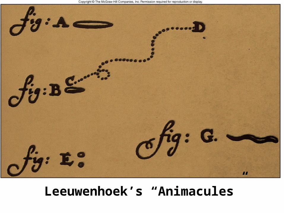
Leeuwenhoek’s “Animacules”
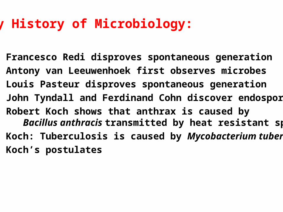
Early History of Microbiology:
1668 – Francesco Redi disproves spontaneous generation
1676 – Antony van Leeuwenhoek first observes microbes
1861 – Louis Pasteur disproves spontaneous generation
1876 – John Tyndall and Ferdinand Cohn discover endospores
1877 – Robert Koch shows that anthrax is caused by Bacillus anthracis transmitted by heat resistant spores
1882 – Koch: Tuberculosis is caused by Mycobacterium tuberculosis
1884 – Koch’s postulates

Fig. 1.4

Theory of Spontaneous Generation
- Organisms arise from non-living material
- Redi showed emergence of flies in rotting meat requiredprevious contact with flies
- Pasteur refuted the theory of spontaneous generationusing careful experiments
- Tyndall and Cohn confirmed Pasteur’s finding by showingthat endospores accounted for sterilization-resistant“spontaneous” bacterial growth

Fig 1.2 - Pasteur’s experiment disproving spontaneous generation

Endospores:
Endospores account for sterilization-resistant life formspresent in soil-derived infusions (from hay, for example)
Predicted by Tyndall (1876) from studies on different infusions
Discovered by Cohn (1876) in soil bacteria
Koch (1877) showed endospores transmit anthrax

Vital Activities and Roles of Microorganisms
- Support all living cells (Bacteria, Archaea, Eucarya)- Involved in nitrogen fixation
- Replenish oxygen on Earth
- Degrade organic waste material
- Serve as models for eukaryotes in study of genetics,
metabolism, and biochemical principles
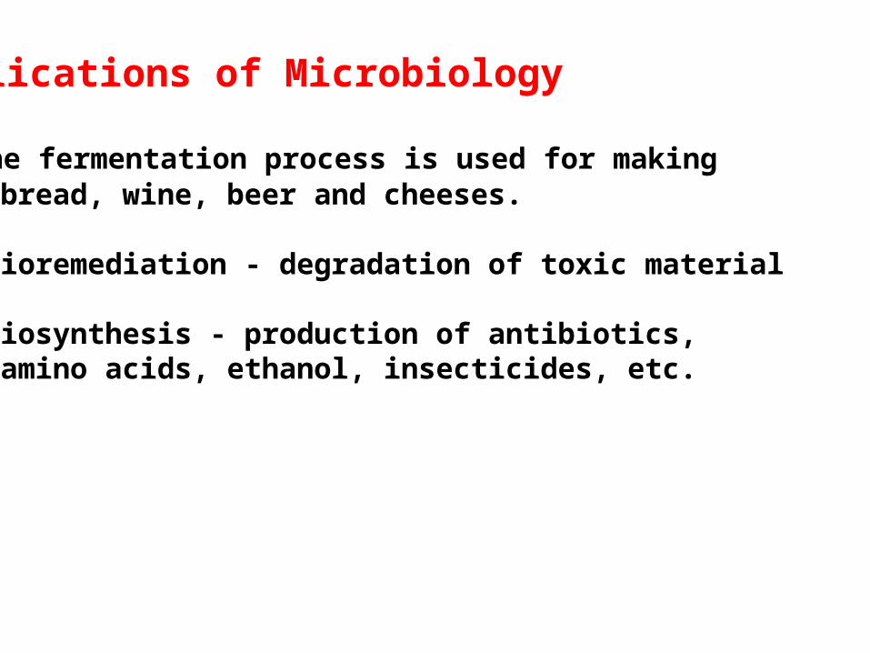
Applications of Microbiology
- The fermentation process is used for making bread, wine, beer and cheeses.
- Bioremediation - degradation of toxic material
- Biosynthesis - production of antibiotics, amino acids, ethanol, insecticides, etc.

Genetic Engineering
The process by which the genes from one organismare introduced into related or unrelated organisms
Examples:
Human growth hormone geneInterferonInsulinBlood clotting and dissolving enzymesVaccine productionGenetically engineered plantsGene therapy with viruses

Medical Microbiology
- Infectious diseases have existed for many years, and affect humans, animals, plants, and microbes
- Emerging infectious diseases
- Re-emerging infectious diseases

Historically important diseases
Small pox - 10 million deaths over last 4000 yearslast case in 1977current bioterrorist threat
Bubonic Plague – 25 million deaths (1346-1350)currently less than 100 per yearrats, carriers of Yersinia pestis, transmitted by fleascontrolled by sanitation, antibiotics
Foot and Mouth Disease (2001)Highly contagious4 million stock animals destroyed to control disease
Infections in US currently at 750 million cases per year
200,000 deaths/year in the US

Fig. 01.03
Figure1.3 “New” infectious Diseases in Humans since 1976

Emerging diseases
Legionaires’ diseaseToxic shock syndromeLyme diseaseAIDSHentavirus pulmonary syndromeHemolytic-uremic syndromeCryptosporidiosisWest Nile virus diseaseSARSAvian flu

Resurging old diseases
Antibiotic resistanceSpread by travelersUnvaccinated childrenOlder peopleAIDS

Three Domains based on ribosomal RNA sequencing:
Bacteria = prokaryotes
Archaea = prokaryotes
Eucarya = eukaryotes

Table 01.02
Table 1.2 – Comparison of Bacteria, Archaea and Eucarya

Bacteria:
Shaped as rods, spheres or spirals
Rigid cell walls containing peptidylglycan
Division by binary fission
Motility via flagella

Figure 1.5 Bacteria viewed through a scanning electron microscope

Archaea:
Life in extreme environments
Thermoplasma – live in burning coal pile tailings
Sulfolobus – live in acidic hot springs
Methanogens – anaerobes, generate methane
Halogens – live in saturated salt solutions
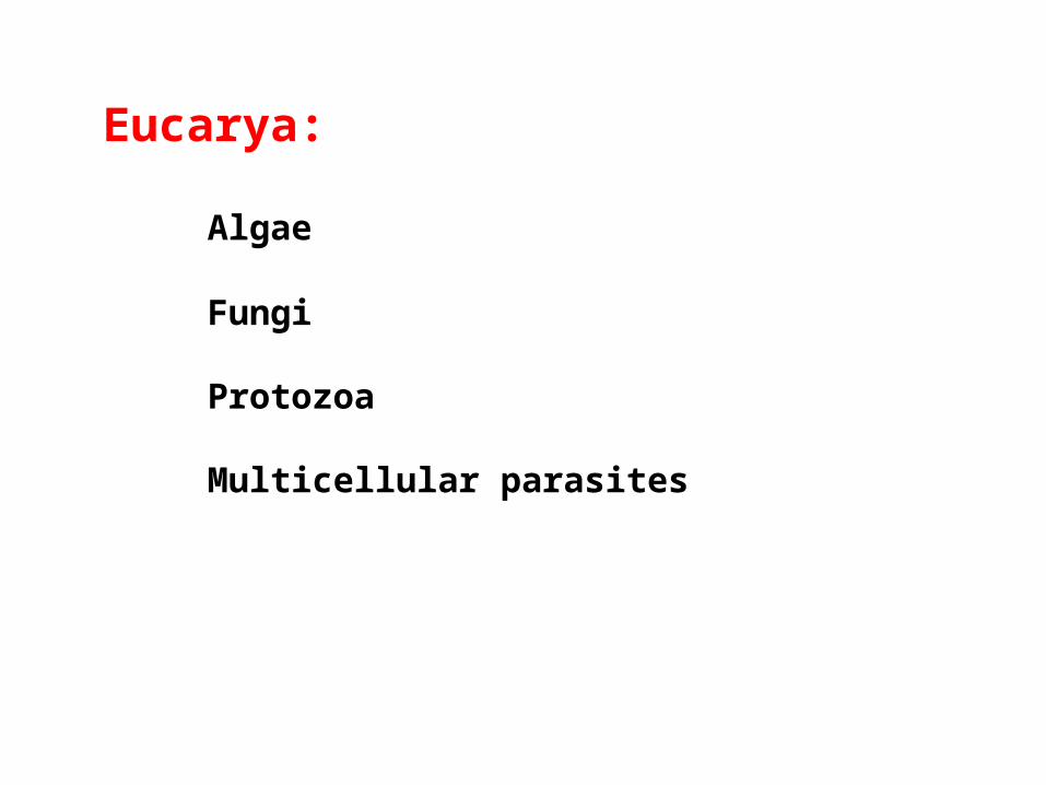
Eucarya:
Algae
Fungi
Protozoa
Multicellular parasites

Figure 1.6 – Micrasterias, a green alga

Figure 1.7 – Two forms of fungi:Cryptococcus (unicellular yeast) stained with india inkAspergillus, multicellular mold viewed with scanning EM

Fig. 01.08
Figure 1.8 – Paramecium, a ciliated protozoan
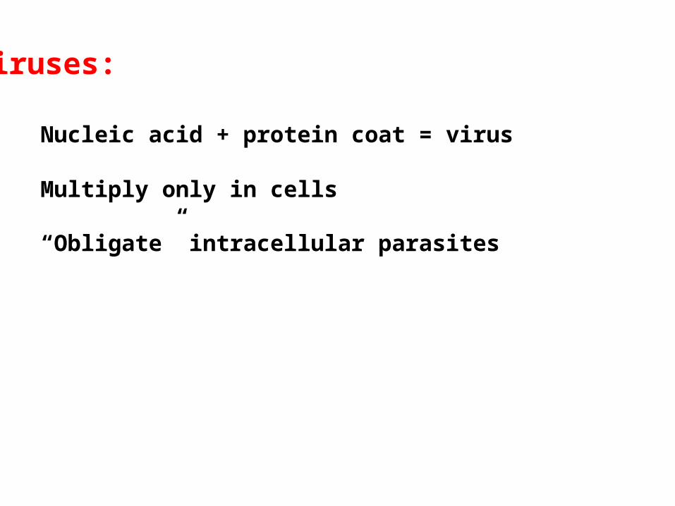
Viruses:
Nucleic acid + protein coat = virus
Multiply only in cells
“Obligate” intracellular parasites

Fig. 01.09
Figure 1.9 – Three kinds of viruses
Tobacco MosaicVirus (TMV)
Bacteriophage Influenza virus

Viroids:
Short pieces of nucleic acid (RNA)Intracellular parasites (plant diseases)

Figure 1.10 – Viroids compared to bacteriophage T7
PSTV = Potato spindle tuber viroids

Prions:
Apparently no nucleic acid; only proteinCause neurodegenerative diseases
such as mad cow disease

Fig. 01.11
Figure1.11 – Prions from a scrapie-infected hampster

Fig. 1.12

Fig. 1.13 – Sizes of Organisms and Viruses