lecture 8 - equine hoof
TRANSCRIPT
AAEP 56th Annual Convention: Baltimore, Maryland 2010
The Equine Hoof
Dr.KathrynCarmalt,DVM,MSc,BA,AS;SlidescourtesyofDr.Anderson
UNIVERSITY OF SASKATCHEWAN Saskatoon, Saskatchewan, Canada. www.usask.ca
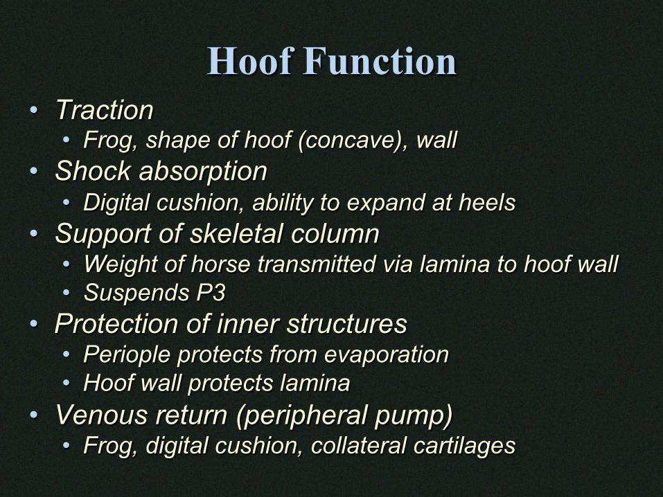
Hoof Function • Traction
• Frog, shape of hoof (concave), wall • Shock absorption
• Digital cushion, ability to expand at heels • Support of skeletal column
• Weight of horse transmitted via lamina to hoof wall • Suspends P3
• Protection of inner structures • Periople protects from evaporation • Hoof wall protects lamina
• Venous return (peripheral pump) • Frog, digital cushion, collateral cartilages
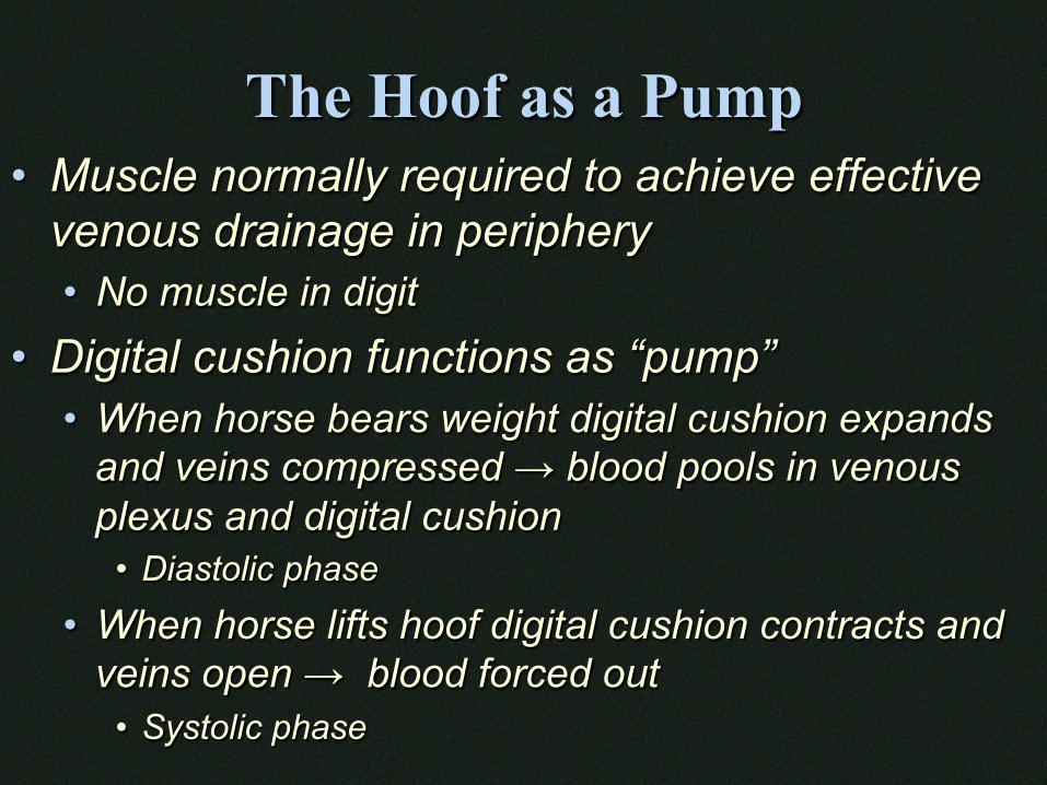
The Hoof as a Pump • Muscle normally required to achieve effective
venous drainage in periphery • No muscle in digit
• Digital cushion functions as “pump” • When horse bears weight digital cushion expands
and veins compressed → blood pools in venous plexus and digital cushion
• Diastolic phase
• When horse lifts hoof digital cushion contracts and veins open → blood forced out
• Systolic phase
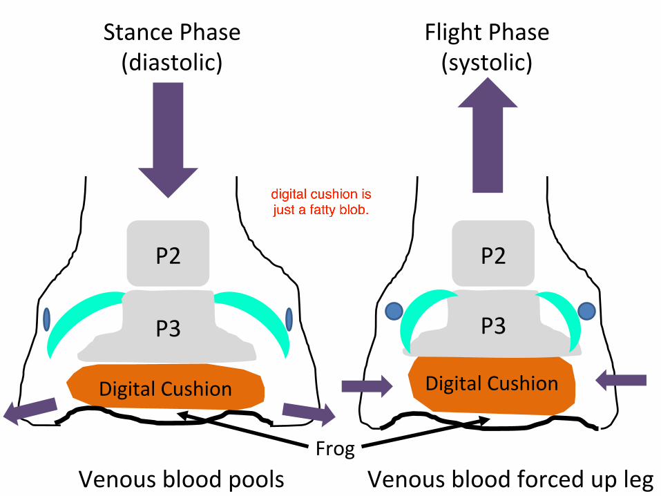
DigitalCushion DigitalCushion
P3 P3
P2 P2
Venousbloodpools Venousbloodforcedupleg
StancePhase(diastolic)
FlightPhase(systolic)
Frog
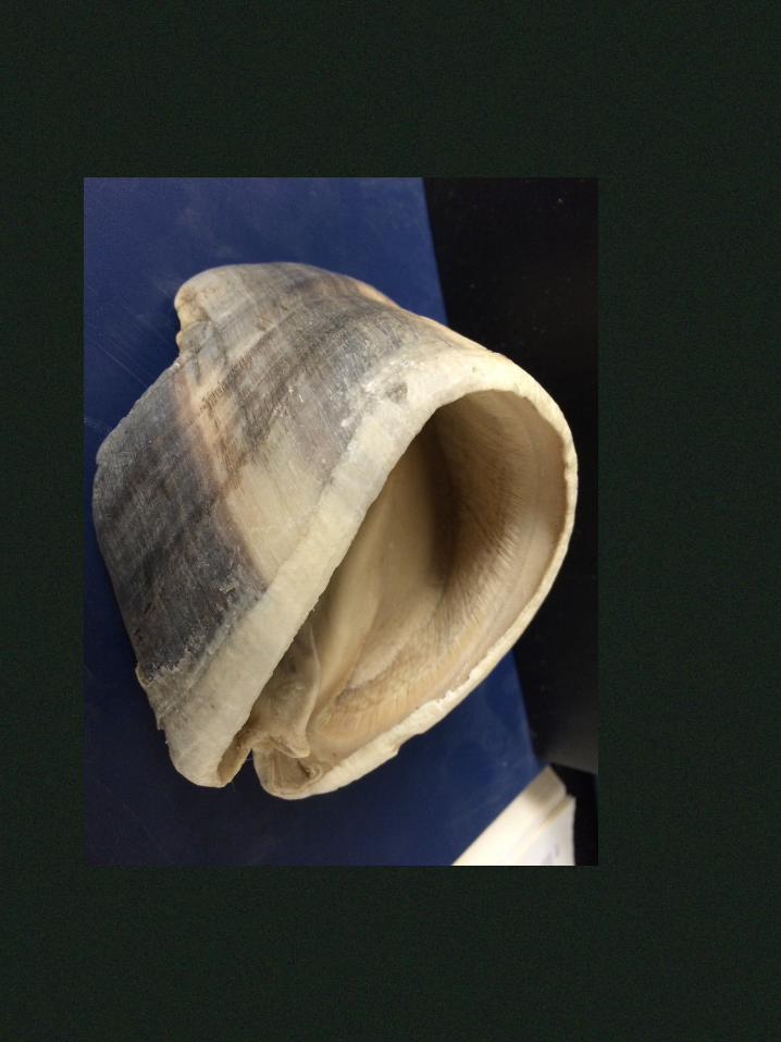
Outer Parts of the Hoof
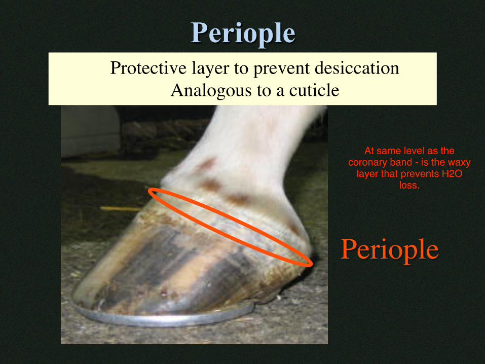
Periople
Periople
Protective layer to prevent desiccationAnalogous to a cuticle
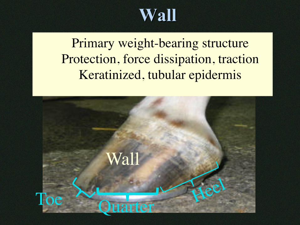
Wall
Wall
Primary weight-bearing structureProtection, force dissipation, traction
Keratinized, tubular epidermis
Toe Quarter
Wall
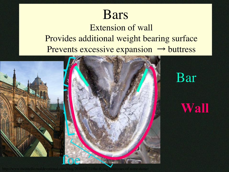
Bar
BarsExtension of wall
Provides additional weight bearing surfacePrevents excessive expansion � buttress
Heel
Quarter
Toehttp://www.theintellectualdevotional.com/blog/2010/02/21/the-flying-buttresses-of-notre-dame/
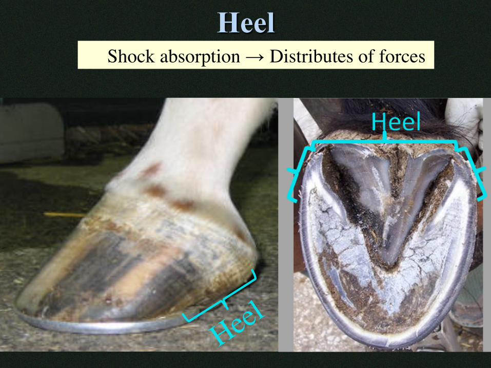
Heel Shock absorption → Distributes of forces
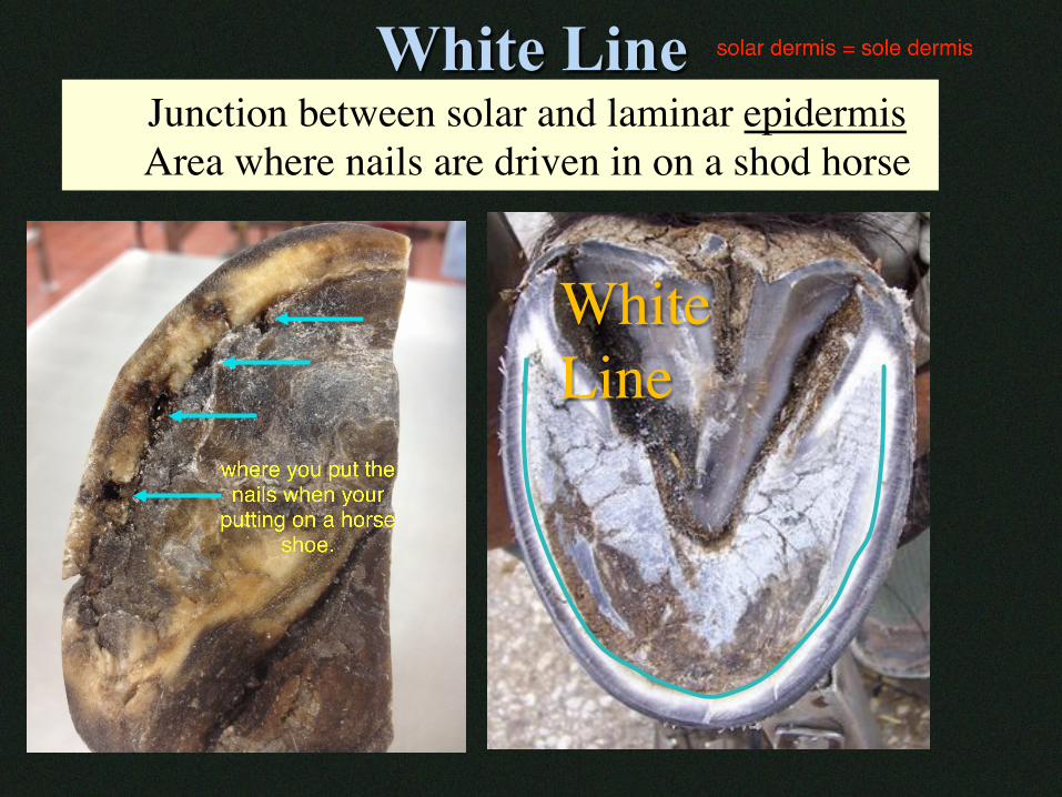
White Line
White Line
Junction between solar and laminar epidermisArea where nails are driven in on a shod horse
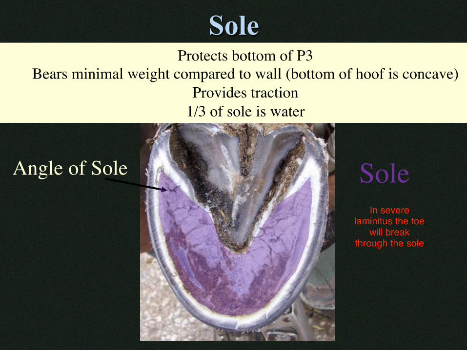
Sole
Angle of Sole
Protects bottom of P3Bears minimal weight compared to wall (bottom of hoof is concave)
Provides traction 1/3 of sole is water
Sole
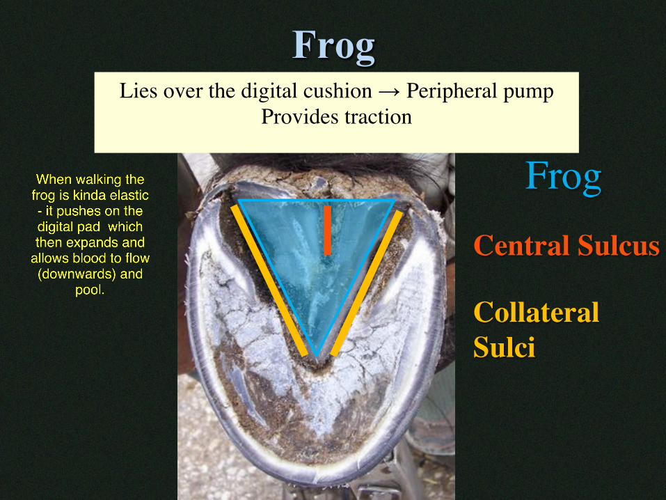
Frog
Frog Central Sulcus
Collateral Sulci
Lies over the digital cushion → Peripheral pumpProvides traction
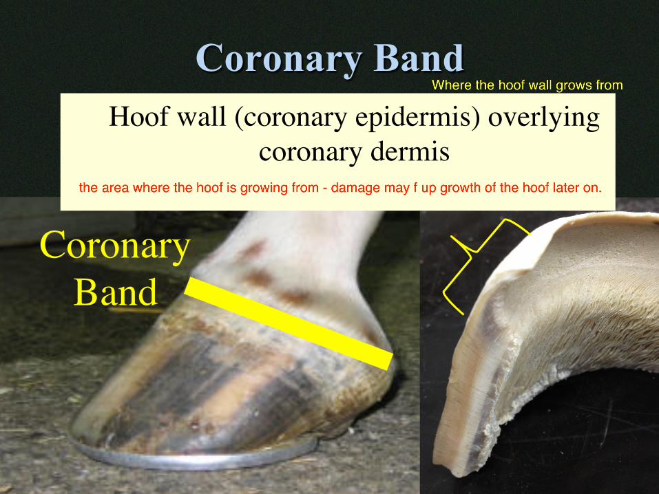
Coronary Band
CoronaryBand
Hoof wall (coronary epidermis) overlying coronary dermis
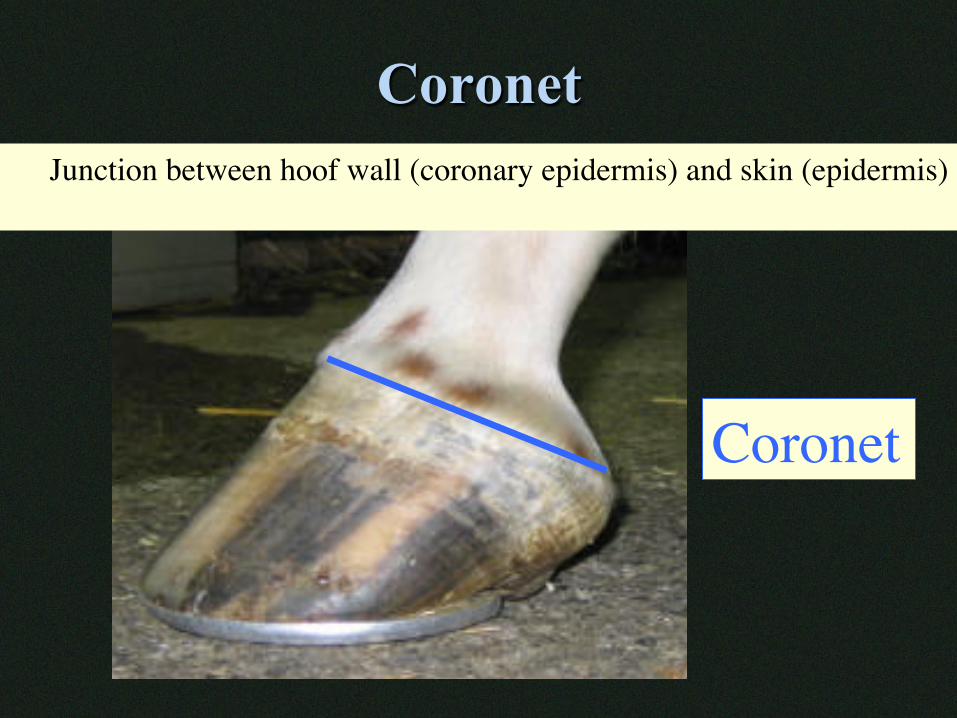
Coronet
Coronet
Junction between hoof wall (coronary epidermis) and skin (epidermis)
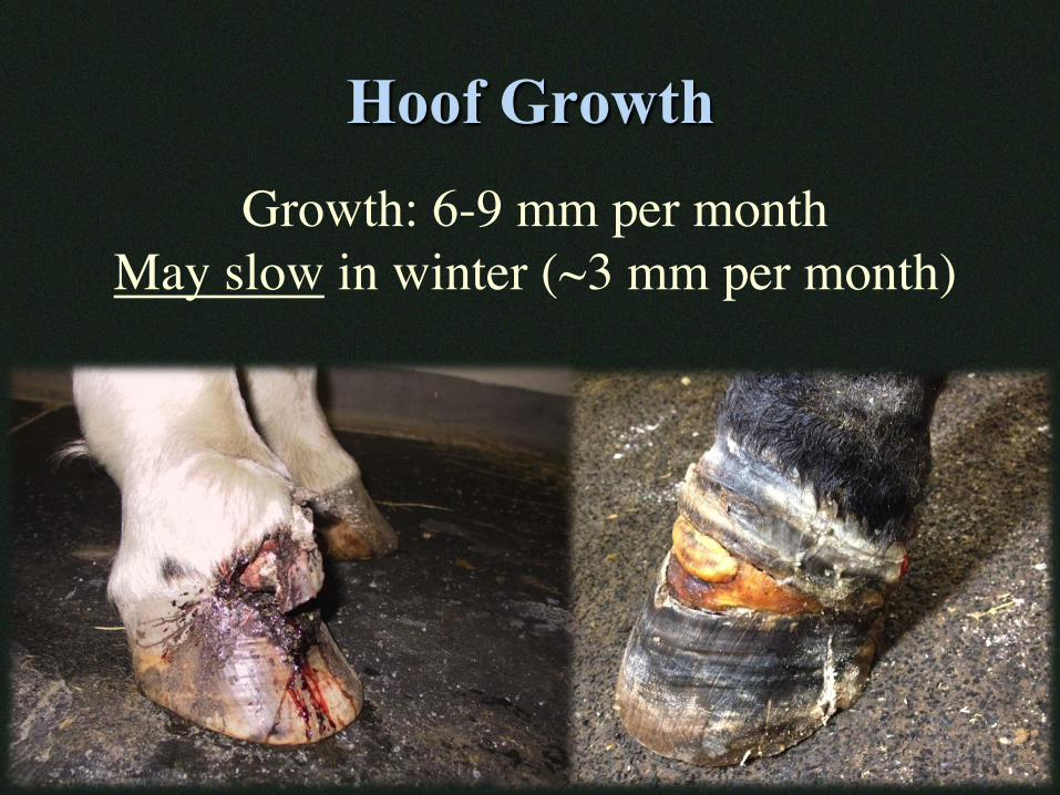
Hoof Growth Growth: 6-9 mm per month
May slow in winter (~3 mm per month)
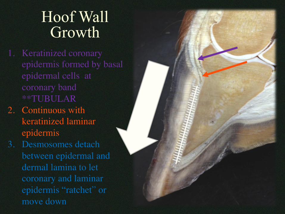
1. Keratinized coronary epidermis formed by basal epidermal cells at coronary band**TUBULAR
2. Continuous with keratinized laminar epidermis
3. Desmosomes detach between epidermal and dermal lamina to let coronary and laminar epidermis “ratchet” or move down
Hoof Wall Growth
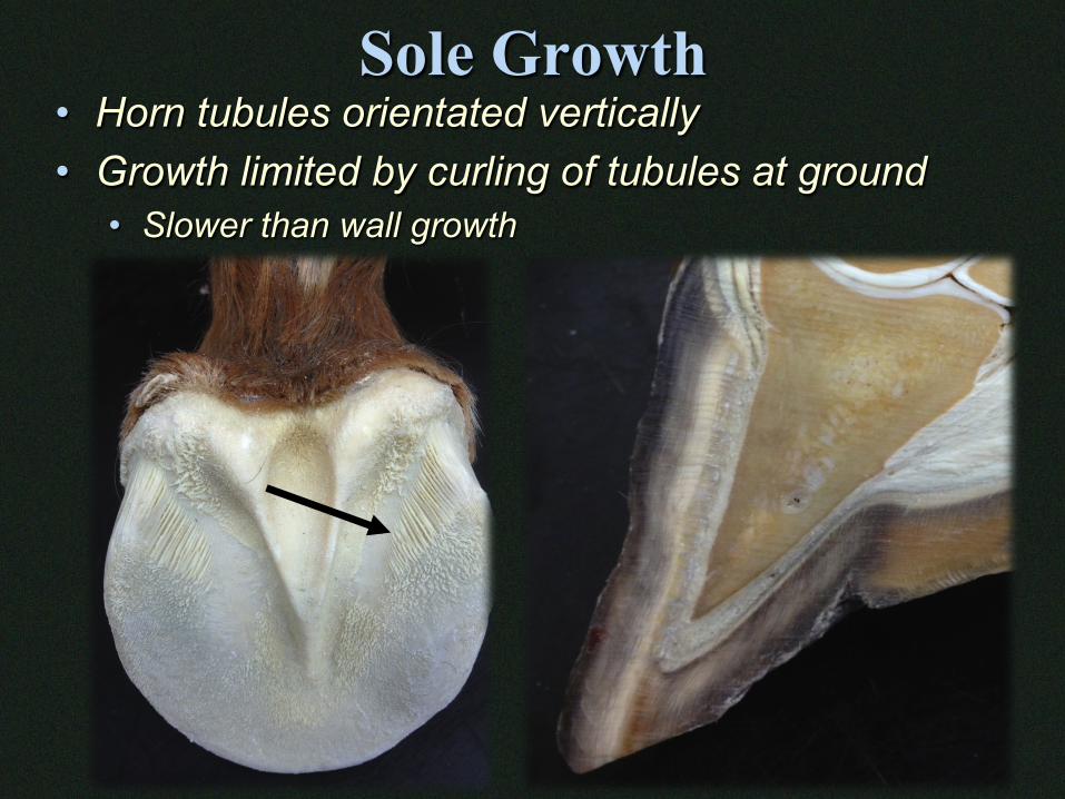
Sole Growth • Horn tubules orientated vertically • Growth limited by curling of tubules at ground
• Slower than wall growth
Inner Parts of the Hoof
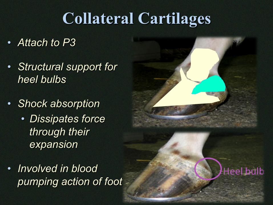
Collateral Cartilages • Attach to P3 • Structural support for
heel bulbs • Shock absorption
• Dissipates force through their expansion
• Involved in blood pumping action of foot
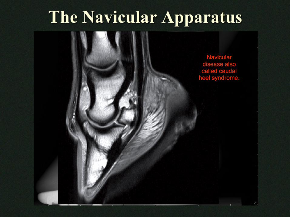
The Navicular Apparatus
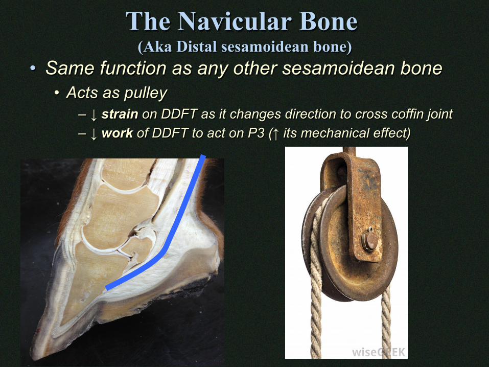
The Navicular Bone (Aka Distal sesamoidean bone)
• Same function as any other sesamoidean bone • Acts as pulley
– ↓ strain on DDFT as it changes direction to cross coffin joint – ↓ work of DDFT to act on P3 (↑ its mechanical effect)
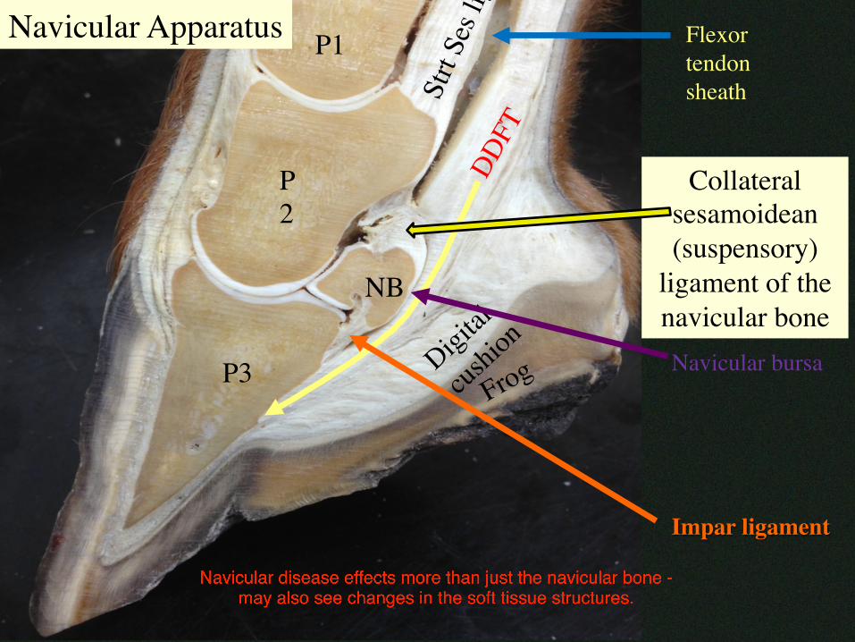
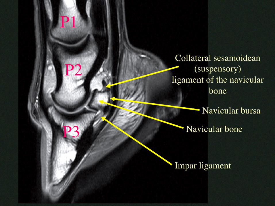
P3
P2
P1
NB
Collateral sesamoidean (suspensory)
ligament of the navicular bone
Impar ligament
Flexor tendon sheath
Navicular Apparatus
Navicular bursa
P1
P3
P2
Navicular bursa
Collateral sesamoidean (suspensory)
ligament of the navicular bone
Navicular bone
Impar ligament
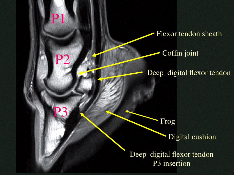
P1
P3
P2Deep digital flexor tendon
Deep digital flexor tendonP3 insertion
Flexor tendon sheath
Coffin joint
Frog
Digital cushion
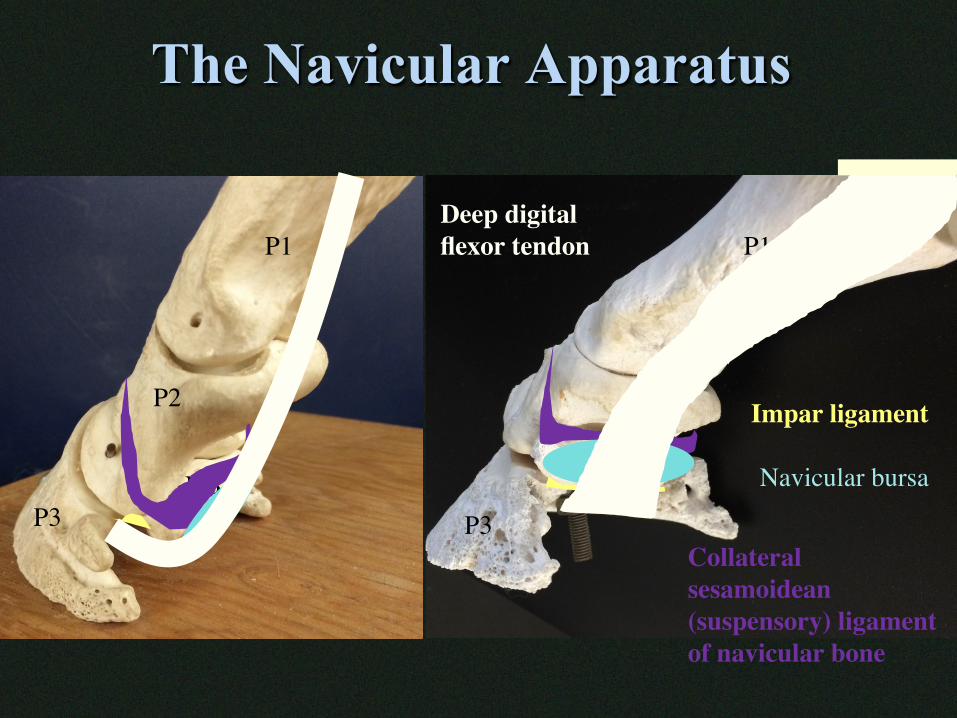
The Navicular Apparatus
P1 P1
P2 P2
P3 P3NB
NB
Impar ligament
Collateral sesamoidean (suspensory) ligament of navicular bone
Navicular bursa
Deep digital flexor tendon
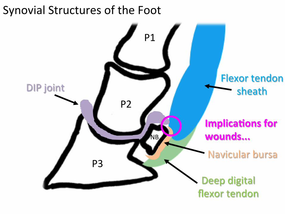
FlexortendonsheathDIPjoint
Navicularbursa
Deepdigitalflexortendon
P3
P2
P1
NB
Implica(onsforwounds...
SynovialStructuresoftheFoot
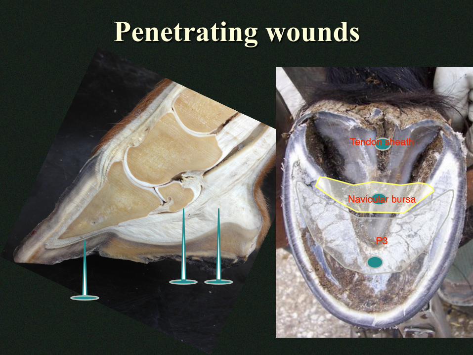
Penetrating wounds
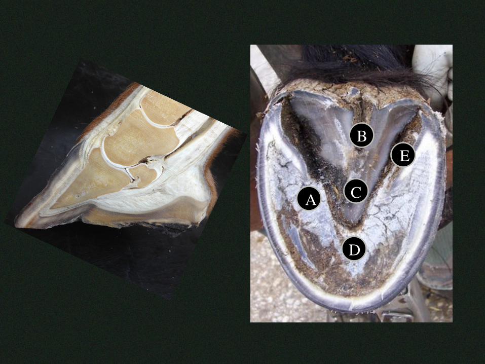
A
E
D
C
B
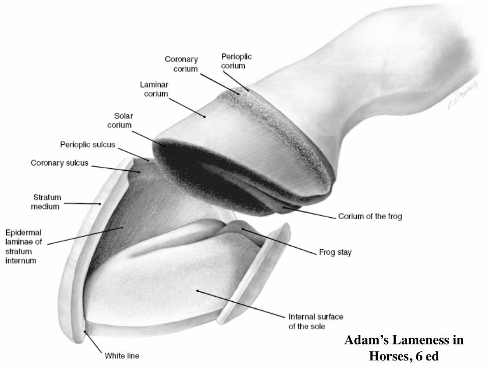
Adam’s Lameness in Horses, 6 ed
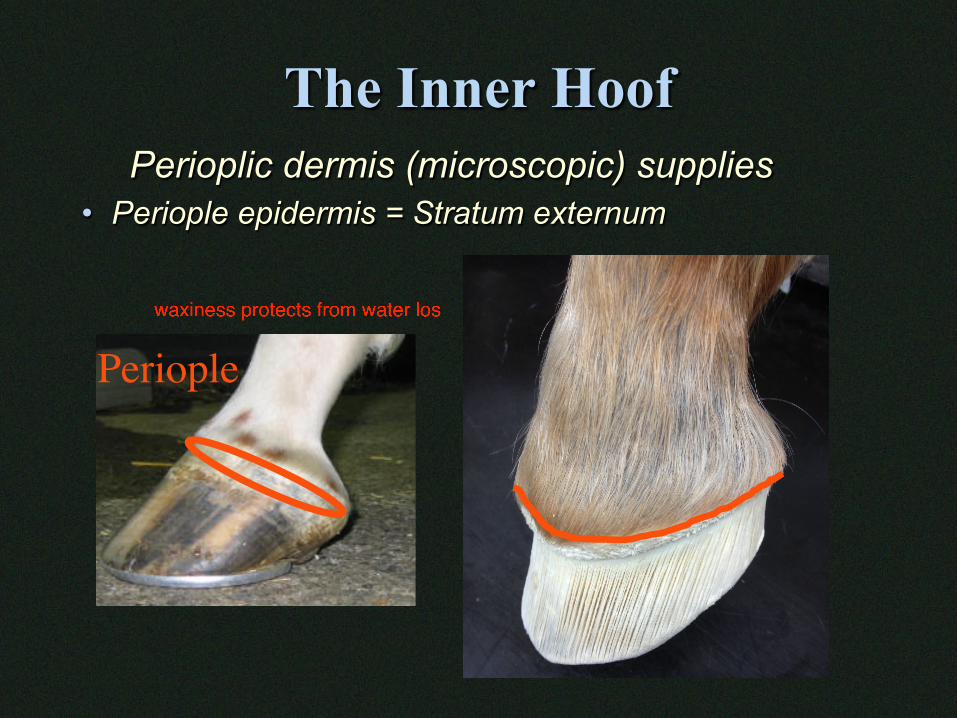
The Inner Hoof Perioplic dermis (microscopic) supplies
• Periople epidermis = Stratum externum
Periople
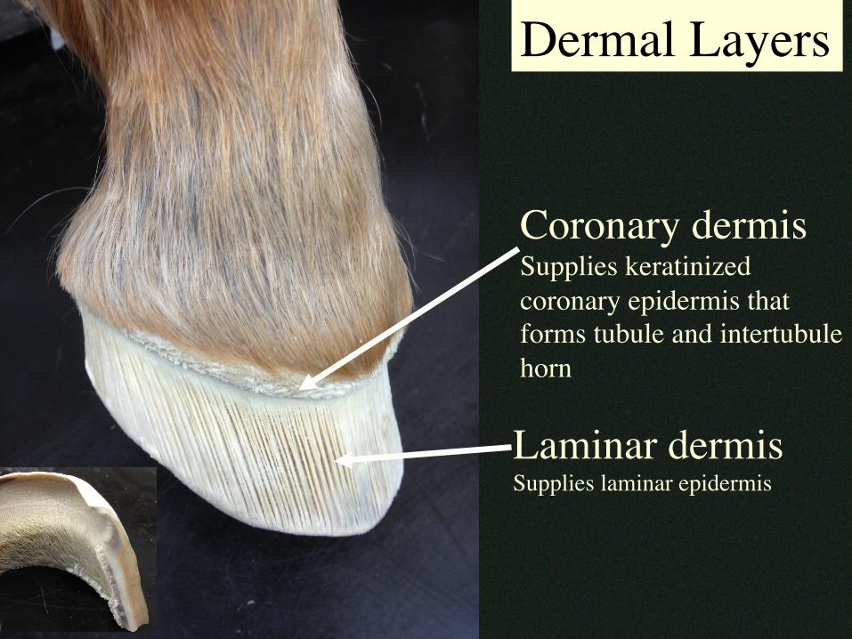
Coronary dermisSupplies keratinized coronary epidermis that forms tubule and intertubule horn
Laminar dermisSupplies laminar epidermis
Dermal Layers
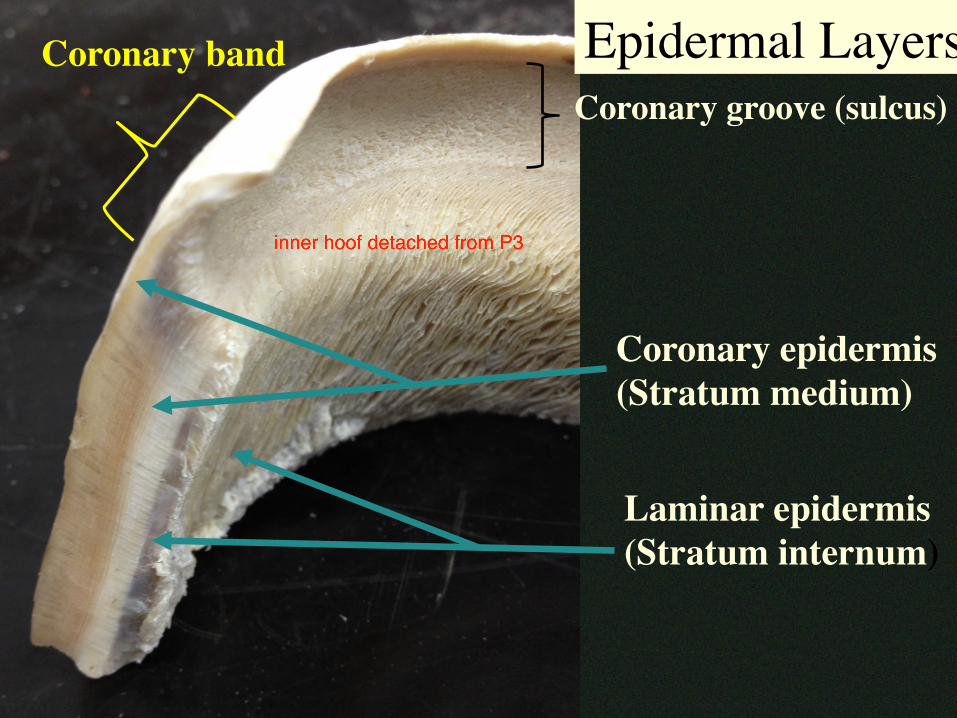
Laminar epidermis(Stratum internum)
Coronary epidermis(Stratum medium)
Coronary groove (sulcus)Coronary band Epidermal Layers
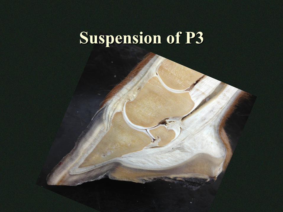
Suspension of P3
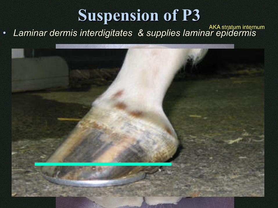
Suspension of P3 • Laminar dermis interdigitates & supplies laminar epidermis
P3
Frog
Hoof Wall
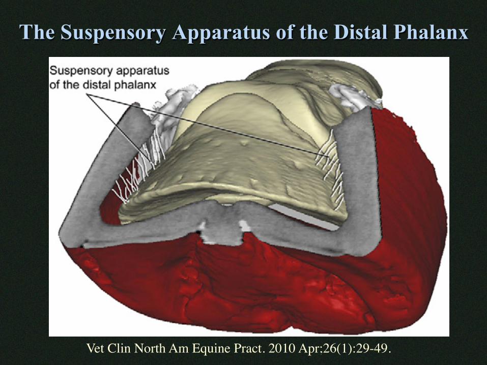
The Suspensory Apparatus of the Distal Phalanx
Vet Clin North Am Equine Pract. 2010 Apr;26(1):29-49.
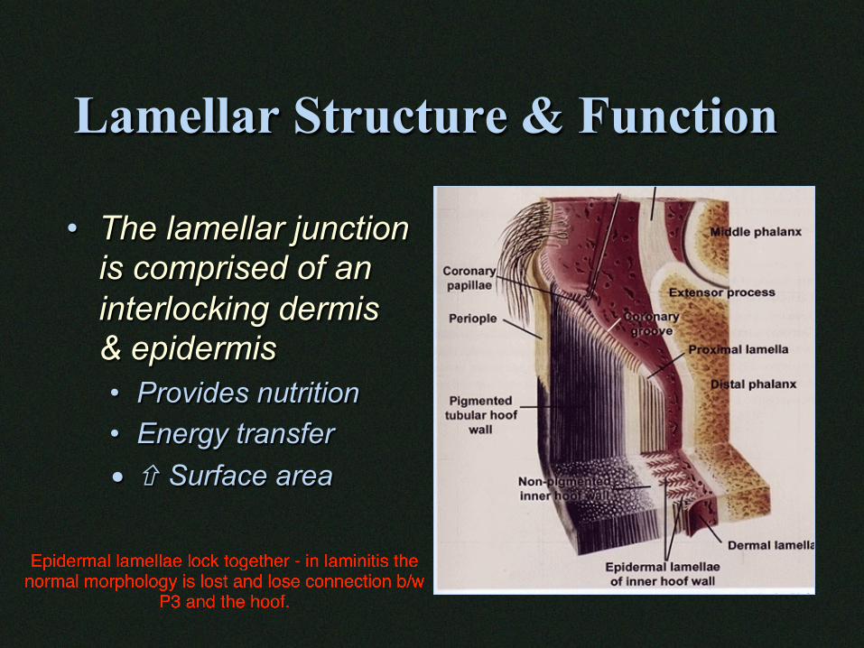
Lamellar Structure & Function
• The lamellar junction is comprised of an interlocking dermis & epidermis • Provides nutrition • Energy transfer • ñ Surface area
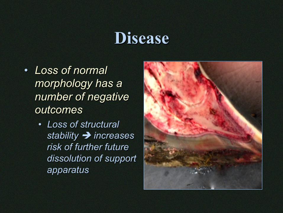
Disease
• Loss of normal morphology has a number of negative outcomes • Loss of structural
stability è increases risk of further future dissolution of support apparatus
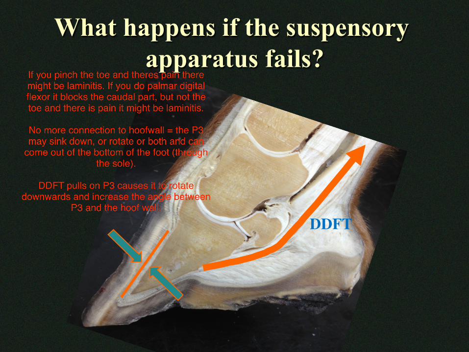
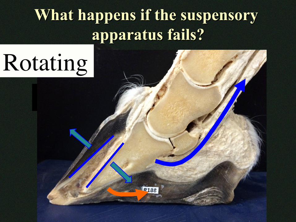
What happens if the suspensory apparatus fails?
DDFT
What happens if the suspensory apparatus fails?
DDFT
Rotating
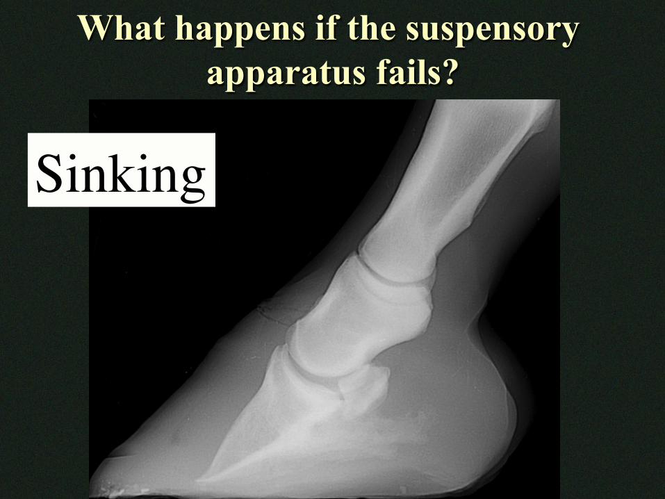
What happens if the suspensory apparatus fails?
Sinking
Blood & Nerve Supply • Medial/Lateral Palmar/Plantar Digital
• Vein, Artery, Nerve
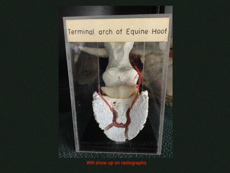
• Vessels enter on palmar/plantar aspect of P3 • Arteries though foramen in P3 • Venous drainage primarily along dorsal border of P3
• Extensive branching • Venous plexuses
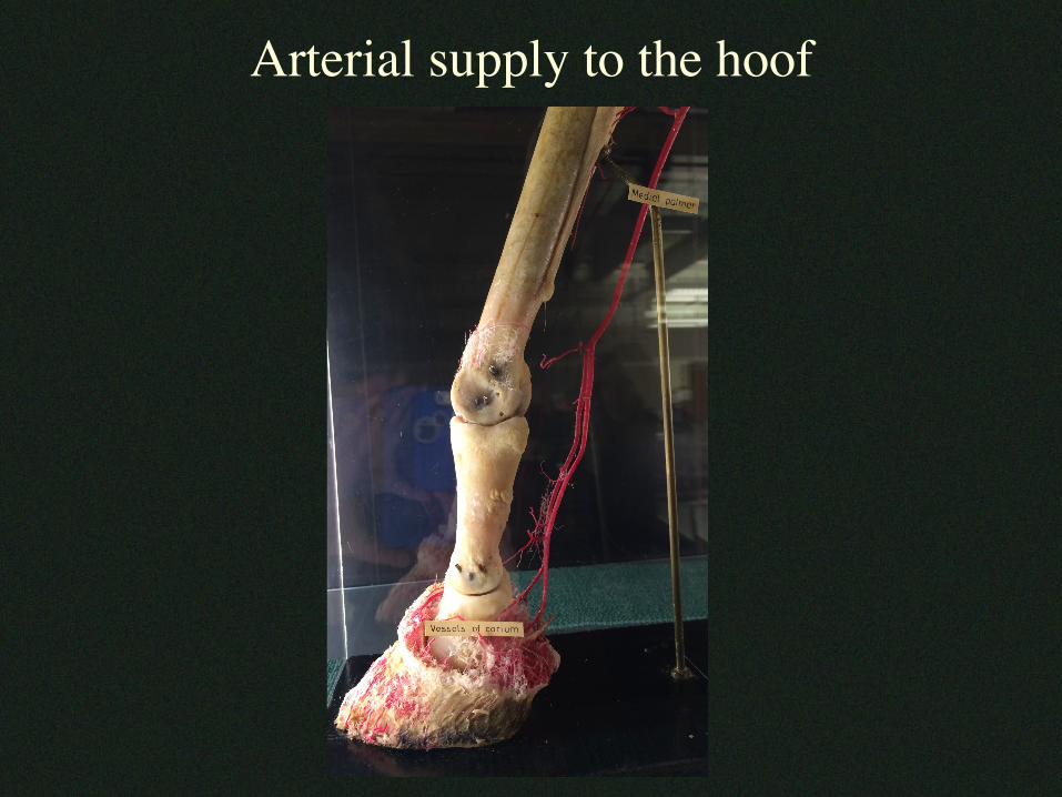
Arterial supply to the hoof
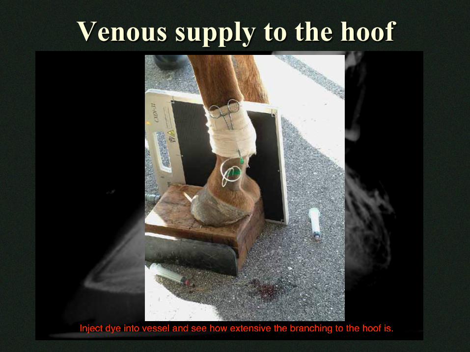
Venous supply to the hoof
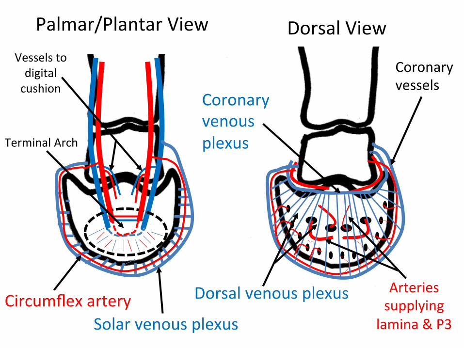
Palmar/PlantarView DorsalView
TerminalArch
Vesselstodigitalcushion
Solarvenousplexus
Arteriessupplying
lamina&P3
Coronaryvessels
DorsalvenousplexusCircumflexartery
Coronaryvenousplexus