lec: 1 cell structure & types
TRANSCRIPT
histology ___________________________ م.م تقى عبد الكريم
1
lec: 1 cell structure & Types
Histology (microscopic anatomy) :
means the microscopic study of tissues of human body. Histology
the Histology is a branch of microscopic anatomy and deals only with
form Different tissues combine to structure of tissues. microscopic
and organ and organs are the units of organs
systems.
The science concerned with the minute structure of cells, tissues and
organs in relation to their function.
Cells
Cells are the tiny living units that form the tissues, organs and structures
within the body. In turn, the body is composed of different types and
varieties of cells to carry out specific functions, but they all have the same
basic structure. All cells contain cytoplasm and are surrounded by
a membrane, and contain the following structures of organelles:
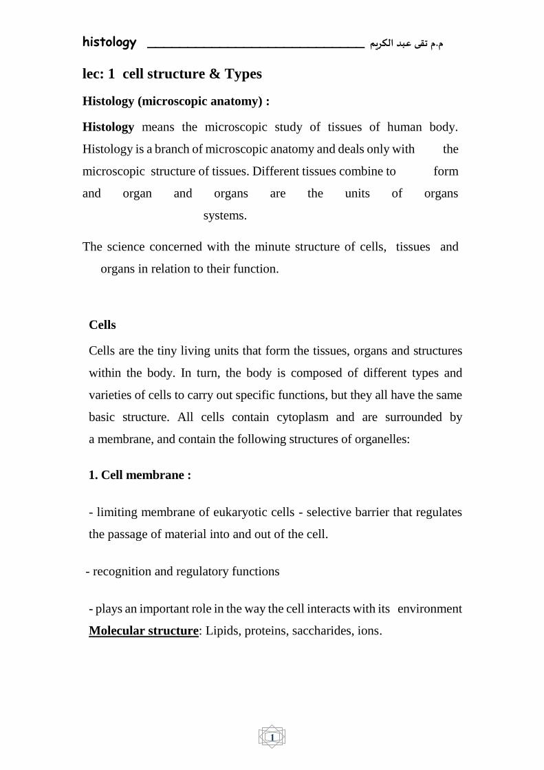
1. Cell membrane :
- limiting membrane of eukaryotic cells - selective barrier that regulates
the passage of material into and out of the cell.
- recognition and regulatory functions
- plays an important role in the way the cell interacts with its environment
Molecular structure: Lipids, proteins, saccharides, ions.

histology ___________________________ م.م تقى عبد الكريم
2
2. Nucleus :
The nucleus, on the other hand, is considered the largest organelle within a
cell. It contains the genetic material in the form of deoxyribonucleic acid
(DNA), along with the enzymes necessary for DNA replication and RNA
transcription.
3. Nucleolus:
Is the structure within the nucleus and help in synthesis of ribosomes.
Surround by nuclear membrane .
Nuclear envelope : thin line
- composed of 2 membranes, between is perinuclear space (cisterna)
- to the inner membrane are attached the fibrous laminae composed
of polypeptides called lamins (ø 80-300 nm)
2 membranes fuse together and form nuclear pores covered by diaphragm
Structure of diaphragm: - 8 peripheral globular proteins molecules + 1
central globular protein

histology ___________________________ م.م تقى عبد الكريم
3
Function: - passage of macromolecules, mRNA, proteins from the
cytoplasm, ions – active transport Outer membrane of nuclear envelope
is covered by ribosomes, perinuclear cisterna is continuous with lumen
of rough endoplasmic reticulum
4. Centrosome : small body located near the nucleus , its has a dense center
and radiating tubules ,centrosome play role in cell division .
5. Mitochondria : spherical or oval organelles in diameter 0,5x10 µm
visualized by iron hematoxylin , Function: transforming of chemical
energy into energy easily accessible to the cell (ATP), production and
storage of energy.
6. Golgi body: (folded membranes that process proteins from the
endoplasmic reticulum) .
o Flattened vesicles in stacks which receive protein from ER
o Form secretory vesicles to transport proteins to different parts
of the cell (vacuole, lysosome, etc) or for secretion.
o cis face - "receiving" side of Golgi apparatus
o trans face - "shipping" side of Golgi apparatus
7. Lysosomes: (contain digestive enzymes)
8. Endoplasmic Reticulum - an extensive membranous network
continuous with the outer nuclear membrane.
Rough ER - has ribosomes and is involved in secreted protein
synthesis
Smooth ER - lacks ribosomes and is involved in membrane lipid
synthesis.
histology ___________________________ م.م تقى عبد الكريم
4
cell components
Tissues :
Body tissues : are collections of cells, grouped in the body according to
structure and function. In histology, human tissues are separated into four
distinct categories:
Muscular: Muscle tissue is made up of long thin muscle cells called
myocytes. Their structure and arrangement allows for muscular
contraction.

histology ___________________________ م.م تقى عبد الكريم
5
Nervous: Nervous tissue forms the nervous system, and is made up of
specialised cells called neurons and neuroglial cells. Neurons conduct
nerve signals from one to another in the form of electrical impulses.
Epithelial: Epithelial tissue comprises epithelial cells arranged together in
sheets. These sheets serve as protective layers, forming coverings like
the skin, and the lining of the small intestine.
Connective: Connective tissue forms a connective web throughout the
body. It fills gaps and connects different organs and body parts, so that the
carefully arranged structure of the body can be maintained.

histology ___________________________ م.م تقى عبد الكريم
6
lec: 2 Epithelium (epithelial tissues)
Epithelium tissue forms continuous layers of cells that cover surfaces
and line cavities of the body.
Epithelial tissue covers the outside of the body and lines organs, vessels
(blood and lymph), and cavities. Epithelial cells form the thin layer of
cells known as the endothelium, which is continuous with the inner
tissue lining of organs such as the brain, lungs, skin, and heart. The free
surface of epithelial tissue is usually exposed to fluid or the air, while
the bottom surface is attached to a basement membrane.
The cells in epithelial tissue are very closely packed together and joined
with little space between them. With its tightly packed structure, we
would expect epithelial tissue to serve some type of barrier and
protective function and that is certainly the case. For example, the skin
is composed of a layer of epithelial tissue (epidermis) that is supported
by a layer of connective tissue. It protects the internal structures of the
body from damage and dehydration.
Epithelial tissue also helps to protect against microorganisms. The
skin is the body's first line of defense against bacteria, viruses, and
other microbes.
Epithelial tissue functions to absorb, secrete, and excrete substances.
In the intestines, this tissue absorbs nutrients during digestion.
Epithelial tissue in glands secrete hormones, enzymes, and other
substances. Epithelial tissue in the kidneys excrete wastes, and in the
sweat glands excrete perspiration.

histology ___________________________ م.م تقى عبد الكريم
7
Epithelial tissue also has a sensory function as it contains sensory
nerves in areas such as the skin, tongue, nose, and ears.
Ciliated epithelial tissue can be found in areas such as the
female reproductive tract and the respiratory tract. Cilia are hair-like
protrusions that help propel substances, such as dust particles or
female gametes, in the proper direction.
Classifying Epithelial Tissue:
Based on:
1) type of cell in which the tissue is made of
2) shape
3) number of layers of cells
1- Covering and lining epithelium : from the outer layers of the
skin ;lines open cavities of the digestive and respiratory
systems; covers the walls of organs of the closed ventral body
cavity.
2- Glandular epithelium: surrounds glands within the body.
Covering and lining epithelium: they can be classified according to
the number of the cell layers into:
a- Simple epithelial tissues: consist of a single layer of cell.
b- Stratified epithelial tissues : consist of multilayer of cells only the
bottom layer touches the basement .
1- Simple epithelial tissues: they can be classified according to the
shape of the constituent cell:
2- Simple squamous epithelium : consist of a single layer of flattened
cells with disc shape central nuclei , is found lining sacs of lung and

histology ___________________________ م.م تقى عبد الكريم
8
wall of blood vessels , its shape and arrangement permit the
exchanges of substance in these locations.
3- Simple cuboidal epithelium : consist of single layer of cube like
cell with large spherical nuclei , this type is found in glands as
salivary gland , or in kidney tubule , its function secretion and
absorption .
4- Simple columnar epithelium : consist of single layer of tall cells
with round to oval nuclei located near the bottom of each cell , this
type of epithelium is found digestive tract as stomach.
5- Pseudostratified ciliated columnar epithelium: one layer of cells
, but appears stratified because cells are off different heights . all
cells are in contact with the basement membrane .
6- Stratified epithelial tissues :
1- Stratified squamous : has multiple layers of cells, the basal cells
are cuboidal or columnar are metabolically active , the surface
layer are flattened (squamous) there are two type a.
nonkeratinized (e.g. lining of esophagus) b. keratinized (e.g.
skin) .
2- Stratified cuboidal : usually has two or three layers of cuboidal
cells. This type of epithelium is largely confined to the lining of
large ducts (sweat gland).
3- Stratified columnar: has several cell layers, the outermost of
which contains columnar cell . this type of epithelium is
relatively rare , found in male urethra .
4- Transitional epithelium : is similar to Stratified squamous
epithelium , except that the outermost cell layer consist of large

histology ___________________________ م.م تقى عبد الكريم
9
Rounded cells (dome- like ). This type of tissue changes in
response to tention . found in urinary bladder .

histology ___________________________ م.م تقى عبد الكريم
10

histology ___________________________ م.م تقى عبد الكريم
11
2- Glandular epithelium : consist of one or more cells that produce
and secrete a specific product . there are two types of glands:
a- Exocrine gland : are gland with ducts that secrete their
product onto the outer surface (sweat gland ) or into
body cavities (pancreas).
b- Endocrine gland: are gland without duct secrete
hormones internally so they are transported by
bloodstream( thyroid gland) .
Glandular epithelium : are classified by the following
morphological characteristic to:
1- Unicellular glands : consisting of only one glandular epithelium
cell e.g. goblet cell of digestive system.
2- Multicellular glands : multiple cell make up one gland ; e.g
salivary glands
Glands are divided according to type of secretion:
1- Serous glands:
a- Secret watery solution of enzyme
b- Have small lumen
c- Have round nucleus located in the basal of the cell
d- Have well stained cytoplasm
e- The secretory units composed of pyramidal cells with unclear
border
f- e.g parotid gland
histology ___________________________ م.م تقى عبد الكريم
12
2- mucous glands :
a- poduce mucous secretion
b- have large lumen
c- have pressed flattened nucleus in the basal limit of the cell
d- have poorly stained cytoplasm
e- the secretory units composed of pyramidal cells with clear boder
f- palatine gland
3- mixed or seromucous glands : a gland in which some secretory
cells are serous and some mucous (salivary glands)
lec: 3 Connective tissues

histology ___________________________ م.م تقى عبد الكريم
13
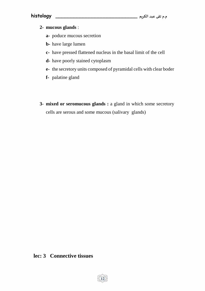
Connective tissue (CT) is a one of the four main classes of tissues.
Although it is the most abundant and widely distributed of the primary
tissues, the amount of connective tissue in a particular organ varies. Like
to the timber framing of a house, the connective tissue provides structure
and support throughout the body.
Structure of Connective Tissue
Connective tissue has three main components:
1. Ground substance
2. Fibers
3. Cells
Together the ground substance and fibers make up the extracellular
matrix. The composition of these three elements vary tremendously
from one organ to the other. This offers great diversity in the types of
connective tissue.
Ground Substance

histology ___________________________ م.م تقى عبد الكريم
14
1. Ground substance – is a gel containing:
• water, salts and
• 3 kinds of molecules containing carbohydrates:
– glycosaminoglycan or GAG,
– proteoglycans and
– glycoproteins
Features of ground substance:
• Permeability
• Barrier to the penetration of bacteria
2.Connective Tissue Fibers
Connective tissue contains three types of fibers: collagen, elastic and
reticular.
1. Collagenous Fibers
Collagenous fibers consist of types I, II, or III collagen and are present
in all types of connective tissue. Collagenous connective tissue is
divided into two types, based upon the ratio of collagen fibers to
ground substance:
Loose (areolar connective tissue) is the most abundant form of
collagenous connective tissue. It occurs in small, elongated bundles
separated by regions that contain ground substance.
Dense connective tissue is enriched in collagen fibers with little
ground substance. If the closely packed bundles of fibers are located
in one direction, it is called regular; if oriented in multiple directions,
it is referred to as irregular. An example of regular dense connective

histology ___________________________ م.م تقى عبد الكريم
15
tissue is that of tendons; an example of irregular dense connective
tissue is that of the dermis.
Collagen I : is found the dermis of skin
Collagen I I : is found in hyaline cartilage
Collagen I I I : found in liver and bone marrow
2.Reticular Fibers
Reticular fibers are composed of type III collagen. Unlike the thick
and coarse collagenous fibers, reticular fibers form a thin reticular
network. Such networks are widespread among different tissues and
form supporting frameworks in the liver, lymphoid organs, capillary
endothelia, and muscle fibers.
3.Elastic Fibers
Elastic fibers contain the protein elastin, which co-polymerizes with
the protein fibrillin. These fibers are often organized into lamellar
sheets, as in the walls of arteries. Dense, regular, elastic tissue
characterizes ligaments. Elastic fibers are stretchable because they are
normally disorganized – stretching these fibers makes them take on an
organized structure.

histology ___________________________ م.م تقى عبد الكريم
16

histology ___________________________ م.م تقى عبد الكريم
17
3.Cells Of Connective Tissue
Although connective tissue has fewer cells than most tissues, the cells
found in connective tissue are still important. Fibroblasts and adipocytes
do not leave connective tissue.

histology ___________________________ م.م تقى عبد الكريم
18
a- Fibroblast
Fibroblasts are widely distributed within connective tissue and
synthesize the components of the extracellular matrix. They are also
capable of differentiating into other types of connective tissue cells.
b- Adipocyte
Adipocytes (or fat cells) are specialized for the synthesis and storage of
lipids. They may occur singly but are more often found as clusters within
loose connective tissue.
c- Mast Cell
Mast cells are widely distributed in connective tissue. They release
molecules that dilate blood vessels and recruit more immune cells to
the site of an infection.
d-Plasma Cell
Plasma cells produce large quantities antibodies against specific
antigens.
e-Macrophage
Monocytes differentiate into macrophages within tissues. Macrophages
are avidly phagocytic cells that engulf and digest microbes, cellular
debris and foreign substances.
histology ___________________________ م.م تقى عبد الكريم
19
The Cells and Fibers of L.I.C.T.
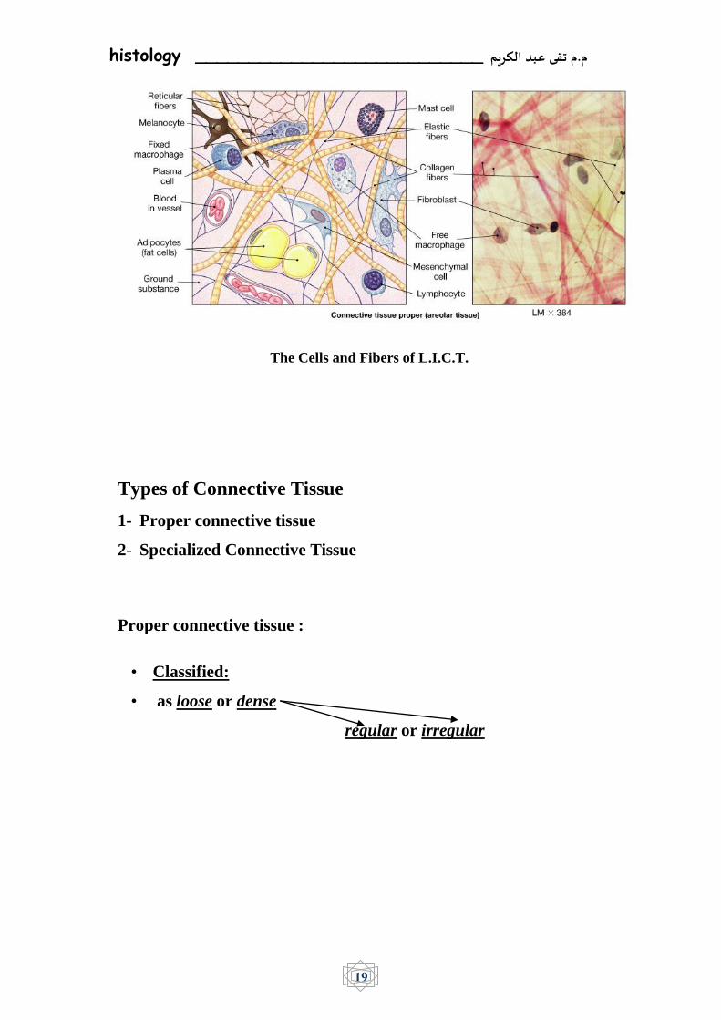
Types of Connective Tissue
1- Proper connective tissue
2- Specialized Connective Tissue
Proper connective tissue :
• Classified:
• as loose or dense
regular or irregular

histology ___________________________ م.م تقى عبد الكريم
20

histology ___________________________ م.م تقى عبد الكريم
21
Loose connective tissue : The loose connective tissue contains cells,
fibers, and ground substance in roughly equal parts. Among the cells,
the fibroblasts are the predominant cells; other types of connective tissue
cells are also present, along with nerves and blood vessels. Collagen
fibers predominate, but elastic and reticular fibers are also present. The
loose connective tissue has moderate amount of ground substance. The
combination of these components gives a delicate consistency to the
loose connective tissue making it flexible and not very resistant to stress.
Loose Connective Tissue Location
1. Lamina propria (areolar tissue)
2. Blood vessels (Areolar tissue)
3. Found in the ducts of glands (Areolar tissue)
4. Bone marrow (Reticular tissue)
5. Spleen (Reticular tissue)
6. Lymph nodes (Reticular tissue)
7. Umbilical cord (Mucoid tissue)
8. Embryo (Mesenchyme - embryonic tissue).
1.Areolar Connective Tissue
These tissues are widely distributed and serve as a universal packing
material between other tissues. The functions of areolar connective
tissue include the support and binding of other tissues.It also helps in
defending against infection.
Is a loosely arranged connective tissue that is widely distributed in the
body such as in gastrointestinal tract, blood vessels and ducts of
glands. The areolar tissue contains collagen fibers, reticular fibers and

histology ___________________________ م.م تقى عبد الكريم
22
a few elastic fibers embedded in a thin and almost fluid-like ground
substance.
2. Adipose Tissue: this type of tissue differs from other connective
tissues in two respects it contains more of fat cells and not the
intercellular substances and secondly, each fat cell is surrounded by
its own basal lamina.
3.Reticular Connective Tissue: the reticular connective tissue is
predominatly made up of reticuler fibers and characterized by a
cellular framework as seen in lymphatic tissues and bone marrow.
Adipose tissue: Yellow adipose tissue in paraffin section with lipids washed out.
Dense connective tissue: this type of tissue is characterized by an
abundance of fibers with fewer cells , as compared to the loose connective
tissue.

histology ___________________________ م.م تقى عبد الكريم
23
a. Regular connective tissue : it is made primarily of parallel collagen
fibers , a few elastic fibers and the major cell type is the fibroblast. found
in tendons
b. Irregular connective tissue : it is made primarily of irregularly
arranged collagen fibers , some elastic and the major cell type is the
fibroblast . found in skin.
Dense connective tissue ( regular)
—
—
—
—

histology ___________________________ م.م تقى عبد الكريم
24
— Lec 4: Specialized Connective Tissue:
Cartilage
is a form of fibrous connective tissue that is composed of closely packed
collagenous fibers in a rubbery gelatinous substance called chondrin.
The skeletons of sharks and human embryos are composed of cartilage.
Cartilage also provides flexible support for certain structures in adult
humans including the nose, trachea, and ears.
There are three different types of cartilage, each with different
characteristics.
1. Hyaline cartilage; is the most common type and is found in areas
such as the trachea, ribs, and nose. Hyaline cartilage is flexible, elastic,
and surrounded by a dense membrane called perichondrium.
2. Fibrocartilage : is the strongest type of cartilage and composed of
hyaline and dense collagen fibers. It is inflexible, tough, and located in
areas such as between vertebrae, in some joints, and in heart valves.
Fibrocartilage does not have perichondrium.
3.Elastic cartilage : contains elastic fibers and is the most flexible type
of cartilage. It is found in locations such as the ear and larynx (voice
box).

histology ___________________________ م.م تقى عبد الكريم
25

histology ___________________________ م.م تقى عبد الكريم
26
Bone :
is a type of mineralized connective tissue that contains collagen and
calcium phosphate, a mineral crystal. Calcium phosphate gives bone its
firmness. There are three types of cells:
Osteoblasts : are responsible for the synthesis of the organic
components o bone matrix , consisting of type I collagen fibers and
osteonectin .
Osteocytes: Individual osteoblasts are gradually surrounded by their
own secretion and become osteocytes enclosed singly within spaces
called lacunae .
Osteoclasts : which are multi-nucleated giant cells involved in the
resorption and remodeling of bone tissue.
There are two types of bone tissue: spongy and compact.
Spongy bone, also called cancellous bone, gets its name because of its
spongy appearance. The large spaces, or vascular cavities, in this type
of bone tissue contain blood vessels and bone marrow. Spongy bone is
the first bone type formed during bone formation and is surrounded by
compact bone, found at the end of long bones.
Compact bone, or cortical bone, is strong, dense, and forms the hard
outer bone surface. Small canals within the tissue allow for the passage
of blood vessels and nerves( Haversian canal ).
Lamellae : are concentric rings of a strong matrix

histology ___________________________ م.م تقى عبد الكريم
27
Lacuna : are the small space between the lamella in which the
osteocytes
Canaliculi: a minute channels that linked the lacuna together which
provide routes for pass the nutrients and waste product for osteocytes.
The bulbous ends of each long bone, known as the epiphyses (or
singularly as an epiphysis), are made up of spongy, or cancellous, bone
tissue covered by a thin layer of compact bone. The diaphysis, or shaft,
contains the medullary cavity and blood cell–producing marrow. A
membrane called the periosteum covers the outer bone to provide
nutrients and oxygen, remove waste, and connect with ligaments and
tendons.

histology ___________________________ م.م تقى عبد الكريم
28

histology ___________________________ م.م تقى عبد الكريم
29
Ossification of Bone
Ossification: is the process of the synthesis of bone from cartilage.
There are two types of ossification- intramembranous and
endochondral ossification. Bone may be synthesized by
intramembranous ossification, endochondral ossification or a
combinationofthetwo.
1- Intramembranous ossification:
Most flat bones are produced by this types of ossification it takes
place within condensations of embryonic mesenchymal tissue such
as the frontal and parietal bones of the skull, temporal bones and the
mandible and maxilla.
2-Endochondral Ossification
In endochondral ossification, bone develops by replacing hyaline
cartilage. Cartilage does not become bone. Instead, cartilage
serves as a template to be completely replaced by new bone.
Endochondral ossification takes much longer than intramembranous
ossification . Bones at the base of the skull and long bones form via
endochondral ossification. This type of ossification is responsible
for the formation of short and long bones.
- Zone of reserve cells: Athin layer (3 – 6 cells wide ) of small,
randomly oriented chondrocytes adjacent to the bony trabeculae
on the articular side of the growth plate.
-Zone of prolifrration : Chrondrocytes are stacked in prominent
rows and the cartilage matrix becomes more basophilic in this

histology ___________________________ م.م تقى عبد الكريم
30
zone. Mitotc figures are present and the axis of the mitotic figure
usually is perpendicular to that of the row of chondrocytes.
-Zone of hypertrophy : Chrondrocytes and their lacunae increase
in size .
-Zone of calcification : Depositin of mineralas in the matrix
surrounding the enlarged lacunae causing cell death.
-Zone of ossification : Osteoblasts deposil bone matrix on the
exposed plates of calcified cartilage.
-Zone of resorption : Osteoclasts absorb the oldest ends of the
bone spicules.

histology ___________________________ م.م تقى عبد الكريم
31