learning from ionising radiation dose errors, advers events and … · 2019. 6. 28. · learning...
TRANSCRIPT
June 2019
Learning from ionising radiation dose errors, adverse events and near misses in UK clinical imaging departments Working party report to clinical imaging board

Contents
Executive summary 3
1. Working party recommendations 5
2. The purpose of this document 6
3. Background 7
4. Patient pathways for all clinical imaging modalities 17
5. Categorisation methods 21
6. Conclusion 33
References 34
Glossary 37
Appendix 1. Working party 38
Appendix 2. Pilot study 42
Appendix 3. Final coding taxonomy including contributory factors taxonomy 58
Appendix 4. Final reporting template 60
3Learning from ionising radiation dose errors, adverse events and near misses in UK clinical imaging departments Working party report to clinical imaging board
www.rcr.ac.uk
Executive summary In 2008, Towards Safer Radiotherapy (a joint document from The Royal College of Radiologists [RCR], Society and College of Radiographers [SCoR], and Institute of Physics and Engineering in Medicine [IPEM]) was published offering guidance to the radiotherapy community on the categorisation of radiotherapy errors (RTE).1 This was, and continues to be, well received, and has become the definitive process for reporting errors and near misses in UK radiotherapy departments. The main recommendation was that each department must have a system for reporting and analysing errors with lessons learnt being fed back to the staff in multidisciplinary team meetings.
Adoption of the methodology outlined in Towards Safer Radiotherapy means that now, all UK RT departments support the voluntary collection of RTE data which are analysed to identify when and at what point in the patient pathway the RTE occurred.1 This enables the identification of regular patterns of practice that may have contributed to these errors/near misses. Recognising and reviewing these patterns supports staff to learn from them with the overall aim to enhance patient safety.
The categorisation described in Towards Safer Radiotherapy has been widely accepted as a national resource in coding and classifying RTE and near misses, with many departments regularly using the classification system to support local and national discussions.1 Quality improvement is further enhanced in radiotherapy services by the voluntary reporting of errors and near misses – the UK National Reporting and Learning System (NRLS) continues to receive voluntary data submissions. Dissemination of the learning from the data review and analyses is undertaken by the multidisciplinary Public Health England (PHE) ‘Patient Safety in Radiotherapy Steering Group’ via biannual reports and newsletters and so on.2
It has become apparent that similar guidance on the standard coding of errors and near misses would also benefit the UK clinical imaging. In response to this, the UK clinical imaging board (CIB), comprising the RCR, SCoR and IPEM commissioned a joint professional body working party to develop guidance to support the UK clinical imaging community in the methodology of identification, classification, coding and reporting of radiation dose errors and near misses.
The safe and accurate delivery of diagnostic clinical imaging services is the responsibility of all staff involved in the clinical imaging patient pathway. Of course, annual reports such as those from the Care Quality Commission (CQC) in England, go a long way in identifying patterns of reportable errors and have a place in supporting the community to review local procedures with the aim of changing practice if required. A robust radiation safety culture involving radiation dose errors/near misses reporting within local departments is imperative in fostering patient safety and ongoing quality improvement of imaging services. It is also, arguably, the national sharing of the learning from such errors, which ultimately highlights and helps to support the potential need for procedural change.
The working party (Appendix 1), chaired by Maria Murray (SCoR), was supported by colleagues from Public Health England and the Care Quality Commission (CQC) and included a lay representative.
Two main tools have been developed to be used by staff in clinical imaging departments to categorise and record errors and near misses. This report and additional user guidance have been published to support clinical imaging departments and nuclear medicine (NM) staff to understand and implement the standard categorisation system.3 Support is also available on as analysing patterns of incidents and methods for staff feedback to ensure that learning takes place.

4Learning from ionising radiation dose errors, adverse events and near misses in UK clinical imaging departments Working party report to clinical imaging board
www.rcr.ac.uk
This report includes:
§ An explanation of the principles behind the factors that could potentially affect the occurrence of errors and near misses in clinical practice
§ Details of the standard coding taxonomy and reporting tool developed by the working party
§ Recommendations for the future implementation of the coding taxonomy and reporting tool across the UK.
User guidance is available with specific detail for all clinical imaging staff groups with the inclusion of supporting scenarios to provide examples and advice on practical issues relating to the coding and reporting systems.3
This report and user guidance do not undermine an employer’s legal responsibilities for reporting accidental or unintended radiation exposures that are ‘clinically significant’ to the appropriate authority.4 It is envisaged that the use of the standard categorisation system could also support UK clinical imaging departments in fulfilling their responsibilities under Regulation 8(3) of the Ionising Radiation (Medical Exposures) Regulations 2017.5,6
We would like to take this opportunity to thank Ms Maria Murray as well as all the members of the working party for their obvious dedication, commitment and positivity in undertaking the task in hand, especially when this type of development has never been done before.
Dr Nicola Strickland President, The Royal College of Radiologists
Ms Sue Webb President, The Society and College of Radiographers
Professor Mark Tooley President, The Institute of Physics and Engineering in Medicine

5Learning from ionising radiation dose errors, adverse events and near misses in UK clinical imaging departments Working party report to clinical imaging board
www.rcr.ac.uk
1. Working party recommendations
The coding taxonomy, the reporting template and the associated user guidance form the standard categorisation system, the aim of which is to enhance patient safety by learning from events involving unintended/accidental exposure to ionising radiations in healthcare.
The data provided by the system allow departments to review potential patterns of errors and near misses. It is a future goal that the system be used across the UK to facilitate interdepartmental comparison of results to support learning, encourage sharing of good practice and prevent repeat occurrence of similar incidents. This national benchmarking would also allow departments to identify areas where patient safety could be improved.
The working party recommends that:
1. The standard categorisation system and associated user guidance be used and adopted locally as a mechanism for categorising events involving unintended exposure to ionising radiation.
2. In line with the Committee on Medical Aspects of Radiation in the Environment (COMARE) 16th Report, a multidisciplinary approach to error and near miss reporting for events involving unintended exposure to ionising radiation is taken both at a local and national level.7 Radiographers, radiologists and physicists should work collaboratively in using the system to develop a culture of learning from errors and near misses.
3. The standard categorisation system is used for the reporting of errors and near misses and its use is embedded into local job plans. This will ensure that duty holders are supported and encouraged to report failures in systems, processes and people without fear of blame. This culture will be positively re-enforced by the sharing and publication of trends leading to actions that improve service user safety.
4. The standard categorisation system should, where possible, integrate with existing incident reporting systems and with the National Reporting and Learning System (NRLS), to avoid unnecessary duplication of work for busy clinical imaging departments.
5. A national workshop or a series of regional roadshows take(s) place involving at least one representative from every clinical imaging organisation to ensure that the standard categorisation system is understood and implemented as widely across the UK as possible.
6. As part of the implementation phase, departments submit their anonymised coded data for overall analysis to a national body whose role is to collate it, undertake consistency checking and highlight actual patterns of errors and near misses across the UK. These would be communicated back to the clinical imaging community. This national body should be led by a national organisation that is able to disseminate the learning, independent from enforcement authorities. This would be a significant move forward for the imaging community to improve patient safety.
7. A multidisciplinary national steering group is set up, led by Public Health England and including professional body representatives and clinical specialists (as users of the system).2 The group would evaluate the progress and impact of the standard categorisation system across the UK and make recommendations for future iterations such as the use of safety barriers.

6Learning from ionising radiation dose errors, adverse events and near misses in UK clinical imaging departments Working party report to clinical imaging board
www.rcr.ac.uk
The primary aim of this report and associated user guidance is to help UK clinical imaging staff to minimise future potential ionising radiation exposures errors/near misses while enhancing ongoing patient safety. The user guidance (separate to this report) is intended to provide a practical approach to implementing the standard categorisation system for the identification of errors and near misses.3 This includes the primary process coding (Tiers 1 and 2 of the coding taxonomy) and any contributory factors with instructions on using the reporting template (an information technology [IT] system to report final codes). It involves a clear objective methodology for highlighting, categorising and recording radiation dose errors and near misses that may occur during any phase of the clinical imaging patient pathway.
This report includes recommendations for implementation as well as the jointly agreed taxonomies and reporting methodologies to mirror the various stages of the clinical imaging patient pathways for clinical imaging.
This report and associated user guidance has been approved by each CIB professional body and are available in electronic format online.
2. The purpose of this document
7Learning from ionising radiation dose errors, adverse events and near misses in UK clinical imaging departments Working party report to clinical imaging board
www.rcr.ac.uk
Working party members undertook a review of the global literature pertaining to errors, adverse events and near misses. Much of the literature stems from the industrial and commercial sectors and it was apparent that there is a dearth within the healthcare sector.8
Clinical imaging errors do occur but tend to be from the point of view of missed diagnoses and misdiagnosis-related harm.9,10 Attempts have been made to classify errors in clinical imaging to enable learning but again the focus was on poor radiological reporting rather than on errors due to systems failures and so on.11 Brook et al in 2010 detailed an interesting approach to classifying errors in clinical imaging using a system in which the patient is at the center of all errors, closely surrounded by other contributors, for example healthcare professionals.12
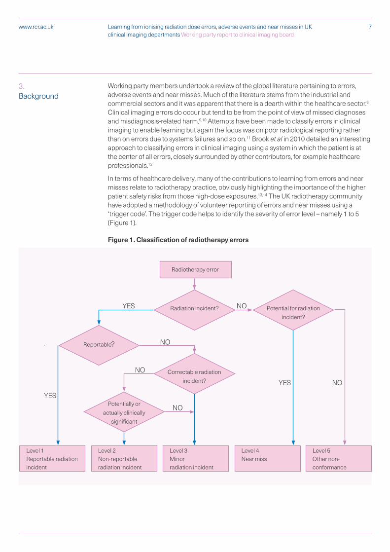
In terms of healthcare delivery, many of the contributions to learning from errors and near misses relate to radiotherapy practice, obviously highlighting the importance of the higher patient safety risks from those high-dose exposures.13,14 The UK radiotherapy community have adopted a methodology of volunteer reporting of errors and near misses using a ‘trigger code’. The trigger code helps to identify the severity of error level – namely 1 to 5 (Figure 1).
Figure 1. Classification of radiotherapy errors
3. Background
Radiotherapy error
Level 1Reportable radiationincident
Level 2Non-reportable radiation incident
Level 4Near miss
Level 3Minor radiation incident
Level 5Other non-conformance
Radiation incident?
Reportable?
Potential for radiation incident?
Correctable radiation incident?
Potentially or actually clinically
significant
YES
YES
YES
NO
NO
NO
NO
NO

8Learning from ionising radiation dose errors, adverse events and near misses in UK clinical imaging departments Working party report to clinical imaging board
www.rcr.ac.uk
Anonymised codes (using the trigger code) are submitted on a voluntary basis by UK radiotherapy departments’ to the national radiotherapy unit in Public Health England (PHE) where data consistency checks take place and patterns of errors are identified.15 The Patient Safety in Radiotherapy Steering Group review and analyse data and disseminate the learning across the UK.2
Clinical imaging errors and near misses and the learning from them has not evolved as much as in radiotherapy. Highlighting the compulsory notification of defined errors to national bodies has been raised in several books as an issue.4,16–18 The Radiology Events Register (RaER) was developed in Australia in 2006 designed to undertake systematic data collection and analysis of adverse incidents and discrepancies in radiology to support quality improvement and patient safety.19 However, in 2013 Hannaford et al reported that the dissemination of patient information into and from medical imaging settings in Australia was ‘fraught with error’.20 Some work was proposed in the United States (US) but it is unclear whether this became operational.10
A multitude of websites is available to the public providing varied and complex accounts of patient safety reporting in the UK.21–24 There is no single resource to illustrate how healthcare workers learn from radiation adverse events or near misses. Many search results signpost to international websites, particularly in the US. To provide assurance to the public and patients that we use information on adverse effects to influence change and improve practice, processes should be more transparant. The information should be accessible and centrally stored. This highlights the need not only for the existence of a reporting system but also for its thoughtful integration into the medical-imaging community in a manner that explains its purpose and promotes its effect.
The National Patient Safety Agency (NPSA) developed a framework for categorising the factors that could contribute to the occurrence of errors and near misses which must also be taken into consideration when analysing patterns of errors.25 Root cause analysis (RCA) is a method of problem solving used for identifying the root causes of faults or problems.26 It may be applied methodically to identify the root causes of events, rather than to simply address the symptomatic result. Although many people are treated safely and successfully daily in the UK NHS, when incidents do happen, it is important that lessons are learned to prevent the same incident occurring elsewhere. RCA investigation is a well-recognised way of doing this and while analysis is normally undertaken after an event, it can be a pre-emptive method to predict events.
Near miss reporting, termed ‘close calls’ or sentinel events is an established process, integral to industrial health and safety.27 Within diagnostic imaging there is currently very limited data available from mandatory reporting systems. In addition to this, there is arguably an ongoing fear of blame associated with adverse event reporting. By including near misses, with which there may be less perceived liability, the aim is to improve the attitude and frequency of reporting. In April 2005, the United States Joint Commission on Accreditation of Healthcare Organisations (JCAHO) developed a Patient Safety Event Taxonomy to address the problems associated with fragmented reporting of patient safety errors, near misses and adverse events.28 The objective was to examine existing reporting systems and create a common pathway that simplified and standardised data entry, subtraction and RCA. The study concluded that the taxonomy provided a common approach, which made it easier to file reports and investigate patient safety events consistently. Having this ability to interpret data on a large scale adds value to our capacity to learn from error. At the same time Shaw et al (2005) published a report concerning

9Learning from ionising radiation dose errors, adverse events and near misses in UK clinical imaging departments Working party report to clinical imaging board
www.rcr.ac.uk
adverse events and near miss reporting across 18 NHS trusts which concluded that ‘voluntary reporting by staff when linked to a multicentre data collecting system can yield information on a large number of incidents.’29 This seems to support the principle of creating a national IT system to collect and analyse incident data.
The COMARE the16th Report supported the establishment of a multidisciplinary team (medical physicists, radiographers and radiologists) whose role would be to optimise examinations, minimise radiation dose and lead a safe, effective radiation protection culture.7 The Department of Health (DoH) published a response in which it defines more precise roles for this multidisciplinary ‘image optimisation team’ including the collation and review of incidents and near misses to inform wider learning, change in practice and improve patient safety.30 It stresses the need for a team approach (radiographer, physicist and radiologist) to radiation protection (RP) risk management and best practice in RP governance. The imaging optimisation team would also be involved in highlighting the need for local review of error reports to enable feedback and learning to staff to ultimately improve patient services.
At the very least, there should be a methodology for standardised reporting of errors and near misses. NHS Improvement operate the NRLS, which collects and collates patient safety reports from healthcare staff across England and Wales. This information is used to develop advice and guidance for the NHS on reducing risks to patients. Every six months patient safety incident report statistics are published nationally.31 This relies upon voluntary reporting, from a variety of different data collecting systems. Data is submitted before incidents are locally investigated and so may not reflect the complete event. Reports are not specific to medical imaging although a safer practice notice was issued in 2007 to advise healthcare organisations to ensure that clinical imaging results are communicated and acted on appropriately.32 This accounts for only one stage in the patient pathway, which begins at referral, includes administration and irradiation, and ends after receiving results and subsequent care. At each stage in this process there is a person entitled with the responsibility of protecting the patient from the effects of ionising radiation. The reporting taxonomy recognises and identifies this.
Regulation 8 of the Ionising Radiation (Medical Exposure) Regulations [IR(ME)R] 2017 requires the reporting of accidental/unintended exposures that are clinically significant that do not occur as a result of equipment failure.5,6 In England, these are reported to the CQC, in Scotland, Northern Ireland and Wales reports are submitted directly to the appropriate devolved IR(ME)R Inspector for that country.33–36 A commonly used reporting tool across UK healthcare in general and imaging departments in particular is DATIX.37 This adverse event system estimates the consequence impact of the event by selecting one of five consequence impact categories:
1. Insignificant
2. Minor
3. Moderate
4. Major
5. Extreme
This existing system may offer the potential to develop a linked reporting system which integrates the reporting taxonomy. The 2016 CQC annual IR(ME)R report established a 3.3% increase in notifications of exposures ‘much greater than intended’ (notification was

10Learning from ionising radiation dose errors, adverse events and near misses in UK clinical imaging departments Working party report to clinical imaging board
www.rcr.ac.uk
under IR(ME)R 2000).38,39 The most commonly reported error was due to ‘wrong patient’ referred or identified within the medical imaging department. This is unchanged from the previous year and suggests inadequate learning from error reporting or insufficient implementation of preventative measures. There was an increase of 17% in notifications received from nuclear medicine. The CQC acknowledge that a proportion of this is likely to be consequent upon increased activity. The report presents no evidence to suggest poor practice and in fact the overall impression is that governance and reporting culture is improving.
Swiss cheese model It is well documented that every step in a process has potential for failure.40 The Swiss cheese model of accident causation is a model used in the risk analysis and risk management of human systems, commonly aviation, engineering and healthcare. It likens human systems to multiple slices of Swiss cheese, stacked together. The risk of a threat becoming a reality is mitigated by the differing layers and types of defenses which are stacked up behind each other. In theory, lapses and weaknesses in one defense do not allow a risk to materialise, since other defenses also exist. The model was originally formally propounded by Dante Orlandella and James T Reason from the University of Manchester and has since gained widespread acceptance. It is sometimes called the cumulative act effect.41
In the Swiss Cheese model, an organisation’s defenses against failure are modeled as a series of barriers, represented as slices of cheese.40 The holes in the slices represent weaknesses in individual parts of the system and are continually varying in size and position across the slices. The system produces failures when holes in each slice momentarily align, permitting (in Reason’s words) ‘a trajectory of accident opportunity’, so that a hazard passes through holes in all of the slices, leading to a failure.42–44 The model includes both active and latent failures. Active failures encompass the unsafe acts that can be directly linked to an accident, such as (in the case of aircraft accidents) pilot error. Latent failures include contributory factors that may lie dormant for days, weeks or months until they contribute to the accident.45
The same framework applies in healthcare – a latent failure could be the similar packaging of two drugs that are then stored close to each other in a pharmacy. Such a failure would be a contributory factor in the administration of the wrong drug to a patient. Errors in healthcare can be the result of ‘system flaws, not character flaws’.46
For a catastrophic error to occur, the holes need to align for each step in the process allowing all defences to be defeated. This represents an inherently flawed system that will allow a problem at the beginning to progress all the way through to affect adversely the outcome. Each slice of cheese is an opportunity to stop an error – the more defences you put up, the better. Also the fewer the holes and the smaller the holes, the more likely you are to catch/stop errors that may occur.
Root cause analysis (RCA)Root cause analysis (RCA) is a method used to identify the root cause(s) of faults or problems rather than to simply address the symptomatic result.47 Analysis is done after an event has occurred. Insights in RCA make it potentially useful as a preemptive method where it can be used to forecast or predict probable events even before they occur. When incidents occur it is important that lessons are learned to prevent the same

11Learning from ionising radiation dose errors, adverse events and near misses in UK clinical imaging departments Working party report to clinical imaging board
www.rcr.ac.uk
incident occurring elsewhere – RCA investigation is a well-recognised way of doing this. Investigations identify how and why patient safety incidents happen.47
Level 1 – Concise investigation
Most commonly used for incidents, claims, complaints or concerns that resulted in no, low or moderate harm to the patient.
Level 2 – Comprehensive investigation
Commonly conducted for actual or potential ‘severe harm or death’ outcomes from incidents, claims, complaints or concerns.
Level 3 – Independent investigation
As per Level 2, but in addition: must be commissioned and conducted by those independent to the provider service and organisation involved.
Conducting a thorough and detailed investigation into how incidents have happened will identify comprehensively the root cause and contributory factors. This will allow a detailed action plan to be developed that will prevent or minimise the risk of it happening again. The process map below describes the basic premise of how this should be carried out (Figure 2).
Figure 2. The RCA process
Getting started
Gathering information and mapping the incident
Indentifying care and service delivery problems
Analysing problems and identifying CFs and RCs
Generating solutions and recommendations
Implementing solution
Writing the report
Contents from NHS Improvement material32

12Learning from ionising radiation dose errors, adverse events and near misses in UK clinical imaging departments Working party report to clinical imaging board
www.rcr.ac.uk
Also within NHS Improvement, work is being undertaken to review the Incident Decision Tree which aims to discover where things go wrong rather than attributing blame to individual(s).48
Human factors‘Human factors is an established scientific discipline used in many other safety critical industries. Human factors approaches underpin current patient safety and quality improvement science, offering an integrated, evidenced and coherent approach to patient safety, quality improvement and clinical excellence’.
By understanding individualsʼ behaviours, interactions and limitations, human factors offers ways to minimise and mitigate human frailties, so reducing medical error and its consequences. Healthcare organisations are already starting to adopt this approach within their governance frameworks and offer human factors training as part of local induction.49
The UK NHS and the independent sector can learn valuable lessons from safety critical industries such as the airline and rail industry.
The report of the Mid Staffordshire NHS Foundation Trust Public Inquiry 2013 emphasised the critical importance of NHS organisations working in partnership to avoid, isolate and/or mitigate risk to high-quality patient care and ensure such widespread systemic failure does not happen again.50
When looking at root cause analysis following incidents it is important to consider that errors occur because the professionals working in healthcare are human and prone to error especially in stressful situations such as a modern clinical imaging department. Consideration of human factors in action plans and recommendations following on from incidents is an important point in order to establish safety barriers, understand the environment and mitigate the risk.
Patient impact ratingsIn their document Seven steps to patient safety, the National Patient Safety Agency has defined levels of harm caused by an incident to be:51
§ No harm – either
– Impact prevented – any patient safety incident that had the potential to cause harm but was prevented, resulting in no harm or
– impact not prevented – any patient safety incident that ran to completion but no harm occurred
§ Low – any patient safety incident that required extra observation or minor treatment and caused minimal harm
§ Moderate – any patient safety incident that resulted in a moderate increase in treatment and which caused significant but not permanent harm
§ Severe – any patient safety incident that appears to have resulted in permanent harm
§ Death – any patient safety incident that directly resulted in death.
There is a need to address how incidents involving unintended exposures to ionising radiation in clinical imaging are categorised according to harm. This applies not only to incidents reportable to the enforcement authority under IR(ME)R and to the Health and Safety Executive (HSE) under the Ionising Radiations Regulations (IRR), but also to
13Learning from ionising radiation dose errors, adverse events and near misses in UK clinical imaging departments Working party report to clinical imaging board
www.rcr.ac.uk
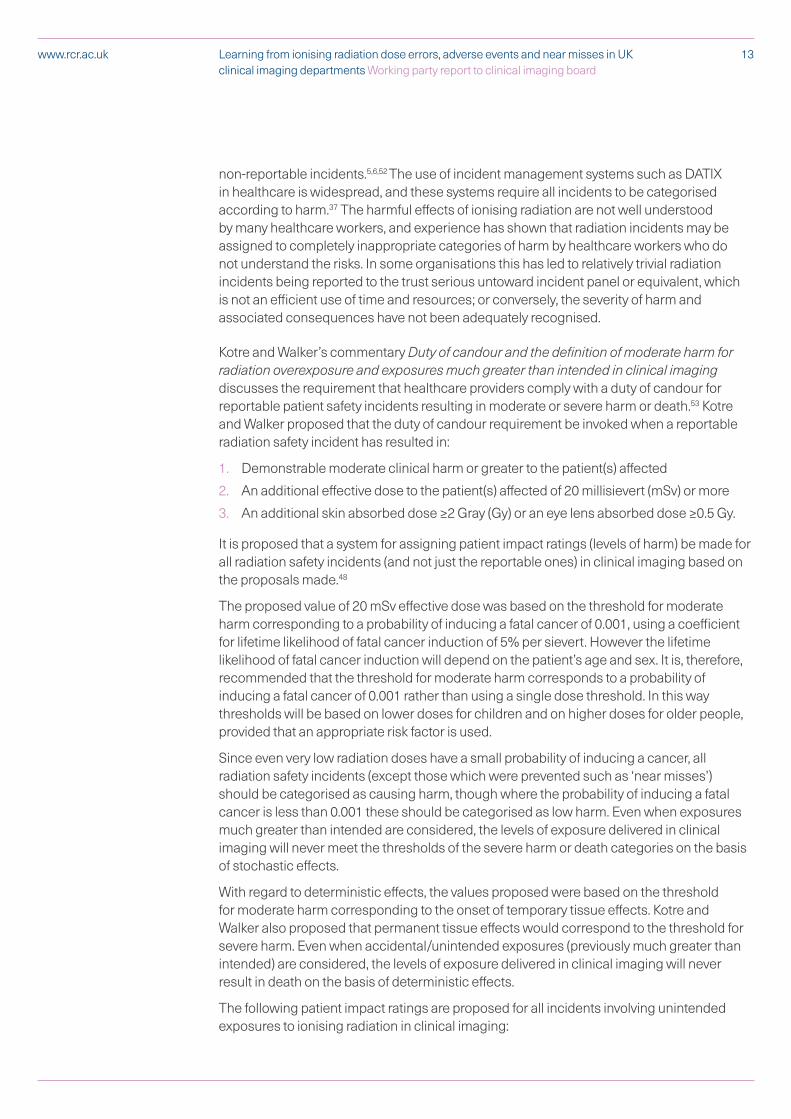
non-reportable incidents.5,6,52 The use of incident management systems such as DATIX in healthcare is widespread, and these systems require all incidents to be categorised according to harm.37 The harmful effects of ionising radiation are not well understood by many healthcare workers, and experience has shown that radiation incidents may be assigned to completely inappropriate categories of harm by healthcare workers who do not understand the risks. In some organisations this has led to relatively trivial radiation incidents being reported to the trust serious untoward incident panel or equivalent, which is not an efficient use of time and resources; or conversely, the severity of harm and associated consequences have not been adequately recognised.
Kotre and Walker’s commentary Duty of candour and the definition of moderate harm for radiation overexposure and exposures much greater than intended in clinical imaging discusses the requirement that healthcare providers comply with a duty of candour for reportable patient safety incidents resulting in moderate or severe harm or death.53 Kotre and Walker proposed that the duty of candour requirement be invoked when a reportable radiation safety incident has resulted in:
1. Demonstrable moderate clinical harm or greater to the patient(s) affected
2. An additional effective dose to the patient(s) affected of 20 millisievert (mSv) or more
3. An additional skin absorbed dose ≥2 Gray (Gy) or an eye lens absorbed dose ≥0.5 Gy.
It is proposed that a system for assigning patient impact ratings (levels of harm) be made for all radiation safety incidents (and not just the reportable ones) in clinical imaging based on the proposals made.48
The proposed value of 20 mSv effective dose was based on the threshold for moderate harm corresponding to a probability of inducing a fatal cancer of 0.001, using a coefficient for lifetime likelihood of fatal cancer induction of 5% per sievert. However the lifetime likelihood of fatal cancer induction will depend on the patient’s age and sex. It is, therefore, recommended that the threshold for moderate harm corresponds to a probability of inducing a fatal cancer of 0.001 rather than using a single dose threshold. In this way thresholds will be based on lower doses for children and on higher doses for older people, provided that an appropriate risk factor is used.
Since even very low radiation doses have a small probability of inducing a cancer, all radiation safety incidents (except those which were prevented such as ‘near misses’) should be categorised as causing harm, though where the probability of inducing a fatal cancer is less than 0.001 these should be categorised as low harm. Even when exposures much greater than intended are considered, the levels of exposure delivered in clinical imaging will never meet the thresholds of the severe harm or death categories on the basis of stochastic effects.
With regard to deterministic effects, the values proposed were based on the threshold for moderate harm corresponding to the onset of temporary tissue effects. Kotre and Walker also proposed that permanent tissue effects would correspond to the threshold for severe harm. Even when accidental/unintended exposures (previously much greater than intended) are considered, the levels of exposure delivered in clinical imaging will never result in death on the basis of deterministic effects.
The following patient impact ratings are proposed for all incidents involving unintended exposures to ionising radiation in clinical imaging:

14Learning from ionising radiation dose errors, adverse events and near misses in UK clinical imaging departments Working party report to clinical imaging board
www.rcr.ac.uk
§ Low harm – incidents resulting in:
1. Demonstrable low clinical harm to the patient(s) affected
2. Any additional radiation dose which is below the thresholds for moderate harm.
§ Moderate harm – incidents resulting in:
1. Demonstrable moderate clinical harm to the patient(s) affected
2. An additional probability of inducing a fatal cancer of 0.001 or more for the patient(s) affected
3. An additional tissue absorbed dose that results in temporary deterministic effects.
§ Severe harm – incidents resulting in:
1. Demonstrable severe clinical harm to the patient(s) affected
2. An additional tissue absorbed dose that results in permanent deterministic effects.
Duty of candourOne very important point, apart from the learning from previous errors and near misses, is the requirement to be open and honest with patients when something goes wrong.54 All healthcare professionals have a duty of candour – a professional responsibility to be open and honest with patients when things go wrong. This is articulated through various professional codes of conduct and specific professional guidance documents.55 Professions regulated by the Health and Care Professions Council (HCPC) should look to the Standards of conduct, performance and ethics: Standard 8.56 Duty of candour applies equally to professions regulated by the Nursing and Midwifery council (NMC) and the General Medical Council (GMC).57,58 In addition, there is a legal duty that applies to regulated professionals working in organisations delivering healthcare; in England, the CQC regulates this. Similar duties exist in Scotland, Wales and Northern Ireland. This duty involves a representative of the organisation informing, supporting and apologising to patients if there have been mistakes in their care that have led to harm. Duty of candour aims to help patients receive accurate, truthful information from healthcare providers.
With regard to radiation incidents, registered healthcare professionals have an obligation to ensure that they are always open and honest with patients regardless of severity of the incident. The only caveat to this would be where it would not be in the best interests of the patient. The HCPC have strengthened the Standards of conduct, performance and ethics to include:57
§ A dedicated standard requiring registrants to be open when things go wrong
§ Informing service users and carers when something goes wrong
§ Taking action where possible to put matters right.
Example
An 88 year old female patient underwent an X-ray of her hand that was not required by the consultant. The referral form had been completed in error by an unauthorised member of staff.
The risk to the patient from this exposure was extremely low. It was agreed with the consultant that it would cause unnecessary distress and anxiety to the patient to try and explain what had gone wrong and the risk involved as it was extremely low. This decision was documented in the patient’s notes.

15Learning from ionising radiation dose errors, adverse events and near misses in UK clinical imaging departments Working party report to clinical imaging board
www.rcr.ac.uk
When recording any radiation error, consideration must be given to the information shared with patients (or their representative) to comply with the professional and statutory duty of candour.5,6 Schedule 2(l) of 2017 IR(ME) Regulations stipulates that the patient (or a representative) is ‘provided with adequate information relating to the benefits and risks associated with the radiation dose from the exposure’ and in Regulation 8(1) be ‘informed of the occurrence of any relevant clinically significant unintended or accidental exposure’ and, in particular details of the outcome of the exposure analysis,6
For England duty of candour is within regulation 20 of the Health and Social Care Act 2008 (Regulated Activities) Regulations 2014.59 For Scotland duty of candour is within part 2 of the Health (Tobacco, Nicotine, Care and so on) (Scotland) Act 2016 and in Northern Ireland, there is a plan to introduce statuary duty of candour as stated in the NI annual report May 2015.60,61
Present situationThe working party members recognise that there is (to their knowledge), presently no national error categorisation system other than for reportable errors under IR(ME)R.4–6 Brook et al in 2010 proposed a categorisation system to support the analysis of near misses or adverse events with the patient at the heart of the system, other healthcare practitioners participating in his/her care and the interlinked role each plays between them in aiming for the successful patient outcome.12 This proposal also introduced the issue of other contributing factors in error analyses. It is unclear whether this proposal became operational.
Members of the working party believe that to prevent errors from occurring, there is a need for a readily available and easy-to-use (operational) system for detecting, classifying and analysing mistakes that can be subject to some form of root cause analysis. It could be argued that errors will continue to occur unless the initial error is properly addressed and potential contributing factors from the individuals involved are resolved.12 Errors may reflect long-standing substandard practices that are often retrospectively recognised and with latent system failures may allow errors to continue. A robust radiation safety culture involving radiation dose errors/near misses reporting within local departments is imperative in fostering patient safety and ongoing quality improvement of imaging services. It is also, arguably, the national sharing of the learning from such errors, which ultimately highlights and helps to support the potential need for procedural change.
Finally, Regulation 8(3) of IR(ME)R stipulates that the employer establishes a system for recording analyses of events involving or potentially involving accidental or unintended exposures.5,6 Although this is to be proportionate to the radiological risk posed by the practice, the working party believe that the proposed standard categorisation system supports compliance with these Regulations.
Before the development of a taxonomy and recording tool could be undertaken, members of the working party made some pre-emptive assumptions.
§ There is a wide variation in local practice of error reporting and review.
§ Some good practice and systems exist but they are not shared.
§ The volume of data that will be created due to the complexity of the various clinical imaging pathways will be a challenge.
§ There is a recognition that it may not able to capture everything.

16Learning from ionising radiation dose errors, adverse events and near misses in UK clinical imaging departments Working party report to clinical imaging board
www.rcr.ac.uk
§ There are four country differences in reporting errors.
§ Errors in referral are generally outside of the clinical imaging (CI) team.
§ Measurement of risk is not standard.
§ Not all radiation errors occur within clinical imaging departments, for example, they can occur in theatres, dental practice, cardiac catheters labs and so on.
§ Some classifications are already in use across the UK, for example:
– Equipment
– Staff training
– Referral error
– Duplicate
– Pregnancy
– Wrong patient
– Timing of referral.
Prior to any work beginning on the creation of a standard categorisation system, working party members felt it necessary to review the various patient pathways evident in clinical imaging services with the aim to illustrate (and standardise as much as possible) the pathways from the perspective of each IR(ME)R duty holder.

17Learning from ionising radiation dose errors, adverse events and near misses in UK clinical imaging departments Working party report to clinical imaging board
www.rcr.ac.uk
Referrer pathway for medical exposures The diagram below shows the steps involved for the referrer as the IR(ME)R duty holder when referring a patient for clinical imaging.
Considering the risk versus benefit principal, ‘benefit’ can only be established after the referrer has reviewed the results and made a decision regarding treatment or further investigation.
Patient correctly identified.Verify pregnancy or breastfeeding status.Previous medical history checked including relevant imaging (including duplicate requests).Patient’s mobility assessed.Confirm patient understands and consents to the examination and understands when/how they will receive the appointment/urgent examination.
Referral guidelines (iRefer or local guidelines) to confirm appropriate examination requested.62
Non-ionising radiation alternative considered.Adequate relevant clinical information supplied on request form as required and including previous imaging.
Correct region/laterality confirmed.Unique identifier confirmed (signature/electronic signature/correct user login).Ensure correct timing is clearly defined.
Mandatory information completed. Check if this is the CORRECT patient again.Complete and send request.Cancellation procedure or exams no longer required.
EXP
OSU
RE
Make and record clinical evaluation of examination in line with local procedures.*
Ensure clinical evaluation is used in the decision to manage.Consider need for further imaging .Discuss with patient.
*All steps, preceding (pre-exam pale pink box) and proceeding (post-exam white box) the medical exposure have been included.
Also see the Society and College of Radiographersʼ IR(ME)R Referrer Pause and Check poster.63
Practitioner pathway for medical exposures The diagram overleaf shows the steps involved for the practitioner as the IR(ME)R duty holder when justifying a diagnostic imaging procedure. Consideration must be given to the risk versus benefit principal, such that a sufficient net benefit should result from the medical exposure.
Where no direct medical benefit is expected for the individual (volunteers participating in research exposures) dose constraints should be adhered to.
All steps, preceding the medical exposure have been included.
Please note: In interventional radiology, the practitioner may be a radiologist, cardiologist, vascular surgeon or a radiographer with advanced practice.
Please note: In nuclear medicine this is always the Administration of Radioactive Substances Advisory Committee (ARSAC) certificate holder (b) in Tier 2 of the coding taxonomy. In IR(ME)R this person must hold a practitioner licence.5,6
4. Patient pathways for all clinical imaging modalities

18Learning from ionising radiation dose errors, adverse events and near misses in UK clinical imaging departments Working party report to clinical imaging board
www.rcr.ac.uk
Other clinicians or radiographers who are entitled (as the operator) may authorise under guidelines produced by the practitioner.
Confirm referrer ID.(Confirm referrer is entitled).Patient correctly identified.Match patient data on referral with RIS.
Check previous medical history, including all relevant imaging.Enquire whether patient is pregnant or breastfeeding if relevant.Establish intended timing of procedure.
Evaluate clinical information supplied by referrer and consider any appropriate alternative procedure not involving ionising radiation.Balance risk v benefit of medical exposure and confirm decision.
Assign modality and protocol. Include any specific requirements for the individual exposure.
Assign urgency.Clarify timing of procedure.
Justify the medical exposure. Authorise the medical exposure.
Please note: Some of these stages may be undertaken by the entitled operator using authorisation guidelines developed by the practitioner.
Operator pathway for medical exposuresThe diagram below shows the steps involved for the operator as the IR(ME)R duty holder when performing the practical aspects of the exposure during a diagnostic imaging procedure.
This pathway assumes that the equipment is fit for purpose, that regular quality-assurance checks have been undertaken and that operators have been adequately trained to use the equipment. Also see the IR(ME)R Operator Pause and Check poster.64
Confirm identity of referrer (check they are entitled).Confirm justification of the exposure and identity of entitled practitioner.ORCompare referral with guidelines produced by a practitioner and authorise request when entitled.Check previous medical imaging for the patient.Confirm timing of the examination is appropriate.Confirm modality is correct.Check blood results as required for intravenous injections/interventional procedures.
Confirm correct patient identity.Confirm previous medical history and relevant imaging with patient.Explain procedure and confirm patient understands.
Confirm no contraindications to examination (follow pregnancy/breastfeeding policy and so on).Confirm consent and record where appropriate.Confirm correct body region/laterality. Confirm patient weight/height when appropriate.Position patient.
Confirm correct product, date, volume, flow-rate, concentration, activity (where appropriate) and route of administration for any intravenous (IV) contrast agent or radiopharmaceutical associated with exposure.Select appropriate examination protocol and equipment settings.Perform optimisation adjustments with due regard to patient age, sex, pregnancy status, BMI and dose constraints.
EXP
OSU
RE
Complete exposure.Check image quality and confirm no further imaging required.Complete post processing.Attend to aftercare needs of patient including appropriate information regarding results.
Send images to image archive system and confirm complete arrival of images on archive system (where possible before proceeding with next patient).
Record exposure factors Complete clerical duties with regard to all documentation including the administration of contrast agent or radio-pharmaceutical.Document a clinical evaluation.

19Learning from ionising radiation dose errors, adverse events and near misses in UK clinical imaging departments Working party report to clinical imaging board
www.rcr.ac.uk
Operator pathway for studies involving radioactive substances (nuclear medicine and nuclear cardiology including single photon emission computed tomography [SPECT]/CT and positron emission tomography [PET]/CT)The diagram overleaf shows the steps involved for the operators when performing the practical aspects of the exposure for studies involving radioactive substances paying special attention to differences/specific requirement that occur in these types of studies compared to other modalities in clinical imaging.
This pathway involves a number of steps and many different professionals. Some of the steps do not directly involve the patient and not all steps are relevant to all patients. More than one operator may be included in each step, for example, when double checking on dispensing or administration is required.
This pathway assumes that the equipment is fit for purpose, that regular quality-assurance checks have been undertaken with regard to the equipment and the radiopharmaceutical and that operators have been adequately trained.

20Learning from ionising radiation dose errors, adverse events and near misses in UK clinical imaging departments Working party report to clinical imaging board
www.rcr.ac.uk
Aut
hori
sati
on u
nder
pr
otoc
olR
eque
stin
g R
adio
phar
mac
euti
cal
Pre
pari
ng a
nd
disp
ensi
ng
Rad
ioph
arm
aceu
tica
l
Che
ckin
g qu
alit
y of
R
adio
phar
mac
euti
cal
Adm
inis
trat
ion
of
radi
oact
ivit
y
EXPOSURE – ADMINISTRATION
Sca
nnin
g th
e pa
tien
t
EXPOSURE – CT SCANNING AS REQUIRED
Taki
ng ra
dioa
ctiv
e bl
ood
sam
ples
Pro
cess
ing
of ra
dioa
ctiv
e bl
ood
sam
ples
Con
firm
refe
rrer
ID.
(Con
firm
refe
rrer
is e
ntitl
ed).
Patie
nt c
orre
ctly
iden
tified
.M
atch
pat
ient
dat
a on
refe
rral
w
ith R
IS.
Che
ck p
revi
ous
med
ical
hi
stor
y, in
clud
ing
all r
elev
ant
imag
ing.
Enqu
ire w
heth
er p
atie
nt is
pr
egna
nt o
r bre
astfe
edin
g if
rele
vant
and
issu
e ad
vice
as
appr
opria
te.
Eval
uate
clin
ical
info
rmat
ion
supp
lied
by re
ferr
er a
nd
com
pare
with
pro
toco
l/gu
idel
ines
pro
duce
d by
A
RSA
C h
olde
r to
esta
blis
h/as
sign
pro
cedu
re to
be
unde
rtak
en.
Esta
blis
h in
tend
ed ti
min
g an
d ra
dioa
ctiv
ity a
s pe
r A
RSA
C h
olde
r’s p
roto
col/
guid
elin
es.
Aut
horis
e th
e m
edic
al
expo
sure
whe
n en
title
d.
Con
firm
aut
horis
atio
n of
th
e ex
posu
re a
nd id
entit
y of
ent
itled
pra
ctiti
oner
/au
thor
isin
g op
erat
or.
Che
ck p
revi
ous
med
ical
im
agin
g fo
r the
pat
ient
.C
onfir
m ti
min
g of
the
exam
inat
ion
is a
ppro
pria
te.
Con
firm
cor
rect
pro
cedu
re.
Che
ck b
lood
resu
lts o
r ot
her s
afet
y pr
ecau
tions
as
appr
opria
te fo
r pro
cedu
re.
Esta
blis
h an
y co
rrec
tions
to
be m
ade
to th
e ra
dioa
ctiv
ity
base
d on
age
/hei
ght/
wei
ght
of p
atie
nt a
s pe
r AR
SAC
ho
lder
’s p
roto
col.
Req
uest
the
corr
ect
radi
oact
ivity
and
ra
diop
harm
aceu
tical
for t
he
corr
ect t
ime,
pro
cedu
re a
nd
patie
nt.
Con
firm
cor
rect
requ
este
d ra
dioa
ctiv
ity a
nd
radi
opha
rmac
eutic
al fo
r co
rrec
t pro
cedu
re.
Ensu
re th
e co
rrec
t m
anuf
actu
ring
of th
e ra
diop
harm
aceu
tical
pro
duct
. M
ake
appr
opria
te c
orre
ctio
ns
for r
adio
activ
ity b
ased
on
age/
heig
ht/w
eigh
t/pr
egna
ncy
stat
us o
f pat
ient
as
per
AR
SAC
hol
der’s
pro
toco
l.D
raw
up
the
corr
ect
prod
uct,
incl
udin
g vo
lum
e,
conc
entr
atio
n, a
nd
radi
oact
ivity
for t
he p
roce
dure
an
d en
sure
this
is c
orre
ct
for t
he re
ques
ted
time
of
adm
inis
trat
ion.
C
orre
ctly
labe
l the
via
l/syr
inge
an
d up
date
reco
rds.
Ensu
re th
e ap
prop
riate
ch
rom
atog
raph
y is
un
dert
aken
for t
he c
orre
ct
radi
opha
rmac
eutic
al a
s pe
r lo
cal p
roto
col t
o en
sure
that
th
e ra
dioc
hem
ical
pur
ity o
f th
e pr
oduc
t con
form
s to
the
guid
elin
es.
Rec
ord
resu
lt as
app
ropr
iate
.
Con
firm
cor
rect
pat
ient
iden
tity.
Con
firm
mod
ality
is c
orre
ct.
Con
firm
med
ical
his
tory
and
cl
inic
al in
form
atio
n w
ith p
atie
nt
and
requ
est.
Con
firm
pre
viou
s m
edic
al
imag
ing
for t
he p
atie
nt.
Con
firm
tim
ing
of th
e ex
amin
atio
n is
app
ropr
iate
.C
onfir
m b
lood
resu
lts o
r ot
her s
afet
y pr
ecau
tions
as
appr
opria
te.
Con
firm
con
sent
and
reco
rd
whe
re a
ppro
pria
te.
Con
firm
no
cont
rain
dica
tions
to
exa
min
atio
n (fo
llow
loca
l pr
egna
ncy/
brea
stfe
edin
g po
licy,
et
c) in
clud
ing
patie
nt a
bilit
y to
un
derg
o sc
an.
Con
firm
adv
ice
as a
ppro
pria
te
(for e
xam
ple,
sto
p br
east
feed
ing
for x
hrs
)C
onfir
m a
ny c
orre
ctio
ns m
ade
to
the
radi
oact
ivity
bas
ed o
n ag
e/he
ight
/wei
ght p
regn
ancy
sta
tus
of p
atie
nt a
re c
orre
ct a
s pe
r A
RSA
C h
olde
r’s p
roto
col.
Expl
ain
proc
edur
e an
d an
y re
stric
tions
to b
e fo
llow
ed
afte
rwar
ds a
nd c
onfir
m p
atie
nt
unde
rsta
nds.
Con
firm
cor
rect
ra
diop
harm
aceu
tical
, dat
e, ti
me,
vo
lum
e, ra
dioa
ctiv
ity a
nd ro
ute
of a
dmin
istr
atio
n an
d re
cord
as
appr
opria
te.
Con
firm
cor
rect
pat
ient
iden
tity.
Con
firm
mod
ality
is c
orre
ct.
Con
firm
cor
rect
radi
opha
rmac
eutic
al
adm
inis
trat
ion
and
that
tim
ing
of th
e ex
amin
atio
n is
app
ropr
iate
.C
onfir
m m
edic
al h
isto
ry a
nd c
linic
al
info
rmat
ion
with
pat
ient
and
requ
est.
Con
firm
pre
viou
s/re
leva
nt im
agin
g.Ex
plai
n pr
oced
ure
and
confi
rm p
atie
nt
unde
rsta
nds.
Con
firm
cor
rect
bod
y re
gion
/lat
eral
ity.
Posi
tion
patie
nt.
Sele
ct a
ppro
pria
te e
xam
inat
ion
prot
ocol
and
equ
ipm
ent s
ettin
gs.
Perf
orm
opt
imis
atio
n ad
just
men
ts w
ith
due
rega
rd to
pat
ient
hei
ght/
wei
ght o
r B
MI,
preg
nanc
y st
atus
.C
onfir
m n
o fu
rthe
r im
agin
g re
quire
d.If
furt
her i
mag
ing
is re
quire
d en
sure
ap
prop
riate
aut
horis
atio
n is
obt
aine
d.
Con
firm
cor
rect
pat
ient
iden
tity.
Con
firm
con
sent
and
reco
rd w
here
ap
prop
riate
.C
onfir
m c
orre
ct p
roce
dure
.C
onfir
m re
leva
nt m
edic
al h
isto
ry/c
linic
al
info
rmat
ion
with
pat
ient
.Ex
plai
n pr
oced
ure
and
confi
rm p
atie
nt
unde
rsta
nds.
Con
firm
cor
rect
sam
plin
g si
te.
Con
firm
cor
rect
radi
opha
rmac
eutic
al
adm
inis
trat
ion
and
that
tim
ing
is
appr
opria
te a
nd re
cord
as
appr
opria
te.
Ensu
re s
ampl
es a
re la
belle
d co
rrec
tly w
ith
patie
nt, s
tudy
and
tim
ing
deta
ils.
Con
firm
cor
rect
pat
ient
sam
ples
Se
lect
app
ropr
iate
pro
toco
l and
equ
ipm
ent
setti
ngs
as p
er re
ques
t.En
sure
all
sam
ples
are
sub
divi
ded
into
cor
rect
ly
labe
lled
vial
s an
d fo
llow
cor
rect
loca
l pro
toco
l. Pr
epar
e st
anda
rd s
ampl
es a
s pe
r loc
al p
roto
col
as a
ppro
pria
te.
Inse
rt c
orre
ct p
atie
nt, p
roce
dure
, ra
diop
harm
aceu
tical
, blo
od a
nd s
tand
ard
sam
ples
dat
a in
to c
alcu
latio
n sh
eet t
o ob
tain
th
e re
sult.

21Learning from ionising radiation dose errors, adverse events and near misses in UK clinical imaging departments Working party report to clinical imaging board
www.rcr.ac.uk
5. Categorisation methods
Coding framework (taxonomy)To support the development of a system that identifies, classifies, codes and reports radiation dose errors, adverse events and near misses, the working party, over a period of time, created many versions of a taxonomy. It was important that this taxonomy identified each element of the typical patient pathways found in both radiological and nuclear medicine services and that the resultant code could identify the root cause. The complex nature of radiological and nuclear medicine services caused widespread discussion (and re-discussion) before a taxonomy could be agreed and prepared for the pilot phase.
The initial pre-pilot coding framework (the taxonomy) detailed each part of the patient pathway from point of referral to final report. It detailed options for the user to identify the nature of the error, adverse event or near miss (incident).
§ The severity level (1–4)
§ The exposure type (1–4)
§ The modality used(1–7)
To support the identification of the root cause of an incident, the coding framework was divided into the four duty holder roles within the Ionising Radiation (Medical Exposure) Regulations 2000 – namely:
§ The employer
§ The referrer
§ The practitioner
§ The operator.
With further sub categories (for example):
§ Wrong anatomy
§ Wrong side and so on.
Within each duty holder section, there were numbers listed to indicate at which point on the pathway (Tier 1) the incident actually occurred together with the identification of the cause of the incident (Tier 2). Incidents often involve a complex chain of events. Whil an oversight or certain action may be viewed as the immediate cause of an incident, subsequent analysis will often expose a series of events or deviations from safe practice. These events are described as root cause and causative (contributory) factors.
Incident
§ Tier 1 – Primary code: the point in the pathway that the error first occurred.
§ Tier 2 – Secondary code: what went wrong? The detail of the error.
These two ‘tiers’ identify the root cause of any given incident.
Additionally, the coding framework also identified potential causative factors (numbered CF1 to CF7) which included headings such as individual; environmental; technical; patient-related and so on. For any given incident, there could have been one or more causative factors. The causative (contributory) factor(s) identify the weakness(es) that allow an incident to occur.

22Learning from ionising radiation dose errors, adverse events and near misses in UK clinical imaging departments Working party report to clinical imaging board
www.rcr.ac.uk
With the use of the coding framework, following the internal reporting of an incident, an alphanumeric code is produced which is then entered into an IT system to support the identification of patterns of errors and near misses. The working party created another tool (a basic IT system) to record the alphanumeric code – namely, the reporting template (RT) which is a basic Excel spreadsheet with drop down boxes for each element of the final alphanumeric code (see Appendix 4).
Example:
As an example of a clinical incident, the following scenario helps to illustrate how the coding framework (pilot) was used to create the final alphanumeric code.
An adult patient presented for a skeletal survey X-ray and this was undertaken by a second year student under supervision.
On the lateral lumbar view the detector was not fully covered by the X-ray beam and a very high exposure – subsequently five times the intended dose – was given for the lateral lumbar spine.
All doses have been recorded.
For this incident, the resultant code is: 2/1/1/DH4f/T14/T2c/CF1a/
Severity – Level 2
Type – 1 – medical exposure
Modality – 1 – general radiology
Duty holder – DH4 – operator – f – trainee under supervision
Tier 1–4 – pre-exposure safety checks
Tier 2 – c – wrong patient position/set up
Causative factor – CF1 – organisational – a – inadequate leadership/supervision
Reporting templateThe reporting template (RT), a basic excel spreadsheet with drop down boxes for each element of the taxonomy, allows the recording of the final alphanumeric code (see Appendix 4). Once the code is agreed, the user merely inserts this into the RT by clicking on the relevant boxes for each column detailed in the RT. The idea is that the RT should be a live document that may be used at any time and by any user, preferably the one which codes the incident. Results from the RT may be analysed for patterns of errors and near misses and can be shared locally or nationally. It is hoped that the information from these patterns would be widely disseminated to support ongoing discussion and learning by staff in UK services.
Members of the working party ultimately hope that this reporting template be either linked to a web-based system or to other present systems, for example, DATIX to allow ease of use and time saving within already busy clinical imaging departments.37 Recommendation number four within this guidance document highlights this aspiration.
To test the validity, reliability and reproducibility of the tools (the coding framework, the causative factors and the reporting template), a pilot study took place towards the end of 2016 and was completed early 2017. Seventeen clinical imaging centres from throughout

23Learning from ionising radiation dose errors, adverse events and near misses in UK clinical imaging departments Working party report to clinical imaging board
www.rcr.ac.uk
the four countries of the UK were invited to participate. Twelve centres responded making a 70% response rate. See Appendix 2 for full details of the pilot study.
The coding framework and the reporting template used in the pilot phase are detailed in these files:
The causative factors taxonomy used in the pilot phase is detailed in Appendix 2d.
Each centre was asked to:
§ Use the pilot coding framework with the pilot contributory factors taxonomy (CF) to code six control scenarios (coded and consistency checked by working party members)
§ To code ten recent radiation incidents from their own department to retrospectively test the coding taxonomy
§ To then insert the final alphanumeric code for each of the sixteen incidents into the pilot reporting template (RT)
§ To complete the pilot participation form to highlight any ambiguities/difficulties encountered in using the two tools.
Control scenarios for coding in pilot study (with working party [WP] code results) § Scenario one: An 86-year-old male in patient received an unintended computed
tomography (CT) abdo pelvis when the radiographer scanned the wrong body area. A CT chest was requested. Concerns were raised around this individual who had been involved with more than one incident. It was identified that there was inadequate training, assessment and supervision of this radiographer which led to three reportable incidents within CT. The individual stated that they had been distracted due to a busy department and also lacked knowledge around protocols and radiologists justification codes.
WP code: 1/1/2/DH4c/T14/T2e/CF1e/CF2c
§ Scenario two: A patient was admitted via the emergency department (ED) and was assessed by a stroke emergency nurse practitioner (ENP) who requested a CT head and brought the request to CT. A second request was submitted by a doctor and this was signed off as justified by a radiologist; the request was then entered onto computerised radiology system (CRIS). The patient was collected from the ED by the operating radiographer and scanned following all procedures and protocols. The patient was re-entered onto CRIS and attended for a CT head by a radiology assistant working in CT – it was not identified that the patient had already had a CT scan. The patient was positioned by a different radiographer who was on their break during the first scan and so did not recognise the patient. The patient did not alert the radiographer to their first scan. The ID procedure was followed and stop/check for the scan took place (where two radiographers scan a patient – all details are confirmed and recalled), however, previous imaging had not been checked. The staff in CT stated that they had been busy that day and must have forgotten to complete the imaging history check appropriately.
WP code: 1/1/2/DH4c/T12/T2a/CF2d/CF3c – this one caused much difference in pilot study – definitely an operator error
§ Scenario three: A patient had a barium swallow examination. It was discovered that the examination was not reported. The examination was given to a radiologist but only two images were on the picture archiving and communication system (PACS), an image

24Learning from ionising radiation dose errors, adverse events and near misses in UK clinical imaging departments Working party report to clinical imaging board
www.rcr.ac.uk
of the stomach and a procedure summary. It transpired that due to a problem with PACS on the day of the procedure, the automatic transfer of images to PACS had not taken place for this patient. There was no written procedure in place for staff to check that images had transferred to PACS correctly. Staff looked for images on the machine where the examination was performed but the images were not on the machine as the exam had been performed many weeks earlier and an engineer had serviced the machine since then. The patient was informed and an immediate appointment made for the patient.
WP code: 2/1/4/DH4c/T17/T2e/CF2a – should definitely be operator error with employers responsibility being a CF
§ Scenario four: The patient was having a SPECT/CT. For the CT, the justifying clinician had specified the following protocols:
– Cervical spine (reference 100 mAs) for skull base to C6/C7
– Thoracic spine (reference 40 mAs) for C6/C7 to T12
– Lumbar spine (reference 65 mAs) for T12 to top of hip joints.
The technologist used the cervical spine protocol to scan from the skull base to T12 and the lumbar spine protocol for T12 to the top of the hip joints, so C6/C7 to T12 was scanned with the cervical spine protocol instead of the lower dose thoracic spine protocol. In addition to the dose from the CT, the patient will have received a dose from administration of 800 MBq of Tc-99m MDP for the SPECT scan. The effective dose from this is estimated as 5 mSv.
WP code: 2/1/2//DH4g/T14/T2b/CF2c – should be NM
§ Scenario five: An inpatient who had undergone recent abdominal surgery subsequently developed chest pain. He was given a CT scan to investigate this, at the request of a consultant cardiologist. An incidental finding of this CT scan was that the patient had wedge compressions of the T6 and T7 vertebrae. A bone mineral density scan was requested to assess this to indicate the likelihood of further fractures. The referring doctor was not familiar with the differences between referral forms for a bone mineral density (DEXA) scan and a nuclear medicine bone scan. The latter was used in error but clearly included clinical indications for a DEXA scan. When vetted by the ARSAC certificate holder, the error was realised due to the clinical indications given for the referral. This was communicated back to the referrer and the correct request was made for a DEXA scan.
WP code: 3/1/3/DH2a/T12/T2g/CF2c/CF3b – should be DEXA – or could be NM – the intended modality should be coded
§ Scenario six: A patient was referred to the inpatient X-ray department for a postoperative examination of the right hip. The radiographer identified the patient correctly and asked if it was their right side that was to be imaged. She discussed that it was a referral for a hip examination and the patient agreed. An anteroposterior (AP) X-ray of both hips and a lateral of the right hip were taken. It was not obvious at this stage that the wrong examination had been performed as the patient had undergone a previous hip replacement. After the procedure it became apparent to the radiographer that it was the knee that had been operated on and not the hip. The radiographer rang the referring doctor and spoke to the staff nurse on the ward who

25Learning from ionising radiation dose errors, adverse events and near misses in UK clinical imaging departments Working party report to clinical imaging board
www.rcr.ac.uk
confirmed that it was the patient’s knee that had been replaced. The correct imaging was then carried out.
WP code: 1/1/1/DH2a/T13/T2b/C
Changes following the pilot studyIt was apparent to the working party that slight changes were required of both the coding framework (re-named coding taxonomy) and the reporting template.
The main ones were:
1. The intended modality should be coded and not the modality requested in error. An additional column was added to reflect this.
2. The need to include a none duty holder at times when an error occurs out-with the control of a duty holder, for example, when equipment fails. Duty holders are now numbered 1–5 and an additional column was added.
The causative factors within the scenarios caused the greatest discrepancies during the pilot study and it was necessary to re-word the taxonomy which was then re-named as the contributory factors (CF) taxonomy (Appendix 3). It was felt necessary to introduce sample scenarios and resultant codes for the CF section to be included within this guidance document to support a deeper understanding for the future user.
The main lessons learned from the pilot study were:
– Many departments already have local reporting procedures
– Changes required of the causative factors
– Very difficult to do well without detailed scenario information
– Equipment incidents need to be factored in
– Subjectivity is natural
– Consistency checking of data coding is required.
The final coding taxonomy is detailed in Appendix 3.
The final contributory factors taxonomy is also summarised in Appendix 3 (see next section for more detail).
The final reporting template is detailed in Appendix 4.
Final contributory factor taxonomy detailsFollowing the pilot phase, the final contributory factors taxonomy was reformed. The working party elected to include contributory factors when developing the error coding taxonomy as an element of their remit to provide a process for the classification of errors and near misses in diagnostic imaging and nuclear medicine. The working party felt inclusion of contributory factor taxonomies would enhance trend analysis.
Future work on the analysis of diagnostic imaging and nuclear medicine errors would seek to improve the learning from these events, subsequently improving patient safety.
Root cause and contributory factor

26Learning from ionising radiation dose errors, adverse events and near misses in UK clinical imaging departments Working party report to clinical imaging board
www.rcr.ac.uk
Incidents and errors often involve a complex chain of events. While an oversight or certain action may be viewed as the immediate cause of an incident, subsequent analysis will often expose a series of events or deviations from safe practice. These events are described as root cause and contributory factors.
Root cause – identified event that leads to an occurrence or incident … the what. (The primary point on the pathway coding – Tier 1 and Tier 2).
Contributory factor – weakness that causes the apparent basis of an event to happen ... the why. (The contributory factors [CF] coding).
Definitions and examples of the clinical imaging CF taxonomies are provided later in this section. CF taxonomy is found in Appendix 3. A description of how to apply the CF coding process is provided below.
Application of error taxonomies
It is intended that both the root cause (Tier 1 and 2) and contributory factor taxonomies are applied by individuals with a clear understanding of clinical imaging processes, and who will have received some training on the application of the taxonomies. Ideally these individuals would include (and be supported by) a multidisciplinary team consisting of medical physicists, radiographers and radiologists.
Application of contributory factor taxonomy
Several studies have shown there is often a complex chain of events that may lead to an adverse outcome.63 Although a particular action or omission may be the immediate cause of an incident, closer analysis usually reveals a series of events and departures from safe practice. The contributory factor (CF) taxonomy has been designed so that each of these events can be captured.
These events are described as root cause and contributory factors.
The root cause has been defined as an identified event that leads to anticipated operational occurrences or accident conditions.64
A contributory factor is defined as the latent weakness that allows or causes the observed cause of an initiating event to happen, including the reasons for the latent weakness.
Examples of the application of the contributory factor taxonomy are provided in the section of this document entitled ‘Scenarios for the application of taxonomies’.
The initial taxonomy code that should be applied is the root cause, primary pathway coding (Tier 1 and Tier 2). The subsequent contributory factor coding taxonomy facilitates the inclusion of up to three CF per event. The entire error coding process (Tier 1 and Tier 2 and CF codes) have been included in the examples provided in this section.
Definitions and examples of contributory factor taxonomy
CF1: Individual
The field of human factors concerns the interaction between humans and the system in which they work.45 Human error occurs when the actions and decisions of individuals result in failures that can immediately or directly impact patient safety. Human or individual factors may be divided into the following categories:
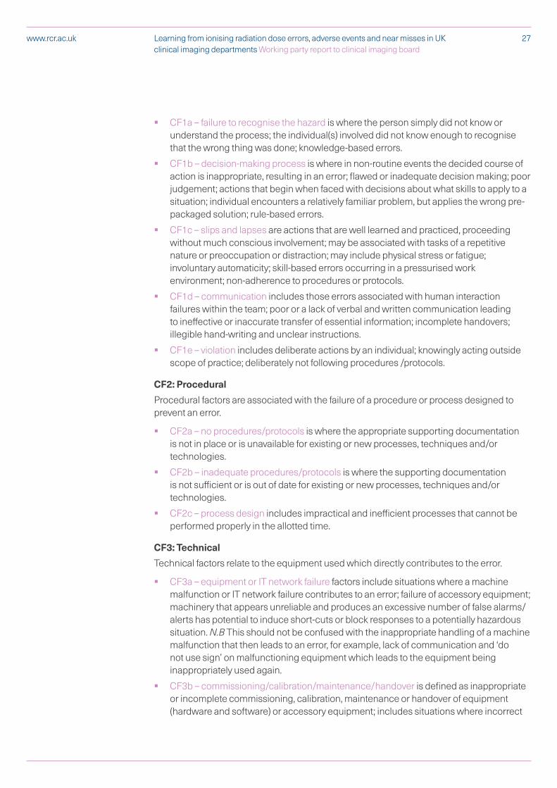
27Learning from ionising radiation dose errors, adverse events and near misses in UK clinical imaging departments Working party report to clinical imaging board
www.rcr.ac.uk
§ CF1a – failure to recognise the hazard is where the person simply did not know or understand the process; the individual(s) involved did not know enough to recognise that the wrong thing was done; knowledge-based errors.
§ CF1b – decision-making process is where in non-routine events the decided course of action is inappropriate, resulting in an error; flawed or inadequate decision making; poor judgement; actions that begin when faced with decisions about what skills to apply to a situation; individual encounters a relatively familiar problem, but applies the wrong pre-packaged solution; rule-based errors.
§ CF1c – slips and lapses are actions that are well learned and practiced, proceeding without much conscious involvement; may be associated with tasks of a repetitive nature or preoccupation or distraction; may include physical stress or fatigue; involuntary automaticity; skill-based errors occurring in a pressurised work environment; non-adherence to procedures or protocols.
§ CF1d – communication includes those errors associated with human interaction failures within the team; poor or a lack of verbal and written communication leading to ineffective or inaccurate transfer of essential information; incomplete handovers; illegible hand-writing and unclear instructions.
§ CF1e – violation includes deliberate actions by an individual; knowingly acting outside scope of practice; deliberately not following procedures /protocols.
CF2: Procedural
Procedural factors are associated with the failure of a procedure or process designed to prevent an error.
§ CF2a – no procedures/protocols is where the appropriate supporting documentation is not in place or is unavailable for existing or new processes, techniques and/or technologies.
§ CF2b – inadequate procedures/protocols is where the supporting documentation is not sufficient or is out of date for existing or new processes, techniques and/or technologies.
§ CF2c – process design includes impractical and inefficient processes that cannot be performed properly in the allotted time.
CF3: Technical
Technical factors relate to the equipment used which directly contributes to the error.
§ CF3a – equipment or IT network failure factors include situations where a machine malfunction or IT network failure contributes to an error; failure of accessory equipment; machinery that appears unreliable and produces an excessive number of false alarms/alerts has potential to induce short-cuts or block responses to a potentially hazardous situation. N.B This should not be confused with the inappropriate handling of a machine malfunction that then leads to an error, for example, lack of communication and ‘do not use sign’ on malfunctioning equipment which leads to the equipment being inappropriately used again.
§ CF3b – commissioning/calibration/maintenance/handover is defined as inappropriate or incomplete commissioning, calibration, maintenance or handover of equipment (hardware and software) or accessory equipment; includes situations where incorrect

28Learning from ionising radiation dose errors, adverse events and near misses in UK clinical imaging departments Working party report to clinical imaging board
www.rcr.ac.uk
data was provided by the vendor or supplier; where equipment was incorrectly calibrated or protocols were adjusted by the vendor or supplier.
– CF3c – device/product design factors include flaws or inadequacies inherent in the design of equipment or ancillary kit used as part of the exposure or to inform the exposure.
CF4: Patient related
Patient factors relate to incidents where the actions or individual circumstances of the patient directly contribute to the error. These are sub-divided into the following categories:
§ CF4a – medical condition relates to where the patient’s physical condition is particularly complex or serious including an inability to remain still.
§ CF4b – communication with the patient includes those errors associated with human interaction failures between the team and the patient; includes language issues, comprehension difficulties; through lack of or miscommunication the patient has misunderstood an instruction leading directly to an error.
§ CF4c – non-compliance is described as being when a patient does not comply with the procedure; this may be through their own volition or through an unknown inability to comply; where cultural, religious and social issues affect the ability of a patient to be consistent with pre-conceived expectations; compliance of paediatrics; where a patient has chosen to purposefully ignore advice which has directly led to an incident – such as deliberately withheld knowledge of a pregnancy.
CF5: Teamwork/management/organisational
Teamwork/management/organisational factors are associated with poor organisational structures and culture. These factors transcend all levels of the organisation from senior management to individual teams working at an operational level. These are sub-divided into the following categories:
§ CF5a – inadequate leadership includes absence of a safety culture at a strategic or operational level; constructive challenging of policies is discouraged; outdated practice; inadequate supervision or consistency; where the emphasis might be to achieve imposed targets or waiting times without review of available resources; workload is not appropriately planned or managed.
§ CF5b – unclear responsibilities and lines of accountability at a strategic or operational level includes undefined roles, responsibilities and lines of accountability within the organisational structure; inconsistent approach to the management of all components of the service and associated processes; service level agreements or contracts are inadequate.
§ CF5c – inadequate capital resources includes equipment and finance and relates to situations where appropriate funding is not available to run the service as proposed; equipment is no longer fit for purpose; service level agreements or contracts are not supported.
§ CF5d – inadequate staffing relates to insufficient staffing levels or skill-mix necessary to meet the demands of a service; inadequate staffing numbers or lack of availability of appropriately skilled staff.
§ CF5e – inadequate training includes inadequate or lack of training on local, new or changed processes, techniques and technologies.

29Learning from ionising radiation dose errors, adverse events and near misses in UK clinical imaging departments Working party report to clinical imaging board
www.rcr.ac.uk
§ CF5f – inadequate risk assessment includes the absence of, out-of-date and poorly maintained risk assessment; ineffective or poorly planned change management or introduction of new processes, techniques and technologies.
CF6: Environmental
Environmental factors are associated with the design of the work area and availability of equipment.
§ CF6a – physical includes poor design of equipment and poor workplace layout; power cuts; area excessively noisy and so on.
§ CF6b – natural factors include situations where a fire, flood and so on have contributed to the error.
CF7a: Other
If none of the codes above accurately describe the contributory factor for the incident, please describe the contributory factors in the free text to inform a future refinement of the taxonomy.
Examples of the application of taxonomies and error coding
CF1: Individual
The field of human factors concerns the interaction between humans and the system in which they work.45 Human error occurs when the actions and decisions of individuals result in failures that can immediately or directly impact patient safety.
Individual scenario 1
The patient was positioned for a CT abdomen/pelvis scan by radiographer A. The topogram was performed and radiographer A positioned the start and end positions to include the required anatomy. The resultant CT scan unexpectedly included a large volume of the lungs and missed the lower portion of the pelvis. Radiographer A noticed the required anatomy was missing and repositioned the start and end positions again using the same topogram. Another two CT abdomen/pelvis scans where performed on the advice of a second radiographer (B) in an effort to demonstrate the required anatomy. It transpired the patient had moved position on the scan table after the initial topogram had been performed. Radiographers A and B realise patient movement should have been considered when the first error was identified and a repeat topogram should have been performed at this point.
Coding: Level 1/1/2B/DH4c/4c/CF1b/CF1c/CF4a
Individual scenario 2
The radiographer was bleeped to go to the intensive care unit (ICU) to perform a portable chest X-ray to check positioning of a nasogastric tube (NGT) insertion on patient A. An electronic request had been made for this patient however the radiographer did not have this to hand. On arrival in ICU the nurse caring for patient B explained to the radiographer their patient (B) required a chest X-ray. The radiographer imaged patient B without following the patient identification procedure. This was not the patient (A) who related to the electronic request. The image of patient B therefore was sent incorrectly into the folder of patient A. When the error was identified patient A was X-rayed and the images sorted into the correct patient’s folder. Patient B had not required a chest X-ray at this time.

30Learning from ionising radiation dose errors, adverse events and near misses in UK clinical imaging departments Working party report to clinical imaging board
www.rcr.ac.uk
Coding: Level 1/1/1A/DH4c/1a/CF1e
CF2: Procedural
Procedural factors are associated with failure of procedure or process to prevent an error.
Procedural scenario 1
A patient was referred for a CT scan of the chest abdomen and pelvis for suspected underlying malignancy. The patient was elderly and had limited capacity but was accompanied by a family member. The patient was correctly identified, prepared and positioned on the CT scanner by the radiographer. It was only when the scan was completed and the patient had left the scan room that the radiographer noticed the patient had undergone a CT chest, abdomen and pelvis three weeks previously as an inpatient. The pause and check procedure had not been followed and it transpired there had been two referrals made for this patient. There were no procedures in place to identify duplicate referrals prior to scheduling the examination appointment.
Coding: Level 1/1/2B/DH4c/2b/CF2a/CF1c
Procedural scenario 2
A radiographer received a referral for a CT head scan from the emergency department (ED). The referral was made by a nurse practitioner who was not trained or entitled to request CT imaging. The radiographer sought a radiologist (practitioner) to justify the examination; the referral was accepted and the examination performed. On investigation it was discovered that the departmental employer’s procedures had not been reviewed or revised for ten years and there was no procedure in place for the operator to follow around identification of referrers and scope of entitlement.
Coding: Level 3/1/2B/DH1/1a/CF2a/CF1e
CF3: Technical
Technical factors relate to the equipment used which directly contributes to the error.
Technical scenario 1
There appeared to be a generator fault on a digital radiography (DR) X-ray unit. The operator had just taken a chest X-ray but following the generator fault these images were no longer available to review on either the X-ray unit study list or the PACS. Despite the operator performing a full shutdown and reboot of the system the image could not be retrieved. A call was logged with IT and the PACS team were asked to provide support with locating the image. It was established that this was not an issue with PACS but an equipment fault which had erased the image permanently from the X-ray unit.
Coding: Level 2/1/1A/DH5/1a/CF3a
Technical scenario 2
Following planned maintenance and software upgrade on a DR chest unit a patient had a posterior-anterior (PA) chest X-ray examination performed. The radiographer checked the images and noticed prior to sending to PACS the image had flipped PA to anterior-posterior (AP). The unit was taken out of use, the engineer recalled and support was requested from the medical physics department. A fault was discovered, rectified and the physics team

31Learning from ionising radiation dose errors, adverse events and near misses in UK clinical imaging departments Working party report to clinical imaging board
www.rcr.ac.uk
performed additional quality-assurance (QA) checks before the unit was handed back to the hospital.
Coding: Level 3/1 /1A/DH5/1b/CF3b
CF4 Patient related
Patient factors relate to incidents where the actions or individual circumstances of the patient directly contribute to the error.
Patient related scenario 1
Patient was administered a 0.37 MBq capsule in order to perform a nuclear medicine SeHCAT bile study. The patient was scheduled to return one week later for the scan to complete the study but did not attend at the appointment time. The patient was contacted by telephone and agreed to attend for the scan later that day but did not arrive or make contact with the department. They were also contacted the following morning by telephone but hung up and did not respond to further requests to attend for their scan. The study was aborted without imaging or a diagnosis.
Coding: Level 1/1/3C/DH5 /1b/CF4c
Patient related scenario 2
In preparation for a PET/CT procedure the patient received an explanation of the examination and what would be involved. The patient was asked whether they would be able to lay flat on their back on the scan table for approximately the 30 minutes the scan would take. The patient confirmed they would be able to complete this task. The injection of 18F-FDG was performed. An hour and half after the injection the patient was taken into the CT scanner room and positioned on the scan table and while lying on their back the patient complained about pain in the left hip. Additional support for hips, knees and neck were positioned to relieve pressure on these areas. The patient stated they were comfortable and ready to proceed with the scan. As the initial scanogram was being performed the patient shouted for help and insisted they could not continue with the procedure. The procedure was aborted and no volume imaging was acquired.
Coding: Level 2/1/3C/DH5/1b/CF4a/CF4c
Teamwork/management/organisational
Organisational/management factors are associated with poor organisational structures and culture. These factors transcend all levels of the organisation from senior management to individual teams working at an operational level.
Teamwork/management/organisational scenario 1
A patient received a CT scan using an incorrect scan protocol to answer the clinical question being asked. The practitioner (SpR radiologist) justified the examination prior to the appointment being scheduled and clearly identified a low-dose CT kidneys, ureters and bladder (KUB) protocol. The CT KUB protocol was incorrect as the clinical information provided indicated that a CT urogram would be the appropriate examination. The scan was scheduled for a different radiologist’s scanning list. The error was only identified after the CT KUB had been performed and reported. As the protocol should have been for a CT urogram the patient was recalled to have the contrast enhanced scan element of the protocol. The operator who performed the scan was an agency radiographer on their

32Learning from ionising radiation dose errors, adverse events and near misses in UK clinical imaging departments Working party report to clinical imaging board
www.rcr.ac.uk
second day working in the department. The departmental policy states all agency and new staff members must be closely supervised by an experienced radiographer until s/he has completed his or her local induction training. On this particular day the supervising radiographer had been called away to another patient on the adjacent scanner, leaving the agency radiographer to continue with the list unsupervised.
Coding: Level 3/1/2B/DH3c/3a/CF5a/CF5d/CF1c
Teamwork/management/organisational scenario 2
An audit to check doses were appropriate for a CT pancreas protocol highlighted that five pancreas patients scanned in the past month had received a higher than predicted dose for this examination. Following investigation it transpired the CT pancreas protocol had been amended a month previously and saved into the protocol list by one of the rotational CT radiographers. By changing one of the parameters the radiographer had not realised they had substantially increased the dose to the patient for these examinations. The lead CT radiographer was unaware these changes had been made to the saved protocol list. When the error was discovered the CT pancreas protocol was changed back to the original parameters by the CT lead radiographer and the medical physics expert (MPE) carried out a follow-up dose audit. The department amended their procedures to authorise only sufficiently experienced CT named staff to make permanent changes to the saved protocol list and a double check system was introduced.
Coding: Level 1/1/2B/DH4c/4f/CF5a/CF5b
CF6: Environmental
Environmental factors are associated with the design of the work area and availability of equipment.
Environmental scenario 1
During a routine mammogram, the mammographer performed an exposure and then stepped backwards towards the edge of the room. The mammographer accidentally walked into the emergency off switch which was positioned on the wall behind operator panel. This then cut all power to the system and once restarted the patient’s images were lost. This room had recently been reconfigured and no guard had been placed on the emergency stop to prevent accidental activation.
Coding: Level 2/1/5E /DH1/1g/CF6a/CF3c
Environmental scenario 2
A patient was injected with 800MBq of Tc99m for a nuclear medicine bone scan at 9 am and was advised to return to the department later in the day to receive their scan. In between the time of injection and the scan appointment a pipe in the ceiling above the gamma camera burst, causing a flood within the scan room. The water was identified to be from the renal department above and after fixing the problem the room required a deep clean. This meant that all the scans due for the afternoon had to be postponed. The patient was therefore injected without having a scan performed and required repeat administration of the radio isotope to complete the imaging.
Coding: Level 2/1/3C/DH5/1b/CF6b
Acknowledgements

33Learning from ionising radiation dose errors, adverse events and near misses in UK clinical imaging departments Working party report to clinical imaging board
www.rcr.ac.uk
On the 22 December 2016 the Patient Safety in Radiotherapy Steering Group (PSRTSG) published supplementary guidance for the purpose of enhancing the development of learning and analysis from radiotherapy errors.2 The guidance included a well-developed and verified causative factor taxonomy intended to augment the learning and analysis. Following agreement from the PSRTSG the radiotherapy causative taxonomy has been used and adapted to reflect diagnostic imaging and nuclear medicine practices. There may, however, be further requirement to revise and expand this taxonomy in the light of more experience. The errors and near misses clinical imaging board working party would like to acknowledge the Patient Safety in Radiotherapy Steering Group (PSRTSG) for their generosity in agreeing to their causative factor taxonomy to be used and adapted for this diagnostic imaging and nuclear medicine error work.
6. Conclusion
The primary aim of this standard categorisation system is to support UK clinical imaging staff to classify and code ionising radiation errors and near misses in a standard format in order that patterns may potentially emerge to enable learning. The ultimate aim of such a system is to minimise future potential errors and near misses to enhance ongoing patient safety.
This report details global literature surrounding this issue and provides the approach to implementing the standard categorisation system for the identification of errors and near misses – this includes the final taxonomies and reporting methodologies:
§ The primary process coding (Tiers 1 and 2 of the coding taxonomy – Appendix 3)
§ The contributory factors (CF – Appendix 3)
§ The reporting template (Appendix 4).
Detailed illustrations of the specific elements of four patient pathways in typical UK clinical imaging services are included for clinical imaging modalities. Following the pilot phase of this work, a clear methodology was created to highlight, categorise and record radiation dose errors and near misses that may occur during any phase of the clinical imaging patient pathway.
This report also includes recommendations (pages 5–6) for UK–wide implementation of the standard categorisation system. The working party are proud of this work and are pleased to propose the standard categorisation system to colleagues across the UK for early implementation.

34Learning from ionising radiation dose errors, adverse events and near misses in UK clinical imaging departments Working party report to clinical imaging board
www.rcr.ac.uk
1. The Royal College of Radiologists, British Institute of Radiologists, National Patient Safety Association, Institute of Physics and Engineering in Medicine and the Society and College of Radiographers. Towards safer radiotherapy. London: The Royal College of Radiologists, 2008.
2. www.gov.uk/government/publications/safer-radiotherapy-error-data-analysis-report (last accessed 9/4/19)
3. Clinical Imaging Board. Learning from ionising radiation dose errors, adverse events and near misses in UK clinical imaging departments. Working party guidance. London: The Royal College of Radiologists, 2019.
4. Department of Health. Guidance on investigation and notification of medical exposures much greater than intended. London: Department of Health, 2017.
5. UK Government. The Ionising Radiation (Medical Exposure) Regulations 2017. London: The Stationery Office, 2018.
6. UK Government. The Ionising Radiation (Medical Exposure) Regulations (Northern Ireland) 2018. London: The Stationery Office, 2018.
7. Committee on Medical Aspects of Radiation in the Environment (COMARE). The 16th Report: Patient radiation dose issues resulting from the use of CT in the UK. London: Public Health England, 2014.
8. Wiegmann DA and Shappell SA. A human error approach to aviation accident analysis: the human factors analysis and classification system. Farnham: Ashgate Publishing Ltd., 2003
9. Crossberry P. Clinical cognition and diagnostic error: applications of a dual process model of reasoning. Adv Health Sci Ed 2009; 14(Suppl 1): 27–35.
10. Newman-Toker DE, Pronovost P. Diagnostic errors—the next frontier for patient safety. JAMA 2009; 301(10): 1060–1062.
11. Renfrew DL et al. Error in radiology: classification and lessons in 182 cases presented at a problem case conference. Radiology 1992; 183(1): 145–150.
12. Brook OR et al. Quality initiatives: anatomy and pathophysiology of errors occurring in clinical radiology practice. RadioGraphics 2017: 30(5): 1401–1410.
13. Williams MV. Improving patient safety in radiotherapy by learning from near misses, incidents and errors. Br J Radiol 2007: 80(953): 297–301.
14. Clark B, Brown R, Ploquin J, Kind A, Grimard L. The management of radiation treatment error through incident learning. Radiother Oncol 2010; 95(3): 344 –349.
15. Public Health England. Development of learning from radiotherapy errors. Supplementary guidance series. London: Public Health England, 2016.
16. Dicker AP, WilliamsT, FordE. Quality and safety in radiation oncology: implementing tools and best practices for patients, providers and payers. New York: Demosmedical, 2017.
17. Lawrence L, Ng KH (Eds). Radiological safety and quality: Paradigms in leadership and innovation. Netherlands: Springer, 2014.
18. Romano L and Pinto A (Eds). Errors in radiology. Milan: Springer-Verlag Mailand, 2012.
19. www.raer.org.au/ (last accessed 2/4/19)
20. Hannaford N, Mandel C, Crock C et al. Learning from incident reports in the Australian medical imaging setting: handover and communication errors. Br J Radiol 2013; 86(1022): 20120336.
21. NHS Healthcare Improvement Scotland. Learning from adverse events through reporting and review A national framework for Scotland, third edition. Edinburgh: Healthcare Improvement Scotland, 2018.
22. NHS Healthcare Improvement Scotland. Learning from adverse events Learning and improvement summary. Edinburgh: Healthcare Improvement Scotland, 2016.
23. www.auntminnieeurope.com/index.aspx?sec=sup&sub=xra&pag=dis&itemId=613132 (last accessed 2/4/19)
24. Department of Health. An organisation with a memory. Report of an expert group on learning from adverse events in the NHS. London: The Stationery Office, 2000.
References

35Learning from ionising radiation dose errors, adverse events and near misses in UK clinical imaging departments Working party report to clinical imaging board
www.rcr.ac.uk
25. NHS England. Serious incident framework. Supporting learning to prevent recurrence. London: NHS England, 2015.
26. Wilson PF, Dell LD, Anderson GF. Root cause analysis: a tool for total quality management. Milwaukee, Wisconsin: ASQ Quality Press, 1993.
27. Barach P, Small SD. Reporting and preventing medical mishaps: lessons from non-medical near miss reporting systems. BMJ 2000; 320(7237): 759–763.
28. Chang A, Schyve PK, Croteau RJ, O'Leary DS and Loeb JM. The JCAHO patient safety event taxonomy: a standardized terminology and classification schema for near misses and adverse events. Int JQual Health Care 2005; 17(2): 95–105.
29. Shaw R, Drever F, Hughes H, Osborn S, Williams S. Adverse events and near miss reporting in the NHS. BMJ Quality and Safety 2005; 14: 279–283.
30. Department of Health. Response to the review of radiation dose issues from CT scans. London: Department of Health, 2016.
31. https://improvement.nhs.uk/resources/organisation-patient-safety-incident-reports-28-september-2016/ (last accessed 4/4/19)
32. https://improvement.nhs.uk/resources/learning-from-patient-safety-incidents/ (last accessed 4/4/19)
33. www.cqc.org.uk/content/reporting-irmer-incidents (last accessed 4/4/19)
34. Scottish Executive. The Ionising Radiation (Medical Exposure) Regulations 2000 – Programme of Inspections (S.I. 2000/1059). Edinburgh, Scottish Executive, 2001.
35. Health and Safety Executive. The Ionising Radiation (Medical Exposure) Regulations (Northern Ireland) 2018. London: The Stationery Office, 2018.
36. http://hiw.org.uk/about/whatwedo/inspect/nhs/irmer/?lang=en (last accessed 4/4/19)
37. www.datix.co.uk/en/ (last accessed 4/4/19)
38. Care Quality Commission. (2017). IR(ME)R annual report 2016. London: Care Quality Commission, 2017.
39. UK Government. The Ionising Radiation (Medical Exposure) Regulations 2000. London: The Stationery Office, 2000.
40. Perneger TV. The Swiss cheese model of safety incidents: are there holes in the metaphor? BMC Health Services Research 2005; 5: 71.
41. Reason J. Human error: models and management. BMJ 2000; 320(7,237): 768–770.
42. Smith DR. Controlling Pilot Error. New York: McGraw-Hill Professional, 2001.
43. Symon A and Wilson JH. Clinical risk management in midwifery: the right to a perfect baby? Oxford: Elsevier Health Sciences, 2019.
44. Amos T and Snowden P. Risk management. In James AJB, Kendall T and Worrall A. Clinical governance in mental health and learning disability services: a practical guide. London: The Royal College of Psychiatrists, 2005.
45. Wiegmann DA and Shappell.SA. A human error approach to aviation accident analysis: the human factors analysis and classification system. Aldershot: Ashgate Publishing Ltd, 2003.
46. Hinton-Walker P et al. The intersection of patient safety and nursing research. In Fitzpatrick JJ and Hinton-Walker P. Annual Review of Nursing Research Volume 24: focus on patient safety. New York: Springer Publishing, 2006.
47. https://improvement.nhs.uk/resources/root-cause-analysis-using-five-whys/ (last accessed 9/4/19)
48. https://improvement.nhs.uk/resources/just-culture-guide/ (last accessed 9/4/19)
49. World Health Organisation.(2008). Learning from error: Patient Safety Workshop. Geneva: World Health Organization, 2008.
50. The Mid Staffordshire NHS Foundation Trust Public Enquiry. Report of the Mid Staffordshire NHS Founration Trust Public Enquiry, Chaired by Robert Francis QC. London: The Stationery Office, 2013.
51. National Patient Safety Agency, (2004) Seven steps to patient safety.

36Learning from ionising radiation dose errors, adverse events and near misses in UK clinical imaging departments Working party report to clinical imaging board
www.rcr.ac.uk
52. Health and Safety Executive. Guidance Note PM77. Equipment used in connection with medical exposure. Suffolk: Health and Safety Executive, 2006.
53. Kotre CJ and Walker A. Duty of candour and the definition of moderate harm for radiation overexposure and exposures much greater than intended in diagnostic radiology. B J Radiol 2014; 87(1034): 20130555.
54. The UK Government. The health and social care act 2008 (regulated activities) regulations 2014. London: The Stationery Office, 2014.
55. Society and College of Radiographers. The code of professional conduct. London: Society and College of Radiographers, 2013.
56. www.hcpc-uk.org/standards/standards-of-conduct-performance-and-ethics/ (last accessed 4/4/19)
57. www.nmc.org.uk/standards/guidance/the-professional-duty-of-candour/ (last accessed 4/4/19)
58. General Medical Council. Openness and honesty when things go wrong. Manchester: General Medical Council, 2015.
59. National Health Service, England, Social Care, England, Public Health, England. The Health and Social Care Act 2008 (Regulated Activities) Regulations 2014. London: The Stationery Office, 2014.
60. UK Government. Health (Tobacco, Nicotine etc. and Care) (Scotland) Act 2016. London: The Stationery Office, 2016.
61. Department of Health, Social Services and Public Safety. The Annual (2014) Report of The Chief Medical Officer For Northern Ireland. Belfast: Department of Health, Social Services and Public Safety, 2014.
62. The Royal College of Radiologist. RCR iRefer guidelines: making the best use of clinical radiology. London: The Royal College of Radiologists, 2017.
63. Society and College of Radiographers. Have you 'paused and checked'? An IR(ME)R referrers checklis for referring a patient for a diagnostic imaging examination. London: Society and College of Radiographers, 2016.
64. The Society and College of Radiographers. Have you 'paused and checked'? A clinical imaging examination IR9ME)R operator checklist. London: Society and College of Radiographers, 2016.
65. Link to the coding taxonomy excel file when online
66. Link to the reporting template when published

37Learning from ionising radiation dose errors, adverse events and near misses in UK clinical imaging departments Working party report to clinical imaging board
www.rcr.ac.uk
Glossary Glossary § Incident: An instance of something happening; an event or occurrence.
§ Error: An error is a deviation from the expected norm, regardless of whether it results in any harm. It is frequently merely a symptom of a flawed underlying process that can be remedied. The failure of a planned action to be completed as intended (such as error of execution) or the use of a wrong plan to achieve an aim (such as error of planning). The standard categorisation system and associated user guidance be used and adopted locally as a mechanism for categorising events involving unintended exposure to ionising radiation.
§ Adverse event: A harmful consequence. An event related to medical management, in contrast to a complication of disease. Medical management includes all aspects of care, including diagnosis and treatment, failure to diagnose or treat, and the systems and equipment used to deliver care. Adverse events may be preventable or non-preventable.
§ Near miss: A near miss or mishap that has the potential to cause an adverse event but fails to do so because of chance or because it is intercepted.
§ Taxonomy: A branch of science that encompasses the description, identification, nomenclature and classification of organisms.
§ Code: A system of rules to convert one form of information into another.

38Learning from ionising radiation dose errors, adverse events and near misses in UK clinical imaging departments Working party report to clinical imaging board
www.rcr.ac.uk
Appendix 1. Working party
Appendix 1a. Working party membershipMs Claire Skinner (IPEM)
Ms Aida Hallam (IPEM)
Ms Nicola MacDonald (IPEM) up to end of 2015
Ms Catrin Ferioli (IPEM) from October 2016
Ms Alison MacDonald (SCoR/Ramsay Health)
Ms Sarah Durkin (SCoR) up to end of 2016
Ms Sue Johnson (SCoR) up to end of 2016
Ms Lynda Johnson (SCoR) from early 2017
Mr Philip Plant (SCoR Lay Representative)
Ms Gail Woodhouse (Public Health England)
Ms Rachael Ward (CQC)
Ms Sarah Peters (CQC) from mid 2016
Mrs Maria Murray (SCoR) – Chair
Appendix 1b. Working party terms of reference
Introduction
Since 2008, there has been a standardised method for classifying and reporting radiotherapy errors (RTE) and near misses within the UK.1 The radiotherapy community have adopted this methodology to the extent that all RT UK departments now support the voluntary collection of RTE data. This data is analysed to identify when and at what point in the patient pathway the RTE occurred with the aim of highlighting regular patterns of practice activities that may have contributed to these errors/near misses. Recognising and reviewing these patterns supports staff to learn from them with the overall aim to enhance patient safety.
A guidance document involving a similar methodology is proposed for UK clinical imaging services, and for any other services that perform procedures using ionising radiation such as dentists, cardiology and various surgical specialties.
Purpose/remit
§ Primary objectives:
a. To develop a taxonomy for categorising errors and near misses, which reflects the various parties and stages of the imaging patient pathway
b. To develop a national process to record errors and near misses that could be applied at a local level, to enable learning and potential practice change to prevent them occurring again.
§ Secondary objectives:
a. To develop a process at the local level to enable measurement and comparison against a national dataset

39Learning from ionising radiation dose errors, adverse events and near misses in UK clinical imaging departments Working party report to clinical imaging board
www.rcr.ac.uk
b. To ensure that all guidance aligns with processes currently used in reporting systems such as DATIX.
Specific tasks
The Clinical Imaging Board (CIB) have agreed that a joint working party (WP) is to be convened with meetings taking place at the Society and College of Radiographers (SCoR) headquarters (HQ) in London. Terms of reference are agreed and a Chair nominated to lead the WP. Agenda setting responsibilities for future meetings (face-to-face or teleconference) will lie with the Chair and administrative support will be provided by SCoR. A communication plan will be set up to ensure wider stakeholder involvement. All email correspondence and WP communications will be securely held at SCoR. Confirmed minutes of meetings will be shared with CIB members.
Subsequently all tasks will be equally shared and agreed which will include specific time limits for task completion. The Chair and administrative support will ensure that members’ work is completed, collated and shared among all WP members. This process will ensure ongoing peer support. A final face-to-face meeting will be required before the final draft is sent to but it is envisaged that much of the work may be undertaken via email correspondence.
Membership
The working party membership will include three representatives from both SCoR and IPEM. At least two of the SCoR representatives should presently be working in a clinical imaging department. The IPEM representatives would normally be working in medical physics departments.
1. Society and College of Radiographers (SCoR)
– Maria Murray, SCoR Professional Officer for Radiation Protection (Chair)
– Sue Johnson, SCoR Professional Officer for Clinical Imaging
– Two clinical diagnostic radiographers from the NHS
– A clinical diagnostic radiographer from the independent sector
2. Institute of Physics and Engineering in Medicine (IPEM)
– A medical physicist representative for the IPEM Radiation Protection Special Interest Group
– A medical physicist representative for the IPEM Diagnostic Radiology Special Interest Group
– A medical physicist representative for the IPEM Nuclear Medicine Special Interest Group
Additionally:
§ The WP will involve a representative from Public Health England (PHE) – Ms Sarah Peters, Senior Clinical Officer.
§ The WP will involve support from the IR(ME)R Inspectors at Care Quality Commission (CQC).
§ Representatives from The Royal College of Radiologists (RCR) will be invited to contribute to the draft document at a later stage.
§ A lay representative from the Public/Patient Liaison Group at SCoR – Mr Philip Plant.

40Learning from ionising radiation dose errors, adverse events and near misses in UK clinical imaging departments Working party report to clinical imaging board
www.rcr.ac.uk
Product
The final guidance will be the joint property of the three professional bodies (SCoR, RCR and IPEM) and will require approval by each professional body. The guidance will be available in electronic format on the various professional body websites.
Costs
Any meeting expenses and travel costs should be met by those professional bodies represented on the working party. Costs for those staff from PHE and CQC should be met by their respective organisations.
All direct costs (for example, editorial and/or design costs) related to the production of the guidance will be met jointly by all three CIB professional bodies.
Timescale
The work is expected to begin summer 2015 and to be completed by the end of 2016.
Maria Murray MPhil, CRadP, MSRP, fHEA, DCR(T)
Chair of the working party
SCoR Professional Officer (Radiation Protection)
22 October 2015

41Learning from ionising radiation dose errors, adverse events and near misses in UK clinical imaging departments Working party report to clinical imaging board
www.rcr.ac.uk
Appendix 1c. Communication plan Who1 Named Person2 What3 Why4 How5 When6
The Clinical Imaging Board (CIB)
Chair of Working Party (presently M Murray)
All Inform and update Written communication Quarterly
The Society and College of Radiographers (SCoR)
Maria Murray and Sue Johnson
All Inform and update All Ad hoc
Institute of Physics and Engineering in Medicine (IPEM) – (Nuclear Medicine SIG)
Aida Hallam Progress/status report, advice/guidance/direction
Inform and update All Ad hoc
The Care Quality Commission (CQC) – IR(ME)R
Rachael Ward Advice/guidance/direction
Inform/consult All All
Public Health England (PHE) Gail Woodhouse Advice/guidance/direction
Good practice/inform/consult
All Ad hoc
SCoR/King's College Hospital Sarah Gower Progress/status report, advice/guidance/direction
All All All
Institute of Physics and Engineering in Medicine (IPEM) – (Digital Radiology SIG)
Claire Skinner Progress/status report Inform and update Report prepared for each meeting of the DR SIG
When requested by the DR SIG for their regular meeting
Association of Independent Healthcare Organisations (AIHO)
Alison McDonald Progress/status report Inform and update Written communication Ad hoc
Institute of Physics and Engineering in Medicine (IPEM) – (Radiation Protection SIG)
Nicola MacDonald Progress/status report Inform and update Report prepared for each meeting of the RP SIG
When requested by the RP SIG for their regular meeting
SCoR Patient Public Liaison Group (PPLG)
Philip Plant Progress/status report Inform and update Written communication At each PPLG meeting
1. Breakdown stakeholders into national/strategic; regional/operational 2. Named person: of listed stakeholders 3. Suggested options: progress/status report; exception report; advice/guidance/direction; finance information; 4. Suggested options: update; good practice; consult; inform; ‘buy in’ 5. Suggested options: meetings; written communication; telephone calls; publication; website 6. Suggested options: weekly; bi-monthly; monthly; quarterly; annually; scheduled meetings; ad hoc

42Learning from ionising radiation dose errors, adverse events and near misses in UK clinical imaging departments Working party report to clinical imaging board
www.rcr.ac.uk
Appendix 2. Pilot study
Appendix 2a. Pilot study participant letterDear
It has long been recognised that there may be a need for professional body guidance pertaining to a standard format for the coding and reporting of radiation dose errors and near misses using ionising radiations within the UK clinical imaging community. Presently, there is no known standard methodology for coding these incidents. The safe and accurate delivery of diagnostic clinical imaging services is the responsibility of all staff involved in the clinical imaging patient pathway. The learning from incidents is paramount to ensure ongoing improved governance and patient safety. Similar work already exists within the UK radiotherapy community (Ref 1) which has proved extremely effective as a learning tool for staff.
On behalf of the UK clinical imaging board (CIB), the Society and College of Radiographers (SCoR) is leading a collaborative working party to develop guidance to support the UK clinical imaging community in the identification, classification, coding and reporting of radiation dose errors, adverse adverse events and near misses. The aims of such guidance would be to help staff:
§ To use a standard coding framework and reporting template to classify incidents in a standard format
§ To enable more effective feedback to staff following an incident
§ To enable learning from incidents to take place both locally and nationally to minimise future incidents.
For the purpose of clarification, the following terms are defined:2
Error The failure of a planned action to be completed as intended (such as error of execution) or the use of a wrong plan to achieve an aim (such as error of planning).
Adverse event
An event related to medical management, in contrast to a complication of disease. Medical management includes all aspects of care, including diagnosis and treatment, failure to diagnose or treat, and the systems and equipment used to deliver care. Adverse events may be preventable or non-preventable.
Near miss An error or mishap that has the potential to cause an adverse event but fails to do so because of chance or because it is intercepted.
The working party consists of clinical and professional body representatives from SCoR and the Institute of Physics and Engineering in Medicine (IPEM) as well as staff from Public Health England (PHE), IR(ME)R Inspectors from the Care Quality Commission (CQC) and a Lay Representative.
As chair of the working party, I am writing to you as you have previously agreed to participate in this pilot study to test the appropriateness and reliability of the two main draft tools that have been developed by the working party. It would be beneficial if two staff members collaborated to respond to the pilot – one being a medical physicist and the other a radiographer.

43Learning from ionising radiation dose errors, adverse events and near misses in UK clinical imaging departments Working party report to clinical imaging board
www.rcr.ac.uk
The two draft tools are Excel spreadsheets:
§ A coding framework of clinical imaging errors, adverse events and near misses involving ionising radiations (Appendix 3)
§ A reporting template (Appendix 4)
How to use the tools
The coding framework (Appendix 3) details each part of the patient pathway with a clinical imaging service (from referral to final report).
It details options for you to choose which identify the nature of the error, adverse event or near miss (known as ‘incident’ from now on):
§ The severity level (1–4)
§ The exposure type (1–4)
§ The modality used(1–7).
To support the identification of the root cause of an incident, the coding framework is divided into the four duty holder roles within the Ionising Radiation (Medical Exposure) Regulations 2000 – namely:
§ The employer
§ The referrer
§ The practitioner
§ The operator.
Within each duty holder section, there are numbers listed to indicate at which point on the pathway (Tier 1) the incident actually occurred together with the identification of the cause of the incident (Tier 2)
Additionally, the coding framework also identifies potential causative factors (numbered CF1 to CF7). For any given incident, there may be one or more causative factors – it is proposed that at least one CF is recorded in the recording template for each incident.
To illustrate the use of the coding framework, the following is a sample ‘incident’.
An adult patient presented for a skeletal survey X-ray and this was undertaken by a second year student under supervision. On the lateral lumbar view the detector was not fully covered by the X-ray beam and a very high exposure – five times the intended dose was given – for the lateral lumbar spine. All doses have been recorded.
The resultant code for this incident is 2/1/1/DH4f/T14/T2c/CF1a/
This code has been recorded into the reporting template for the purposes of clarification (see Excel 2).
Responding to the pilot studyThere is a participant response form for you to complete. Please be assured that, even though your name will be included in the response form, your responses will be anonymised before a summary of the pilot results will be shared with members of the Clinical Imaging Board.

44Learning from ionising radiation dose errors, adverse events and near misses in UK clinical imaging departments Working party report to clinical imaging board
www.rcr.ac.uk
When reviewing the tools (especially the coding framework which helps to identify the error code), you are asked:
§ To code the six sample radiation incidents detailing your final code result for each within the reporting template. A sample code has already been placed into the reporting template to clarify what is required (based on the sample incident above). For ease of use, there are drop down boxes inserted into each column of the template.
§ To choose ten recent radiation incidents from your own department to retrospectively test the coding framework.
§ To number your radiation incidents (7–16); to input your final code result for each into the reporting template and to remember to include a brief summary of your numbered anonymised incidents within your participant response form. These will be sense checked by members of the working party.
§ To respond to the questions posed in the participant response form.
§ To highlight any ambiguities/difficulties you have encountered in using the tools.
§ To identify any potential gaps within either of the tools.
§ To make any additional comments as you see fit.
Your response will be reviewed by working party members who will summarise the results and report back to the Clinical Imaging Board. Any necessary modifications to either (or both) of the tools will be discussed and undertaken. The final guidance will involve a clear objective methodology for highlighting and recording ionising radiation dose errors and near misses. It will include the modified tools – it is envisaged that the final coding framework will be a resource within the department and the reporting template will be a live Excel spreadsheet constantly being updated (and shared).
I thank you for your interest in this work and for your time and efforts in responding to this pilot study.
I look forward to hearing from you and on behalf of the working party.
Yours sincerely
Maria Murray MPhil; CRadP; MSRP; fHEA; DCR(T)
Professional Officer (Radiation Protection)
Chair of the working party on behalf of the clinical imaging board
The Society and College of Radiographers
26 September 2016
References:
1. The Royal College of Radiologists, British Institute of Radiologists, National Patient Safety Agency and Institute of Physics and Engineering in Medicine. Towards safer radiotherapy. London: THe Royal College of Radiologists, 2008. RCR, BIR, NPSA, IPEM, SCoR – accessed 13 Sept 2016 http://www.rcr.ac.uk/docs/oncology/pdf/Towards_saferRT_final.pdf
2. World Health Organization. World alliance for patient safety : WHO draft guidelines for adverse event reporting and learning systems : from information to action. Geneva : World Health Organization, 2005.

45Learning from ionising radiation dose errors, adverse events and near misses in UK clinical imaging departments Working party report to clinical imaging board
www.rcr.ac.uk
3. The Royal College of Radiologist. RCR iRefer guidelines: making the best use of clinical radiology. London: The Royal College of Radiologists, 2017.
4. UK Government. The Ionising Radiation (Medical Exposure) Regulations 2000. London: The Stationery Office, 2000.
Appendix 2b. Participant response form1. Name of clinical imaging department: ___________________________________
2. Names and contact details of those responding:
Medical physicist:______________________________________
Radiographer:_________________________________________
3. Pease state your initial thoughts on this work and how useful it may be in practice:
4. Have you recorded your final codes for the six sample incidents and ten of your previous departmental incidents within the reporting template?
YES __________
NO ___________
5. Please give a brief summary of each of the ten radiation incidents from your department that you have coded into the reporting template.
1.
2.
3.
4.
5.
6.
7.
8.
9.
10.
6. Please list any elements you liked/disliked when using the coding framework.
7. Please list any elements you liked/disliked when using the reporting template.
8. Are there any missing categories/improvements within:
The coding framework?
The reporting template?
If so, please state these:
9. Please comment on how these tools could be used in practice, both locally and nationally.
10. Please add any additional comments if you wish.

46Learning from ionising radiation dose errors, adverse events and near misses in UK clinical imaging departments Working party report to clinical imaging board
www.rcr.ac.uk
Appendix 2c. Sample incidents for coding in pilot studyScenario one: An 86-year-old male inpatient received an unintended computed tomography (CT) abdo pelvis when the radiographer scanned the wrong body area. A CT chest was requested. Concerns were raised around this individual who had been involved with more than one incident, it was identified that there was inadequate training, assessment and supervision of this radiographer which led to three reportable incidents within CT. The individual stated that they had been distracted due to a busy department and also lacked knowledge around protocols and radiologists justification codes.
Scenario two: A patient was admitted via the emergency department (ED) and was assessed by a stroke emergency nurse practitioner (ENP) who requested a CT head and brought the request to CT. A second request was submitted by a doctor and this was signed off as justified by a radiologist; the request was then entered onto the computerised radiology information system (CRIS). The patient was collected from the ED by the operating radiographer and scanned following all procedures and protocols. The patient was re-entered onto the CRIS and attended for a CT head by a radiology assistant working in CT – it was not identified that the patient had already had a CT scan. The patient was positioned by a different radiographer who was on their break during the first scan and so did not recognise the patient. The patient did not alert the radiographer to their first scan. The ID procedure was followed and stop/check for the scan took place (where two radiographers scan a patient – all details are confirmed and recalled), however, previous imaging had not been checked. The staff in CT stated that they had been busy that day and must have forgotten to complete the imaging history check appropriately.
Scenario three: A patient had a barium swallow examination. It was discovered that the examination was not reported. The examination was given to a radiologist but only two images were on the picture archiving and communication system (PACS), an image of the stomach and a procedure summary. It transpired that due to a problem with PACS on the day of the procedure, the automatic transfer of images to PACS had not taken place for this patient. There was no written procedure in place for staff to check that images had transferred to PACS correctly. Staff looked for images on the fluoroscopy equipment where the examination was performed but the images were not on the machine as the exam had been performed many weeks earlier and an engineer has serviced the machine since then. The patient was informed and an immediate appointment made for the patient.
Scenario four: The patient was having a SPECT/CT. For the CT, the justifying clinician had specified the following protocols:
§ Cervical spine (reference 100 mAs) for skull base to C6/C7
§ Thoracic spine (reference 40 mAs) for C6/C7 to T12
§ Lumbar spine (reference 65 mAs) for T12 to top of hip joints.
The technologist used the cervical spine protocol to scan from the skull base to T12 and the lumbar spine protocol for T12 to the top of the hip joints, so C6/C7 to T12 was scanned with the cervical spine protocol instead of the lower dose thoracic spine protocol. In addition to the dose from the CT, the patient will have received a dose from administration of 800 MBq of Tc-99m MDP for the SPECT scan. The effective dose from this is estimated as 5 mSv.
Scenario five: An inpatient who had undergone recent abdominal surgery subsequently developed chest pain. He was given a CT scan to investigate this, at the request of a

47Learning from ionising radiation dose errors, adverse events and near misses in UK clinical imaging departments Working party report to clinical imaging board
www.rcr.ac.uk
consultant cardiologist. An incidental finding of this CT scan was that the patient had wedge compressions of the T6 and T7 vertebrae. A bone mineral density scan was then requested to assess this to indicate the likelihood of further fractures. The referring doctor was not familiar with the differences between referral forms for a bone mineral density (DEXA) scan and a nuclear medicine bone scan. The latter was used in error but clearly included clinical indications for a DEXA scan.
When vetted by the ARSAC certificate holder, the error was realised due to the clinical indications given for the referral. This was communicated back to the referrer and the correct request was made for a DEXA scan.
Scenario six: A patient was referred to the inpatient X-ray department for a postoperative examination of the right hip. The radiographer identified the patient correctly and asked if it was their right side that was to be imaged. She discussed that it was a referral for a hip examination and the patient agreed. An AP X-ray of both hips and a lateral of the right hip were taken. It was not obvious at this stage that the wrong examination had been performed as the patient had undergone a previous hip replacement.
After the procedure it became apparent to the radiographer that it was the knee that had been operated on and not the hip.
The radiographer rang the referring doctor and spoke to the staff nurse on the ward who confirmed that it was the patient’s knee that had been replaced. The correct imaging was then carried out.
Appendix 2d. Causative factor taxonomy descriptions for pilot studyCausative factor taxonomy has been included in the coding framework to help identify system problems or root causes that could trigger a range of different incidents. To help with choosing an appropriate causative factor, explanations are provided below.
CF 1: Management/organisational
These factors are associated with poor organisational structure and culture. They can be found throughout all levels of the organisation from senior management to individual teams at an operational level.
§ CF1a – inadequate leadership: Inadequate leadership/supervision, outdated practice, workload not planned or managed.
§ CF1b – unclear lines of responsibility/accountability: Strategic or operational level undefined roles, lines of accountability, service level agreements/contracts inadequate.
§ CF1c – inadequate resources, equipment and finance: Appropriate funding not available to run the quality of service, equipment not fit for purpose, SLAs/contracts not supported.
§ CF1d – inadequate staffing: Insufficient staffing levels/skill-mix to meet demands of service. Lack of appropriately trained staff. Includes ‘out of hours’.
§ CF1e – inadequate training: Inadequate/lack of training on local new, changed processes, techniques and technology.
§ CF1f – outsourcing: Includes administrative, reporting and clinical work.

48Learning from ionising radiation dose errors, adverse events and near misses in UK clinical imaging departments Working party report to clinical imaging board
www.rcr.ac.uk
CF2: Procedural
Factors associated with failure of procedure or process.
§ CF2a – no procedures/protocols: Supporting documentation not in place or unavailable for existing or new processes, techniques and technologies.
§ CF2b – inadequate procedures/protocols: Supporting documentation out of date or insufficient.
§ CF2c – adherence to procedures/protocols: Local process not adhered to. For example prefilled referral form, shared login details.
§ CF2d – process failure: Impractical/inefficient processes which cannot be performed in the required time, failure of planned action.
CF3: Individual
Human error occurs when action or decision of an individual leads to a failure.
§ CF3a – failure to recognise hazard: Knowledge-based errors – person simply did not know or understand the process.
§ CF3b – decision-making process: In non-routine events the course of action is inappropriate and results in an error. Flawed decision making.
§ CF3c – communication: Poor or lack of verbal /written communication, incomplete handovers, illegible handwriting, unclear instructions.
§ CF3d – violation: Knowingly acting outside of scope of practice/deliberate action.
CF4: Technical
Equipment used directly contributes to error.
§ CF4a – equipment or IT failure: Includes network failure – IT systems not communicating, equipment unreliable and produces a number of false alarms. This taxonomy does not relate to inappropriate handling of a machine malfunction.
§ CF4b – equipment/IT inadequacy: Includes failure of accessory equipment and network inadequacy.
§ CF4c – commissioning/maintenance/QA: Incomplete or inappropriate commissioning, calibration or maintenance of equipment (hard and software) and includes accessory equipment (for example, contrast injectors).
CF5: Patient related
Actions or individual circumstances of the patient directly contribute to the error.
§ CF5a – medical condition: For example inability to remain still, complex/serious health condition.
§ CF5b – communication: Language issues, comprehension difficulties, lack of or miscommunication – patient has misunderstood. Human interaction failures between team and patient.
§ CF5c – non-compliance: When patient does not comply with the procedure – through their own choice or for example cultural religious and social issues affect the ability of the patient to be consistent. Compliance of paediatrics, patient chosen to ignore advice for example withheld knowledge of pregnancy.

49Learning from ionising radiation dose errors, adverse events and near misses in UK clinical imaging departments Working party report to clinical imaging board
www.rcr.ac.uk
CF6: Environmental
Associated with design of the work area and availability of equipment.
§ CF6a – physical: Includes poor design of equipment, power cut, distractions due to work area excessively noisy.
§ CF6b – design/layout of work areas: Poor workplace layout.
§ CF6c – natural factors: Include situations where fire/flood and so on have contributed to error.
CF7: Other
§ CF7a
NOTE:
If none of the codes above accurately describe the causative factor for the incidents that you have coded, please give further information in response to question ten of the participant response form.
Thank You
Appendix 2e. Pilot study resultsSeventeen centres were invited to participate from across the UK – each sent information as detailed in Appendices 2a–2d.
Each centre was asked to:
§ Use coding framework to code six control scenarios (numbered 1–6)
§ To code ten recent radiation incidents (numbered 7–16) from their own department to retrospectively test the coding framework
§ Insert codes into the reporting template (1–16)
§ Complete the pilot participation form to highlight any ambiguities/difficulties encountered/gaps and so on.
Twelve centres responded giving a response rate of 70%.
Participants’ comments
Positive comments
§ The development of a standardised format for categorising incidents is long overdue and I am very optimistic about this initiative. There remains the risk of subjective elements, but I can’t see that they can be eliminated entirely. I think that the coding of incidents using a format such as this should be restricted to key people from each organisation to maintain the quality of the data, open access may dilute the efficacy.
§ Liked the structure and focus on the patient pathway.
§ It’s very well thought out and very thorough. Despite this, we foresee some likely variation in coding for the same incident due to differences in opinion/interpretation.
§ The coding framework is intuitive and easy to follow. I was able to successfully find categories for all of the incidents we assessed.
§ I really welcome the greater detail of the coding system which I feel has been long overdue in coming. The patient pathway for X-ray imaging is complex and errors can be

50Learning from ionising radiation dose errors, adverse events and near misses in UK clinical imaging departments Working party report to clinical imaging board
www.rcr.ac.uk
of very many types at the many stages. Being able to accurately categorise incidents is the first step to understanding where and how fault occurs and thus leading to effective corrective action and improvement of service. By having a standardised method it will be much easier to engage in wider learning.
§ The template was clear and easy to use. A minor improvement would be if the details of the secondary code could be included next to the letter (such as ‘a. Lack of procedures’ instead of just ‘a.’). We think this would reduce instances of incorrect selection that you can get with drop down menus.
§ I think this is a very valuable project with far reaching possibilities and implications to improving safety. I would be very keen to be involved in any group that takes this project forward, should the opportunity be available.
§ As a department we already hold a similar database. This would provide a consistent approach across all trusts that could be filtered for trends and so on.
§ I like the idea of collating this sort of data; a lot of errors tend to slip under the radar because they are non-reportable, so having a way to log these could allow analysis of trends and so on both a local and national level. The coding took a bit of getting my head around at first.
§ Excellent piece of work, this project will allow a national coding system for radiation incidents which can be understood across all sites. Initially it does take a little time to understand the coding framework.
§ It was a very interesting exercise.
Negative comments
§ We thought this was useful but a bit complicated at first.
§ It is a good idea to try and standardise incident reporting/recording. However, the spreadsheet has been unnecessarily detailed.
§ It is complex, but understandable with practice.
§ Disliked inability to print.
§ I note it is not possible to code more than one error, particularly for tier 2 where there could be multiple errors at the same point (although rare I accept).
§ Didn’t like having to choose one operator role when there may have been more.
§ I don’t think both ~Tier 1 and Tier 2 descriptions are necessary; it would be easier to fill in the sheet if you had only one.
§ The coding is too detailed and adds up significant time to our already busy schedule.
§ We feel causative factors need to include consideration of human factors; human error, distraction.
§ ‘Drop down menu’ – you select the various relevant descriptors, the code would be automatically generated? The code could then drive a summary report of that specific incident.

51Learning from ionising radiation dose errors, adverse events and near misses in UK clinical imaging departments Working party report to clinical imaging board
www.rcr.ac.uk
Participants’ comments on coding framework and reporting template
Positive coding framework
§ I like the introduction of the causative factors. I feel here is where the most explanation is needed.
§ Liked the structure and focus on the patient pathway.
§ I like the structure of the coding, the categories and subcategories. The coding is easy to grasp and understand.
Negative coding framework
§ There are a lot of categories and this could be refined.
§ When coding, I worked without the descriptive sheet, while my colleague worked with it. On occasion we came to different conclusions, and when discussed felt that some of the CF descriptions could have been improved.
§ It’s very well thought out and very thorough. Despite this, we foresee some likely variation in coding for the same incident due to differences in opinion/interpretation. We had particular problems with operator ‘human errors’, which felt like they should be coded under CF3 individual, but none of the sub-categories seemed to fit.
Positive reporting template
§ The template was clear and easy to use.
§ The data is clearly laid out in the template and it is easy to see the key info.
§ Liked use of drop down menus – easy to use.
Negative reporting template
§ This works for this exercise but for departmental review it would need to have the information with each category to allow trend analysis.
§ We are using human factors more and more frequently in our internal investigation reporting, and as a way of improving the safety culture of the organisation. I think there should be CF’s that reflect some of these elements. For example in our incident (No 11) we identified that the radiographer did not feel empowered to refuse the direction of the registrar, despite knowing that the scan was highly likely to be unsuccessful and it was not in the patient’s best interests to try at that time. We couldn’t find a way of reflecting this in the coding, but a more extensive human factors option would be a great improvement.
§ It would be nice to have the full text written out in the drop down boxes when selecting the subcategories (such as the a, b, c, and so on bits) and thus displayed in the table.
Appendix 2f. Coded scenarios (from centres)Table 1 below highlights the comparison of all centre’s resultant codes for the six sample (control) incidents. The bold red text illustrates the differences (discrepancies) in the resultant codes from the centres and those from the working party members. Most of the discrepancies were evident in the causative factor codes.

52Learning from ionising radiation dose errors, adverse events and near misses in UK clinical imaging departments Working party report to clinical imaging board
www.rcr.ac.uk

53Learning from ionising radiation dose errors, adverse events and near misses in UK clinical imaging departments Working party report to clinical imaging board
www.rcr.ac.uk
Consistency checking
Teams from the working party each analysed different pilot centres’ error coding to establish the accuracy of the coding. From these analyses, it was agreed that the main coding taxonomy required only a few minor changes. Causative factors coding underwent more major changes and these were discussed and agreed. The new Contributory Factors Taxonomy (Appendix 3) was created to address these changes. The table below provides an illustration of these issues.
Com
ments
Severity Level (1–4)
Exposure type (1–4
Modality (1–7)
Duty holder
(DH
) (1–4)
Referral type or role
involved (from grey
boxes in Coding
Framew
ork)
(T) – P
rimary
code (Tier 1)
(T) – Secondary
code(Tier 2))
Causative Factor
(CF) (1–7)
Tier 2
Tier 2
Third Causative
Factor (CF) (1–7)
if required
Tier 2
Scenario seven:Patient A and patient B referred for nuclear medicine bone scans. Patient A scanned correctly. Patient B imaged immediately after A with images recorded under Patient A’s name. Operator realised error and arranged for images to be synchronised to correct CRIS data prior to sending to PACS and reporting.
3. Near miss
1. Medical exposure
3. Nuclear medicine and/SPECT/PET-CT
Operator g 8. Post-processing
b CF2 –procedural
c
3. Near miss
1. Medical exposure
4. Nuclear medicine and/SPECT/PET-CT
Operator 8. Post-processing
b CF2 –procedural
c

54Learning from ionising radiation dose errors, adverse events and near misses in UK clinical imaging departments Working party report to clinical imaging board
www.rcr.ac.uk
Com
ments
Severity Level (1–4)
Exposure type (1–4
Modality (1–7)
Duty holder
(DH
) (1–4)
Referral type or role
involved (from grey
boxes in Coding
Framew
ork)
(T) – P
rimary
code (Tier 1)
(T) – Secondary
code(Tier 2))
Causative Factor
(CF) (1–7)
Tier 2
Tier 2
Third Causative
Factor (CF) (1–7)
if required
Tier 2
Scenario nine:Patient enrolled on research study that includes preoperative imaging only. Study protocol is amended to include postoperative imaging (using identical exam protocol), which was approved through an ethics committee but had not been approved locally (at that stage). Patient referred for post-op CT hip, patient expected exam (having received updated PIS), operator proceeded without realising this was not included in locally approved research protocol
1. Notifiable 3. Research 2. Computed tomography only
Operator c 3. Exam authorisation
c CF2 –procedural
c
1. Notifiable 3. Research 2. Computed tomography only
Operator c 3. Exam authorisation
c CF2 – procedural
c

55Learning from ionising radiation dose errors, adverse events and near misses in UK clinical imaging departments Working party report to clinical imaging board
www.rcr.ac.uk
Com
ments
Severity Level (1–4)
Exposure type (1–4
Modality (1–7)
Duty holder
(DH
) (1–4)
Referral type or role
involved (from grey
boxes in Coding
Framew
ork)
(T) – P
rimary
code (Tier 1)
(T) – Secondary
code(Tier 2))
Causative Factor
(CF) (1–7)
Tier 2
Tier 2
Third Causative
Factor (CF) (1–7)
if required
Tier 2
Scenario eleven:Patient referred for right shoulder X-ray, should have been referred for left shoulder. On questioning patient, radiographer was told of historical problems with right shoulder, so completed exam stated in referral. Error noted and reporting stage and patient was recalled for exam of left shoulder.
2. Not notifiable
1. Medical exposure
1. General radiology
Referrer b 3. Wrong anatomy
a CF3 – individual
c
AH: this may now enter as CF3 slips and lapses
2. Not notifiable
1. Medical exposure
1. General radiology
Referrer b 3. Wrong anatomy
a CF3 – individual
c
It is imperative that once the standard categorisation system is operational among UK clinical imaging departments that the coding is undertaken by a multidisciplinary team (within each department) to ensure consistency of final codes. It must be stressed that there will be an inherent element of subjectivity in any analysis. It is imperative that there is adequate clinical information detailed in all error/near miss reports to ensure that an appropriate code may be applied.
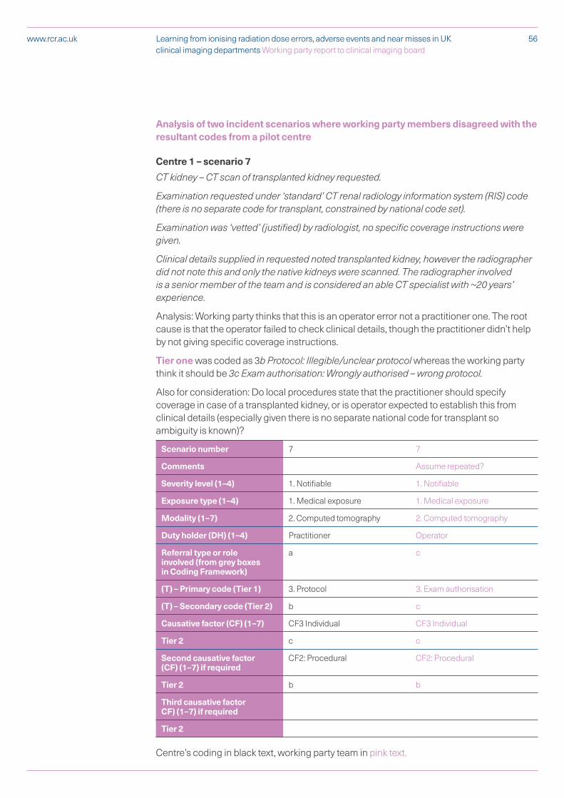
56Learning from ionising radiation dose errors, adverse events and near misses in UK clinical imaging departments Working party report to clinical imaging board
www.rcr.ac.uk
Analysis of two incident scenarios where working party members disagreed with the resultant codes from a pilot centre
Centre 1 – scenario 7
CT kidney – CT scan of transplanted kidney requested.
Examination requested under ‘standard’ CT renal radiology information system (RIS) code (there is no separate code for transplant, constrained by national code set).
Examination was ‘vetted’ (justified) by radiologist, no specific coverage instructions were given.
Clinical details supplied in requested noted transplanted kidney, however the radiographer did not note this and only the native kidneys were scanned. The radiographer involved is a senior member of the team and is considered an able CT specialist with ~20 years’ experience.
Analysis: Working party thinks that this is an operator error not a practitioner one. The root cause is that the operator failed to check clinical details, though the practitioner didn’t help by not giving specific coverage instructions.
Tier one was coded as 3b Protocol: Illegible/unclear protocol whereas the working party think it should be 3c Exam authorisation: Wrongly authorised – wrong protocol.
Also for consideration: Do local procedures state that the practitioner should specify coverage in case of a transplanted kidney, or is operator expected to establish this from clinical details (especially given there is no separate national code for transplant so ambiguity is known)?
Scenario number 7 7
Comments Assume repeated?
Severity level (1–4) 1. Notifiable 1. Notifiable
Exposure type (1–4) 1. Medical exposure 1. Medical exposure
Modality (1–7) 2. Computed tomography 2. Computed tomography
Duty holder (DH) (1–4) Practitioner Operator
Referral type or role involved (from grey boxes in Coding Framework)
a c
(T) – Primary code (Tier 1) 3. Protocol 3. Exam authorisation
(T) – Secondary code (Tier 2) b c
Causative factor (CF) (1–7) CF3 Individual CF3 Individual
Tier 2 c c
Second causative factor (CF) (1–7) if required
CF2: Procedural CF2: Procedural
Tier 2 b b
Third causative factor CF) (1–7) if required
Tier 2
Centre’s coding in black text, working party team in pink text.

57Learning from ionising radiation dose errors, adverse events and near misses in UK clinical imaging departments Working party report to clinical imaging board
www.rcr.ac.uk
Centre 3 – scenario 12
An outpatient was referred for a non-contrast renal CT scan. The radiographer read the patient's clinical history and noted that the patient had kidney problems. The radiographer proceeded to undertake a non-contrast CT scan, but when processing the images saw that the scan had been protocolled by a consultant radiologist as requiring contrast. The examination code on CRIS had not been changed to reflect this. The patient required a repeat scan with contrast which was completed the same day.
Analysis: Working party thinks 3c Exam authorisation: Wrongly authorised – wrong protocol means getting the protocol wrong because all the available information has not been fully checked (though that could also be 3a Exam authorisation: Misinterpretation of clinical information), whereas 4b Pre-exposure safety checks – wrong protocol selection is simply setting the wrong protocol on the machine by mistake.
Scenario number 12 12
Severity Level (1–4) 1. Notifiable 1. Notifiable
Exposure type (1–4) 1. Medical exposure 1. Medical exposure
Modality (1–7) 2. Computed tomography only 1. Computed tomography only
Duty holder (DH) (1 – 4) Operator Operator
Referral type or role involved (from grey boxes in Coding Framework)
c c
(T) – Primary code (Tier 1) 4. Pre-exposure safety checks 3.Exam authorisation
(T) – Secondary code (Tier 2) b c
Causative Factor (CF) (1 – 4) CF3. Individual CF3. Individual
Tier 2 a a
Second Causative Factor (CF) (1 – 7) if required
CF2. Procedual CF2. Procedual
Tier 2 c c
Third Causative Factor (CF) (1 – 4)Tier
Tier 2
Centre’s coding in black text, Working Party team in pink text.

58Learning from ionising radiation dose errors, adverse events and near misses in UK clinical imaging departments Working party report to clinical imaging board
www.rcr.ac.uk
Appendix 3. Final coding taxonomy including contributory factors taxonomy
Final coding taxonomy Please see separate excel spreadsheet entitled ‘Coding taxonomy’65
The proposal is that each clinical imaging department prints out this taxonomy, laminates it and makes it available in relevant area(s) to be a source of reference. The taxonomy is colour coded for ease of use. See separate user guidance for additional support in using the taxonomy.
Contributory factor taxonomy The final coding taxonomy was further enhanced by the addition of these contributory factors (CF). It was felt that inclusion of the contributory factor taxonomies would enhance subsequent error/near miss trend analysis.
Category Code Description
Category CF1 Individual
Sub-category CF1a Failure to recognise hazard (knowledge-based and so on).
CF1b Decision-making process (rule-based or old or invalid rule used and so on).
CF1c Slips and lapses (skill-based, involuntary automaticity and so on).
CF1d Communication (inaccuracy or omission of verbal, written and so on).
CF1e Violation (deliberate action, acting outside scope and so on).
Category CF2 Procedural
Sub-category CF2a No procedures/protocols (not in place or unavailable and so on).
CF2b Inadequate procedures/protocols.
CF2c Process design (impractical and inefficient processes and so on).
Category CF3 Technical
Sub-category CF3a Equipment or IT network failure (including accessories) .
CF3b Commissioning/calibration/ maintenance/handover (including accessories).
CF3c Device/product design.
Category CF4 Patient related
Sub-category CF4a Medical condition (inability to remain still etc).
CF4b Communication with the patient (language issues, comprehension and so on).
CF4c Non-compliance.
Category CF5 Teamwork/management/organisational
Sub-category CF5a Inadequate leadership (inadequate supervision, congruence or consistency and so on).
CF5b Unclear responsibilities and lines of accountability.
CF5c Inadequate capital resources (equipment in use no longer fit for purpose and so on).

59Learning from ionising radiation dose errors, adverse events and near misses in UK clinical imaging departments Working party report to clinical imaging board
www.rcr.ac.uk
Category Code Description
CF5d Inadequate staffing (insufficient staffing levels or skill-mix necessary to meet the demands of a service and so on).
CF5e Inadequate training (inadequate or lack of training and so on).
CF5f Inadequate risk assessment (poor change management and so on).
Category CF6 Environmental
Sub-category CF6a Physical (power cut, control area excessively noisy, distractions and so on).
CF6b Natural factors (fire, flood and so on).
Category CF7 Other
CF7a Other.

60Learning from ionising radiation dose errors, adverse events and near misses in UK clinical imaging departments Working party report to clinical imaging board
www.rcr.ac.uk
Appendix 4. Final reporting template
Please see separate excel spreadsheet entitled Reporting template66’
The working party recognise that there is still some work to be undertaken to make this template more user friendly.
The proposal is that this is a web-based tool that is live – perhaps even being incorporated into an incident management system, for example, DATIX.


The Royal College of Radiologists 63 Lincoln’s Inn Fields London WC2A 3JW
+44 (0)20 7405 1282 [email protected] www.rcr.ac.uk
@RCRadiologists
The Royal College of Radiologists is a Charity registered with the Charity Commission No 211540.
The Royal College of Radiologists. Learning from ionising radiation dose errors and near misses in UK clinical imaging departments. Working party report to Clinical Imaging Board.. London: The Royal College of Radiologists, 2019.
RCR 2019
© The Royal College of Radiologists, June 2019.
For permission to reproduce any of the content contained herein, please email: [email protected]
This material has been produced by The Royal College of Radiologists (RCR) for use internally within the specialties of clinical oncology and clinical radiology in the United Kingdom. It is provided for use by appropriately qualified professionals, and the making of any decision regarding the applicability and suitability of the material in any particular circumstance is subject to the user’s professional judgement.
While every reasonable care has been taken to ensure the accuracy of the material, RCR cannot accept any responsibility for any action taken, or not taken, on the basis of it. As publisher, RCR shall not be liable to any person for any loss or damage, which may arise from the use of any of the material. The RCR does not exclude or limit liability for death or personal injury to the extent only that the same arises as a result of the negligence of RCR, its employees, Officers, members and Fellows, or any other person contributing to the formulation of the material.