laparoscopic and endoscopic co-operative surgery for … suturing after esd. ... with malignant...
TRANSCRIPT

Daisuke Ichikawa, Shuhei Komatsu, Toshiyuki Kosuga, Kazuma Okamoto, Eigo Otsuji, Division of Digestive Surgery, Department of Surgery, Kyoto Prefectural University of Medicine, Kamigyo-ku, Kyoto 6028566, Japan
Osamu Dohi, Yuji Naito, Kazuhiro Kamada, Yoshito Itoh, Department of Gastroenterology and Hepatology, Kyoto Prefectural University of Medicine, Kamigyo-ku, Kyoto 6028566, Japan
Author contributions: Ichikawa D, Komatsu S and Dohi O designed the study, performed the operations, and contributed equally to this work; all of other authors followed up the patients and collected the clinical data; Ichikawa D wrote the manuscript.
Institutional review board statement: The study was reviewed and approved by the Institutional Review Board of the Kyoto Prefectural University of Medicine.
Informed consent statement: All study participants provided written informed consent prior to study enrollment.
Conflict-of-interest statement: The authors have no conflicts of interest to disclose.
Data sharing statement: No additional data are available.
Open-Access: This article is an open-access article which was selected by an in-house editor and fully peer-reviewed by external reviewers. It is distributed in accordance with the Creative Commons Attribution Non Commercial (CC BY-NC 4.0) license, which permits others to distribute, remix, adapt, build upon this work non-commercially, and license their derivative works on different terms, provided the original work is properly cited and the use is non-commercial. See: http://creativecommons.org/licenses/by-nc/4.0/
Manuscript source: Unsolicited manuscript
Correspondence to: Daisuke Ichikawa, MD, PhD, Division of Digestive Surgery, Department of Surgery, Kyoto Prefectural University of Medicine, 465 Kajii-cho, Kamigyo-ku, Kyoto
6028566, Japan. [email protected]: +81-75-2515527Fax: +81-75-2515522
Received: July 29, 2016Peer-review started: August 1, 2016First decision: September 28, 2016Revised: October 10, 2016Accepted: November 13, 2016Article in press: November 13, 2016Published online: December 21, 2016
AbstractAIMTo assess the safety and feasibility of laparoscopic and endoscopic co-operative surgery (LECS) for early non-ampullary duodenal tumors.
METHODSTwelve patients with a non-ampullary duodenal tumor underwent LECS at our hospital. One patient had two mucosal lesions in the duodenum. The indication for this procedure was the presence of duodenal tumors with a low risk for lymph node metastasis. In particular, the tumors included small (less than 10 mm) submucosal tumors (SMT) and epithelial mucosal tumors, such as mucosal cancers or large mucosal adenomas with malignant suspicion. The LECS procedures, such as full-thickness dissection for SMT and laparoscopic reinforcement after endoscopic submucosal dissection (ESD) for epithelial tumors, were performed for the 13 early duodenal lesions in 12 patients. Here we present the short-term outcomes and evaluate the safety and feasibility of this new technique.
Submit a Manuscript: http://www.wjgnet.com/esps/Help Desk: http://www.wjgnet.com/esps/helpdesk.aspxDOI: 10.3748/wjg.v22.i47.10424
10424 December 21, 2016|Volume 22|Issue 47|WJG|www.wjgnet.com
World J Gastroenterol 2016 December 21; 22(47): 10424-10431 ISSN 1007-9327 (print) ISSN 2219-2840 (online)
© 2016 Baishideng Publishing Group Inc. All rights reserved.
ORIGINAL ARTICLE
Laparoscopic and endoscopic co-operative surgery for non-ampullary duodenal tumors
Clinical Trials Study
Daisuke Ichikawa, Shuhei Komatsu, Osamu Dohi, Yuji Naito, Toshiyuki Kosuga, Kazuhiro Kamada, Kazuma Okamoto, Yoshito Itoh, Eigo Otsuji
RESULTSTwo SMT-like lesions and eleven superficial epithelial tumor-like lesions were observed. Seven and Six lesions were located in the second and third parts of the duodenum, respectively. All lesions were successfully resected en bloc . The defect in the duodenal wall was manually sutured after resection of the duodenal SMT. For epithelial duodenal tumors, the ulcer bed was laparoscopically reinforced via manual suturing after ESD. Intraoperative perforation occurred in two out of eleven epithelial tumor-like lesions during ESD; however, they were successfully laparoscopically repaired. The median operative time and intraoperative estimated blood loss were 322 min and 0 mL, respectively. Histological examination of the tumors revealed one adenoma with moderate atypia, ten adenocarcinomas, and two neuroendocrine tumors. No severe postoperative complications (Clavien-Dindo classification grade Ⅲ or higher) were reported in this series, but minor leakage secondary to pancreatic fistula occurred in one patient.
CONCLUSIONLECS can be a safe and minimally invasive treatment option for non-ampullary early duodenal tumors.
Key words: Non-ampullary tumor; Laparoscopic and endoscopic cooperative surgery; Early duodenal cancer
© The Author(s) 2016. Published by Baishideng Publishing Group Inc. All rights reserved.
Core tip: We performed laparoscopic and endoscopic co-operative surgery (LECS) procedures, such as full-thickness dissection for submucosal tumors and laparoscopic reinforcement after endoscopic submu-cosal dissection for epithelial tumors, for 13 early duodenal lesions in 12 patients, and analyzed the safety and feasibility of LECS for early non-ampullary duodenal tumors. All lesions were successfully resec-ted en bloc . No severe postoperative complications (Clavien-Dindo classification grade Ⅲ or higher) were reported in this series, but minor leakage secondary to pancreatic fistula occurred in one patient. LECS can be a safe and minimally invasive treatment option for non-ampullary early duodenal tumors.
Ichikawa D, Komatsu S, Dohi O, Naito Y, Kosuga T, Kamada K, Okamoto K, Itoh Y, Otsuji E. Laparoscopic and endoscopic co-operative surgery for non-ampullary duodenal tumors. World J Gastroenterol 2016; 22(47): 10424-10431 Available from: URL: http://www.wjgnet.com/1007-9327/full/v22/i47/10424.htm DOI: http://dx.doi.org/10.3748/wjg.v22.i47.10424
INTRODUCTIONPancreatoduodenectomy is considered as the standard approach for the resection of duodenal tumors[1,2].
Partial duodenal resection has also been attempted as an alternative treatment option for duodenal tumors such as mucosal adenocarcinoma and small submucosal tumors (SMT), which do not require lymph node dissection[3-6]. Conventional surgery, however, is still invasive especially for patients with such early duodenal tumors.
Recently, postoperative quality of life has received considerable attention, in addition to the oncological outcomes of patients with malignant tumors. There-fore, endoscopic treatments, such as endoscopic mucosal resection (EMR) and endoscopic submucosal dissection (ESD), are being increasingly performed for early epithelial duodenal tumors. However, endoscopic treatments for duodenal tumors are still controversial. The anatomical features of the duodenum, such as the narrow lumen and thin wall, make endoscopic resection of tumors very difficult. In fact, several recent reports have demonstrated that severe complications, such as perforation and bleeding, frequently occur during and after endoscopic treatments[7-11]. Recently, Hiki et al[12,13] reported on laparoscopic and endoscopic co-operative surgery (LECS) for gastrointestinal stromal tumors (GIST). This revolutionary procedure enables the precise assessment of tumor boundaries, thereby facilitating adequate tumor resection.
Taking the abovementioned factors into conside-ration, we have recently developed an LECS concept for the treatment of early non-ampullary duodenal tumors. In this report, we present the details of our procedure and its short-term outcomes and evaluate the safety and feasibility of this new technique.
MATERIALS AND METHODSPatientsBetween 2015 and 2016, twelve patients (7 males and 5 females) with a non-ampullary duodenal tumor underwent LECS at Kyoto Prefectural University of Medicine. One patient had two mucosal lesions in the second and third parts of the duodenum. All patients underwent diagnostic endoscopy with biopsy. Indications for endoscopic and/or laparoscopic local resection of the duodenal tumors were those with a low risk for lymph node metastasis. Our therapeutic strategies for the early duodenal epithelial tumors, such as mucosal cancers or large mucosal adenomas with malignant suspicion, were as follows: (1) epi-thelial mucosal tumors of less than 10 mm diameter in the duodenum were treated with EMR; (2) epithelial mucosal tumors of less than 20 mm diameter in the first part of the duodenum were generally treated with common ESD; and (3) epithelial mucosal tumors of more than 10 mm diameter in the second and third parts of the duodenum and those of more than 20 mm in the first part of the duodenum were treated with LECS under general anesthesia. LECS procedure was also indicated for small SMTs (less than 10 mm
10425 December 21, 2016|Volume 22|Issue 47|WJG|www.wjgnet.com
Ichikawa D et al . LECS for duodenal tumors

diameter) of the duodenum with negligible risk for lymph node metastasis in principle. All subjects gave written informed consent prior to undergoing treatment by LECS.
The clinicopathological features of these patients were retrospectively reviewed from their hospital records. Macroscopic and microscopic classifications of tumors were based on the International Union against Cancer/ Tumor, Node, Metastasis (UICC/TMN) staging system. Postoperative functional evaluation using cine-magnetic resonance imaging examination (cine-MRI) was also performed in patients who one year had passed after operation as previously reported[14]. In brief, patients fasted for 4 h prior to the examination. After intake of a jelly drink, imaging was performed for 30 min with the patients in a supine position. Serial images were obtained in a single slice of the plane along the long axis of the second and third parts of the duodenum including the resected area.
Surgical proceduresUnder general anesthesia, a 12-mm laparoscopy port was inserted through the umbilicus using the open technique. After pneumoperitoneum with carbon dioxide, four additional trocars were inserted as shown in Figure 1. The trocars, except the trocar for the laparoscope, were symmetrically located along the line connecting the location of target tumor and the umbilicus.
From the left side of the patient, the greater omentum was divided at 3 cm from the right gastroe-piploic vessels, using laparoscopic coagulating shears (Harmonic Scalpel, Ethicon Endo-Surgery, Cincinnati, OH, United States).
The attachment of the transverse mesocolon was freed from the pancreatic head and retroperitoneal tissues. During exfoliation, the accessory right colic vein was clipped and divided to avoid unnecessary bleeding due to intraoperative tensions during ESD procedures. For tumors located in the third part of the duodenum, the hepatocolic ligament was also
dissected, and the right colic flexure was mobilized from the retroperitoneum to achieve a good visual field for the horizontal part of duodenum. Then, the duodenum was mobilized along with the pancreatic head from the retroperitoneum.
Before the endoscopic procedure, the jejunum was clamped using laparoscopic removal forceps. The tumor location was confirmed using both endoscopy and laparoscopy, and the periphery of the tumor was carefully marked using the endoscope. Using ESD technique, a circumferential mucosal incision was made around the tumor with a flush knife and/or a clutch cutter (Fujifilm Co., Tokyo, Japan) after the injection of sodium hyaluronate solution (MucoUp; Johnson and Johnson K.K., Tokyo, Japan). Full-thickness circumferential dissection was subsequently performed endoscopically and laparoscopically for SMT. On the other hand, the common ESD procedure was performed for epithelial tumors such as early duodenal cancers and large adenomas.
Thereafter, the edge of the duodenal wall defect was closed using a laparoscopic hand-sewn suturing technique for cases with SMT and for perforation during ESD. Even after successful ESD, we reinforced the ESD ulcer bed of the duodenum using the laparoscopic hand-sewn suturing technique. After tumor resection, intraluminal endoscopic lavage was performed with a copious amount of saline. Then, the precise location of the ulcer bed was confirmed based on the transmitted light of the endoscope and the pressure exerted from the serosal surface using laparoscopy forceps. Interrupted full-thickness and sero-muscular sutures were added to this site, with taken care to avoid any stenosis or deformity in this series.
After completing the procedure, the endoscope was inserted and passed over the resected location to confirm that there was neither stenosis nor leakage.
RESULTSPatient and tumor characteristics are shown in Table 1. All patients had no symptoms related to the duodenal lesions, and all the lesions were detected on endoscopy performed as a part of a routine physical examination.
During the study period, EMR was indicated for 30 cases of epithelial mucosal tumors of less than 10 mm diameter in the several parts of the duodenum, and common ESD was indicated for three cases of epithelial mucosal tumors of less than 20 mm diameter in the first part of the duodenum. On the other hand, a total of 12 patients with 13 duodenal lesions were treated by the LECS procedures. Two SMT-like lesions and eleven superficial epithelial tumor-like lesions were observed. Seven and six lesions were located in the second and third parts of the duodenum, respectively. Five lesions were located on the oral side of ampulla of Vater, while eight lesions were on the anal side.
Figure 2 shows intraoperative views of the LECS procedure for duodenal SMT. After tumor resection, the
10426 December 21, 2016|Volume 22|Issue 47|WJG|www.wjgnet.com
Figure 1 Trocar arrangement for duodenal laparoscopic and endoscopic co-operative surgery. A 12-mm trocar was placed through the umbilicus as a camera port. Four additional trocars were located symmetrically along the line connecting the location of the target tumor and the umbilicus.
Ichikawa D et al . LECS for duodenal tumors

Table 1 Patient and tumor characteristics as well as surgical results
1Median (range); 2Clavien-Dindo classification. NET: Neuroendocrine tumor.
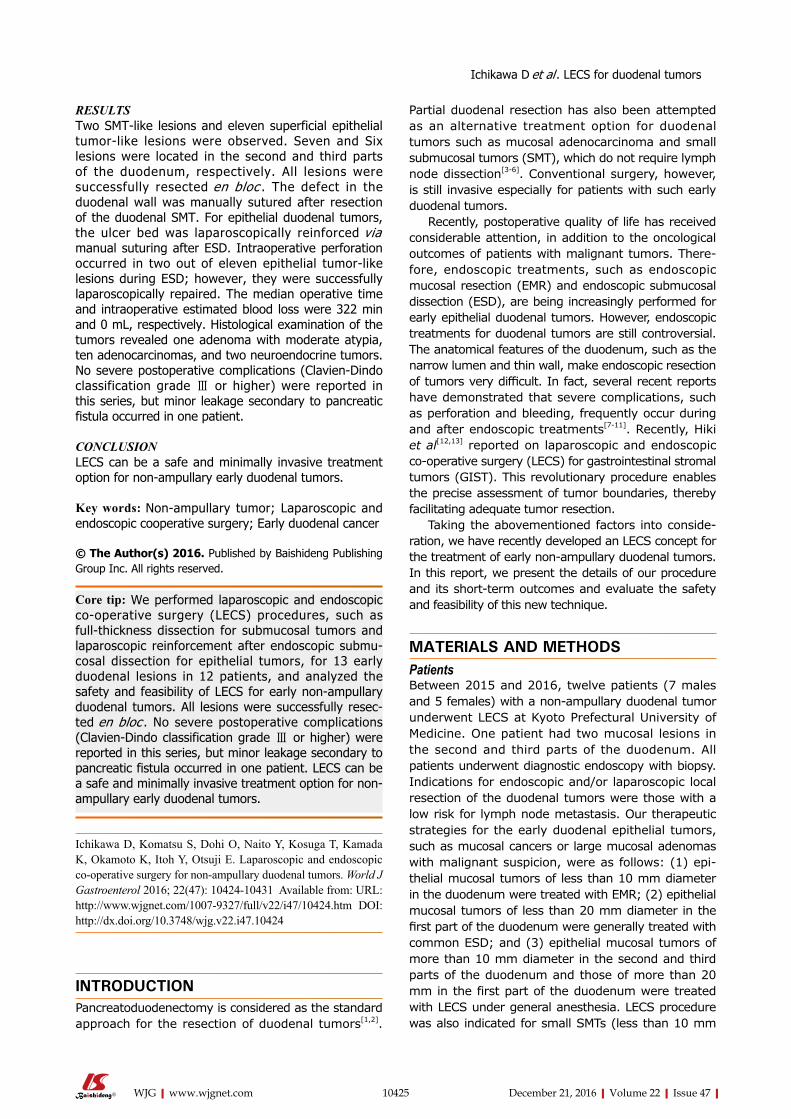
defect in the duodenal wall was closed by a hand-sewn suturing procedure. Figure 3 shows intraoperative views of the LECS procedure for epithelial duodenal tumors, in which the resulting ulcer after ESD was laparoscopically reinforced. The ulcer bed was very thin and easily recognized by the transmitted light of the endoscope, even after successful ESD. Intraoperative perforation was found in two out of the eleven epithelial tumor-like lesions of ESD. These perforations were successfully repaired laparoscopically by hand-sewn sutures. All lesions were successfully resected en bloc. The median operative time and intraoperative estimated blood loss were 322 min and 0 mL, respectively. Conversion to open surgery was not required in this series.
The median time for restarting oral intake was 1 d. The median postoperative hospital stay was 9 d. No severe postoperative complications (Clavien-Dindo classification grade Ⅲ or higher) were reported in this series, but minor leakage secondary to a pancreatic fistula occurred in one patient. The leakage was minor and asymptomatic and was conservatively managed. One patient experienced obstruction of the food passage after discharge. Endoscopic examina-tion revealed near circumferential duodenal wall edema, which resolved over a week with conserva-tive management.
Histological examination of the resected tumors revealed one adenoma with moderate atypia, ten aden-ocarcinomas, and two neuroendocrine tumors. The
10427 December 21, 2016|Volume 22|Issue 47|WJG|www.wjgnet.com
median follow-up periods of these patients were 14 mo (range 3-19 mo). No local or distant recurrence was detected in any patient during the short postoperative follow-up period.
Parameters Statistics
No. of patients 12No. of lesions 13Age (yr) 70 (63-79)1
Sex (male/female) 7/5Location
2nd 73rd 6
Macroscopic typesSubmucosal tumor 2Epithelial (elevated) 11
Size (mm) 22 (11-38)1
Operation time (min) 322 (220-570)1
ESD time (min) 105 (20-210)1
Blood loss (mL) 0 (0-150)1
Postoperative complications2
Minor (≤ grade Ⅱ) 2Major (≥ grade Ⅲ) 0
Pathological diagnosisNET 2Adenoma 1Adenocarcinoma 10
Depth of tumorMucosal 10Submucosal 3
Postoperative hospital stay (d) 9 (7-49)1
Follow-up period (mo) 14 (3-19)1
A
B
C
D
Figure 2 laparoscopic and endoscopic co-operative surgery procedure for a duodenal submucosal tumors. A: Endoscopic view of a duodenal neuroendocrine tumor; B: Endoscopic view of a full-thickness dissection; C: Laparoscopic view after full-thickness excision; D: Laparoscopic view after closure using hand-sewn sutures.
Ichikawa D et al . LECS for duodenal tumors
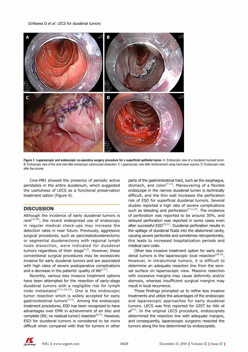
Cine-MRI showed the presence of periodic active peristalsis in the entire duodenum, which suggested the usefulness of LECS as a functional preservation treatment option (Figure 4).
DISCUSSIONAlthough the incidence of early duodenal tumors is rare[15,16], the recent widespread use of endoscopy in regular medical check-ups may increase the detection rates in near future. Previously, aggressive surgical procedures, such as pancreatoduodenectomy or segmental duodenectomy with regional lymph node dissection, were indicated for duodenal tumors regardless of their stage[1,2]. However, these conventional surgical procedures may be excessively invasive for early duodenal tumors and are associated with high rates of severe postoperative complications and a decrease in the patients’ quality of life[1,17].
Recently, various less invasive treatment options have been attempted for the resection of early-stage duodenal tumors with a negligible risk for lymph node metastasis[3-11,18-23]. One is the endoscopic tumor resection which is widely accepted for early gastrointestinal tumors[7-11]. Among the endoscopic treatment procedures, ESD has been recognized to have advantages over EMR in achievement of en bloc and complete (R0; no residual tumor) resection[9,11]. However, ESD for duodenal tumors is considered to be more difficult when compared with that for tumors in other
parts of the gastrointestinal tract, such as the esophagus, stomach, and colon[7-11]. Maneuvering of a flexible endoscope in the narrow duodenal lumen is technically difficult, and the thin wall increases the perforation risk of ESD for superficial duodenal tumors. Several studies reported a high rate of severe complications such as bleeding and perforation[7-11,15]. The incidence of perforation was reported to be around 30%, and delayed perforation was reported in some cases even after successful ESD[10,11]. Duodenal perforation results in the spillage of duodenal fluids into the abdominal cavity, causing severe peritonitis and sometimes retroperitonitis; this leads to increased hospitalization periods and medical care costs.
Other less invasive treatment option for early duo-denal tumors is the laparoscopic local resection[18-23]. However, in intraluminal tumors, it is difficult to determine an adequate resection line from the sero-sal surface on laparoscopic view. Massive resection with excessive margins may cause deformity and/or stenosis, whereas insufficient surgical margins may result in local recurrence.
These findings prompted us to refine less invasive treatments and utilize the advantages of the endoscopic and laparoscopic approaches for early duodenal tumors. LECS was first reported for GIST by Hiki et al[12]. In the original LECS procedure, endoscopists determined the resection line with adequate margins, and consequently, laparoscopic surgeons resected the tumors along the line determined by endoscopists.
10428 December 21, 2016|Volume 22|Issue 47|WJG|www.wjgnet.com
A B
C D
Figure 3 laparoscopic and endoscopic co-operative surgery procedure for a superficial epithelial tumor. A: Endoscopic view of a duodenal mucosal tumor; B: Endoscopic view of the ulcer bed after endoscopic submucosal dissection; C: Laparoscopic view after reinforcement using hand-sewn sutures; D: Endoscopic view after the sutures.
Ichikawa D et al . LECS for duodenal tumors
In the present study, we applied LECS techniques for the local resection of early-stage non-ampullary duodenal tumors. Local resection without destroying surrounding tissues was not only a less invasive but also function-preserving treatment, as shown in the results of the duodenal motility assay.
The conventional LECS procedure was applied to SMT with a low potential for lymph node metastasis. The indication includes GIST of less than 20 mm, carcinoid tumors of less than 10 mm, and other small SMT (less than 20 mm) with a low malignant potential[13,24]. An endoscopic circumferential incision for duodenal SMT was difficult and time-consuming due to a narrow duodenal lumen, and therefore, laparoscopic resection was finally performed with endoscopic-marking guidance. In this study, the original LECS procedure enabled the resection of submucosal tumors with minimal margins and less postoperative duodenal deformity. On the other hand, we also applied the LECS technique for mucosal cancers and large adenomas with a suspected focal malignancy. These superficial epithelial tumors generally have no risk for lymph node metastasis and are considered an indication for ESD. However, bleeding and perforation during and after ESD occurred at a considerable frequency in our hospital; this outcome was similar to that of other reports, and the procedure was sometimes associated with severe complications[7-11]. Therefore, we applied the LECS technique for the superficial tumors as a safety-sensitive strategy. Our method involved ESD
and subsequent reinforcement of the thinned ulcer bed by hand-sewn stiches. Firstly, we used single-layer seromuscular sutures for the reinforcement of the ESD ulcer. However, reinforcement by seromuscular sutures alone required strict recognition of the ESD ulcer boundaries and resulted in intraluminal protrusion of the ulcer bed between the sutured mucosa. This kind of reinforcement was assumed to be effective for perforation but not for hemorrhagic complications. In fact, Irino et al[23] have reported that postoperative bleeding occurred after this kind of LECS procedure. Therefore, we sutured the ESD ulcer bed with two layers of stitches, full-thickness and seromuscular, using a laparo-endoscopic view to decrease the rate of postoperative complications, with care to avoid any stenosis or deformity in this series. The LECS procedure achieved a good short-term outcome, except for 1 case with minor leakage secondary to a pancreatic fistula, which resolved after a week of conservative management. The fistula was suspected to be due to damage to the pancreatic tissues caused by the sutures. From our experience, preoperative diagnostic endoscopic ultrasonography is advocated for the circumferential localization of tumors. If the tumor is found to extend to the pancreatic side of duodenum, endoscopic closure using clips should be considered.
Our LECS procedure had another issue to consider. A full-thickness suture may possibly cause the intra-abdominal dissemination of tumor cells detached from the tumor. However, previous studies have
10429 December 21, 2016|Volume 22|Issue 47|WJG|www.wjgnet.com
A B C
D E F
Figure 4 Cine-magnetic resonance imaging findings of duodenal contraction. These figures are in the following order: A→B→C→D→E→F. White arrows show the second part of the duodenum. They show the presence of periodic active peristalsis in the duodenum.
Ichikawa D et al . LECS for duodenal tumors
reported that no recurrence was recorded in 90 cases of perforation during ESD treatment[25]. Therefore, we assumed that the small amount of tumor cells or tumor debris from superficial tumors was unlikely to adhere to the peritoneum and develop into peritoneal recurrences. Nevertheless, in order to decrease intraluminal free tumor cells, we performed a lavage of the duodenal lumen with an adequate amount of saline (more than 1000 mL) after ESD and thereafter stitched the ulcer bed. Another advantage of the LECS procedure is that it enabled the immediate closure of perforation occurring during the ESD technique. In fact, perforation during ESD occurred in three cases in this series. Among them, one case developed a micro-perforation that was not recognized by the endoscopist during ESD, yet was detected by careful observation of the serosal surface. This kind of latent micro-perforation may be the cause of delayed perforation that sometimes results in lethal complications after ESD.
We should give further consideration to the indi-cations of laparoscopic reinforcement after duodenal ESD. Some reports demonstrated that most delayed perforations after ESD develop in tumors on the anal side of the ampulla of Vater because of the direct exposure of the ESD ulcer bed to pancreatic juice and biliary enzymes[8,10,11]. Therefore, the additional reinforcement by laparoscopic sutures might be required only after ESD for tumors on the anal side of the ampulla of Vater. Although future large trials are needed for confirmation, the LECS procedure could be safe, minimally invasive, and represent an adequate treatment option for non-ampullary early duodenal tumors with an ignorable risk of lymph node metastasis.
COMMENTSBackgroundConventional surgery for early duodenal tumors is excessively invasive. Endoscopic submucosal dissection has been increasingly performed for early duodenal tumors. However, severe complications, such as bleeding and perforation, frequently occur during the perioperative period of endoscopic submucosal dissection (ESD).
Research frontiersVarious less invasive treatment options have been attempted for the resection of early duodenal tumors with a negligible risk for lymph node metastasis. One of the less invasive approaches is the laparoscopic local resection. Recently, laparoscopic and endoscopic co-operative surgery (LECS) has been attempted for the treatments of early duodenal tumors. However, laparoscopy-based local resection can expose the epithelial tumor to the abdominal cavity, which may cause an opportunity for peritoneal metastasis.
Innovations and breakthroughsIn this study, the authors applied the LECS technique for non-ampullary early duodenal tumors. The common LECS procedure - full-thickness dissection - was performed for small submucosal tumors (SMT), whereas a modified LECS procedure - laparoscopic reinforcement after ESD - was performed for mucosal epithelial tumors. LECS procedures could be a safe, minimally invasive and function preserving treatment option for non-ampullary early duodenal tumors with a negligible risk of lymph node metastasis.
ApplicationsThis study suggested that LECS can be a safe and minimally invasive treatment option for non-ampullary early duodenal tumors with a negligible risk of lymph node metastasis although future large trials are needed for confirmation.
TerminologyEndoscopic mucosal resection (EMR) and ESD are well-established techniques of endoscopic resection for the treatment of gastrointestinal epithelial lesions. LECS, which stands for laparoscopy and endoscopy co-operative surgery, was developed by Hiki et al. A SMT is a tumor derived from the submucosal or deeper layer and includes gastrointestinal stromal tumor and neuroendocrine tumor.
Peer-reviewThis paper is an intriguing manuscript showing clinical results and usefulness of LECS procedures for non-ampullary early duodenal tumors. The objective is very curious and important. Few centers would have such an opportunity and this unusual experience is worth recording.
REFERENCES1 Chok AY, Koh YX, Ow MY, Allen JC, Goh BK. A systematic
review and meta-analysis comparing pancreaticoduodenectomy versus limited resection for duodenal gastrointestinal stromal tumors. Ann Surg Oncol 2014; 21: 3429-3438 [PMID: 24854490 DOI: 10.1245/s10434-014-3788-1]
2 Cloyd JM, George E, Visser BC. Duodenal adenocarcinoma: Advances in diagnosis and surgical management. World J Gastrointest Surg 2016; 8: 212-221 [PMID: 27022448 DOI: 10.4240/wjgs.v8.i3.212]
3 Stauffer JA, Raimondo M, Woodward TA, Goldberg RF, Bowers SP, Asbun HJ. Laparoscopic partial sleeve duodenec-tomy (PSD) for nonampullary duodenal neoplasms: avoiding a whipple by separating the duodenum from the pancreatic head. Pancreas 2013; 42: 461-466 [PMID: 23462322 DOI: 10.1097/MPA.0b013e3182649956]
4 Kanaji S, Nakamura T, Nishi M, Yamamoto M, Kanemitu K, Yamashiita K, Imanishi T, Sumi Y, Suzuki S, Tanaka K, Kakeji Y. Laparoscopic partial resection for hemangioma in the third portion of the duodenum. World J Gastroenterol 2014; 20: 12341-12345 [PMID: 25232270 DOI: 10.3748/wjg.v20.i34.12341]
5 Tanaka E, Kim M, Lim JS, Choi YY, Saklani A, Noh SH, Hyung WJ. Usefulness of laparoscopic side-to-side duodenojejunostomy for gastrointestinal stromal tumors located at the duodenojejunal junction. J Gastrointest Surg 2015; 19: 313-318 [PMID: 25421359 DOI: 10.1007/s11605-014-2699-6]
6 Chung JC, Kim HC, Hur SM. Limited resections for duodenal gastrointestinal stromal tumors and their oncologic outcomes. Surg Today 2016; 46 : 110-116 [PMID: 25860591 DOI: 10.1007/s00595-015-1163-x]
7 Takimoto K, Imai Y, Matsuyama K. Endoscopic tissue shielding method with polyglycolic acid sheets and fibrin glue to prevent delayed perforation after duodenal endoscopic submucosal dissec-tion. Dig Endosc 2014; 26 Suppl 2: 46-49 [PMID: 24750148 DOI: 10.1111/den.12280]
8 Inoue T, Uedo N, Yamashina T, Yamamoto S, Hanaoka N, Takeuchi Y, Higashino K, Ishihara R, Iishi H, Tatsuta M, Takahashi H, Eguchi H, Ohigashi H. Delayed perforation: a hazardous complication of endoscopic resection for non-ampullary duodenal neoplasm. Dig Endosc 2014; 26: 220-227 [PMID: 23621427 DOI: 10.1111/den.12104]
9 Matsumoto S, Yoshida Y. Selection of appropriate endoscopic therapies for duodenal tumors: an open-label study, single-center experience. World J Gastroenterol 2014; 20: 8624-8630 [PMID: 25024618 DOI: 10.3748/wjg.v20.i26.8624]
10 Kakushima N, Kanemoto H, Tanaka M, Takizawa K, Ono H. Treatment for superficial non-ampullary duodenal epithelial tumors. World J Gastroenterol 2014; 20: 12501-12508 [PMID: 25253950
10430 December 21, 2016|Volume 22|Issue 47|WJG|www.wjgnet.com
COMMENTS
Ichikawa D et al . LECS for duodenal tumors

DOI: 10.3748/wjg.v20.i35.12501]11 Marques J, Baldaque-Silva F, Pereira P, Arnelo U, Yahagi
N, Macedo G. Endoscopic mucosal resection and endoscopic submucosal dissection in the treatment of sporadic nonampullary duodenal adenomatous polyps. World J Gastrointest Endosc 2015; 7: 720-727 [PMID: 26140099 DOI: 10.4253/wjge.v7.i7.720]
12 Hiki N, Yamamoto Y, Fukunaga T, Yamaguchi T, Nunobe S, Tokunaga M, Miki A, Ohyama S, Seto Y. Laparoscopic and endoscopic cooperative surgery for gastrointestinal stromal tumor dissection. Surg Endosc 2008; 22: 1729-1735 [PMID: 18074180 DOI: 10.1007/s00464-007-9696-8]
13 Hiki N, Nunobe S, Matsuda T, Hirasawa T, Yamamoto Y, Yamaguchi T. Laparoscopic endoscopic cooperative surgery. Dig Endosc 2015; 27: 197-204 [PMID: 25394216 DOI: 10.1111/den.12404]
14 Ichikawa D, Komatsu S, Okamoto K, Shiozaki A, Fujiwara H, Otsuji E. Esophagogastrostomy using a circular stapler in laparoscopy-assisted proximal gastrectomy with an incision in the left abdomen. Langenbecks Arch Surg 2012; 397: 57-62 [PMID: 21853418 DOI: 10.1007/s00423-011-0840-5]
15 Honda T, Yamamoto H, Osawa H, Yoshizawa M, Nakano H, Sunada K, Hanatsuka K, Sugano K. Endoscopic submucosal dissection for superficial duodenal neoplasms. Dig Endosc 2009; 21: 270-274 [PMID: 19961529 DOI: 10.1111/j.1443-1661.2009.00908.x]
16 Buchbjerg T, Fristrup C, Mortensen MB. The incidence and prog-nosis of true duodenal carcinomas. Surg Oncol 2015; 24: 110-116 [PMID: 25936244 DOI: 10.1016/j.suronc.2015.04.004]
17 Müller MW, Dahmen R, Köninger J, Michalski CW, Hinz U, Hartel M, Kadmon M, Kleeff J, Büchler MW, Friess H. Is there an advantage in performing a pancreas-preserving total duodenectomy in duodenal adenomatosis? Am J Surg 2008; 195: 741-748 [PMID: 18436175 DOI: 10.1016/j.amjsurg.2007.08.061]
18 Sakon M, Takata M, Seki H, Hayashi K, Munakata Y, Tateiwa N. A novel combined laparoscopic-endoscopic cooperative approach for duodenal lesions. J Laparoendosc Adv Surg Tech A 2010; 20: 555-558 [PMID: 20578925 DOI: 10.1089/lap.2009.0392]
19 Ohata K, Murakami M, Yamazaki K, Nonaka K, Misumi N, Tashima T, Minato Y, Shozushima M, Mitsui T, Matsuhashi N, Fu K. Feasibility of endoscopy-assisted laparoscopic full-thickness resec-tion for superficial duodenal neoplasms. ScientificWorldJournal 2014; 2014: 239627 [PMID: 24550694 DOI: 10.1155/2014/239627]
20 Tsushimi T, Mori H, Harada T, Nagase T, Iked Y, Ohnishi H. Laparoscopic and endoscopic cooperative surgery for duodenal neuroendocrine tumor (NET) G1: Report of a case. Int J Surg Case Rep 2014; 5: 1021-1024 [PMID: 25460463 DOI: 10.1016/j.ijscr.2014.10.051]
21 Tamaki I, Obama K, Matsuo K, Kami K, Uemoto Y, Sato T, Ito T, Tamaki N, Kubota K, Inoue H, Yamamoto E, Morimoto T. A case of primary adenocarcinoma of the third portion of the duodenum resected by laparoscopic and endoscopic cooperating surgery. Int J Surg Case Rep 2015; 9: 34-38 [PMID: 25723745 DOI: 10.1016/j.ijscr.2015.02.031]
22 Kyuno D, Ohno K, Katsuki S, Fujita T, Konno A, Murakami T, Waga E, Takanashi K, Kitaoka K, Komatsu Y, Sasaki K, Hirata K. Laparoscopic-endoscopic cooperative surgery is a safe and effective treatment for superficial nonampullary duodenal tumors. Asian J Endosc Surg 2015; 8: 461-464 [PMID: 26708586 DOI: 10.1111/ases.12211]
23 Irino T, Nunobe S, Hiki N, Yamamoto Y, Hirasawa T, Ohashi M, Fujisaki J, Sano T, Yamaguchi T. Laparoscopic-endoscopic coopera-tive surgery for duodenal tumors: a unique procedure that helps ensure the safety of endoscopic submucosal dissection. Endoscopy 2015; 47: 349-351 [PMID: 25479560 DOI: 10.1055/s-0034-1390909]
24 Kachare SD, Liner KR, Vohra NA, Zervos EE, Fitzgerald TL. A modified duodenal neuroendocrine tumor staging schema better defines the risk of lymph node metastasis and disease-free survival. Am Surg 2014; 80: 821-826 [PMID: 25105406]
25 Ikehara H, Gotoda T, Ono H, Oda I, Saito D. Gastric perforation during endoscopic resection for gastric carcinoma and the risk of peritoneal dissemination. Br J Surg 2007; 94: 992-995 [PMID: 17535014 DOI: 10.1002/bjs.5636]
P- Reviewer: Hiki N, Shibata T, Slomiany BL, Tovey FI S- Editor: Gong ZM L- Editor: A E- Editor: Liu WX
10431 December 21, 2016|Volume 22|Issue 47|WJG|www.wjgnet.com
Ichikawa D et al . LECS for duodenal tumors

© 2016 Baishideng Publishing Group Inc. All rights reserved.
Published by Baishideng Publishing Group Inc8226 Regency Drive, Pleasanton, CA 94588, USA
Telephone: +1-925-223-8242Fax: +1-925-223-8243
E-mail: [email protected] Desk: http://www.wjgnet.com/esps/helpdesk.aspx
http://www.wjgnet.com
I S S N 1 0 0 7 - 9 3 2 7
9 7 7 1 0 07 9 3 2 0 45
4 7