journal of the association for research in otolaryngology · journal of the association for...
TRANSCRIPT

Relative Time Course of Degeneration of Different CochlearStructures in the CD/1 Mouse Model of Accelerated Aging
SHANTHINI MAHENDRASINGAM, JAMIE A. MACDONALD, AND DAVID N. FURNESS
Institute for Science and Technology in Medicine and the School of Life Sciences, Keele University, Keele, Staffordshire ST5 5BG,UK
Received: 19 September 2010; Accepted: 22 February 2011; Online publication: 12 March 2011
ABSTRACT
Presbycusis (age-related hearing loss) can result fromvarious cochlear pathologies. We have studied thetime course of degeneration in a mouse that showsaccelerated presbycusis, the CD/1 mouse, as a possi-ble model to investigate stem-cell strategies to preventor ameliorate presbycusic changes. CD/1 mice from 0to 72 weeks old were examined by light and electronmicroscopy. Early pathological changes were detectedin basal turn spiral ligament fibrocytes and spiralganglion, but the latter was variable as both satellitecells and neurons were normal in some cochleae.Light microscopic counts in the spiral ligament of 20-week-old mice revealed that of the five main types(types I–V), only type V fibrocytes showed noreduction in numbers compared with 3-week-oldanimals, and type IV showed the greatest losses.However, all types of fibrocyte showed subtle damagewhen examined using electron microscopy, in theform of swollen mitochondria, as early as 2 weeks. Theextent of mitochondrial damage showed a degree ofcorrespondence with the light microscopic pattern offibrocyte loss in that types III and IV fibrocytes hadthe most abnormal mitochondria and type V the least,especially at early stages. By 10–15 weeks, ultrastruc-tural features of fibrocyte damage were similar tolonger term changes reported in gerbils. Stria vascularis,spiral ganglion and hair cells showed few consistentearly signs of damage but became increasingly affected,lagging behind the fibrocyte damage. Our data suggestthat fibrocyte pathology may precede other presbycusic
changes; breakdown of homeostatic mechanisms towhich they contribute may cause the subsequentdegeneration of the hair cells. Overall, there were manysimilarities to presbycusic changes in other rodents andhumans. Therefore, the features of accelerated aging inthis mouse make it a suitable model for rapidly assessingpossible strategies to prevent or ameliorate presbycusicchanges.
Keywords: cochlea, spiral ligament, fibrocytes, age-related hearing loss, presbycusis
INTRODUCTION
There are four main forms of age-related hearing loss(presbycusis) in humans, three of which are sensory,neural (affecting primarily the hair cells and spiralganglion (SG)) and strial degeneration (Schuknechtand Gacek 1993), where the lateral wall (striavascularis (SV) and spiral ligament (SL)) deteriorates.In lateral wall degeneration, the SL degeneratesbefore the SV (Kusunoki et al. 2004) or hair cells.
The development of stem-cell technology offers ameans to repair the damaged or presbycusic cochlea(Hildebrand et al. 2008) or prevent presbycusis withtimely intervention. The outbred CD/1 mouse straincommonly shows accelerated presbycusis (Shone et al.1991) with hearing loss at about 4 weeks, prior to haircell loss (Le Calvez et al. 1998a, b). Studies of thesemice have shown different possible causes: onesuggests that SL damage precedes hair cell lossinitially in basal turn, with a reduction in endolym-phatic K+ concentration (Wu and Marcus 2003).However, others report that the SG degenerates first
Correspondence to: David N. Furness & Institute for Science andTechnology in Medicine and the School of Life Sciences & KeeleUniversity & Keele, Staffordshire ST5 5BG, UK. Telephone: +44-1782-733496; fax: +44-1782-733516; email: [email protected]
JARO 12: 437–453 (2011)DOI: 10.1007/s10162-011-0263-6D 2011 Association for Research in Otolaryngology
437
JAROJournal of the Association for Research in Otolaryngology
(Riva et al. 2005, 2007). These differences may reflectstrain variations in different facilities.
The SG consists primarily of afferent neuronal cellbodies with satellite cells surrounding them. Thesatellite cells express proteins indicative of oxidativestress, mitochondrial defects and apoptosis by 4 weeksof age (Donadieu et al. 2007; Riva et al. 2007).
The SL consists of five main types of fibrocyteembedded in extensive extracellular matrix in mice(Furness et al. 2009) and gerbil (Spicer and Schulte2002). Type I fibrocytes are lateral to the SV, type IIlateral to the spiral prominence epithelium, type IIIalong the external edge of SL, type IV in the basilarcrest region and type V are supra-strial (Spicer andSchulte 1991, 2002; Nakazawa et al. 1995; Ichimiya etal. 2000; Suko et al. 2000; Weber et al. 2001). Fibrocytedegeneration occurs in various mouse strains(Hequembourg and Liberman 2001; Wu and Marcus2003), in labyrinthitis (Ichimiya et al. 1998), otitismedia (Ichimiya et al. 1999), genetic hearing lossDFN3 (Minowa et al. 1999), noise exposure (Hiroseand Liberman 2003) and otospiralin knock-outs(Delprat et al. 2005).
Mitochondrial degeneration in the SL, as in the SG,plays a crucial role; in a mouse model of DFN3,fibrocytes had fewer mitochondria (Minowa et al.1999), and in mice exposed to a mitochondrial toxin,3-nitropropionic acid (3-NP) (Okamoto et al. 2005), SLpathology was accompanied by hearing loss. Systemicadministration of a caspase inhibitor prior to andduring 3-NP administration suggested that caspase-dependent apoptosis of fibrocytes takes place as a resultof mitochondrial damage (Mizutari et al. 2008).
The present study was motivated by the possibility ofattempting SL cell replacement therapy using CD/1mice bred in our animal facility. We therefore haveinvestigated the time course of degeneration of theircochlear structures, specifically: (1) the pattern of cellloss, (2) the ultrastructural signs of early pathology and(3) the extent of mitochondrial damage using structuralquantification and visualization of the cytochromeoxidase system (Seligman et al. 1968, 1973; Perotti etal. 1983).
MATERIALS AND METHODS
Animals
CD/1 mice were bred and maintained in KeeleUniversity’s Central Animal Facility. To avoid thestrain from becoming significantly inbred, stock wasrefreshed periodically by introducing new mice fromthe suppliers (Charles Rivers). All animals weretreated in accordance with the UK Animals (ScientificProcedures) Act of 1986, and the project was givenapproval by Keele University’s ethical committee.
Hair cell counts
To determine the time course of hair cell loss, twoanimals each at 10, 15 and 20 weeks old were used toobtain cochleae for hair cell counts. Animals were givenan overdose of sodium pentobarbitone (IP; Pentoject,Animalcare Ltd, York), decapitated, the bullae removedand opened to expose the cochleae. These were fixed byintralabyrinthine perfusion and subsequent immersionfor 2 h in 4% paraformaldehyde (PFA) in 0.1 Mphosphate buffer (PB) at pH 7.4, decalcified with 5.5%ethylenediaminetetraacetic acid (EDTA) containing0.1% PFA in PB for 3–10 days and dissected into two tothree portions using needles and iris scissors. They werethen stained with 1:100 phalloidin-FITC in phosphatebuffered saline (PBS) containing 0.2% Triton-X-100 for1 h, mounted onto slides and examined with a ×60objective lens with a 2-mmworking distance on a BioRadMRC 1024 confocal microscope. Stacks of images wereacquired along the length of the cochlea, with eachimage representing a square of 226×226 μm. Hair cellswere assessed quantitatively in the most completedissection from each age; the others being used toconfirm the general pattern. The number present wasdetermined by identifying intact apical cuticular plateand stereocilia, and the number of gaps estimated toprovide a total expected and so calculate a percentagesurvival per image. Where there was very substantial haircell loss, numbers expected could not be estimatedaccurately from the large gaps, so the average obtainedper image from the 10-week-old sample (where few weremissing) was used as the expected value.
Fixation and embedding for light and electronmicroscopy
To evaluate other regions and to assess cellularcondition at the electron microscopic level, mice of0, 2, 3, 5, 10, 15 and 20 weeks old (four animals ineach age group) were prepared for sectioning. Priorto preparation for electron microscopy, mice weretested for the presence or absence of hearing byeliciting an acoustic startle reflex by making a snap oftwo fingers directly above their cage. Whilst notquantitative, this gave some indication of hearingability. Animals were anesthetised and the cochleaexposed as above. Each cochlea was fixed by perfusionwith 2.5% glutaraldehyde (GTA) in 0.1 M sodiumcacodylate buffer containing 2 mM calcium chloride(pH 7.4) through the round window and a small holemade in the apex, and immersed in the same fixativefor 2 h. Cochleae were then washed in 0.1 M sodiumcacodylate buffer containing 2 mM calcium chloride(pH 7.4), fixed in 1% osmium tetroxide in the samebuffer for 1 h and decalcified in 5.5% EDTA/0.1%PFA solution for 3–4 days at 4°C. The decalcified
438 MAHENDRASINGAM ET AL.: Aging in CD/1 Mice

cochleae were dehydrated in a graded series ofethanols (70%, 80%, 90%, 100% and dry 100%) for30 min in each concentration, infiltrated with mixturesof ethanol and Spurr resin followed by pure Spurr resinover a period of 8 h and polymerized in pure Spurr resinat 60°C for 36 h.
Some cochleae from 52- (n=2) to 72- (n=2) week-old CD/1 mice available from a separate study wereevaluated for fibrocyte loss. These were fixed in 4%PFA, decalcified, dehydrated and embedded in LR-White resin according to the methods described inFurness et al. (2009).
Light microscopy and quantification of cell losswith age
Two midmodiolar semithin sections (1–2 μm) werecut from the embedded cochlea of each of thefour animals from the 3- and 20-week-old groups.LR-white embedded cochlear sections from 72-week-old mice from a previous project were alsoused for qualitative assessment of cell survival. Toquantify the number of fibrocytes of each type, thenumber of cells in the SV the number of capil-laries, and the survival of SG neurones in the 3-and 20-week mice, the sections were stained withtoluidine blue and a montage of images acquiredof the basal lateral wall from both sides of the coil,using a ×40 objective on a Leitz Dialux lightmicroscope (LM) fitted with an Infinity digitalcamera. After assembling the lateral wall montage,the number of cell nuclei in each category, andthe number of patent capillary profiles in eacharea was counted. This method thus analyses arelatively large but equivalent area in all samplesrepresenting the lower basal half turn of thecochlea. A single image of the SG was alsoobtained from one section from each mouse, a75×75 μm square was placed approximately overthe centre at low magnification, then enlarged, andthe number of ganglion-cell nuclei within thesquare was counted. To avoid overcounting orundercounting, only cells that were fully in thesquare or bisected by the right and top sides werecounted, whilst those bisected by the left andbottom sides of the square were not counted.
Transmission electron microscopy. To examine the SL,SV, SG and organ of Corti at the ultrastructurallevel by transmission electron microscopy (TEM),ultrathin midmodiolar sections (120 nm) were cut,stained in 2% ethanolic uranyl acetate (20 min)and lead citrate (2 min) and examined using aJEOL JEM-1230 transmission electron microscopeoperated at 100 kV.
Mitochondrial degeneration. To assess degeneration ofmitochondria in the different types of fibrocyte and inSG in aging CD/1 mice (3, 5 and 10 weeks old; n=3from each age group), random micrographs weretaken of the different types of fibrocyte and regions ofSG and a rating scale was used to designate the extentof mitochondrial damage: 1=normal, 2=minimaldamage with small white holes, 3=more damage withlarge area of white holes, 4=approximately half of thearea degenerated with larger white holes, and 5=maximal damage, mitochondria expanded dramatically.
Diaminobenzidine cytochrome oxidase reaction productin mitochondria. Because mitochondrial damage wasmost evident in fibrocytes, mitochondrial cytochromeoxidase reaction was assessed in them from 3- (n=2)and 10- (n=2) week-old CD/1 mice. Animals wereanaesthetised as above, decapitated, the bullaeremoved and opened and the protocol of Perotti etal. (1983) employed. Each cochlea was fixed byperfusion with 1% PFA and 2% GTA in 0.1 Msodium cacodylate buffer containing 2 mM calciumchloride (pH 7.4) through the round window and asmall hole made in the apex and immersed in thesame fixative for 15 min. The cochleae were thenwashed overnight at 4°C in 0.1 M sodium cacodylatebuffer, followed by three changes in the same bufferover a period of 4 h, dissected partially to expose thecochlear spiral and incubated in 10 mg of 3,3′-diaminobenzidine tetrahydrochloride (DAB, Sigma-Aldrich) in 10 ml of 0.05 M tris-HCl buffer (pH 7.4) at37°C in a water bath for 3 h (every 30 min, the DABmedium was replaced with freshly prepared DABsolution). The cochleae were then washed in 0.05 Mtris-HCl buffer, fixed in 2.5% glutaraldehyde in 0.1 Msodium cacodylate buffer for 2 h, washed in 0.1 Msodium cacodylate buffer, fixed in 2%osmium tetroxidein sodium cacodylate buffer for 2 h, and washed in thesame buffer. They were then dehydrated in ethanol andembedded and polymerised in Spurr resin for TEM.Midmodiolar ultrathin sections were cut and stained inlead citrate for 2 min and examined using the JEOLJEM-1230 electron microscope operated at 100 kV.
RESULTS
Location and identification of the differentcochlear regions
A LM cross section of the cochlear duct in the basalturn of a 3-week-old cochlea, comprising scala media,SG and lateral wall, and the location of differentfibrocyte types in the latter, is shown in Figure 1 fororientation purposes. The lateral wall consists of SVand SL the latter including the basilar crest and spiralprominence. The different fibrocyte types in these
MAHENDRASINGAM ET AL.: Aging in CD/1 Mice 439

locations have been described previously at the EMlevel for the CD/1 mouse (Furness et al. 2009) and wehave used the same criteria here for identification.
Equivalent images are shown for 5-, 10-, 15-, 20- and72-week-old cochleae (Fig. 1B–F). By 20 weeks theorgan of Corti was substantially damaged. The SV hada normal thickness even at 20 weeks, but appearedlighter stained in some samples. The SL appeared tohave a reduced cell density and the SG showedevidence of loss of peripheral processes as theyapproached the organ of Corti. By 72 weeks, signifi-cant degeneration had occurred in all tissues aroundthe cochlear duct, in the organ of Corti, SG and SL,although the extracellular matrix in which the fibro-
cytes are normally embedded appeared still to bepresent. The LM images did not show substantialqualitative changes before 20 weeks, although themice clearly became deaf or nearly so by 20 weeks.Acoustic startle reflexes were present in all the miceexcept 2 at 20 weeks, but reduced hearing (in termsof limited reaction to the clicking sounds made abovethe cage) was noted in one out of four animals at 5and 10 weeks, two out of four at 15 weeks and theremaining two out of four at 20 weeks, indicating aprogressive decrease in hearing ability over thisperiod. This prompted us to evaluate the time courseof degenerative changes by cell counts and electronmicroscopy.
FIG. 1. Radial toluidine blue-stained sections of the basal turn cochlear duct at various ages (3–72 weeks). A The 3-week section illustrates theorgan of Corti (asterisk) spiral ganglion (SG) and lateral wall, the latter consisting of spiral prominence (sp), SV (sv), basilar crest (bc) and SL withfive distinct fibrocyte regions (I, II, III, IV and V). Note that type III cells run along the boundary of the SL with the bony wall. The remaining crosssections (B–E) of 5-, 10-, 15- and 20-week-old mice show progressive changes with age. At 20 weeks, regions of the lateral wall are lighter stained(arrow), there is loss of peripheral processes projecting from the SG to the hair cells (asterisk) and total loss of hair cells (arrowhead). FA 72-week-old section shows substantial loss of fibrocytes in the SL, degeneration of the organ of Corti and SG. Note, however, that the extracellular matrixof the SL is preserved. Bar=50 μm.
440 MAHENDRASINGAM ET AL.: Aging in CD/1 Mice

Quantitative analysis of cell losses
Hair cell counts (Fig. 2) from phalloidin-stainedsurface preparations showed very few missing cells by10 weeks but progressive loss subsequently, from thebasal end. By 20 weeks, more than half of the outerhair cells in all three rows, and significant numbers ofinner hair cells, were missing.
To determine how much fibrocytes loss occurs, andwhich types were most affected, quantitative analysisof the number of each of the different types offibrocytes was carried out in 3 and 20 weeks old CD/1mice (n=4 per group). At 3 weeks, the SL appearednormal at the LM level. Between the two time periods,there was a reduction in mean number of all of thefibrocyte types except type V, with greatest percentagereduction in type IV (Fig. 3). Statistical analysis usinga Wilcoxon’s sign rank test indicated that the reduc-tion in type I fibrocytes was not significant (P90.05),but it was significant for types II, III and IV (PG0.05).The SG cells were also counted in the central portionof the ganglion, with no evidence of cell loss despitethe loss of peripheral processes noted above (Fig. 3;Wilcoxon’s sign rank test, P90.05).
We also analysed other structures in the lateral wallby counting cells and capillary profiles. The numberof cells in the three layers of the SV (basal cells,intermediate cells and marginal cells) did not change
significantly, nor did the number of capillary profilesin either the SL or the SV (Fig. 3).
Ultrastructural observations
LM observations indicated that all except type Vfibrocytes consistently begin to degenerate between3 and 20 weeks, but subtle changes will not bedetected by this method. We therefore evaluated theultrastructure of hair cells, SL, SV and SG at 0, 2, 3, 10,15 and 20 weeks. Since hair cell damage or lossappears first in the high-frequency region, we exam-ined mainly the basal locations of the CD/1 cochlea,but where damage was evident more apically, wecomment on that also.
Zero- to three-week-old mice. At 0 weeks old, fibrocyteswere undifferentiated and showed no evidence ofdamage (data not shown). At 2 weeks old, fibrocytescould be distinguished from each other and hadvirtually adult morphology. They showed minorevidence of mitochondrial damage whilst minimalchanges were noted in the organ of Corti, althoughsome vacuolization was occasionally seen in outer haircells. Overall SL, SV, SG and organ of Corti wererelatively normal (data not shown). In 3-week-oldmice, the organ of Corti (Fig. 4A) again appearednormal. In SL mitochondria showed greater, and
FIG. 2. Cytocochleograms at 10, 15 and 20 weeks old; each bar represents a count from one of a series of images taken sequentially along the organof Corti from apex (bin 1) to base.Grey bars are estimates from smaller areas or adjacent images where the organ of Corti was damaged by dissection orobscured. Note the virtually intact organ of Corti at 10 weeks, with progressively larger loss of hair cells from the basal turn to the apex in all rows from15 to 20 weeks. (Labels 10A, 15C and 20B refer to the specific cochlea chosen for counting of the four examined at each age).
MAHENDRASINGAM ET AL.: Aging in CD/1 Mice 441

varying, degrees of degeneration in all types offibrocyte in all samples, and to a lesser extent in theSV (Fig. 4B–F). The SG appeared undamaged in onesample (Fig. 5A), damaged to only a minor extent in asecond (Fig. 5B) and in a third showed evidence ofextensive swollen Golgi and endoplasmic reticulumcisternae in the neurons but little damage tomitochondria (Fig. 5C). Satellite cells in this sampleshowed vacuolization.
To test whether mitochondrial damage was anartefact of fixation, we also examined two other strainsof mice fixed and embedded using the same method.Fibrocytes were examined from young adults (3–4 weeks) of C57BL/6 (data not shown) and C3HeB/FeJ (Fig. 6). Neither showed evidence of mitochondrialdamage.
Five-week-old mice. In 5-week-old mice, TEM analysissuggested that the organ of Corti was normal (Fig. 7A).The SV showed an increased degree of degeneratingmitochondria in some animals compared with 3-weekmice (Fig. 7B) although it was normal in others. Infibrocytes in basal locations of all animals, however,there was evidence of cytoplasmic degeneration in alltypes in the form of vacuoles that were also foundassociated with mitochondria (Fig. 7D, E and G), andnuclear disruption in some of the type V cells (Fig. 7G).Some of the type IV fibrocytes showed a greater degreeof degeneration as manifested by remnants ofdisintegrating cytoplasm and nuclei (Fig. 7F). A few
type IV fibrocytes in such a late stage of degenerationwere also found in the apical turn of the cochlea (datanot shown). The SG was normal in two out of threemice, and in the third, mild evidence of cytoplasmicextraction was noted around the margins of SGneurones (data not shown). Mild damage was noted inmitochondria of the neurones.
Ten-week-old mice. Mild to extensive cytoplasmicdegeneration was noticed in the hair cells of the basalturn of 10-week-old mice (Fig. 8A) whilst the hair cellsin the apical location appeared normal. Damage tookthe form of vacuolization and cellular distortion, but didnot appear to involve specifically the mitochondria as aprimary feature. The SV showed little evidence ofdegeneration but some mitochondrial swelling wasobserved (Fig. 8B). At this age, more substantialchanges were observed in all of the different types offibrocytes in the basal turn. The nuclei of some of thefibrocytes appeared to have reduced chromatin, thenucleoplasm appearing less dense, the cell bodyappeared shrunken and distorted and the cytoplasmless dense in type I and type II especially (Fig. 8C–G).The type II fibrocytes also had more pronouncedplasma membrane folding (compare Fig. 8D withFig. 7D). Although spaces remained in the type IV area,the fibrocytes were reduced to remnants of disintegratingcytoplasm and nuclei (Fig. 8F). Moderate to severecytoplasmic vacuolization was noticed in the type Vfibrocytes which, however, retained some normalappearing cytoplasm and mitochondria (Fig. 8G).Despite the degeneration and shrinkage of fibrocytes,extracellular matrix was still present and appearednormal around all of them (Fig. 8C–G).
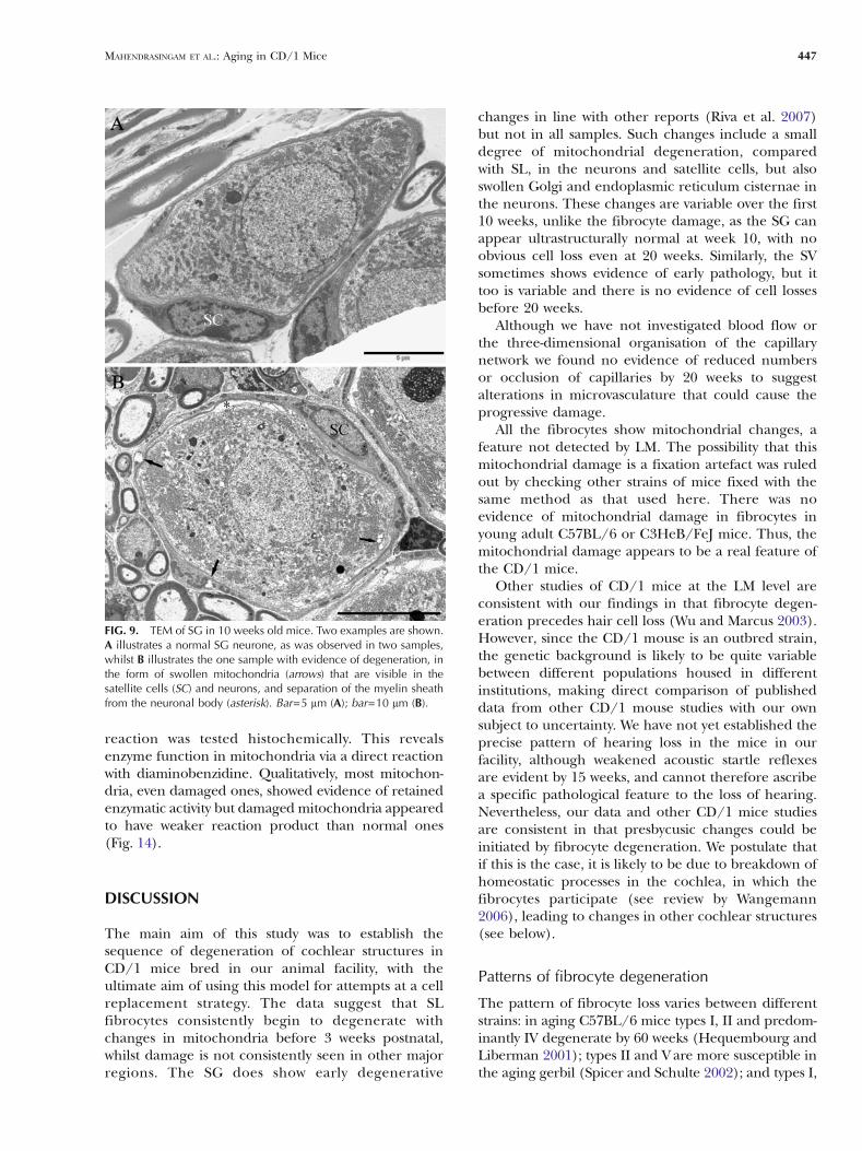
The SG again appeared normal in one sample,mildly disrupted in a second and with more majordisruption in the third (Fig. 9), with similar pathologyto that described previously at 3 and 5 weeks althoughwith greater evidence of mitochondrial pathology inneurones and satellite cells (see below).
Fifteen-week-old mice. In 15-week-old mice, bothouter hair cells and fibrocytes showed moreextensive degeneration (Fig. 10). In the basallocations of the cochlea, outer hair cell damage andloss was evident and Deiters’ cells were seenextending into the space previously occupied by theouter hair cells (Fig. 10A). The hair cells in the apicalturn of the cochlea did not show any substantialchanges. SV in the basal turn at this age showedlocalised areas of damage, but mostly normalmorphology (Fig. 10B). In the basal locations, emptyspaces were seen in the type I fibrocyte area(Fig. 10B) and in the basilar crest area (Fig. 10C),suggesting loss of types I and IV fibrocytes,respectively. The cell bodies of type V fibrocytescontained large vacuoles with mitochondria in them,and there was evidence of degeneration of the large
FIG. 3. Histograms showing the mean number of nuclei of the fivedifferent types of fibrocyte, mean number of capillary profiles in SVand SL (SVcap and SLcap, respectively), mean number of marginalcell (MC), intermediate cell (IC) and basal cell (BC) nuclei and meannumber of SG neurones per 75×75 μm area in 3- and 20-week-oldCD/1 mice. The mean number of types I, II, III and IV fibrocyte is lessin the 20-week-old mice compared with the 3-week-old ones, butthere is no difference in the type V fibrocyte number. Differences thatwere significant (PG0.05) on a Wilcoxon’s sign rank test areindicated by asterisks. There are no significant differences in numberof capillaries in the SV or SL or cells in SV or SG. Bars indicate thestandard error of the means.
442 MAHENDRASINGAM ET AL.: Aging in CD/1 Mice

processes extending to the edge of the scala vestibuli(Fig. 10D). Extracellular matrix was still present inapparently normal distribution as there were largefibres adjacent to gaps that generally were devoid ofmaterial except for cell remnants (Fig. 10B–D). The
SG showed similar degrees of pathology to the worstobserved at 10 weeks (data not shown).
Twenty-week-old mice. In the 20-week-old mice,outer hair cells were damaged and lost in basallocations, inner hair cells were distorted and
FIG. 4. TEM of radial sections of mice at 3 weeks old. AThe organ of Corti has a normal appearance with the inner hair cell (ihc) and three rowsof outer hair cells (ohc1, ohc2 and ohc3) visible. B The SV is relatively normal but there is evidence of swollen mitochondria; marginal cell (mc),intermediate cell (ic). C–G In all fibrocytes, a varying number of mitochondria (arrowheads) appear swollen and distorted to different degrees,with reduced cristae, but the nuclei (nu) and cytoplasm (cy) appear normal, with the exception of some evidence of white spaces in addition tothe mitochondrial swelling in type IV cells. Note the characteristically extensive plasma membrane folding (arrows) in type II and V cells. Bar=10 μm (A); bar=2 μm (B, C, E, F and G); bar=1 μm (E).
MAHENDRASINGAM ET AL.: Aging in CD/1 Mice 443

supporting cells surrounding the inner hair cell werealso damaged (Fig. 11A); outer hair cell damage andloss was also observed in the apical locations (data notshown). The SV showed some localised areas of
damage (Fig. 11B), but substantial regions ofnormality. In the basal locations, some type Ifibrocytes were lost and the remaining ones had veryfew organelles (Fig. 11C); some type III fibrocyteswere lost and the cytoplasm of the remaining ones wassubstantially reduced (Fig. 11D). Gaps were seen inthe extracellular matrix of the basilar crest region dueto the loss of type IV fibrocytes (Fig. 11E), and thesupra-strial region was reduced in extent with type Vcells, though present, appearing to be furtherdegenerated (Fig. 11F). The SG neurons showedmore evidence of dilated cisternal membranes andseparation from satellite cells, though the latterappeared relatively normal (data not shown).
Fifty-two-week-old mice. In the 52 week-old CD/1mice, no hair cells were observed in the basal turn(Fig. 12A). The SV of upper basal turn showed somevacuolization in some of the animals, whilst in thelower basal turn it was reduced in thickness with fewercells remaining (Fig. 12B). Many fibrocytes weremissing but a few remaining cells were located in theregions corresponding to type I, II, III and V; virtuallyall type IV cells had degenerated. The extracellularmatrix formed a framework around the gaps thatresembled the matrix of younger stages (Fig. 12C–F).The SG also had few cells left.
Quantitative changes in mitochondrial damage
The ultrastructural data provided good evidence thatdegenerative changes occur in fibrocytes with ageprior to substantial or consistent damage in othertissues. Themost obvious changes were tomitochondria,although vacuolization also occurred (the latter poten-tially caused by extreme mitochondrial swelling) andeventual loss of cells. To evaluate further, the extent ofdegeneration at different stages, mitochondrial damagewas assessed quantitatively by determining the propor-tion of mitochondria showing one of five different
FIG. 5. TEM of SG in 3-week-old mice. Three examples are shown(A–C) with increasing amounts of abnormality. The example in Ashows no evidence of degeneration, whereas mild cytoplasmicdisruption is visible in B, and swollen cisternae of endoplasmicreticulum and Golgi body (arrow) are visible in C. The satellite cells(SC) are all normal except in C where vacuolization has occurred (v),and there is a small degree of separation of the myelin sheath fromthe neuron in places (asterisk). Bar=5 μm (A and C); bar=10 μm (B).
FIG. 6. TEM of 4-week C3HeB/FeJ mouse type II fibrocyte preparedin the same way as the CD/1 mice. Mitochondria appear to be wellpreserved (e.g. arrow), indicating that the mitochondrial damage inthe CD/1 mice is not a fixation artefact. Bar=2 μm.
444 MAHENDRASINGAM ET AL.: Aging in CD/1 Mice

categories of damage in 3-, 5- and 10-week-old mice(Fig. 13). This analysis revealed that even at 3 weeks, lessthan half the mitochondria were considered normal inall the different fibrocyte types. Types III and IV
fibrocytes showed the least proportion of normalmitochondria, types I and V the most.
After 5 weeks, the proportion of normal mitochon-dria had decreased in types I, II and V fibrocytes,
FIG. 7. TEM of radial sections of mice at 5 weeks old. A The organ of Corti still appears normal with inner hair cell (ihc) and three rows of outerhair cells (1, 2 and 3). B The SV shows increased evidence of mitochondrial swelling in some but not all of the animals investigated; basal cell(bc). C The cytoplasm of type I cells contains fewer organelles and is of lower density than preceding stages, but the nucleus (nu) appears normal;note the collagen fibres (arrow). D Type II cells with characteristic plasma membrane folding (large arrow), contain vacuoles of different sizes(small arrows) and some of the vacuoles are associated with mitochondria (arrowheads). E Vacuoles associated with mitochondria (arrowheads) inthe cytoplasm of a type III cell. F Disintegrating type IV cells (arrows) with remnants of cytoplasm and nucleus. G The type V cell with normalplasma membrane folding (arrow) contains large vacuoles associated with mitochondria (arrowheads) and the nucleus (nu) is disrupted. Bar=10 μm (A and D); bar=5 μm (C and F); bar=2 μm (B, E and G).
MAHENDRASINGAM ET AL.: Aging in CD/1 Mice 445

whilst the proportions showing a small level ofdamage (category 2) had increased. Little changewas noted in types III and IV although the proportionof heavily damaged mitochondria (category 5) hadincreased slightly in all fibrocytes.
After 10 weeks, a clear decrease in normal mito-chondria was seen in all fibrocyte types, with between5% and 15% considered normal, the most in type Vfibrocytes. Most of the damaged mitochondria showedrelatively mild disruption (category 2).
By contrast, mitochondrial damagewas absent in haircells and much less prominent in the SG. Counts in thelatter showed that few defective mitochondria weredetected in satellite cells or neurons until week 10, witha minor increase in mitochondrial degeneration being
noted in the neurons (Fig. 13) although substantiallyless than in SL. A statistical test of the change inproportion of normal mitochondria between weeks 3and 10 was performed using the Wilcoxon’s sign ranktest. For pooled fibrocyte data, the proportion ofnormal mitochondria decreased significantly (pG0.001;n=15) whereas for pooled SG-satellite cell data, therewas no significant change (p90.05; n=6).
Cytochemistry of diaminobenzidine cytochromeoxidase reaction product in mitochondriaof fibrocytes
In an effort to determine whether swollen mitochon-dria were dysfunctional, the cytochrome oxidase
FIG. 8. TEM of radial sections of mice at 10 weeks old. A Radial section of an organ of Corti with inner hair cell (ihc) and three rows of outerhair cells (ohc1, ohc2 and ohc3). Vesiculation is evident in the cytoplasm (arrows) of the inner hair cell (ihc) and first row of outer hair cells(ohc1). B The SV retains a similar level of damage in some mice as at 5 weeks; marginal cell (mc). (C, D and E) The nuclei (nu) of types I, II and IIIfibrocyte have less chromatin and the cell bodies appear shrunken, especially type III (E, arrows), containing much thinner cytoplasm and veryfew mitochondria. Type II fibrocytes additionally have more pronounced membrane folding (D, arrows). F Large spaces previously occupied bytype IV fibrocytes contain remnants of disintegrating cytoplasm (small arrows) and nucleus (large arrow). G Type V fibrocytes show severevacuolization in the cytoplasm (arrows) but retain many normal mitochondria (arrowheads). Extracellular matrix (ecm) fibres were found in allfibrocyte locations and appeared normal. Bar=5 μm (A); bar=2 μm (B–G).
446 MAHENDRASINGAM ET AL.: Aging in CD/1 Mice
reaction was tested histochemically. This revealsenzyme function in mitochondria via a direct reactionwith diaminobenzidine. Qualitatively, most mitochon-dria, even damaged ones, showed evidence of retainedenzymatic activity but damaged mitochondria appearedto have weaker reaction product than normal ones(Fig. 14).
DISCUSSION
The main aim of this study was to establish thesequence of degeneration of cochlear structures inCD/1 mice bred in our animal facility, with theultimate aim of using this model for attempts at a cellreplacement strategy. The data suggest that SLfibrocytes consistently begin to degenerate withchanges in mitochondria before 3 weeks postnatal,whilst damage is not consistently seen in other majorregions. The SG does show early degenerative
changes in line with other reports (Riva et al. 2007)but not in all samples. Such changes include a smalldegree of mitochondrial degeneration, comparedwith SL, in the neurons and satellite cells, but alsoswollen Golgi and endoplasmic reticulum cisternae inthe neurons. These changes are variable over the first10 weeks, unlike the fibrocyte damage, as the SG canappear ultrastructurally normal at week 10, with noobvious cell loss even at 20 weeks. Similarly, the SVsometimes shows evidence of early pathology, but ittoo is variable and there is no evidence of cell lossesbefore 20 weeks.
Although we have not investigated blood flow orthe three-dimensional organisation of the capillarynetwork we found no evidence of reduced numbersor occlusion of capillaries by 20 weeks to suggestalterations in microvasculature that could cause theprogressive damage.
All the fibrocytes show mitochondrial changes, afeature not detected by LM. The possibility that thismitochondrial damage is a fixation artefact was ruledout by checking other strains of mice fixed with thesame method as that used here. There was noevidence of mitochondrial damage in fibrocytes inyoung adult C57BL/6 or C3HeB/FeJ mice. Thus, themitochondrial damage appears to be a real feature ofthe CD/1 mice.
Other studies of CD/1 mice at the LM level areconsistent with our findings in that fibrocyte degen-eration precedes hair cell loss (Wu and Marcus 2003).However, since the CD/1 mouse is an outbred strain,the genetic background is likely to be quite variablebetween different populations housed in differentinstitutions, making direct comparison of publisheddata from other CD/1 mouse studies with our ownsubject to uncertainty. We have not yet established theprecise pattern of hearing loss in the mice in ourfacility, although weakened acoustic startle reflexesare evident by 15 weeks, and cannot therefore ascribea specific pathological feature to the loss of hearing.Nevertheless, our data and other CD/1 mice studiesare consistent in that presbycusic changes could beinitiated by fibrocyte degeneration. We postulate thatif this is the case, it is likely to be due to breakdown ofhomeostatic processes in the cochlea, in which thefibrocytes participate (see review by Wangemann2006), leading to changes in other cochlear structures(see below).
Patterns of fibrocyte degeneration
The pattern of fibrocyte loss varies between differentstrains: in aging C57BL/6 mice types I, II and predom-inantly IV degenerate by 60 weeks (Hequembourg andLiberman 2001); types II and V are more susceptible inthe aging gerbil (Spicer and Schulte 2002); and types I,
FIG. 9. TEM of SG in 10 weeks old mice. Two examples are shown.A illustrates a normal SG neurone, as was observed in two samples,whilst B illustrates the one sample with evidence of degeneration, inthe form of swollen mitochondria (arrows) that are visible in thesatellite cells (SC) and neurons, and separation of the myelin sheathfrom the neuronal body (asterisk). Bar=5 μm (A); bar=10 μm (B).
MAHENDRASINGAM ET AL.: Aging in CD/1 Mice 447

II, III and IV, predominantly types II and IV, in the aginghuman cochlea (Kusunoki et al. 2004). In humans, theloss of types II and IV fibrocytes occurs in all turns but ismost severe in the apical turn even in infants (Kusunokiet al. 2004). The most consistent feature of these data isthat types II and IV cells are the most likely todegenerate, but there are clearly exceptions.
In the CD/1 mouse, our counts of fibrocytes at20 weeks old show significant reductions in types II,III and IV compared with 3 weeks, consistent withobservations by Wu and Marcus (2003) where basaltype IV loss was noted as early as 5 weeks. Type Vfibrocytes are the least affected up to 20 weeks. Moreadvanced changes culminate in 72-week-old mice inloss of virtually all fibrocytes in the type IV region andsevere reductions in the others. Loss of type IVfibrocytes has also been noted as one of the forms ofpathology in Fisher 344 rats (Buckiova et al. 2006),suggesting that it may be a common feature ofpresbycusis.
To assess whether this pattern of fibrocyte loss isconsistent with the extent of early mitochondrial
damage, we analysed the proportion of mitochondriashowing varying extents of disruption from normal toextreme in the different fibrocyte types. The type Vfibrocytes have fewer damaged mitochondria at leastup to 10 weeks, consistent with the fact that they areleast likely to go missing. Types III and IV fibrocytesshow at the earliest stage the most abnormal mitochon-dria, consistent with their more rapid degenerationinitially. Thus, there is a potential correspondencebetween mitochondrial damage and the susceptibilityof the fibrocytes but further data are needed to confirmthis.
Ultrastructural features of fibrocyte degeneration
As well as early mitochondrial damage, subsequentultrastructural changes include vacuolization andchromatin alterations that probably indicate an apop-totic process, in line with observations by Mizutari etal. (2008). The latter study implicated mitochondrialdysfunction. Similar cytoplasmic vacuolization andmitochondrial degeneration were also evident in
FIG. 10. TEM of radial sections of mice at 15 weeks old. A The organ of Corti has a normal inner hair cell (ihc) and first row outer hair cell(ohc1) but the second and third rows of outer hair cells are missing and a Deiters’ cell (dc) can be seen expanded (arrow) into the space normallyoccupied by the latter. B The SV (sv) appeared relatively normal; the upper part of the SL contains empty spaces (asterisk) where type III fibrocytes(small arrows) and type I fibrocytes are missing; some type III (arrowhead) and type I fibrocytes are still present. Extracellular matrix fibres (largearrows) can still be seen adjacent to the empty spaces where the cells are missing. C The basilar crest area of the SL contained large spaces(arrows) with small remnants of cytoplasm and disintegrating nuclei (arrowhead) of the type IV fibrocytes in a late stage of degeneration, as well asa normal type IV fibrocyte. D Supra-strial region of the SL showing a type V fibrocyte with large vacuoles containing mitochondria (arrows) anddegenerating processes (arrowhead) that extend into the scala vestibuli (scv). Bar=10 μm (A and B); bar=2 μm (C and D).
448 MAHENDRASINGAM ET AL.: Aging in CD/1 Mice

some of the fibrocytes types in 33–36 months oldgerbil cochlea (Spicer and Schulte 2002) suggestingthat age-relate changes in the CD/1 mouse, althoughaccelerated, have similar features to longer term agingin gerbils.
The most prominent degeneration we observedwas in the basilar crest area of the SL where type IVfibrocytes appear to disintegrate resulting in largespaces containing remnants of cytoplasm/nucleus,and by 15 weeks gaps can be seen due to loss of thecells. Clear spaces with narrowed processes of type IIfibrocytes have been observed in the 10-week-old CD/1mice, similar to the aging gerbil cochlea (Spicer andSchulte 2002). Gaps are also seen in the type I area dueto loss of type I fibrocytes in the 15-week-old CD/1 miceand the type III area after 20 weeks.
The causes of fibrocyte degeneration
Fibrocyte degeneration is not a general phenomenonin mice; moreover, genetic factors play a role inaccelerated age-related changes in many mice: a genefor age-related hearing loss (ahl) has been found inC57BL/6J and in nine other inbred mouse strains(Johnson et al. 2000) but not in the CBA/CaJ strainwhich has minimal hearing loss and normal organ ofCorti at 34 weeks and 25 months old (Hequembourgand Liberman 2001; Wu and Marcus 2003). A geneticbasis for presbycusis has not been investigated in CD/1mice.
In another model of aging, it has been reportedthat hair cell loss is exacerbated by copper/zincsuperoxide dismutase deficiency (McFadden et al.1999). This implies involvement of the superoxideradical, failure to eradicate which may lead tomitochondrial damage. The morphological changeswe observe in mitochondria in fibrocytes and SVwhich show quite extreme levels of damage could berepresentative of a similar deficiency. The appearanceof vacuoles within the mitochondria implies breakdownof the ion and water balances within them. With greaterdamage, the enzymatic activity of the mitochondria
FIG. 11. TEM of 20-week-old mice. A Radial section of an organ ofCorti with damaged outer hair cell region (ohc) and loss of the thirdrow of outer hair cells (arrow). The supporting cell (arrowhead)surrounding the inner hair cell (ihc) also appears damaged. B Someregions of the SV show degeneration; the region illustrated hereshows white vacuoles associated with swollen mitochondria. C Thecytoplasm (cy) of the type I cell has very few organelles; nucleus (nu).D The cytoplasm of type III fibrocyte is substantially reduced leavinga large space surrounding the cell; nu. E Large gaps (arrows) in theextracellular matrix are due to loss of some of the type IV fibrocytesin the basilar crest region of the SL. F The cytoplasm of type Vfibrocytes appear shrunken, shows significant disruption (arrows) andcontains a few mitochondria. Bar=10 μm (A and E); bar=5 μm (C);bar=2 μm (B, D and F).
R
MAHENDRASINGAM ET AL.: Aging in CD/1 Mice 449

FIG. 12. TEM of 52-week-old mice. A Radial section of a degenerated organ of Corti with loss of inner and outer hair cells, and the supportingcells (sc) occupying the region of degenerated outer hair cells; inner pillar cell (ipc). B SV in the basal turn is thinner than in younger animals withpossible cell depletion. However, the normal three layers of cells can usually be distinguished, including basal cells (bc). C Loss of a type II cell(asterisk), the extracellular matrix around the gap is still present. D Region of the SL lateral to the SV with loss of type I (asterisk) and type III(arrows) cells but with remaining extracellular matrix (ecm). E Basilar crest region with a remaining type IV cell (arrow), and the majority of thetype IV cells degenerated (asterisk). F Supra-SV region with degenerated type V cells (asterisk) but with remaining ecm. rm Reissner’s membrane,scv scala vestibuli. Bar=10 μm (A), 1 μm (C), 2 μm (B and D), 5 μm (E and F).
450 MAHENDRASINGAM ET AL.: Aging in CD/1 Mice

seems to be reduced. Thus, we postulate that themitochondrial damage is a contributory factor tofibrocyte degeneration and subsequently to the haircell loss. Mutations in mitochondrial genes are acommon cause of human deafness (Fischel-Ghodsian1999), and it is possible that the CD/1 mouse alsosuffers an mtDNA mutation that generates the accel-erated age-related pathology. Mutations in mtDNA arealso common in general in aging and can accumulateover time until they reach a critical level (Seo et al.2010).
The lack of mitochondrial damage in hair cells,and the weak level detected in SG neurons suggeststhat there is a tissue-specific difference as well asgenetic susceptibility; it may be that higher energydemands in the SL result in greater damage tosusceptible mitochondria through the generation ofgreater amounts of free radicals (Seo et al. 2010). Inaddition, cells can show heteroplasmy where mutated
FIG. 13. A TEM of a region of fibrocyte cytoplasm showing thecategorisation of damage levels used for quantitative assessment ofmitochondria, indicated by numbers 1 (undamaged) to 5 (grossdamage). Bar=1 μm. B Histograms showing how the levels ofdamage in SL and SG change between 3-, 5- and 10-week-oldanimals. Note that the proportion of category 1 (undamaged)mitochondria decreases whilst the proportion of category 2, inparticular, increases with age in the fibrocytes (Tye I–V). Mitochon-drial damage was limited in satellite cells (SC) and in SG neurons,increasing slightly at 10 weeks in the latter.
R
FIG. 14. Examples of the cytochrome oxidase-DAB reaction inmitochondria. More damaged mitochondria (double asterisks) fre-quently show a lower level of reaction product than normal ones(asterisk). Bar=500 nm.
MAHENDRASINGAM ET AL.: Aging in CD/1 Mice 451

and wild type mtDNA coexist. Thus variable degreesof tissue-specific effect are not surprising.
Mechanisms of presbycusis caused by fibrocytedegeneration
In this study, mitochondrial degeneration is clearlypresent early on in the presbycusic process and mayproduce functional loss before actual loss of thefibrocytes. It could explain reports that hearing impair-ment is present in some CD/1 mice younger than5 weeks (Shone et al. 1991; Le Calvez et al. 1998a, b)when in our mice there is no major loss of cells at thisage and it may contribute by causing specific lateral wallpathophysiology such as loss of the EP. How this earlymitochondrial damage relates to the subsequent pro-gressive hearing loss is not known. It is not certain that itleads to the degeneration of the fibrocytes, andpotential breakdown of homeostatic mechanisms dueto fibrocyte loss, but it may be one factor.
As with other presbycusic models, there is alsoincreasing SV, SG and hair cell damage with time, herelagging behind fibrocyte damage. The SV varies consid-erably in its degenerative state, with some normalstructure even at 52 weeks, but also showsmitochondrialdamage. Outer hair cells became substantially damagedor lost between 15 and 20 weeks in the basal turn; innerhair cells and their supporting cells also begin todegenerate by this age. This is consistent with otherstudies showing that degeneration of the organ of Cortiin the aging CD/1mice progresses from base to apex; in28-week-old CD/1 mice the organ of Corti in basal turnis replaced by simple epithelial cells (Wu and Marcus2003). It seems likely that the increasingly substantialhearing loss seen later in most CD/1 mice mostlyreflects the hair cell loss.
The lack of mitochondrial pathology and the lateronset of damage to hair cells and SG are consistentwith our hypothesis that fibrocyte degeneration couldinitiate the pathology of the organ of Corti andauditory nerve. A possible process leading to hair celldegeneration is breakdown of K+ recycling from theperilymph surrounding the hair cell bases due todegeneration of the SL and subsequently SV. Exces-sive K+ in hair cells appears to result in hair cell death(Nouvian et al. 2003). This toxicity is probablyavoided only if K+ is continuously pumped out of theperilymph, a process dependent on fibrocytes (seereview by Wangemann 2006).
CONCLUSIONS
We have observed that subtle changes, primarily inmitochondria, occur quite soon after birth in allfibrocytes in CD/1 mice, and these degenerative
changes progress to loss particularly of types II, IIIand IV fibrocytes. The early changes, we suggest, are aprelude to SV, SG and hair cell damage. Later featuresof the aging process, although accelerated, are similarto other rodent models and to humans. This meansthe CD/1 mouse could be a model for presbycusiscaused potentially by fibrocyte loss, although functionalstudies are required to confirm this mechanism. Earlyintervention to prevent or repair fibrocyte damage, e.g.with stem cells, is a possible way forward to prevent thepresbycusic changes.
ACKNOWLEDGEMENTS
Work supported by Deafness Research UK, the GrandCharity and the Henry Smith Charity. We are indebted toKaren Walker for technical help and to Dr. DM Lawton forsupplying micrograph F for Fig. 1.
REFERENCES
BUCKIOVA D, POPELAR J, SYKA J (2006) Collagen changes in thecochlea of aged Fischer 344 rats. Exp Gerontol 41:296–302
DELPRAT B, RUEL J, GUITTON MJ, HAMARD G, LENOIR M, PUJOL R, PUEL
JL, BRABET P, HAMEL CP (2005) Deafness and cochlear fibrocytealterations in mice deficient for the inner ear protein otospir-alin. Mol Cell Biol 25:847–853
DONADIEU E, HAMDI W, DEVEZE A, LUCCIANO M, LAVIEILLE JP, MAGNAN J,RIVA C (2007) Improved cryosections and specific immunohis-tochemical methods for detecting hypoxia in mouse and ratcochleae. Acta Histochem 109:177–184
FISCHEL-GHODSIAN N (1999) Mitochondrial deafness mutationsreviewed. Hum Mutat 13:261–270
FURNESS DN, LAWTON DM, MAHENDRASINGAM S, HODIERNE L, JAGGER DJ(2009) Quantitative analysis of the expression of the glutamate-aspartate transporter and identification of functional glutamateuptake reveal a role for cochlear fibrocytes in glutamatehomeostasis. Neurosci 162:1307–1321
HEQUEMBOURG S, LIBERMAN MC (2001) Spiral ligament pathology: amajor aspect of age-related cochlear degeneration in C57BL/6mice. JARO 2:118–129
HILDEBRAND MS, TACK D, MCMORDIE SJ, DELUCA A, HUR IA, NISHIMURA
C, HUYGEN P, CASAVANT TL, SMITH RJ (2008) Audioprofile-directedscreening identifies novel mutations in KCNQ4 causing hearingloss at the DFNA2 locus. Genet Med 10:797–804
HIROSE K, LIBERMAN MC (2003) Lateral wall histopathology andendocochlear potential in the noise-damaged mouse cochlea.JARO 4:339–352
ICHIMIYA I, KURUNO Y, HIRANO T, MOGI G (1998) Changes inimmunostaining of inner ears after antigen challenge into thescala tympani. Laryngoscope 108:585–591
ICHIMIYA I, SUZUKI M, HIRANO T, MOGI G (1999) The influence ofpneumococcal otitis media on the cochlear lateral wall. HearRes 131:128–134
ICHIMIYA I, YOSHIDA K, HIRANO T, SUZUKI M, MOGI G (2000)Significance of spiral ligament fibrocytes with cochlear inflam-mation. Int J Paediatr Otolaryngol 56:45–51
JOHNSON KR, ZHENG QY, ERWAY LC (2000) A major gene affecting age-related hearing loss is common to at least ten inbred strains ofmice. Genomics 70:171–180
452 MAHENDRASINGAM ET AL.: Aging in CD/1 Mice

KUSUNOKI T, CUREOGLU S, SCHACHERN PA, BABA K, KARIYA S, PAPARELLAMM (2004) Age-related pathologic changes in the humancochlea: a temporal bone study. Otolaryngol Head Neck Surg131:897–903
LE CALVEZ S, AVAN P, GILAIN L, ROMAND R (1998A) CD1 hearingimpaired mice. I: distortion product otoacoustic emission levels,cochlear function and morphology. Hear Res 120:37–50
LE CALVEZ S, GUILHAUME A, ROMAND R, ARAN JM, AVAN P (1998B) CD/1hearing impaired mice. II: group latencies and optimal f2/f1ratios of distortion-product otoacoustic emissions, and scanningelectron microscopy. Hear Res 120:51–61
MCFADDEN SL, DING D, REAUME AG, FLOOD DG, SALVI RJ (1999) Age-related cochlear hair cell loss is enhanced in mice lackingcopper/zinc superoxide dismutase. Neurobiol Aging 20:1–8
MINOWA O, IKEDA K, SUGITANI Y, OSHIMA T, NAKAI S, KATORI Y, SUZUKI M,FURUKAWA M, KAWASE T, ZHENG Y, OGURA M, ASADA Y, WATANABE K,YAMANAKA H, GOTOH S, NISHI-TAKESHIMA M, SUGIMOTO T, KIKUCHI T,TAKASAKA T, NODA (1999) Altered cochlear fibrocytes in a mousemodel of DFN3 nonsyndromic deafness. Science 285:1408–1411
MIZUTARI K, MATSUNAGA T, KAMIYA K, FUGINAMI Y, FUGII M, OGAWA K(2008) Caspase inhibitor facilitates recovery of hearing byprotecting the cochlear lateral wall from acute cochlearmitochondrial dysfunction. J Neurosci Res 86:215–222
NAKAZAWA K, SPICER SS, SCHULTE BA (1995) Ultrastructural local-ization of Na, K-ATPase in the gerbil cochlea. J HistochemCytochem 43:981–991
NOUVIAN R, RUEL J, WANG J, GUITTON MJ, PUJOL R, PUEL JL (2003)Degeneration of sensory outer hair cells following pharmaco-logical blockade of cochlear KCNQ channels in the adult guineapig. Eur J Neurosci 17:2553–2562
OKAMOTO Y, HOYA N, KAMIYA K, FUJII M, OGAWA K, MATSUNAGA T (2005)Permanent threshold shift caused by acute cochlear mitochondrialdysfunction is primarily mediated by degeneration of the lateralwall of the cochlea. Audiol Neurootol 10:220–233
PEROTTI ME, ANDERSON WA, SWIFT H (1983) Quantitative cytochem-istry of the diaminobenzidine cytochrome oxidase reactionproduct in mitochondria of cardiac muscle and pancreas. JHistochem Cytochem 31:351–365
RIVA C, LONGUET M, LUCCIANO M, MAGNAN J, LAVIEILLE JP (2005)Implication of mitochondrial apoptosis in neural degenerationin a murin model for presbycusis. Rev Laryngol Otol Rhinol126:67–74
RIVA C, DONADIEU E, MAGNAN J, LAVIEILLE JP (2007) Age-relatedhearing loss in CD/1 mice is associated to ROS formation andHIF target proteins up-regulation in the cochlea. Exp Gerontol42:327–336
SCHUKNECHT HF, GACEK MR (1993) Cochlear pathology in presby-cusis. Ann Otol Rhinol Laryngol 102:1–16
SELIGMAN AM, KARNOVSKY MJ, WASSERKRUG HL, HANKER JS (1968)Nondroplet ultrastructural demonstration of cytochrome oxi-dase activity with a polymerising osmiophilic reagent, diamino-benzidine (DAB). J Cell Biol 38:1–14
SELIGMAN AM, SHANNON WA JR, HOSHINO Y, PLAPINGER RE (1973) Someimportant principles in 3,3′-diaminobenzidine ultrastructuralcytochemistry. J Histochem Cytochem 21:756–758
SEO AY, JOSEPH AM, DUTTA D, HWANG JCY, ARIS JP, LEEUWENBURGH C(2010) New insights into the role of mitochondria in aging:mitochondrial dynamics and more. J Cell Sci 123:2533–2542
SHONE G, RAPHAEL Y, MILLER JM (1991) Hereditary deafness occur-ring in CD/1 mice. Hear Res 57:153–156
SPICER SS, SCHULTE BA (1991) Differentiation of inner ear fibrocytesaccording to their ion transport related activity. Hear Res 56:53–64
SPICER SS, SCHULTE BA (2002) Spiral ligament pathology in quiet-aged gerbils. Hear Res 172:172–185
SUKO T, ICHIMIYA I, YOSHIDA K, SUZUKI M, MOGI G (2000) Classificationand culture of spiral ligament fibrocytes from mice. Hear Res140:137–144
WANGEMANN P (2006) Supporting sensory transduction: cochlearfluid homeostasis and the endocochlear potential. J Physiol576:11–21
WEBER PC, CUNNINGHAM CD, SCHULTE BA (2001) Potassium recyclingpathways in the human cochlea. Laryngoscope 111:1156–1165
WU T, MARCUS DC (2003) Age-related changes in cochlearendolymphatic potassium and potential in CD/1 and CBA/CaJmice. JARO 4:353–362
MAHENDRASINGAM ET AL.: Aging in CD/1 Mice 453