journal of marine science: research &...
TRANSCRIPT

ISSN: 2155-9910
Journal of Marine Science: Research & Development
The International Open AccessJournal of Marine Science: Research & Development
Executive Editors
Viney P. AnejaNorth Carolina State University, USA
Stephen C. JewettUniversity of Alaska Fairbanks, USA
Lee Ann J. ClementsJacksonville University, USA
Masahiro SakaiUniversity of Miyazaki,USA
Richard A. SnyderUniversity of West Florida, USA
This article was originally published in a journal by OMICS Publishing Group, and the attached copy is provided by OMICS
Publishing Group for the author’s benefi t and for the benefi t of the author’s institution, for commercial/research/educational use including without limitation use in instruction at your institution, sending it to specifi c colleagues that you know, and providing a copy to your institution’s administrator.
All other uses, reproduction and distribution, including without limitation commercial reprints, selling or licensing copies or access, or posting on open internet sites, your personal or institution’s website or repository, are requested to cite properly.
Available online at: OMICS Publishing Group (www.omicsonline.org)
Digital Object Identifi er: http://dx.doi.org/10.4172/2155-9910.1000109

Research Article Open Access
Volume 2 • Issue 3 • 1000109J Marine Sci Res DevISSN:2155-9910 JMSRD an open access journal
Open AccessResearch Article
Marine ScienceResearch & Development
Salazar, Brooks, J Marine Sci Res Dev 2012, 2:3http://dx.doi.org/10.4172/2155-9910.1000109
*Corresponding author: Dr. W. Randy Brooks, Department of Biological Sciences, Charles E. Schmidt College of Science, Florida Atlantic University, 777 Glades Road, Boca Raton FL, 33431, USA, Tel: (561) 297-3320; Fax: (561) 297-2749; E-mail: [email protected]
Received May 07, 2012; Accepted July 18, 2012; Published July 22, 2012
Citation: Salazar MA, Brooks WR (2012) Morphology, Distribution and Comparative Functional Morphology of Setae on the Carapace of the Florida Speck Claw Decorator Crab Microphrys bicornutus (Decapoda, Brachyura). J Marine Sci Res Dev 2:109. doi:10.4172/2155-9910.1000109
Copyright: © 2012 Salazar MA et al,. This is an open-access article distributed under the terms of the Creative Commons Attribution License, which permits unrestricted use, distribution, and reproduction in any medium, provided the original author and source are credited.
Morphology, Distribution and Comparative Functional Morphology of Setae on the Carapace of the Florida Speck Claw Decorator Crab Microphrys bicornutus (Decapoda, Brachyura)Monique A. Salazar and W. Randy Brooks*
Department of Biological Sciences, Florida Atlantic University, Boca Raton, Florida, USA
Keywords: Electron microscopy; Hooked setae; Sensory setae; Masking crab; Decorating; Decorator crab
Abbreviations: SEM: Scanning Electron Microscopy; CW: Carapace Width; CL: Carapace Length; L: Length; W: Width
IntroductionAbout 25% of the major animal phyla have representatives that attach
material from their environment onto themselves. This behavior has been called covering, ornamenting, masking, hatting, carrying, shield-carrying or trash-carrying, but is most commonly called decorating [1]. Morphological pre-adaptations are not seen in most taxa that decorate and, hence, are not prerequisites for decoration; e.g., sea urchins use tube feet, which all echinoderms possess; polychaete tube worms use mucus, which both decorators and non-decorator worms secrete; and snails use calcium carbonate secretions, which are normally used by decorators and non-decorators to make their shells [1].
Decorating is common in at least three families of brachyuran crustaceans, which include crabs [1]. Crabs are typically inconspicuous in the marine environment, being more active nocturnally, presumably to minimize the risk of predation [2-4]. Masking or decorator crabs can achieve a subtle presence by attaching materials from their environment to their exoskeleton, and are morphologically well adept for decorating behavior; e.g., having hooked setae that are unique to the family Majidae [1,5-11].
Additionally, crustaceans possess sensory setae that are used to communicate with the external environment. These cuticular articulations show great morphological diversity indicative of their sensory functions [12,13]. Most classification systems and studies of crustacean setal morphology have focused specifically on seta present on the antennae and mouthparts [14-27]. Relatively few studies have focused on setal morphology on the carapace, pereopods and rostrum of crabs [1,5,6,8-10,28]. Using scanning electron microscopy (SEM), the aims of this study were to describe setal morphology and distribution on the exoskeleton of Microphrys bicornutus, and provide a possible correlation between these structures and decorating activity.
Materials and MethodsSEM
28 male and 12 female specimens ranging from 0.5 m-2.0 cm CW of M. bicornutus were chosen in order to account for any variation in sex and maturity stages when possible post molt samples were used. Individual crabs were placed in a 50 mL beakers, covered in seawater, and freeze anesthetized (-4°C). Crabs were then fixed in 3% gluteraldehyde/0.1M sodium cacodylate buffered seawater (pH 8) for 24 hours and rinsed in 3 changes of 0.1M sodium cacodylate buffer for 5 minutes each. Prior to dehydration, for removal of debris, epibionts, and bacteria, crabs were cleaned using several techniques adapted from Felgenhauer, 1987. Post cleaning, crabs were dehydrated in an Ethanol series as follows: 3x 20% EtOH for 5 minutes, 3x 40% EtOH for 5 minutes, 3x 60% EtOH for 10 minutes, 3x 70% EtOH for 5 minutes, 3x 80% EtOH for 5 minutes, 3x 100% EtoH for 5 minutes, and critical point dried using 3x Hexamethyldisilazane (HMDS) for 5 minutes. Samples were mounted onto aluminum SEM stubs with carbon tape and sputter coated with palladium using a Canon sputter coater.
The dehydrated/critical point dried, mounted, and palladium-coated crab samples were examined and SEM images collected using a Phillips XL-30 FEG Environmental Scanning Electron Microscope (ESEM). Digitally-collected images were manipulated using Corel Paint Shop Pro Photo X2® and setae measure using Image J®.
AbstractSome species of crab are known to “decorate” or attach various materials to their exoskeleton. Little is known
about the functional morphology that facilitates such activities. In this study, ultrastructural morphology and distribution of setae on the exoskeleton of the Florida Speck Claw Decorator Crab Microphrys bicornutus were examined using scanning electron microscopy. Eleven morphologically complex structures were identified and mapped on the exoskeleton. Hooked setae were the primary structures used to attach algae to the crab’s body, as confirmed by ablation experiments in which successful decoration by the crab was practically nonexistent. Ten additional setal structures were present, including pappose, cuspidate, connate, plumose, plumodenticulate, two types of serrate, and simple setae. Within the set of these 10 additional setae, two novel types of setae were discovered and possible nomenclature suggested. On the basis of location and the high degree of morphological variation exhibited among these structures, a primary sensory function may be inferred with mechanical decoration playing a minimal role.

Citation: Salazar MA, Brooks WR (2012) Morphology, Distribution and Comparative Functional Morphology of Setae on the Carapace of the Florida Speck Claw Decorator Crab Microphrys bicornutus (Decapoda, Brachyura). J Marine Sci Res Dev 2:109. doi:10.4172/2155-9910.1000109
Page 2 of 8
Volume 2 • Issue 3 • 1000109J Marine Sci Res DevISSN:2155-9910 JMSRD an open access journal
To describe setal distribution, the exoskeleton was divided according to Rathbum [6]. Setae are characterized according to Watling [12] and Garm [13]. Possible nomenclature based on morphology is suggested for structures that have not been previously described.
Hooked setae ablation experiments
Algae attached to the exoskelton of individual crabs (n=12) were removed. The crabs were weighed, allowed to decorate for a 24 h period, reweighed, and the amount of algae added to the carapace quantified. In a subsequent experiment, the flange of the hooked setae that contained denticles (i.e., morphological feature most likely involved in maintaining direct contact and attachment of decorated materials) was ablated. The crabs were weighed, and again allowed to decorate for a 24 h period, reweighed, and the amount of algae added to the exoskeleton quantified. The Mann Whitney Rank Sum Test was used to statistically analyze these data.
Results SEM
The carapace of M. bicornutus (Figure 1) was analyzed using SEM to describe setal morphology and distribution in this crab species. Eleven types of setae were discovered and photographed for M. bicornutus. No notable difference was observed between males and females and among the size ranges. The only notable difference was the wear (i.e., setae with clearly degraded structures, presumably the result of extensive use) of the setae between non-recently molted crabs and recent post-molts; the latter having unworn substructures. For each seta type consistently encountered a description is provided, including average lengths and widths, substructures present, average lengths for the substructures and areas of the carapace where the setal type was predominantly encountered.
Type 1: Hooked setae (Figure 2)
Characteristics:
1. L 589 μm, W 37 μm, length increases towards anterior tips of rostrum and posterior margins of carapace
2. No pore present
3. Show infracuticular articulation
4. On carapace organized into 2-3 rows (Figure 2A) extending from rostrum to frontal region, continuing onto right and left protogastric regions and anterolateral, epibranchial,
mesobranchial, and metabranchial regions
5. On pereopods organized into 2 rows on dorsal side of the merus, carpus, and propodus
6. Setal shaft straight with distal curve/flange
7. Setal shaft tapers towards distal end
8. Underside of distal curve bears denticles (L 3 μm) (Figure 2B) organized into 2-8 rows decreasing towards terminal end
9. Denticles on older setae blunted from wear
Type 2: Pappose setae (Figure 3)
Characteristics:
1. L 794 µm, W 23 µm, long and slender (Figure 3A)
2. No pore present
3. Show infracuticular articulation
4. Setal shaft straight
5. Setal shaft tapers towards distal end
6. Grouped near hooked setae and present in high densities on lateral margins of exoskeleton
7. Setules (L 28 µm) (Figure 3B) randomly arranged along the setal shaft becoming more closely arranged and shorter (minimum measured 5 µm), towards the distal end
8. Distal edges of setules serrated (Figure 3C).
Type 3: Connate setae (Figure 4)
Characteristics:
1. L 66 µm, W 29 µm, found covering the entire exoskeleton except for the dactylus of the pereopods
2. No pore present
3. Show supracuticular articulation
4. Setal shaft straight
5. Setal shaft drastically tapers from base to tip
6. No outgrowths present on setal shaft
Type 4: Cuspidate setae (Figure 5)
Characteristics:
1. L 106 µm, W 22 µm, found covering the entire exoskeleton except for dactylus of pereopods
2. No pore present
3. Show infracuticular articulation
4. Setal shaft is straight
5. Setal shaft gradually tapers from base to tip
6. No outgrowths present on setal shaft
Type 5: Plumose setae (Figure 6)
Characteristics:
1. L 310 µm, W 10 µm, found extending from orbital region near eye stalk and base of pereopods (Figure 6A)
Figure 1: Image of Microphrys bicornutus showing the high density of setae present on the first pereopods, rostrum, and frontal and orbital regions of the exoskeleton.
Citation: Salazar MA, Brooks WR (2012) Morphology, Distribution and Comparative Functional Morphology of Setae on the Carapace of the Florida Speck Claw Decorator Crab Microphrys bicornutus (Decapoda, Brachyura). J Marine Sci Res Dev 2:109. doi:10.4172/2155-9910.1000109
Page 3 of 8
Volume 2 • Issue 3 • 1000109J Marine Sci Res DevISSN:2155-9910 JMSRD an open access journal
2. No pore present
3. Show infracuticular articulation
4. Setal shaft straight with long setules (L 29 µm) along entire shaft
5. Setules organized in a row on each side of the seta (Figure 6B)
Type 6: Simple setae (Figure 7)
Characteristics:
1. L 454 µm, W 13 µm, long and slender setae (Figure 7A) commonly encountered along lateral margins of exoskeleton
2. Pore may or may not be present
3. Show infracuticular articulation
4. Setal shaft is straight with a slight curve towards the distal end (Figure 7B)
5. No outgrowths present on setal shaft
Type 7: Plumodenticulate setae (Figure 8)
Characteristics:
1. L 70 µm, W 4 µm, found on ventral surface along abdominal and thoracic region divisions (Figure 8A)
2. No pore present
3. Show infracuticular articulation
4. Setal shaft is straight with setules (L 17 µm) on proximal half
5. Denticles (L 4 µm) present on distal half (Figure 8B) of setal shaft
Type 8: Serrate setae (Figure 9)
Characteristics:
1. L 177 µm, W 11 µm, found in rows on abdominal and thoracic region of the ventral surface (Figure 9A)
2. No pore present
3. Show infracuticular articulation
4. Setal shaft is straight with denticles (L 3 µm) organized in rows on each side of shaft (Figure 9B)
5. Setal shaft slightly tapers towards distal end
Type 9: Triserrate setae (Figure 10)
Characteristics:
1. L 94 µm, W 6 µm, found on the abdominal and thoracic region of ventral surface
2. No pore present
3. Show infracuticular articulation
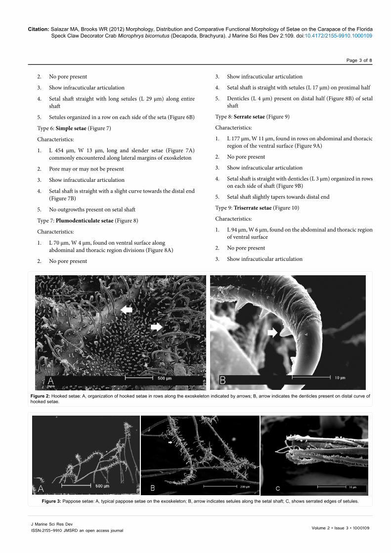
Figure 2: Hooked setae: A, organization of hooked setae in rows along the exoskeleton indicated by arrows; B, arrow indicates the denticles present on distal curve of hooked setae.
Figure 3: Pappose setae: A, typical pappose setae on the exoskeleton; B, arrow indicates setules along the setal shaft; C, shows serrated edges of setules.

Citation: Salazar MA, Brooks WR (2012) Morphology, Distribution and Comparative Functional Morphology of Setae on the Carapace of the Florida Speck Claw Decorator Crab Microphrys bicornutus (Decapoda, Brachyura). J Marine Sci Res Dev 2:109. doi:10.4172/2155-9910.1000109
Page 4 of 8
Volume 2 • Issue 3 • 1000109J Marine Sci Res DevISSN:2155-9910 JMSRD an open access journal
4. Setal shaft is straight with denticles (L 5 µm) arising towards distal half
5. Setal shaft slightly tapers towards distal end
Type 10: Peniculate setae (proposed name) (Figure 11)
Characteristics:
1. L 346 µm, W 9 µm, found randomly on entire exoskeleton, except for dactylus of pereopods, commonly encountered near hooked setae and lateral margins
2. Subterminal/terminal pore present
3. Show infracuticular articulation
4. Found randomly on entire exoskeleton, commonly encountered near hooked setae and lateral margins
5. Setal shaft is straight with two-four rows of denticles (L 1.5 µm) within grooves along left and right underside of shaft (Figure 11A)
6. Denticles start half way along setal shaft and merge at a cluster
surrounding a subterminal/terminal pore (Figure 11B) on distal end
Type 11: Barbed setae (proposed name) (Figure 12)
Characteristics:
1. L 124 µm, W 4 µm, found randomly on entire exoskeleton, except for dactylus of pereopods, commonly encountered near hooked setae and lateral margins
2. Terminal pore may be present
3. Show infracuticular articulatoion
4. Setal shaft is straight with barbs (1 µm) randomly arranged along bottom half and sides of seta (Figure 12A) forming a cluster a tip (Figure 12B)
5. Top half of distal portion of shaft is free of outgrowths except for a pair of barbs that arise near distal tip (Figure 12C)
Hooked setae ablation experiments
When all setae on the exoskeleton were left intact crabs added an average of 0.022 grams per cm2 of algae. When hooked setae were ablated, a significant decrease was measured, with amount of algae added for decoration being reduce to an average of 0.001 grams per cm2 (Mann-Whitney Rank Sum Test, P< 0.001).
DiscussionIn general crustacean exoskeletons are composed of a durable
cuticle. Setae traverse the cuticle and come in direct contact with the external environment, potentially providing the central nervous system with information about chemical and mechanical stimuli [18,29]. Seta may be defined as “an articulated cuticular extension” and can exhibit a number of diverse shapes and sizes [12]. This morphological variation provides not only information on the function, but may also provide information on the phylogeny of the group with distribution varying from species to species [9,10,13].
This study provides the first account of setal morphology and distribution on the carapace of the Florida Speck Claw Decorator Crab Microphrys bicornutus, with an emphasis on how these structures are involved in decorating behavior.
Decorating is a multifaceted behavior that generally begins once the crab selects a piece of material of appropriate size from its surrounding, using its chelae [30]. M. bicornutus uses algae as decorating material. One of the chelae is used to transfer the algae to the area near the front of the crab where the maxillae chew the material to an appropriate size and shape for preparation for attachment. The crab now takes the material from its mouthparts using one of the chelae and rubs the material against the area of the carapace covered with hooked setae until the material is securely attached. Hooked setae contain denticles that pierce into the algae, like Velcro®, to assist in holding the algae in place.
In decorator crabs, setae are generally found on the rostrum, carapace, and pereopods [9,11,30]. A growing body of evidence supports mechanical attachment of decorating material using hooked setae as the primary mechanisms for attachment of decorated materials [6,9,11,31]. Wicksten [9] showed that in L. crispatus, when all hooked setae and epidermal projections were removed, the crabs were not able to decorate. When all structures except for hooked setae were removed, crabs were able to decorate normally, demonstrating that hooked
Figure 4: Typical connate setae on the exoskeleton.
Figure 5: Typical cuspidate setae on the exoskeleton indicated by arrow.

Citation: Salazar MA, Brooks WR (2012) Morphology, Distribution and Comparative Functional Morphology of Setae on the Carapace of the Florida Speck Claw Decorator Crab Microphrys bicornutus (Decapoda, Brachyura). J Marine Sci Res Dev 2:109. doi:10.4172/2155-9910.1000109
Page 5 of 8
Volume 2 • Issue 3 • 1000109J Marine Sci Res DevISSN:2155-9910 JMSRD an open access journal
Figure 6: Plumose setae: A, arrow indicates plumose setae extending from the orbital region of the exoskeleton; B, long slender setules present on opposite sides of the setae indicated by arrow.
Figure 7: Simple setae: A, typical simple setae extending from the lateral margins on the exoskeleton; B, distal end of the setal shaft is slightly curved.
Figure 8: Plumodenticulate setae: A, arrow indicates a typical plumodenticulate setae on the ventral surface of the exoskeleton; B, setules present on the proximal end of the setal shaft and denticles present on the distal, terminal end of the setal shaft.

Citation: Salazar MA, Brooks WR (2012) Morphology, Distribution and Comparative Functional Morphology of Setae on the Carapace of the Florida Speck Claw Decorator Crab Microphrys bicornutus (Decapoda, Brachyura). J Marine Sci Res Dev 2:109. doi:10.4172/2155-9910.1000109
Page 6 of 8
Volume 2 • Issue 3 • 1000109J Marine Sci Res DevISSN:2155-9910 JMSRD an open access journal
setae are used for mechanical attachment of decorating materials. This finding was confirmed in the present study for M. bicornutus. When all hooked setae were ablated, attempts made to attach algae to the exoskeleton were unsuccessful because crabs lacked the structures that physically pierce into the algae and hold it in place.
While hooked setae were used to attach material onto the body, playing an active role in decoration, 8 of the remaining 10 other setae described fall into several categories of chemical and mechanical receptors described by Watling [12]. Plumose, pappose, and plumodenticulate setae are considered annulate setae with setules, which are always mechanoreceptors. Cuspidate, connate, simple, and serrate setae are considered annulate setae without setules, which are usually chemoreceptors, but may play a role as mechanoreceptors when found in large groups.
In addition to the eight previously categorized seta types, two new setae, not previously described in the literature, were photographed and described. Nomenclature for these setae is proposed, and is based on external morphological features. Penniculate setae are named after the latin term for brush, penniculus, as denticles on the shaft of these setae are organized in such a way as to give them a brush-like appearance. Barbed setae are named after the barbs present along the setal shaft. Similarities in size, location and morphology exist between these and previously classified mechanical receptors and chemoreceptors; therefore, it may be presumed that these setae play such a role.
In M. bicornutus hooked setae were used to attach algae to the exoskeleton during decoration, but the other ten setal types – including two novel setae for this species - present on the exoskeleton showed a high degree of morphological variation and likely play an auxiliary role in this behavior. These setae are often present near the hooked setae and may provide information regarding material quality or quantity on the
Figure 9: Serrate setae: A, serrate setae organized in rows on the ventral surface of the exoskeleton; B, denticles in rows on each side of the setal shaft indicated by arrow.
Figure 10: Typical triserrate setae on the ventral surface of the exoskeleton.

Citation: Salazar MA, Brooks WR (2012) Morphology, Distribution and Comparative Functional Morphology of Setae on the Carapace of the Florida Speck Claw Decorator Crab Microphrys bicornutus (Decapoda, Brachyura). J Marine Sci Res Dev 2:109. doi:10.4172/2155-9910.1000109
Page 7 of 8
Volume 2 • Issue 3 • 1000109J Marine Sci Res DevISSN:2155-9910 JMSRD an open access journal
carapace, or lack thereof [11]. SEM analysis revealed that small algal/detrital fragments were present on or near these non-hooked setae. Compared to the large pieces of algae attached to the hooked setae, these small remnants would not likely provide sufficient amount of materials to maintain crypsis. Based on the mechano- and chemo-receptive functions, these latter types of setae (i.e., non-hooked) could extract different stimulus features, cumulatively building an “image” of the chemical and mechanical features of the crab’s external environment. Such information would facilitate the successful completion of the complex behaviors associated with decorating by M. bicornutus [18].
Acknowledgements
The authors wish to thank the University of Miami Center for Advanced Microscopy (UMCAM) for providing the facilities and equipment to produce the SEM images as well as Dr. Patricia Blackwelder and Husain Alsayegh at UMCAM for all of their generosity, time, and help in producing the SEM images; Lisa Tips word and the staff at the Keys Marine Lab for scheduling the boat trips, site selection and assistance in collection; and Sonia Salazar for assistance in collection.
References
1. Berke SK, Miller M, Woodin SA (2006) Modelling the energy-mortality trade-offs of invertebrate decorating behaviour. Evol Ecol Res 8: 1409-1425.
2. Bauer RT (1985) Diel and seasonal variation in species composition and abundance of Caridean shrimps (Crustacea, Decapoda) from seagrass meadows on the north coast of Puerto Rico. Bull Mar Sci 36: 150-162.
3. Bauer RT (1985) Penaeoid shrimp fauna from tropical seagrass meadows: species composition, diurnal and seasonal variation in abundance. Proc Biol Soc Wash 98: 177-190.
4. Bauer RT (1985) Hermit crab fauna from sea grass meadows in Puerto Rico: species composition, diel and seasonal variation in abundance. J Crustacean Biol 5: 249-257.
5. Aurivillus CW (1889) Die Maskirung der oxyrrhynchen Decapoden. K Svenska Vetensk Akad Handl 23: 1-72.
6. Rathbun M J (1925) The spider crabs of America. Bull US Natl Mus 129: 1-599.
7. Jones LM (1938) Decorative algae on a crab, Naxia tumida. Ecology 19: 81-88.
8. Wicksten MK (1976) Studies on hooked setae of Hyas lyratus (Brachyura-Majidae). Syesis 9: 367-368.
9. Wicksten MK (1978) Attachment of decorating materials in Loxorhynchus crispatus (Brachyura: Majidae). Trans Am Microsc Soc 97: 217-220.
10. Wicksten MK (1978) The exterior decorator. Sea Front 24: 277-280.
11. Wicksten M (1993) A review and a model of decorating behavior in Spider Crabs (Decapoda, Brachyura, Majidae). Crustaceana 64: 314-325.
Figure 11: Peniculate setae (proposed name): A, arrow indicates denticles along the setal shaft; B, terminal pore indicated by arrow.
Figure 12: Barbed setae (proposed name): A, arrow indicates barbs along the setal shaft; B, organization of barbs at terminal end of setal shaft; C, arrow indicates a pair of barbs present on the back, terminal end of the setal shaft.

Citation: Salazar MA, Brooks WR (2012) Morphology, Distribution and Comparative Functional Morphology of Setae on the Carapace of the Florida Speck Claw Decorator Crab Microphrys bicornutus (Decapoda, Brachyura). J Marine Sci Res Dev 2:109. doi:10.4172/2155-9910.1000109
Page 8 of 8
Volume 2 • Issue 3 • 1000109J Marine Sci Res DevISSN:2155-9910 JMSRD an open access journal
12. Watling L (1989) A classification system for crustacean setae based on the homology concept. In: Functional morphology of feeding and grooming in Crustacea. Felgenhauer BE, Watling L & Thistle AB, eds., A.A. Balkema, Rotterdam, the Netherlands 5-26.
13. Garm A (2004) Revising the definition of the crustacean seta and setal classification systems based on examinations of the mouthpart setae of seven species of decapods. Zool J Linn Soc 142: 233-252.
14. Derby CD (1982) Structure and function of cuticular sensilla of the lobster Homarus americanus. J Crustacean Biol 2: 1-21.
15. Derby CD, Atema J (1982) The function of chemo- and mechanoreceptors in lobster (Homarus americanus) feeding behaviour. J Exp Biol 98: 317-327.
16. Lavalli KL, Factor JR (1992). Functional morphology of the mouthparts of juvenile lobsters, Homarus americanus (Decapoda: Nephropidae), and comparison with larval stages. J Crustacean Biol 12: 467-510.
17. Weatherby TM, Wong KK, Lenz PH (1994) Fine structure of the distal sensory setae on the first antennae of Pleuromamma xiphias (Giesbrecht) (Copepoda). J Crustacean Biol 14: 670-685.
18. Derby CD, Steullet P (2001) Why do animals have so many receptors? The role of multiple chemosensors in animal perception. Bio Bull 200: 211-215.
19. Garm A, Høeg JT (2001) Function and functional groupings of the complex mouth apparatus of the squat lobsters Munida sarsi (Huus) and M. tenuimana (G.O. Sars) (Crustacea: Decapoda). Biol Bull 200: 281-297.
20. Mead KS, Weatherby TM (2002) Morphology of stomatopod chemosensory sensilla facilitates fluid sampling. Invertebr Biol 121: 148-157.
21. Garm A, Hallberg E, Høeg JT (2003) Role of maxilla 2 and its setae during feeding in the shrimp Palaemon adspersus (Crustacea: Decapoda). Biol Bull 204: 126-137.
22. Garm A, Derby CD, Hoeg JT (2004) Mechanosensory neurons with bend- and osmo- sensitivity in mouthpart setae from the spiny lobster Panulirus argus. Biol Bull 207: 195-208.
23. Weisbaum D, Lavalli KL (2004) Morphology and distribution of antennular setae of Scyllarid Lobsters (Scyllarides aequinoctialis, S. latus, and S. nodifer) with comments on their possible function. Invertebr Biol 123: 324-342.
24. Drumm DT (2005) Comparative morphology of the mouthparts, chelipeds and foregut of two kalliapseudid apseudomorphans (Crustacea: Tanaidacea). Proce Acad Nat Sci Philadelphia 154: 137-147.
25. Garm A (2005) Mechanosensory properties of the mouthpart setae of the European shore crab Carcinus maenas. Mar Biol 147: 1179-1190.
26. Garm A, Shabani S, Hoeg JT, Derby CD (2005) Chemosensory neurons in the mouthparts of the spiny lobsters Panulirus argus and Panulirus interruptus (Crustacea: Decapoda). J Exp Mar Biol Ecol 314: 175-186.
27. Hesselberg T, Vincent JFV (2005) A comparative study of the functional morphology of parapodia and setae in nereids (Polychaeta: Nereididae). Animal Biology 56: 103-120.
28. Szebeni T, Hartnoll RG (2005) Structure and distribution of carapace setae in British spider crabs. J Nat Hist 39: 3795-3809.
29. Grasso FW, Basil JA (2002) How lobsters, crayfishes, and crabs locate sources of odor: current perspectives and future directions. Curr Opin Neurobiol 12: 721-727.
30. Wicksten MK (1980) Decorator crabs. Sci Am 242: 116-122.
31. Schmitt W (1965) Crustaceans. Ann Arbor: University of Michigan, Press 204.
Submit your next manuscript and get advantages of OMICS Group submissionsUnique features:
• Userfriendly/feasiblewebsite-translationofyourpaperto50world’sleadinglanguages• AudioVersionofpublishedpaper• Digitalarticlestoshareandexplore
Special features:
• 200OpenAccessJournals• 15,000editorialteam• 21daysrapidreviewprocess• Qualityandquickeditorial,reviewandpublicationprocessing• IndexingatPubMed(partial),Scopus,DOAJ,EBSCO,IndexCopernicusandGoogleScholaretc• SharingOption:SocialNetworkingEnabled• Authors,ReviewersandEditorsrewardedwithonlineScientificCredits• Betterdiscountforyoursubsequentarticles
Submityourmanuscriptat:www.editorialmanager.com/environsci