journal of forensic and legal medicine · the osteological parameters of the skeleton of individual...
TRANSCRIPT
lable at ScienceDirect
Journal of Forensic and Legal Medicine 20 (2013) 1018e1023
Contents lists avai
Journal of Forensic and Legal Medicine
journal homepage: www.elsevier .com/locate/ jflm
Case report
Fatal cranial injury in an individual from Messina (Sicily) during thetimes of the Roman Empire
Andrea Dario Messina, Ph.D, Anthropologist *, Giuseppe Carotenuto, MD, Physician,Roberto Miccichè, BA, Anthropologist, Luca Sìneo, Ph.D., Professor of AnthropologyDipartimento di “Scienze e Tecnologie biologiche, chimiche e farmaceutiche” LabHomo, Laboratori di Antropologia, Universita’ di Palermo (I), Via Archirafi,18, 90123, Italy
a r t i c l e i n f o
Article history:Received 4 January 2013Received in revised form25 July 2013Accepted 25 September 2013Available online 5 October 2013
Keywords:Forensic sciencesForensic anthropologyCranial traumaPerimortem lesionEpidural hematomaRoman Empire
* Corresponding author. Tel.: þ39 (0)9123891806.E-mail address: [email protected] (A.D. Mes
1752-928X/$ e see front matter � 2013 Elsevier Ltdhttp://dx.doi.org/10.1016/j.jflm.2013.09.023
a b s t r a c t
Forensic and archaeological examinations of human skeletons can provide us with evidence of violence.In this paper, we present the patterns of two cranial lesions found on an adult male (T173) buried in agrave in the necropolis ‘Isolato 96’, Messina, Sicily, dating back to the Roman Empire (1st century BC - 1stcentury AD). The skull reveals two perimortem traumatic lesions, one produced by a sharp object on theright parietal bone and the other one on the left parietal bone, presumably the result of a fall. Theinterpretation of fracture patterns found in this cranium are an illustration of how forensic approachescan be applied with great benefit to archaeological specimens.
� 2013 Elsevier Ltd and Faculty of Forensic and Legal Medicine. All rights reserved.
1. Introduction
During the research project ‘From Zancle to Messana’, whichcovered the history of Messina from its Greek foundations to Ro-man domination, archaeological explorations concentrated uponnecropolis ‘Isolato 96’. The excavations, supervised by the Sovrin-tendenza di Messina, which began in 1998 and were completed in1999, have uncovered a wealth of archaeological finds. Strati-graphic information, together with the analysis of burial apparatusand funerary objects, have allowed the identification of differentchronological stages which embrace an extensive period of time,from the Hellenistic era to Late Antiquity (4th century BC e 5thcentury AD). Especially during Roman Imperial period Messanaseems to be characterized by strong social and institutional crises1
and radical changes in the arrangement of space for burials.2
Traditional sources for evidence of interpersonal violence andwarfare in historical times are comprised documents and archae-ological finds, including human remains. Usually, evidence ofskeletal trauma cited in archaeological literature is primarily basedon observations of the bone healing processes and remodeling,
sina).
and Faculty of Forensic and Legal M
with little attention given to perimortem injuries.3 Indeed, when atrauma pattern has been observed in either forensic or archaeo-logical settings, the following step would be to evaluate the timingof this injury and its possible association with the cause andmanner of death.4
The cranium is often subjected to weapon-related trauma andinjury patterns can be very complicated and difficult to evaluate.Moreover, cranial trauma can be caused by daily activities, as wellas by all sorts of accidents.5e7 Several recent studies have addressedtype and position of cranial traumatic injuries in relation to epi-sodes of violence.8e18
This paper describes two cranial injuries observed in individualT173, which date to the Roman Empire (1st century BCe1st centuryAD), based on grave goods (Piriform Unguentarium) recovered in thegrave.2
For examining skeletal trauma we analyze the affected areaaccording to the following traditional categories: fracture angle andoutline, color and surface morphology.19e21 Moreover, we applied3D CT imaging to obtain additional features for distinguishingperimortem trauma and postmortem damage: preponderanttexture, preponderant outline, relationship to the path of leastresistance, signs of plastic response and the presence of hinging.22
Indeed, non-invasive imaging techniques are of great value in
edicine. All rights reserved.

Fig. 1. Frontal and superior view of T173 skull.
Fig. 2. Lateral view of T173 skull. Trauma on right parietal is visible.
A.D. Messina et al. / Journal of Forensic and Legal Medicine 20 (2013) 1018e1023 1019
forensic anthropology when applied to lesions with doubtful peri-mortem or postmortem features.23
The cranial lesions in T173 do not appear to indicate post-depositional alterations. Our own anthropological examination ofthe skull suggest that the lesions are of traumatic nature and wereinflicted perimortem: the first trauma on the right parietal bonewascaused by a sharp object, while the second, on the left parietal bone,could be the consequence of a fall.
The aim of this study was to examine these lesions with regardto their possible etiology and to formulate a hypotheses about theevents that led to their occurrence, to establishwhether thesewere,in fact, the cause of death of individual T173.
2. Materials and methods
The skull, labeled T173, derives from a complete inhumationburial.
Age estimation was based on pubic symphyseal facemorphology24; metamorphosis at the sternal rib end25;morphology of auricular surface of the ilium.26 Sex was assigned viastandard metric analysis of the pelvic bone traits.27 The followingmeasurements have been taken: PUM, SPU, DCOX, IIMT, ISMM,SCOX, SS, SA, SIS and VEAC.
For the description of cranial fractures, we referred to the cur-rent criteria.3,28e30 In addition, we performed a CT scanning of theskull, using a multi detector scanner General Electric LightSpeedVCT 64 Slice CT, gantry rotation time 0.6 s, slice thickness 0.6 mm,high-resolution protocol, integrated with MIP and VRT-3D recon-struction. Data were saved as Digital Imaging and Commutations inMedicine format bitmap files (DICOM), which were later convertedinto High Dynamic Range image format files through the use of thesoftware AMIRA 5.0.31,32
Questions related to the weapon used were approached usingtechniques developed in forensic pathology and anthropology33e35; we also compared our results with sources of literature on theMediterranean area in the Roman and Modern Ages.36e38
3. Results
The osteological parameters of the skeleton of individual T173indicate a young adult (20e34 years).
All measurements detected from the T173 coxal bones un-equivocally supported the assignation of male (with a 99.78% and99.75% posterior probabilities, for left and right sides respectively).
The skull is almost complete and appears to be asymmetric, dueto a light depression of the left parietal bone (Fig. 1).
All mandibular teeth are present, except for the left M1 and M3which were lost postmortem; the maxillary central incisors, theright lateral incisor, the right P4 and M1 were lost antemortem,while right P3 was lost postmortem.
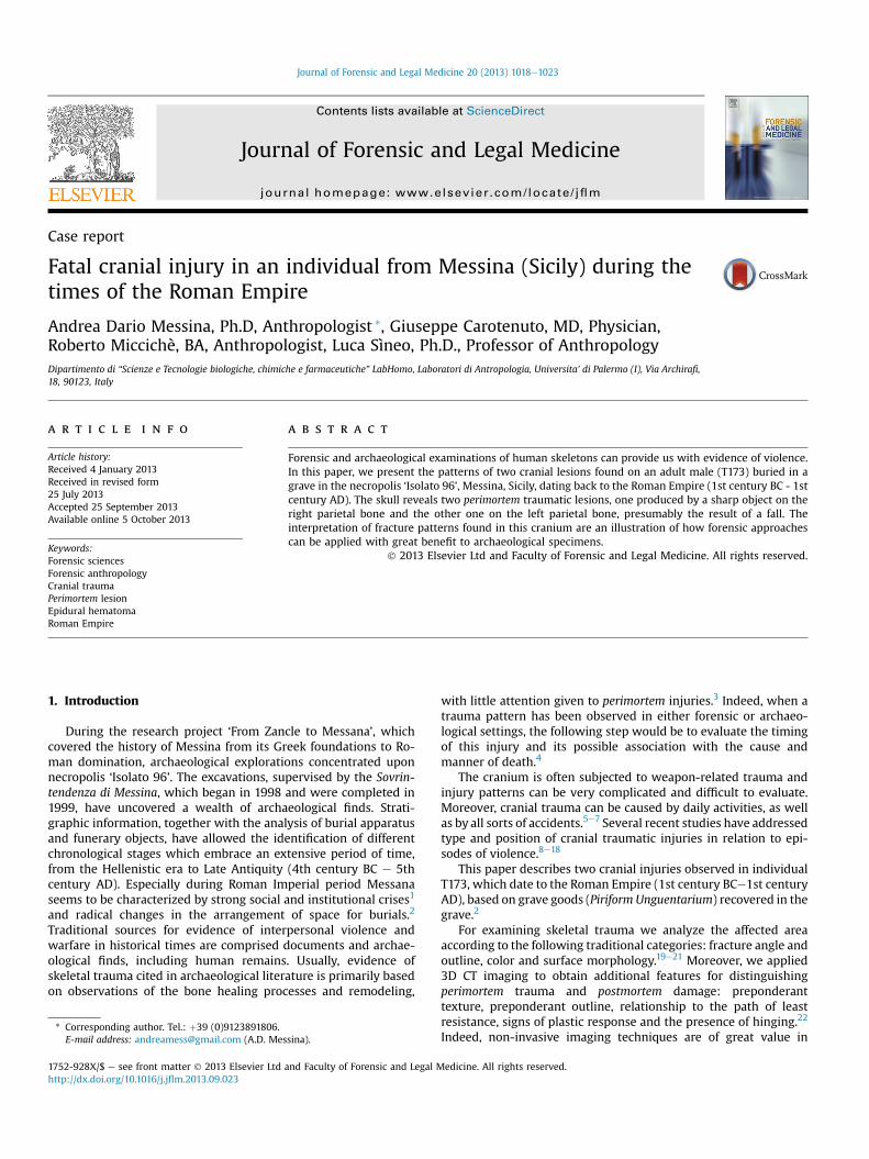
On the external surface of the right parietal bone, near thecoronal suture, an oval-shaped lesion is present bounded above bythe temporal lines: the minor diameter measures 11 mm (a), whilethe major diameter measures 21 mm (A). In this area there is aperforation (Fig. 2), shaped like a rectangular trapezoid, whit thefollowing measurements: minor base (b) 3.93 mm, major base (B)6.0 mm, oblique side (l) 5.55 mm, while the height (h) measures11 mm (Fig. 3). The perforation is delimited by bone fragments,introflexed but still attached to the surrounding bone and visible onthe cerebral surface (Fig. 4). There are no signs of bony remodeling.On the endocranial surface the perforation affects some of thegrooves of the right middle meningeal vessels.
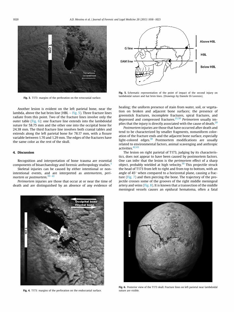
Fig. 3. T173: margins of the perforation on the ectocranial surface.
Fig. 5. Schematic representation of the point of impact of the second injury onlambdoidal suture and hat brim lines. (Drawings by Daniele Di Lorenzo).
A.D. Messina et al. / Journal of Forensic and Legal Medicine 20 (2013) 1018e10231020
Another lesion is evident on the left parietal bone, near thelambda, above the hat brim line (HBL e Fig. 5). Three fracture linesradiate from this point. Two of the fracture lines involve only theouter table (Fig. 6): one fracture line extends into the lambdoidalsuture for 58.75 mm and the other one into the occipital bone for24.38 mm. The third fracture line involves both cranial tables andextends along the left parietal bone for 78.17 mm, with a fissurevariable between 1.70 and 1.29 mm. The edges of the fractures havethe same color as the rest of the skull.
4. Discussion
Recognition and interpretation of bone trauma are essentialcomponents of bioarchaeology and forensic anthropology studies.3
Skeletal injuries can be caused by either intentional or non-intentional events, and are interpreted as antemortem, peri-mortem or postmortem.39e41
Perimortem injuries are those that occur at or near the time ofdeath and are distinguished by an absence of any evidence of
Fig. 4. T173: margins of the perforation on the endocranial surface.
healing; the uniform presence of stain from water, soil, or vegeta-tion on broken and adjacent bone surfaces; the presence ofgreenstick fractures, incomplete fractures, spiral fractures, anddepressed and compressed fractures.21,30 Perimortem usually im-plies that the injury is directly associated with the cause of death.20
Postmortem injuries are those that have occurred after death andtend to be characterized by smaller fragments, nonuniform color-ation of the fracture ends and the adjacent bone surface, especiallylight-colored edges.30 Postmortem modifications are usuallyrelated to environmental factors, animal scavenging and anthropicactivities.42,43
The lesion on right parietal of T173, judging by its characteris-tics, does not appear to have been caused by postmortem factors.One can infer that the lesion is the perimortem effect of a sharpobject, probably wielded at high velocity.30 This projectile struckthe head of T173 from left to right and from top to bottom, with anangle of 45� when compared to a horizontal plane, causing a frac-ture (Fig. 7) and then piercing the bone. The trajectory of the pro-jectile crosses some of the grooves of the right middle meningealartery and veins (Fig. 8). It is known that a transection of the middlemeningeal vessels causes an epidural hematoma, often a fatal
Fig. 6. Posterior view of the T173 skull. Fracture lines on left parietal near lambdoidalsuture are visible.

Fig. 7. Reconstruction of 3D trajectory of the projectile and lateral skull radiograph.
A.D. Messina et al. / Journal of Forensic and Legal Medicine 20 (2013) 1018e1023 1021
condition with the build up of blood trapped between the endo-cranial surface and the dura mater, in the ‘Marchant Zone’, i.e., thearea from which the dura mater is readily detached.44 Because ofthe location of the middle meningeal vessels, the blood masstypically lies over the lateral hemisphere (temporal and/or parietallobes).
Since the epidural hematoma is under pressure, it typicallycontinues to grow unless evacuated. Classically, cranial trauma areassociated with concussion and loss of consciousness; the indi-vidualmaywake up after a lucid interval, only to lose consciousnessagain from brain stem distortion as a result of increased intracranialpressure. If the bleeding is very severe, there is no lucid interval.The individual has no time to awake from the concussion beforecompressive brain stem deterioration begins.45
Technically, the epidural hematoma could have been the causeof death of T173.
The pathological consequences of penetrating head woundsdepend on the circumstances of the injury, including the propertiesof the weapon or missile, the energy of the impact, and the locationand characteristics of the intracranial trajectory.46e48 Following theprimary injury or impact, secondary injuries may develop.49,50
Secondary injury mechanisms are defined as pathological pro-cesses (traumatic and neurological) that occur after the time of theinjury and adversely affect the ability of the brain to recover fromthe primary insult.51e53
Fig. 8. T173: the perforation crosses some groves of middle meningeal artery.
Cranial computed tomography scanning (Fig. 9) clearlydemonstrate the presence of splinter bone attached to the fracturemargins confirming that the lesionwas made on ‘fresh’ bone,21 andexcludes the possibility that the lesionwas caused a long time afterdeath on dry bone by taphonomic factors. The characteristics andposition of the lesion on the right parietal, the absence of a bonyreaction on the margins of the perforation, and the presence ofinternal bevelling, all seem to indicate a perimortem trauma,occurring immediately before death (possibly the cause of death) orimmediately after death.
In addition, the use of reconstructed 3D CT imaging allowed usto examine the endocranial surface without jeopardizing theintegrity of the sample. For the lesion described in the right parietalbone we observed the presence of plastic response and hinging(Fig. 10a), typically associated with perimortem fractures. This resultis consistent with the findings of macroscopic visual analysis.
The cause of the various fracture patterns lies in the changingbiomechanical properties of the bone after death.30 Fresh or livingbone contains fluid-filled vessels, grease and collagen fibers, whichmake it significantly more pliable and more resistant to tensileforces than dry bone. In consequence, fresh bones are likely tosplinter with fragments tending to remain attached to one another,and with fractures producing irregular edges. After the death, onthe other hand, the bone becomes harder and more brittle withtime, and as a result shatters into small, more regularfragments.35,54
However, the bones retain ‘fresh’ properties for a considerabletime after death.21,55,56 Therefore caution should be exercisedwhendetermining the timing of fractures. There are some analyticalmethods for studying the timing of a bone fracture of the post-cranial skeleton,57,58 but no reference is available for the skull. Inany case, the timing of injuries can be determined in a probabilisticmanner.4
The lesion on the left parietal bone shows a pattern character-istic of a blunt force trauma, with radiating fracture lines from thepoint where the skull was struck.59 The 3D CT reconstruction of thecranium reveals a smooth preponderant texture and a regularoutline (Fig. 10b). The lines of the fracture that radiate from thepoint of impact are not associated with plastic deformation of theskull and were probably produced at the same time as the trauma.
The features of the left parietal injury indicate that they werecaused by a fall or a blow.
Distinguishing between falls and blows in blunt head trauma isa common and difficult problem in both forensic anthropology andbioarchaeology studies.5
Several authors59e65 have suggested methods for the discrimi-nation of falls and blows, comprising three main indicators: the hatbrim line rule, the side lateralization of fractures, and the number of
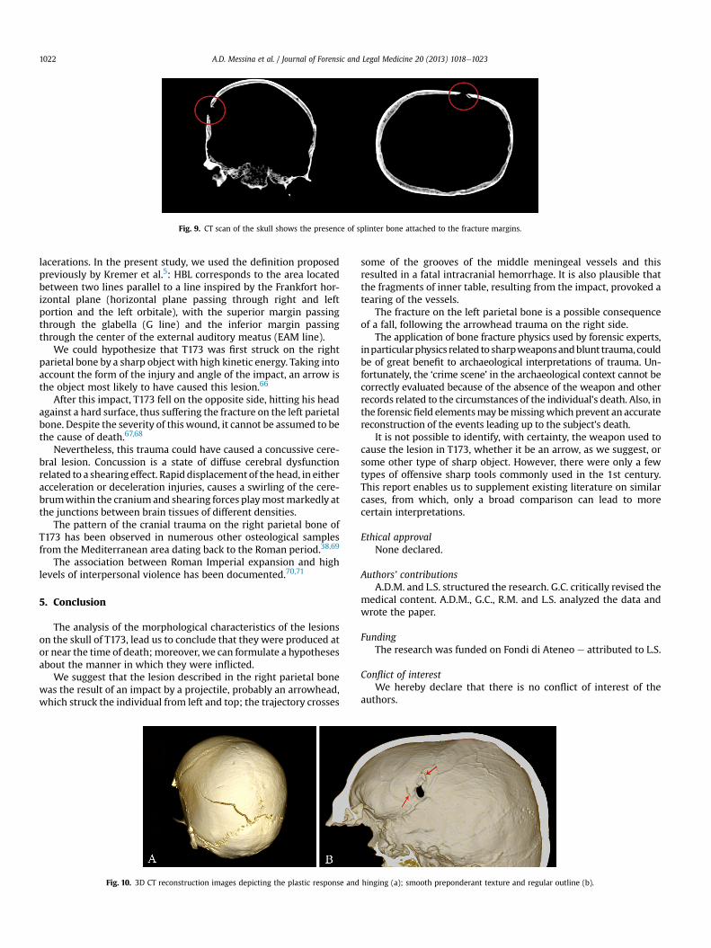
Fig. 9. CT scan of the skull shows the presence of splinter bone attached to the fracture margins.
A.D. Messina et al. / Journal of Forensic and Legal Medicine 20 (2013) 1018e10231022
lacerations. In the present study, we used the definition proposedpreviously by Kremer et al.5: HBL corresponds to the area locatedbetween two lines parallel to a line inspired by the Frankfort hor-izontal plane (horizontal plane passing through right and leftportion and the left orbitale), with the superior margin passingthrough the glabella (G line) and the inferior margin passingthrough the center of the external auditory meatus (EAM line).
We could hypothesize that T173 was first struck on the rightparietal bone by a sharp object with high kinetic energy. Taking intoaccount the form of the injury and angle of the impact, an arrow isthe object most likely to have caused this lesion.66
After this impact, T173 fell on the opposite side, hitting his headagainst a hard surface, thus suffering the fracture on the left parietalbone. Despite the severity of this wound, it cannot be assumed to bethe cause of death.67,68
Nevertheless, this trauma could have caused a concussive cere-bral lesion. Concussion is a state of diffuse cerebral dysfunctionrelated to a shearing effect. Rapid displacement of the head, in eitheracceleration or deceleration injuries, causes a swirling of the cere-brumwithin the craniumand shearing forces playmostmarkedly atthe junctions between brain tissues of different densities.
The pattern of the cranial trauma on the right parietal bone ofT173 has been observed in numerous other osteological samplesfrom the Mediterranean area dating back to the Roman period.38,69
The association between Roman Imperial expansion and highlevels of interpersonal violence has been documented.70,71
5. Conclusion
The analysis of the morphological characteristics of the lesionson the skull of T173, lead us to conclude that they were produced ator near the time of death; moreover, we can formulate a hypothesesabout the manner in which they were inflicted.
We suggest that the lesion described in the right parietal bonewas the result of an impact by a projectile, probably an arrowhead,which struck the individual from left and top; the trajectory crosses
Fig. 10. 3D CT reconstruction images depicting the plastic response and
some of the grooves of the middle meningeal vessels and thisresulted in a fatal intracranial hemorrhage. It is also plausible thatthe fragments of inner table, resulting from the impact, provoked atearing of the vessels.
The fracture on the left parietal bone is a possible consequenceof a fall, following the arrowhead trauma on the right side.
The application of bone fracture physics used by forensic experts,inparticularphysics related to sharpweaponsandblunt trauma,couldbe of great benefit to archaeological interpretations of trauma. Un-fortunately, the ‘crime scene’ in the archaeological context cannot becorrectly evaluated because of the absence of the weapon and otherrecords related to the circumstances of the individual’s death. Also, inthe forensicfield elementsmay bemissingwhich prevent an accuratereconstruction of the events leading up to the subject’s death.
It is not possible to identify, with certainty, the weapon used tocause the lesion in T173, whether it be an arrow, as we suggest, orsome other type of sharp object. However, there were only a fewtypes of offensive sharp tools commonly used in the 1st century.This report enables us to supplement existing literature on similarcases, from which, only a broad comparison can lead to morecertain interpretations.
Ethical approvalNone declared.
Authors’ contributionsA.D.M. and L.S. structured the research. G.C. critically revised the
medical content. A.D.M., G.C., R.M. and L.S. analyzed the data andwrote the paper.
FundingThe research was funded on Fondi di Ateneo e attributed to L.S.
Conflict of interestWe hereby declare that there is no conflict of interest of the
authors.
hinging (a); smooth preponderant texture and regular outline (b).

A.D. Messina et al. / Journal of Forensic and Legal Medicine 20 (2013) 1018e1023 1023
References
1. Finley MI. History of ancient Sicily. Roma: Ed. Laterza; 2009.2. Bacci MG, Tigano G. Da Zancle a MessinaIn Un percorso archeologico attraverso
gli scavi. Messina: Ed. Sicania; 2003.3. Berryman HE, Haun SJ. Applying forensic techniques to interpret cranial frac-
ture patterns in an archaeological specimen. Int J Osteoarchaeol 1996;6:2e9.4. Jordana F, Colat-Parros J, Bénézech M. Diagnosis of skull fractures according to
postmortem interval: an experimental approach in a porcine model. J ForensicSci 2013;58:156e62.
5. Kremer C, Racette S, Dionne C-A, Sauvageau A. Discrimination of falls andblows in blunt head trauma: systematic study of the hat brim line rule inrelation to skull fractures. J Forensic Sci 2008;53:716e9.
6. Aufderheide AC, Rodríguez-Martín C. The Cambridge encyclopedia of humanpaleopathology. Cambridge: Cambridge University Press; 1998.
7. Ortner J. Identification of pathological conditions in human skeletal remains. 2nded. San Diego: Academic Press; 2003.
8. Walker PL. A bioarchaeological perspective on the history of violence. Annu RevAnthropol 2001;30:573e96.
9. Djuri�c MP, Roberts CA, Rako�ccevi�c ZB, Djoni�c DD, Le�csi�c AR. Fractures in latemedieval skeletal populations fromSerbia.AmJPhysAnthropol2006;130:167e78.
10. Roksandic M, Djuri�c M, Rako�sevi�c Z, Seguin K. Interpersonal violence at Lep-enski Vir Mesolithic/Neolithic complex of the iron Gates gorge (Serbia-Romania). Am J Phys Anthropol 2006;129:339e48.
11. Roksandic M, Wood C, Vlak D. Death in the line of duty: late medieval burials atthe site of Lepenski Vir, Serbia. Int J Osteoarchaeol 2007;17:635e42.
12. Paine RR, Mancinelli D, Ruggieri M, Coppa A. Cranial trauma in iron ageSamnite agriculturists, Alfedena, Italy: implications for biocultural and eco-nomic stress. Am J Phys Anthropol 2007;132:48e58.
13. Tung TA. Trauma and violence in the War Empire of the Peruvian Andes:warfare, raids and ritual fights. Am J Phys Anthropol 2007;133:941e56.
14. Jiménez-Brobeil SA, du Souich P, Al Oumaoui I. Possible relationship of cranialtraumatic injuries with violence in the south-east Iberian Peninsula from theNeolithic to the Bronze Age. Am J Phys Anthropol 2009;140:465e75.
15. Jordana X, Galte I, Turbat T, Batsukh D, Garcý C, Isidro A, et al. The warriors ofthe steppes: osteological evidence of warfare and violence from Pazyryk tumuliin the Mongolian Altai. J Archaeol Sci 2009;36:1319e27.
16. Standen VG, Arriaza BG, Santoro CM, Romero Á, Rothammer F. Perimortemtrauma in the Atacama desert and social violence during the Late FormativePeriod (2500e1700 years BP). Int J Osteoarchaeol 2010;20:693e707.
17. Rubini M, Zaio P. Warriors from the East. Skeletal evidence of warfare from aLombard-Avar cemetery in central Italy (Campochiaro, Molise, 6the8th cen-tury AD). J Archaeol Sci 2011;38:1551e9.
18. Erdal ÖD. A possible massacre at early Bronze age Titris Höyük, Anatolia. Int JOsteoarchaeol 2012;22:1e21.
19. Ubelaker DH, Adams BJ. Differentiation of perimortem and postmortem traumausing taphonomic indicators. J Forensic Sci 1995;40:509e12.
20. Sauer N. The timing of injuries and manner of death: distinguishing amongantemortem, perimortem and postmortem trauma. In: Reichs KJ, Bass WM,editors. Forensic osteology. Springfield: Charles Thomas; 1998. p. 321e32.
21. Wieberg DAM, Wescott DJ. Estimating the timing of long bone fractures: cor-relation between the postmortem interval, bone moisture content, and bluntforce trauma fracture characteristics. J Forensic Sci 2008;53:1028e34.
22. Fleming-Farrell D, Michailidis K, Karantanas A, Roberts N, Kranioti EF. Virtualassessment of perimortem and postmortem blunt force cranial trauma.Forensic Sci Int 2013. http://dx.doi.org/10.1016/j.forsciint.2013.03.032.
23. Ruhli FJ, Lanz C, Ulrich-Bochsler S, Alt KW. State-of-the-art imaging in palaeo-pathology: the value of multislice computed tomography in visualizingdoubtful cranial lesions. Int J Osteoarchaeol 2002;12:372e9.
24. Katz D, Suchey JM. Age determination of the male os pubis. Am J Phys Anthropol1986;69:427e35.
25. Iscan MY, Loth SR, Wright RK. Metamorphosis at the sternal rib end: a newmethod to estimate age at death in white males. Am J Phys Anthropol 1984;65:147e56.
26. Buckberry JL, Chamberlain AT. Age estimation from the auricular surface of theilium: a revised method. Am J Phys Anthropol 2002;119:231e9.
27. Murail P, Bruzek J, Houet F, Cunha E. DSP: a tool for probabilistic sex diagnosisusingworldwide variability in hip bonemeasurements.BMSAP2005;17:167e76.
28. Maples WR. Trauma analysis by the forensic anthropologist. In: Reichs KJ,editor. Forensic osteology: advances in the identification of human remains 1986.p. 218e28. Springfield.
29. Merbs CF. Trauma. In: Iscan MY, Kennedy KAR, editors. Reconstruction of lifefrom the skeleton. New York: Wiley-Liss; 1989. p. 23e40.
30. Lovell NC. Trauma analysis in paleopathology. Yearb Phys Anthropol 1997;40:139e70.
31. Hu H. Multi-slice helical CT: scan and reconstruction. Med Phys 1999;26:5e18.32. Hu H, He HD, Foley WD, Fox SH. Four multidetector-row helical CT: image
quality and volume coverage speed. Radiology 2000;215:55e62.33. Spitz WU. Spitz and Fisher’s medicolegal investigation of death: guidelines for the
application of pathology to crime investigation. Springfield: Charles C Thomas;1992.
34. Lewis JE. Identifying sword marks on bone: criteria for distinguishing betweencut marks made by different classes of bladed weapon. J Archaeol Sci 2008;35:2001e8.
35. Kanz F, Grossschmidt K. Head injuries of Roman gladiators. Forensic Sci Int2006;160:207e16.
36. Reichs KJ. Forensic osteology: advances in the identification of human remains.Springfield: Charles C Thomas; 1998.
37. Bauer M, Patzelt D. Intracranial stab injuries: case report and case study.Forensic Sci Int 2002;129:122e7.
38. Erfan M, El-Sawaf A, Al-Tohamy Soliman M, El-Din AS, Kandeel WA, El-ShafyEl-Banna RA, Azab A. Cranial trauma in Ancient Egyptians from the BahariyahOasis, Greco-Roman period. Res J Med Med Sci 4 2009:78e84.
39. Walker PL. Cranial injuries as evidence of violence in prehistoric southernCalifornia. Am J Phys Anthropol 1989;80:313e23.
40. Kaufman MH, Whitaker D, McTavish J. Differential diagnosis of holes in thecalvarium: application of modern clinical data to palaeopathology. J ArchaeolSci 1997;24:193e218.
41. Nagaoka T. Cranial traumatic injuries caused by weapons in Tokugawa Japan.Int J Osteoarchaeol 2012;22:138e44.
42. Quatrehomme GMY, Iscan MY. Postmortem skeletal lesions. Forensic Sci Int1997;89:155e65.
43. Calce S, Rogers T. Taphonomic changes to blunt force trauma: a preliminarystudy. J Forensic Sci 2007;52:1e9.
44. Jennett B, Lindsay K. In: An introduction to neurosurgery. 5th ed. Oxford: But-terworth- Heinemann; 1994.
45. Lindsay KW, Bone I, Callander R. In: Neurology and neurosurgery illustrated. 2nded. New York: Churchill Livingstone; 1991.
46. Grahm TW, Williams Jr FC, Harrington T, Spetzler RF. Civilian gunshot woundsto the head: a prospective study. Neurosurgery 1990;27:696e700.
47. Siccardi D, Cavaliere R, Pau A, Lubinu F, Turtas S, Viale GL. Penetratingcraniocerebral missile injuries in civilians: a retrospective analysis of 314 cases.Surg Neurol 1991;35:455e60.
48. Ardill W, Gidado S. Penetrating head wound: a remarkable case. Surg Neurol2003;60:120e3.
49. Feldman Z, Narayan RK, Robertson CS. Secondary insults associated with severeclosed head injury. Contemp Neurosurg 1992;14:1e8.
50. Cosar A, Gonul E, Kurt E, Gönül M, Tasar M, Yetiser S. Craniocerebral gunshotwounds: results of less aggressive surgery and complications. Minim InvasiveNeurosurg 2005;48:113e8.
51. Aarabi B. Causes of infections in penetrating head wounds in the Iran-Iraq War.Neurosurgery 1989;25:923e6.
52. Tudor M, Tudor L, Tudor KI. Complications of missile craniocerebral injuriesduring the Croatian Homeland War. Mil Med 2005;170:422e6.
53. Cox MW, Whittaker DR, Martinez C, Fox CJ, Feuerstein IM, Gillespie DL. Trau-matic pseudoaneurysms of the head and neck: early endovascular interven-tion. J Vasc Surg 2007;46:1227e33.
54. Evans FG. Mechanical properties of bone. Springfield, IL: Charles C. Thomas;1973.
55. Fitzgerald ER. Dynamic mechanical measurements during the life to deathtransition in animal tissues. Biorheology 1975;12:397e408.
56. Galloway A. The biomechanics of fracture production. In: Galloway A, editor.Broken bones: anthropological analysis of blunt force trauma. Springfield, IL:Charles C. Thomas; 1999. p. 35e62.
57. Villa P, Mahieu E. Breakage patterns of human long bones. J Hum Evol 1991;21:27e48.
58. Boulestin B. Approche taphonomique des restes humainsIn Le cas des Méso-lithiques de la grotte des Perrats et le problème du cannibalisme en préhistoirerécente européenne. Oxford: Archeopress; 1999.
59. Kroman A, Kress TA, Porta D. Fracture propagation in the human cranium: a re-testing of popular theories. Clin Anat 2011;24:309e18.
60. Knight B. Forensic pathology. Oxford: Oxford University Press; 1991.61. Galloway A. The circumstances of blunt force trauma. In: Galloway A, editor.
Broken bones anthropological analysis of blunt force trauma. Springfield: CharlesC. Thomas; 1999. p. 224e54.
62. Ehrlich E, Maxeiner H. External injury marks (wound) on the head in differenttypes of blunt trauma in an autopsy series. Med Law 2002;2:773e82.
63. Spitz WU. Blunt force injury. In: Spitz WU, Spitz DJ, editors. Spitz and Fisher’smedicolegal investigation of death: guidelines for the application of pathology tocrime investigation. 4th ed. Springfield IL: Charles C. Thomas; 2006. p. 199e251.
64. Kremer C, Sauvageau A. Discrimination of falls and blows in blunt head trauma:assessment of predictability through combined criteria. J Forensic Sci 2009;54:923e6.
65. Guyomarc’h P, Campagna-Vaillancourt M, Kremer C, Sauvageau A. Discrimi-nation of falls and blows in blunt head trauma: a multi-criteria approach.J Forensic Sci 2010;55:423e7.
66. Southern P. The Roman army: a social and institutional history. Oxford: OxfordUniversity Press; 2007.
67. Venkatesh VT, Kumar MV, Jagannatha SR, Radhika RH, Pushpalatha K. Patternof skeletal injuries in cases of falls from a height. Med Sci Law 2007;47:330e4.
68. Preuss J, Padosch SA, Dettmeyer R, Driever F, Lignitz E, Madea B. Injuries in fatalcases of falls downstairs. Forensic Sci Int 2004;14:121e6.
69. Novak M, Slaus M. Bone traumas in late antique populations from Croatia. CollAntropol 2010;4:1239e48.
70. Harris WV. War and imperialism in Republican Rome, 327e70 B.C. Oxford: Ox-ford University Press; 1979.
71. Rich J, Shipley G.War and society in the Roman world. London: Routledge; 1993.