journal of cereal science€¦ · gluten and non-gluten proteins of wheat as target antigens in...
TRANSCRIPT

lable at ScienceDirect
Journal of Cereal Science 75 (2017) 252e260
Contents lists avai
Journal of Cereal Science
journal homepage: www.elsevier .com/locate/ jcs
Gluten and non-gluten proteins of wheat as target antigens in autism,Crohn’s and celiac disease
Aristo Vojdani a, b, *, Elroy Vojdani c
a Immunosciences Lab., Inc., 822 S. Robertson Blvd., Ste. 312, Los Angeles, CA 90035, USAb Dept. of Preventive Medicine, Loma Linda University, Loma Linda, CA 92350, USAc Private Practice, West Los Angeles, CA 90025, USA
a r t i c l e i n f o
Article history:Received 7 March 2017Received in revised form17 April 2017Accepted 20 April 2017Available online 21 April 2017
Keywords:Wheat proteinsGlutenNon-glutenCXCR3AutismCrohn’sCeliac disease
* Corresponding author. Immunosciences Lab., Inc.312, Los Angeles, CA 90035, USA.
E-mail address: [email protected] (A. Vojdani).
http://dx.doi.org/10.1016/j.jcs.2017.04.0100733-5210/© 2017 The Authors. Published by Elsevie
a b s t r a c t
Studies show that patients with celiac disease react not only with gluten wheat proteins but also withnon-gluten wheat components. Our goal was to measure IgG or IgA antibodies against wheat proteins orpeptides that would provide the most sensitive method for the detection of wheat immune reaction inchildren with autism spectrum disorder, and patients with Crohn’s and celiac disease (CD). Using ELISA,we measured these antibodies against various gluten and non-gluten wheat proteins. Compared tocontrols in all three conditions, the strongest reaction was against CXCR3-binding gliadin peptide, fol-lowed by a wheat protein mixture, and a-gliadin 33-mer peptide. We determined that a sample thatstrongly reacted against non-gluten proteins also reacted strongly against gluten proteins. We also foundthat IgA antibodies against CXCR3-binding gliadin peptide were strongly reactive in a subgroup of 33% inthe autism group, 42% in the Crohn’s group, and all tested samples with CD. The results indicate thatmeasuring IgG and IgA antibodies against non-gluten proteins adds nothing to the pathologic relevanceof these antibodies. Further research is needed on CXCR3-binding gliadin peptide antibodies as a possiblebiomarker and as a guide for dietary elimination in CD, Crohn’s disease and a subgroup of children withASD.© 2017 The Authors. Published by Elsevier Ltd. This is an open access article under the CC BY license
(http://creativecommons.org/licenses/by/4.0/).
1. Introduction
Immunologic reactivity to gluten proteins has been researchedextensively in celiac disease and, to amuch lesser degree, in Crohn’sdisease (Balakireva and Zamyatnin, 2016; Cimaglia et al., 2014;Schedel et al., 2005; Yang et al., 2005). Wheat in general com-prises about 100 different proteins, the majority of which arealcohol-soluble, with the remainder being water-soluble(Chattopadhyay and Kumar, 2016; Kasarda et al., 2013; Kiefferet al., 1982; Ostergaard et al., 2000). Together, a-gliadins, g-glia-dins, u-gliadins, and low and high molecular weight glutenins arethe major alcohol-soluble proteins called gluten proteins, whichrepresent about 75% of the total proteins of wheat grains (Yanget al., 2005). The remainder of wheat proteins, which aregenerally soluble in water or salt solutions, including serine pro-tease inhibitors (serpins), purinins, farinins, a-amylase/protease
, 822 S. Robertson Blvd., Ste.
r Ltd. This is an open access article
inhibitors and globulins are called non-gluten proteins (Kenrickand Walker Smith, 1970; Moneret-Vautrin et al., 2011; Sotkovskyet al., 2008; Stern et al., 1979). Intestinal T cells from celiac disease(CD) patients respond to a heterogenous array of peptides derivedfrom a-, g-, u-gliadins and glutenins, and produce a significantamount of interferon-g (Camarca et al., 2009).
Interestingly, Camarca et al. showed that the immune system ofsome patients recognized many peptides from single or multiplegliadin families, while others reacted to only one peptide. Thismeans that a large number of gluten epitopes may be involved inthe development of gluten sensitivity, CD, and associated diseases.It is important to understand the nature and properties of immu-nodominant epitopes; not only can they aid in diagnosis, but theyalso have tolerance-related therapeutic applications in several T-cell-mediated diseases. The great majority of CD patients in thestudy (Camarca et al., 2009) reacted to at least one g-gliadin-derived peptide, with half recognizing DQ2-g-I. This suggests thatthe contribution of g-gliadin peptides to CD pathogenesis may begreater than previously thought. Strong and frequent recognitionwas also found from the u-gliadin-derived peptides (Arentz-Hansen et al., 2002; Camarca et al., 2009; Shan et al., 2002).
under the CC BY license (http://creativecommons.org/licenses/by/4.0/).

A. Vojdani, E. Vojdani / Journal of Cereal Science 75 (2017) 252e260 253
Overall, 86% of the CD patients recognized a different array ofpeptides. This indicates that other gliadin peptides not tested in thestudy could be relevant in some CD patients (Camarca et al., 2009).
For this reason, immune response to non-gluten proteins ofwheat was also investigated in celiac disease in a different study(Huebener et al., 2015). The results demonstrated that in additionto the well-recognized immune reaction to gluten proteins, celiacdisease was also associated with humoral immune responsedirected against serpins, purinins, farinins, a-amylase/protease in-hibitor and globulins. However, we found that blood samples fromCD patients that reacted to gluten proteins uniformly also reactedto non-gluten proteins (Huebener et al., 2015). Apart from theirinvolvement in CD, antibodies to these gluten and non-proteinproteins have not been examined in the context of Crohn’s dis-ease and autism.
Crohn’s disease and ulcerative colitis fall under the classificationof inflammatory bowel disease (IBD). They are triggered by envi-ronmental factors, including microbial antigens and food(Huebener et al., 2015). The serologic response in Crohn’s diseaseincludes antibodies against specific components of Saccharomycescerevisiae, mycobacteria, Bacteroides, and Escherichia coli (Bartaet al., 2003; Giaffer et al., 1992; Knoflach et al., 1987; Main et al.,1988). In fact, the measurement of antibodies to baker’s andbrewer’s yeasts directed against cell wall oligomannoside epitope(ASCA) has been proposed as a serological marker for Crohn’s dis-ease (Stern et al., 1979). These antibodies have a sensitivity of60e70% for differentiating Crohn’s disease from controls and aspecificity of 80e95% (Quinton et al., 1988; Yang et al., 2005). Due tooverlapping symptomatologies between CD and Crohn’s disease,ASCA antibodies were alsomeasured in a group of patients with CD.In patients with gluten sensitivity enteropathy (GSE), high in-cidences of ASCAwere reported. This high prevalence of ASCA in CDpatients stimulated us into investigating whether indeed thesegluten and non-gluten proteins of wheat, in addition to playing arole in CD, also had some sort of involvement in Crohn’s disease andautism.
Autism spectrum disorder (ASD) is a group of neuroimmunedisorders in which genes and environmental triggers such as in-fections, toxic chemicals, and dietary components play a role. In ourearlier study we measured antibodies against gliadin in childrenwith ASD (Vojdani et al., 2003). Analysis of the blood samplesrevealed that a significant number of autistic children produced IgGand IgA antibodies against a-gliadin 33-mer peptide (Vojdani et al.,2003). Moreover, in a different study, the effectiveness of a gluten-free diet was tested on children with ASD, and a significantimprovement in behavioral symptomatologies was observed in asubgroup (Elder et al., 2006). Similar to what was done with CD,this other study with ASD was conducted with a-gliadin 33-mer,but not with non-gluten or other gluten peptides, especiallyCXCR3-binding gliadin peptide.
CXCR3 is a chemokine receptor that is expressed in monocytes,eosinophils, NK cells, B cells, and T cells, particularly in CD4þ TH1cells (Groom and Luster, 2011). During the inflammatory process,CXCR3 promotes the recruitment of immune cells into the inflamedtissues by interacting with its three different ligands: CXCL9,CXCL10, and CXCL11. This CXCR3 interaction with its ligands be-comes over-activated in different chronic inflammatory processessuch as inflammatory bowel diseases, rheumatoid arthritis(Hosomi et al., 2011; Laragione et al., 2011; Lee et al., 2009), and inthe small intestinal mucosa of untreated patients with CD (Bondaret al., 2014). The number of TH1 cells is increased in the duodenalmucosa of these patients, and these cells express an increasedamount of CXCR3. Therefore, in the study just cited (Bondar et al.,2014), blood levels of soluble CXCL10 and CXCR3þ cells induodenal biopsies were measured and found to be significantly
elevated. Interestingly, CD patients on gluten-free diets presentedlevels of CXCL10 and numbers of CXCR3þ cells that were similar tothose found in controls (Bondar et al., 2014). This over-activation ofCXCR3 and its ligands and the infiltration of TH1 cells and plasmacells into the small intestine have been shown to be associated withtwo gliadin peptides that bind specifically to CXCR3 on the surfaceof these lymphocytes (Lammers et al., 2008).
Consequently, in this present study we tested IgG and IgA an-tibodies against the major gluten proteins, a-gliadin 33-mer, g-gliadin 15-mer, glutenin 21-mer, and gliadin peptides that bind toCXCR3; and for the non-gluten wheat proteins, purinin, farinin, a-amylase, serpin and globulin. We examined IgG and IgA antibodiesagainst these proteins in patients with CD, Crohn’s disease and inchildrenwith ASD in comparison to healthy controls in order to findout whether or not measurements of humoral immune responseagainst CXCR3-binding gliadin peptide and non-gluten proteinswill add to the clinical efficacy of wheat proteome antibody testingin these disorders.
2. Experimental section
2.1. Materials and methods
2.1.1. Blood samplesForty-eight sera from healthy control subjects aged 18e65 were
obtained from Innovative Research (Novi, MI, USA). Commerciallyavailable sera of 24 patients with Crohn’s disease and 24 sera frompatients with CD were purchased from The Binding Site (San Diego,CA, USA), Inova (San Diego, CA, USA), Trina International Nanikon(Switzerland), Diamedix (Fl, USA) and Innovative Research (Novi,MI, USA). We also used sera from 48 children with ASD aged 2e15that we had used in our earlier study that was approved by theInstitutional Review Board of the Center for Autism and RelatedDisorders (Tarzana, CA, USA) and which had been stored at �80 �C.
The Crohn’s disease sera samples were confirmed using theSaccharomyces cerevisiae (ASCA) IgA kit from Inova Diagnostics (SanDiego, USA), and the degree of positive samples for celiac diseasewas determined by using gliadin IgA and transglutaminase-2 IgAkits also purchased from Inova Diagnostics (San Diego, USA).
The samples that were obtained from commercial sources werefrom regulated and certified providers who strictly maintain theanonymity of their sample donors and who are compliant with allrequired appropriate ethical practices.
The healthy subjects were tested according to FDA guidelines forthe detection of hepatitis B surface antigen, antibodies to HIV, HIV-IRNA, Hepatitis-C RNA, and syphilis. All samples yielded non-reactive or negative results for each test performed.
2.1.2. Proteins and peptidesA whole-wheat antigen was prepared by combining water-
soluble and alcohol-soluble proteins.Different gliadin peptides including a-gliadin 33-mer, deami-
dated a-gliadin 33-mer, g-gliadin 15-mer, glutenin 21-mer, CXCR3-binding gliadin peptides, purinin, farinin, serpin, and globulinpeptides were synthesized by Bio-Synthesis Inc. (Lewisville, TX,USA). a-amylase inhibitor was purchased from Sigma-Aldrich (St.Louis, MO, USA).
2.1.3. Measurement of IgG and IgA by ELISAAntigens and peptides from gluten proteins were dissolved in
methanol, and the non-gluten proteins and peptides were dis-solved in 0.1 M phosphate buffer saline (PBS) pH 7.4 at a concen-tration of 1.0 mg/mL, then diluted 1:100 in 0.1 M carbonate-bicarbonate buffer, pH 9.5. 100 mL each of the wheat mixture ofwater- and alcohol-soluble components were added to different

A. Vojdani, E. Vojdani / Journal of Cereal Science 75 (2017) 252e260254
rows of a microtiter plate. Several wells were also coated with 2% ofbovine serum albumin (BSA) or human serum albumin (HSA) andused as controls. Plates were incubated overnight at 4 �C and thenwashed three times with 200 mL Tris-buffered Saline (TBS) 0.05%Tween 20, pH 7.4. The non-specific binding of immunoglobulinswas prevented by adding 200 mL of 2% BSA in TBS, and incubatedovernight at 4 �C. Plates were washed as described above, and thenserum samples diluted 1:50 for determination of IgA antibody and1:100 for determination of IgG antibody in 1% BSA in TBS containing0.05% Tween 20 were added to duplicate wells and incubated for1 h at room temperature.
Plates were washed, and then alkaline phosphatase goat anti-human IgG or IgA F(ab’)2 fragments (KPI, Gaithersburg, MD, USA)at an optimal dilution of 1:400 for IgA and 1:800 for IgG in 1% BSA-TBSwere added to each appropriatewell; plates were incubated foran additional 1 h at room temperature. After washing five timeswith TBS-Tween buffer, the enzyme reaction was started by adding100 mL of paranitrophenylphosphate in 0.1 mL diethanolaminebuffer 1 mg/mL containing 1 mM MgCl2 and sodium azide pH 9.8.The reaction was stopped 45 min later with 50 mL of 1 N NaOH. Theoptical density (OD) was read at 405 nm with a microtiter platereader. To exclude non-specific binding, the ODs of the controlwells containing HSA or BSA were subtracted from all other wells.Sera from patients with celiac disease with known high titers of IgGand IgA against gliadin and transglutaminase-2 were used as pos-itive controls. Additionally, the calibrators, negative and positivecontrols from Gliadin IgG and IgA ELISA kits from Inova Diagnostics(San Diego, USA) were used for additional levels of quality controland for examining the reproducibility of the ELISA assay.
2.1.4. Statistical methods used in the data analysisThe TTEST function in Microsoft Excel was used to determine
the P values, comparing the data for the patients with the data forcontrols. These P values were then used to determine levels ofsignificance.
3. Results
3.1. Number of patients and tests
The data for IgG and IgA antibodies against an array of glutenand non-glutenwheat antigens and peptides were derived from thesera of 48 healthy control subjects ages 18e65, 50% male and 50%female, with no history of GI disorder, including gluten sensitivityand inflammatory bowel disease. For comparison, these antibodieswere also measured in 48 sera, which, based on elevations ingliadin and transglutaminase IgA (24 sera) and anti-SaccharomycesIgA (24 sera), were classified with the possibility of celiac diseaseand Crohn’s disease, respectively. Also, IgG and IgA antibodies weretested in 48 children ages 2e15 who, based on the Diagnostic andStatistical Manual of Mental Disorders 5, were classified as havingASD.
3.2. Prevalence of IgG and IgA antibodies against gluten and non-gluten proteins in sera of healthy control subjects
We selected water-soluble and alcohol-soluble components ofwheat to represent major antigens of wheat: four panels of pep-tides representing gluten proteins, and four peptides and oneprotein representing non-gluten components of wheat. In healthycontrols we found that at 3 standard deviations (3SD) above themean or 0.5 OD, 3 (Samples #2, 9 and 14) out of the supposedlyhealthy 48 control specimens reacted to IgG with reactions rangingfrom moderate to strong against the wheat mixture, gluten andnon-gluten proteins. The strongest reaction was observed against
the mixture of wheat proteins, followed by CXCR3-binding gliadinpeptide, and then serpin or globulin. For simplification and clarityFig. 1 shows results only for 24 out of the 48 control samples.Sample #2 reacted strongly against the wheat mixture, CXCR3-binding gliadin peptide and serpin, Sample #9 reacted stronglyagainst the wheat mixture, a-gliadin 33-mer, CXCR3-bindinggliadin peptide, purinin and serpin, and Sample #14 against thewheat mixture, a-gliadin 33-mer, and globulin. Sample #18 reactedweakly only against the wheat mixture, and Sample #24 reactedmoderately to the wheat mixture and weakly against a-gliadin 33-mer and a-amylase. In relation to IgA immune reaction, Sample #9reacted most against the wheat mixture, a-gliadin 33-mer, andCXCR3-binding gliadin peptide, to a lesser degree against serpinand globulin, and thenweakest against glutenin, g-gliadin, purinin,farinin and a-amylase inhibitor. Sample #14 reacted stronglyagainst the wheat mixture and weakly against glutenin, CXCR3-binding gliadin peptide, and serpin. Sample #5 reacted weaklyagainst the wheat mixture, a-gliadin 33-mer, glutenin CXCR3-binding gliadin peptide and serpin. Interestingly, out of 24 sam-ples, Sample #7 reacted only against serpin (OD 0.76), Sample #11reacted only against CXCR3-binding gliadin peptide (OD 0.98), #19reacted only against a-amylase inhibitor (OD 0.55), and #22 reactedonly against the wheat mixture (OD 0.61), as shown in Fig. 1 (IgA).
3.3. Detection of IgG and IgA antibodies against gluten and non-gluten proteins in the sera of children with ASD
IgG and IgA antibodies were measured against gluten and non-gluten proteins in 48 sera samples from childrenwith ASD. Analysisof data showed that 16 out of 48 samples or about 33% of thesamples reacted strongly with IgG against the mixture of wheatproteins and a-gliadin 33-mer. 12 or about 25% of the samplesreacted with IgG against non-gluten proteins, and Sample #5reacted weakly only with glutenin and serpin as shown in Fig. 2(IgG). In this figure, only 24 out of 48 sample results are shownindividually. In relation to IgA, 11 out of 48 samples or 23% showeda robust reaction against both gluten and non-gluten proteins withIgA immune reactivity against the mixture of wheat proteins beingthe strongest, followed by CXCR3-binding gliadin and serpin asshown in Fig. 2 (IgA).
3.4. Detection of IgG and IgA antibodies against gluten and non-gluten proteins in the sera of patients with Crohn’s disease
IgG and IgA antibodies were measured against gluten and non-gluten proteins in 24 sera samples from patients with Crohn’sdisease. For IgG antibody, at the OD cutoff of 0.5, 11 out of 24 (46%)of the specimens reacted with the mixture of wheat proteins, and 9specimens out of 24 (38%) reacted very strongly with both glutenand non-gluten proteins as shown in Fig. 3 (IgG). In comparison toIgG, the prevalence of IgA-positive specimens in patients withCrohn’s disease was much lower. Over all, 6 out of 24 specimens(25%) reacted strongly with both gluten and non-gluten proteins,with the gluten protein CXCR3-binding gliadin peptide being thestrongest.
3.5. Detection of IgG and IgA antibodies against gluten and non-gluten proteins in the sera of patients with celiac disease
IgG and IgA antibodies were measured against gluten and non-gluten proteins in 24 sera samples from patients with celiac dis-ease. Results of these peptides and antigen recognition are illus-trated in Fig. 4. At ELISA OD of 0.5 or 3SD above the mean, the valueof IgG antibody was most reactive against CXCR3-binding gliadinpeptides, followed by the mixture of wheat proteins and serpin.

Fig. 1. Antibody against gluten and non-gluten proteins in healthy subjects. Only 24/48 individual test results are shown. (IgG) At 3SD above the mean of all healthy controlsubjects or 0.5 OD, 3 out of 24 specimens (Samples #2, 9 and 14) reacted with IgG from moderate to strong reaction against wheat, gluten and non-gluten proteins. The strongestreaction was observed against the mixture of wheat proteins, followed by CXCR3-binding gliadin, and then serpin or globulin. (IgA) Samples #9 and 14 reacted most against themixture of wheat proteins, to a lesser degree against CXCR3-binding gliadin, and then weakest against the non-gluten protein, serpin. Similarly, Sample #5 reacted moderatelyagainst the wheat mixture and a-gliadin, followed by serpin and CXCR3-binding gliadin. Interestingly, out of 24 samples, only one (Sample #11) reacted only against CXCR3-bindinggliadin, Sample #22 reacted only against the mixture of wheat proteins (OD 0.61), and Sample #19 had a very weak reaction against a-amylase (OD 0.55).
A. Vojdani, E. Vojdani / Journal of Cereal Science 75 (2017) 252e260 255
Overall, the majority of samples reacted strongly against bothgluten or non-gluten proteins (Fig. 4, Table 1). The pattern of IgAantibodies against these same antigens and peptides was differentfrom the pattern for IgG. All 24 specimens showed reactivity tomore than one antigen or peptide. The most prominent reactions indescending order were against CXCR3-binding gliadin, a-gliadin33-mer, serpin, purinin and then a mixture of wheat proteins asshown in Fig. 4 (IgA).
The statistical differences between the levels of IgG antibodiesagainst a mixture of wheat proteins, gluten and non-gluten pro-teins comparing controls versus ASD, Crohn’s and celiac diseasegroups are shown as means and p values in Table 1. The mean OD ofIgG in control specimens for all 10 antigens varied from 0.22 ± 0.13for g-gliadin 15-mer to 0.48 ± 0.58 for the wheat protein mixture.In children with ASD the mean OD values for IgG were the lowest(0.26 ± 0.23) for g-gliadin 15-mer as well, and 0.71 ± 0.82 for wheatmixture IgG, 0.43 ± 0.44 for a-gliadin 33-mer, and 0.53 ± 0.65 forCXCR3-binding gliadin peptide IgG. The P values for IgG antibodyelevation against these 3 antigens were non-significant. Analysis of
data from Crohn’s disease patients showed the mean values for IgGagainst wheat mixture, a-gliadin 33-mer, purinin, farinin, a-amylase inhibitor, serpin, and globulin were the most significant(P < 0.001). Comparison of IgG data in controls versus CD were verysignificant for all tested 10 antigens (P < 0.0001). Moreover, thestatistical differences between the mean of IgA antibodies inhealthy controls versus children with autism were the most sig-nificant against a-gliadin 33-mer, g-gliadin 15-mer, CXCR3-bindinggliadin peptide, farinin, and a-amylase inhibitor (P < 0.05), but notwith the other tested antigens. In comparison to controls, the IgAantibody against CXCR3-binding gliadin peptides was the mostsignificant (P < 0.0001), followed by the wheat protein mixture,serpin and a-amylase inhibitor (P < 0.001) in patients with Crohn’sdisease. Finally, comparison of levels of IgA antibodies against amixture of wheat proteins, gluten and non-gluten proteinscomparing controls versus ASD, Crohn’s and celiac disease groupsshown as means and p values revealed robust immune reactionagainst all tested 10 antigens (P < 0.0001) with CXCR3-bindinggliadin peptide, a-gliadin 33-mer, purinin and wheat protein
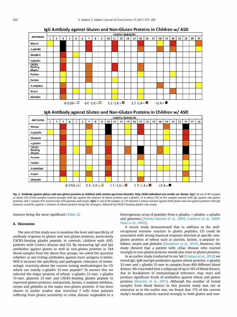
Fig. 2. Antibody against gluten and non-gluten proteins in children with autism spectrum disorder. Only 24/48 individual test results are shown. (IgG) 16 out of 48 samplesor about 33% of the samples reacted strongly with IgG against the mixture of wheat proteins and a-gliadin. 12 or about 25% of the samples reacted with IgG against non-glutenproteins, and 1 sample (#5) reacted only with glutenin and serpin. (IgA) 11 out of 48 samples or 23% showed a robust reaction against both gluten and non-gluten proteins with IgAimmune reactivity against a mixture of wheat proteins being the strongest, followed by CXCR3-binding gliadin and serpin.
A. Vojdani, E. Vojdani / Journal of Cereal Science 75 (2017) 252e260256
mixture being the most significant (Table 2).
4. Discussion
The aim of this study was to examine the level and specificity ofantibody response to gluten and non-gluten proteins, particularlyCXCR3-binding gliadin peptide, in controls, children with ASD,patients with Crohn’s disease and CD. By measuring IgG and IgAantibodies against gluten as well as non-gluten proteins in 144blood samples from the above four groups, we asked the questionwhether or not testing antibodies against more antigens is better.Will it increase the specificity and pathogenic relevance of immu-nologic reactivity above the current testing methodologies for CDwhich use mainly a-gliadin 33-mer peptide? To answer this weselected the major proteins of wheat: a-gliadin 33-mer, g-gliadin15-mer, glutenin 21-mer and CXCR3-binding gliadin peptide torepresent gluten proteins; and purinin, farinin, a-amylase inhibitor,serpin and globulin as the major non-gluten proteins. It has beenshown in earlier studies that intestinal T cells from patientssuffering from gluten sensitivity or celiac disease responded to a
heterogenous array of peptides from a-gliadin, g-gliadin, u-gliadinand glutenins (Arentz-Hansen et al., 2002; Camarca et al., 2009;Shan et al., 2002).
A recent study demonstrated that in addition to the well-recognized immune reaction to gluten peptides, CD could beassociated with strong humoral response directed at specific non-gluten proteins of wheat such as purinin, farinin, a-amylase in-hibitor, serpin and globulin (Huebener et al., 2015). However, thestudy showed that a patient with celiac disease who reactedstrongly to non-gluten proteins would also react to gluten proteins.
In an earlier study conducted in our lab (Vojdani et al., 2014) wetested IgG, IgM and IgA antibodies against wheat proteins, a-gliadin33-mer and g-gliadin 15-mer in samples from 400 different blooddonors.We concluded that a subgroup of up to 16% of blood donors,due to breakdown of immunological tolerance, may react andproduce significant levels of antibodies against wheat and glutenproteins (Hosomi et al., 2011). Although the number of testedsamples from blood donors in this present study was not asextensive as in the earlier one, we found that 17% of the currentstudy’s healthy controls reacted strongly to both gluten and non-

Fig. 3. Antibody against gluten and non-gluten proteins in patients with Crohn’s disease. (IgG) 11 out of 24 (46%) of the specimens reacted with wheat, and 9 specimens out of24 (38%) reacted very strongly with both gluten and non-gluten proteins. (IgA) 6 out of 24 specimens (25%) reacted strongly with both gluten and non-gluten proteins, with thegluten protein CXCR3-binding gliadin peptide being the strongest. 8 (33%) of the samples did not exhibit any IgA antibody against the wheat antigens, gluten and non-glutenpeptides.
A. Vojdani, E. Vojdani / Journal of Cereal Science 75 (2017) 252e260 257
gluten proteins IgG, while 10% reacted strongly for IgA (Fig. 1).Testing for non-gluten proteins did not add any information to theantigenic specificity and the immunologic reactivity in the controls.We then analyzed the data from the sera of children with ASD, andfound that up to 33% of the blood samples reacted strongly againstgluten and non-gluten proteins (Fig. 2). Again we observed thatstrong immune reaction against non-gluten proteins correlatedwith reaction against gluten proteins, except for Sample #13, whichwas the only one that in fact produced IgA antibody against glutenproteins, but not against non-gluten proteins (Fig. 2-IgA). We notedwith interest that among the supposedly healthy control samples,Patient 9 showed very strong reactivity with both IgG and IgA an-tibodies against the wheat protein mixture, CXCR3-binding gliadinpeptide, a-gliadin 33-mer, and, to a lesser extent, serpin andglobulin (Fig. 1). We reiterate that this could indicate that Patient 9may actually have what is variously known as silent, hidden, or theearly stages of CD and NCGS.
Due to some symptomatology overlap between Crohn’s diseaseand CD, we applied IgG and IgA measurements against variouswheat antigens and associated peptides to the sera of patients with
Crohn’s disease to examine the possibility of immune reaction tonon-gluten proteins but not to gluten proteins. In comparison withhealthy controls, IgG antibody in the sera of patients with Crohn’sdisease was found to be highly elevated against antigens from bothgluten and non-gluten proteins in 38% of tested specimens. Simi-larly, IgA antibody reactivity was strong against both gluten andnon-gluten proteins (Fig. 3). Finally, Fig. 4 presents data from CDpatients with antibodies against gluten proteins, which have beendemonstrated to be closely associated with CD and are widelyutilized as serologic markers of this condition (Schedel et al., 2005);in this group, IgG and, especially, IgA antibodies were detectedstrongly against both gluten and non-gluten proteins. The robustIgA response was greatest against CXCR3-binding gliadin peptide,followed by the a-gliadin 33-mer peptide, and then to a lesserdegree against serpin and other gluten and non-gluten proteins(Fig. 4).
Huebener et al. showed in their study (Huebener et al., 2015)that compared with healthy controls, patients with CD exhibitedhigher levels of antibody reactivity to non-gluten proteins. Our owndata from CD patients is in full agreement with their findings that

Fig. 4. Antibody against gluten and non-gluten proteins in patients with celiac disease. (IgG) At ELISA OD of 0.5 or 3SD above the mean, the value of IgG antibody was mostreactive against CXCR3-binding gliadin peptides, followed by the mixture of wheat proteins and serpin. (IgA) All 24 specimens showed reactivity to more than one antigen orpeptide. Overall, the majority of samples reacted strongly against both gluten or non-gluten proteins.
A. Vojdani, E. Vojdani / Journal of Cereal Science 75 (2017) 252e260258
non-gluten proteins of wheat are indeed additional target antigensin celiac disease; however, based on our data from controls, chil-dren with ASD, and patients with CD and Crohn’s disease, we didnot observe that non-gluten proteins are novel target antigens(Barta et al., 2003). This is because in all 144 tested blood speci-mens, as previously stated, we found that if a sample reactedagainst gluten proteins it would also react strongly against non-gluten proteins. This similarity in IgG and IgA response againstgluten and non-gluten proteins may be related to the possiblecross-reactivity and epitope similarities between the two groups ofproteins (Huebener et al., 2015).
For example, a homology analysis indicates that g-gliadins areclose in sequence to purinin proteins. Short sequences of a-amylase/protease inhibitors and the farinin group of proteins havebeen found to be similar to those in certain g-gliadin and lowmolecular weight glutenin proteins. In addition, the reactive cen-ters of some of the identified serpin antigens share homology withglutamine-rich repeats in gluten proteins such as CXCR3-bindinggliadin peptide (Lammers et al., 2008; Ostergaard et al., 2000).
Indeed, we observed a robust IgA immune response againstCXCR3-binding gliadin peptide, a-gliadin 33-mer, then the mixtureof wheat proteins, followed by the other gluten and non-gluten
proteins. This robust immune response against CXCR3-bindinggliadin peptide merits great interest, since it has been shown thatdue to its resistance to digestion and its binding to CXCR3, it acti-vates the inflammatory cascade that induces an increase in bothintestinal permeability and zonulin release, which is the hallmarkof CD (Lammers et al., 2008).
Therefore, additional research should be conducted on a muchlarger number of specimens from various GI disorders in order toexamine the possibility of adding the anti-CXCR3-binding gliadinpeptide to the current repertoire of gluten antibody testing, notonly in patients with CD, but with ASD and Crohn’s disease andpossibly other disorders.
5. Conclusions
The results of this study demonstrate that although humoralimmune response to wheat proteins in children with ASD and pa-tients with Crohn’s disease and CD is not limited only to glutenproteins, measuring IgG and IgA antibodies against non-glutenproteins does not add anything to the pathologic relevance ofthese antibodies. In fact, measuring IgG and IgA antibodies againstgluten proteins, in particular, CXCR3-binding gliadin peptide is

Table 1The statistical differences between the levels of IgG antibodies against a mixture ofwheat proteins, gluten and non-gluten proteins comparing controls versus ASD,Crohn’s and CD groups are shown as means and p values. The patterns of reactivityto the gluten and non-gluten proteins differ from one disorder to another. In the CDand ASD groups the strongest reactions were with CXCR3-binding gliadin, followedby the wheat protein mixture. In the Crohn’s group the strongest reactionwas to thewheat mixture.
Control ASD Crohn’s Celiac
WheatMean 0.48 0.71 0.76 1.20p values 0.1404 0.0010 0.0001a-GliadinMean 0.42 0.43 0.65 0.84p values 0.4554 0.0010 0.0001GluteninMean 0.25 0.32 0.45 0.77p values 0.1837 0.0500 0.0001g-GliadinMean 0.22 0.26 0.46 0.94p values 0.2424 0.0200 0.0001CXCR3 b gliMean 0.29 0.53 0.43 1.23p values 0.0813 0.0500 0.0001PurininMean 0.27 0.35 0.73 0.71p values 0.1441 0.0010 0.0001FarininMean 0.26 0.31 0.61 0.86p values 0.2594 0.0010 0.0001a-Amylase inhibitorMean 0.27 0.35 0.65 0.91p values 0.1832 0.0010 0.0001SerpinMean 0.36 0.43 0.75 1.10p values 0.3009 0.0010 0.0001GlobulinMean 0.26 0.29 0.59 0.82p values 0.3253 0.0010 0.0001
Table 2The statistical differences between the levels of IgA antibodies against a mixture ofwheat proteins, gluten and non-gluten proteins comparing controls versus ASD,Crohn’s and celiac disease groups are shown as means and p values. All three diseasegroups reacted strongest against CXCR3-binding gliadin.
Control ASD Crohn’s Celiac
WheatMean 0.38 0.74 0.48 1.27p values 0.0557 0.0010 0.0001a-GliadinMean 0.31 0.63 0.46 1.42p values 0.0344 0.0500 0.0001GluteninMean 0.25 0.45 0.41 1.25p values 0.0678 0.0500 0.0001g-GliadinMean 0.18 0.42 0.50 1.10p values 0.0132 0.0010 0.0001CXCR3 b gliMean 0.33 0.77 0.79 1.43p values 0.0200 0.0001 0.0001PurininMean 0.22 0.37 0.44 1.31p values 0.0569 0.0500 0.0001FarininMean 0.17 0.34 0.48 1.15p values 0.0377 0.0500 0.0001a-Amylase inhibitorMean 0.24 0.40 0.57 1.12p values 0.0359 0.0010 0.0001SerpinMean 0.28 0.48 0.59 1.26p values 0.0872 0.0010 0.0001GlobulinMean 0.23 0.34 0.39 1.10p values 0.1142 0.0600 0.0001
A. Vojdani, E. Vojdani / Journal of Cereal Science 75 (2017) 252e260 259
sufficient to detect celiac disease with high specificity and sensi-tivity. Therefore, measuring more isn’t always better.
Conflicts of interest
This research did not receive any specific grant from fundingagencies in the public, commercial, or not-for-profit sectors.
Acknowledgments
Acknowledgment is given to Joel Bautista for the preparation ofthis manuscript for publication as well as for the creation of thetables and figures.
References
Arentz-Hansen, H., McAdam, S.N., Molberg, Ø., Fleckenstein, B., Lundin, K.E.,Jørgensen, T.J., Jung, G., Roepstorff, P., Sollid, L.M., 2002. Celiac lesion T cellsrecognize epitopes that cluster in regions of gliadin rich proline residues.Gastroenterology 3, 803e809.
Balakireva, A.V., Zamyatnin Jr., A.A., 2016. Properties of gluten intolerance: glutenstructure, evolution. Nutrients 8, 644.
Barta, Z., Csipo, I., Szabo, G.G., Szegedi, G., 2003. Sensitivity against Saccharomycescerevisiae in patient with Crohn’s disease and celiac disease. World J. Gastro-enterol. 9, 2308e2312.
Bondar, C., Araya, R.E., Guzman, L., Rua, E.C., Chopita, N., Chirdo, F.G., 2014. Role ofCXCR3/CXCL10 axis in immune cell recruitment into the small intestine in celiacdisease. Public Libr. Sci. One 9, e89068. http://dx.doi.org/10.1371/journal.pone.0089068.
Camarca, A., Anderson, R.P., Mamone, G., Fierro, O., Facchiano, A., Costantini, S.,Zanzi, D., Sidney, J., Auricchio, S., Sette, A., Troncone, R., Gianfrani, C., 2009.Intestinal T cell response to gluten peptides are largely heterogeneous: impli-cations for a peptide-based therapy in celiac disease. J. Immunol. 182,4158e4166.
Chattopadhyay, T., Kumar, S., 2016. Diversity analysis of wheat cultivars on the basisof solubility-fractioned seed storage protein polymorphism. Indian J. Bio-technol. 15, 190e194.
Cimaglia, F., Potente, G., Chiesa, M., Mita, G., Bleve, G., 2014. Study of a new gliadincapture agent and development of a protein microarray as a new approach forgliadin detection. Proteom. Bioinforma. 7, 9.
Elder, J.H., Shankar, M., Shuster, J., Theriaque, D., Burns, S., Sherrill, L., 2006. Thegluten-free, casein-free diet in autism: results of a preliminary double blindclinical trial. J. Autism Dev. Disord. 36, 413e421.
Giaffer, M.H., Clark, A., Holdsworth, C.D., 1992. Antibodies to Saccharomyces cer-evisiae in patients with Crohn’s disease and their possible pathogenic impor-tance. Gut 33, 1071e1075.
Groom Jr., Luster, A.D., 2011. CXCR3 in T cell function. Exp. Cell Res. 317, 620e631.Hosomi, S., Oshitani, N., Kamata, N., Sogawa, M., Okazaki, H., Tanigawa, T.,
Yamagami, H., Watanabe, K., Tominaga, K., Watanabe, T., Fujiwara, Y., Maeda, K.,Hirakawa, K., Arakawa, T., 2011. Increased numbers of immature plasma cells inperipheral blood specifically overexpress chemokine receptor CXCR3 andCXCR4 in patients with ulcerative colitis. Clin. Exp. Immunol. 163, 215e224.
Huebener, S., Tanaka, C.K., Uhde, M., Zone, J.J., Vensel, W.H., Kasarda, D.D., Beams, L.,Briani, C., Green, P.H., Altenbach, S.B., Alaedini, A., 2015. Specific non-glutenproteins of wheat are novel target antigens in celiac disease humoralresponse. J. Proteome Res. 14, 503e511.
Kieffer, M., Fraizer, P., Daniels, N.W., Coombs, R.R., 1982. Wheat gliadin fractions andother cereal antigens reactive with antibodies in the sera of coeliac patient. Clin.Exp. Immunol. 50, 651e660.
Kasarda, D.D., Adalsteins, E., Lew, E.J., Lazo, G.R., Altenbach, S.B., 2013. Farinin:characterization of a novel wheat endosperm protein belonging to the prolaminsuper family. J. Agric. Food Chem. 61, 2407e2417.
Kenrick, K.G., Walker Smith, J.A., 1970. Immunoglobulins and dietary protein anti-bodies in childhood coeliac disease. Gut 11, 635e640.
Knoflach, P., Park, B.H., Cunningham, R., 1987. Serum antibodies to cows’ milkproteins in ulcerative colitis and Crohn’s disease. Gastroenterology 92,479e485.
Lammers, K.M., Lu, R., Browley, J., Lu, B., Gerard, C., Thomas, K., Rallabhandi, P., Shea-Donohue, T., Tamiz, A., Alkan, S., Netzel-Arnett, S., Antalis, T., Vogel, S.N.,Fasano, A., 2008. Gliadin induces an increase in intestinal permeability andzonulin release by binding to the chemokine receptor CXCR3. Gastroenterology135, 194e204.
Laragione, T., Brenner, M., Sherry, B., Gulko, P.S., 2011. CXCL10 and its receptorCXCR3 regulate synovial fibroblast invasion in rheumatoid arthritis. Arthritis &Rheumatol. 63, 3274e3283.
Lee, E.Y., Lee, Z.H., Song, Y.W., 2009. CXCL10 and autoimmune diseases. Autoimmun.Rev. 8, 379e383.
Main, J., McKenzie, H., Yeaman, G.R., Kerr, M.A., Robson, D., Pennington, C.R.,Parratt, D., 1988. Antibody to Saccharomyces cerevisiae (bakers’ yeast) in Crohn’sdisease. Br. Med. J. 297, 1105e1106.

A. Vojdani, E. Vojdani / Journal of Cereal Science 75 (2017) 252e260260
Moneret-Vautrin, D.A., Rogniaux, H., Denery-Papini, S., 2011. Assessment of aller-genicity of diploid and hexaploid wheat genotypes: identification of all aller-gens in the albumin/globulin fraction. J. Proteom. 74 (8), 1279e1289.
Ostergaard, H., Rasmussen, S.K., Roberts, T.H., Hejgaard, J., 2000. Inhibitory serpinsfrom wheat grain with reactive centers resembling glutamine-rich repeats ofprolamin storage proteins. Cloning and characterization of five major molecularforms. J. Biol. Chem. 275, 33272e33279.
Quinton, J.F., Sendid, B., Reumaux, D., Duthilleul, P., Cortot, A., Grandbastien, B.,Charrier, G., Targan, S., Colombel, J., Poulain, D., 1988. Anti-Saccharomyces cer-evisiae manna antibodies combined with antineutrophil cytoplasmic autoanti-bodies in inflammatory bowel disease: prevalence and diagnostic role. Gut 42,788e791.
Schedel, J., Rockmann, F., Bongartz, T., Woenkhaus, M., Sch€olmerich, J., Kullmann, F.,2005. Association of Crohn’s disease and latent celiac disease: a case report andreview of the literature. Int. J. Colorectal Dis. 20, 376e380.
Shan, L., Molberg, Ø., Parrot, I., Felix, H., Filiz, F., Gray, G.M., Sollid, L.M., Khosla, C.,2002. Structural basis for gluten intolerance in celiac sprue. Science 297,2275e2279.
Sotkovsky, P., Hubalek, M., Hernychova, L., Novak, P., Havranova, M., Setinova, I.,
Kitanovocova, A., Fuchs, M., Stulik, J., Tuckova, L., 2008. Proteomic analysis ofwheat proteins recognized by IgE antibodies of allergic patients. Proteomics 8,1677e1691.
Stern, M., Fischer, K., Gruttner, R., 1979. Immunoflourescent serum gliadin, anti-bodies in children with coeliac disease and various malabsorptive disorders. II.Specificity of Gliadin antibodies: immunoglobulin classes, immunogenicproperties of wheat protein fractions, and pathogenic significance of food an-tibodies in coeliac disease. Eur. J. Pediatr. 130, 165e172.
Vojdani, A., Kharrazian, D., Partha Sarathi, Mukherjee, 2014. The prevalence ofantibodies against wheat and milk protein in blood donors and their contri-bution to neuroimmune reactivities. Nutrients 6, 15e36.
Vojdani, A., Pangborn, J.B., Vojdani, E., Cooper, E.L., 2003. Infections, toxic chemicalsand dietary peptides binding to lymphocyte receptors and tissue enzymes aremajor instigators of autoimmunity in autism. Int. J. Immunopathol. Pharmacol.16, 189e199.
Yang, A., Chen, Y., Scherl, E., Neugut, A.I., Bhagat, G., Green, P.H.R., 2005. Inflam-matory bowel disease in patients with celiac disease. Inflamm. Bowel Dis. 11,523e528.