it’s not just ct
TRANSCRIPT

It’s not just CT.It’s spectral-detector CT.
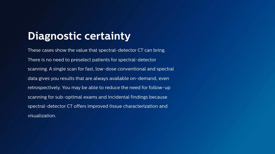
Diagnostic certaintyThese cases show the value that spectral-detector CT can bring.
There is no need to preselect patients for spectral-detector
scanning. A single scan for fast, low-dose conventional and spectral
data gives you results that are always available on-demand, even
retrospectively. You may be able to reduce the need for follow-up
scanning for sub-optimal exams and incidental findings because
spectral-detector CT offers improved tissue characterization and
visualization.

Mystery diagnosis cases
Case Case Case Case Case
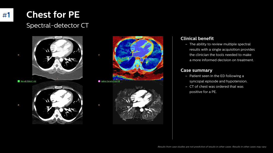
Clinical benefit – The ability to review multiple spectral
results with a single acquisition provides
the clinician the tools needed to make
a more informed decision on treatment.
Case summary– Patient seen in the ED following a
syncopal episode and hypotension.
– CT of chest was ordered that was
positive for a PE.
Results from case studies are not predictive of results in other cases. Results in other cases may vary.
Chest for PESpectral-detector CT

Abdomen pelvisSpectral-detector CT
Clinical benefit – Having multiple results allows the
radiologist to improve their diagnostic
confidence by quantifying the area of
anatomy.
Case summary– On conventional CT, an enlarged
inhomogeneous area was seen in the pelvis.
– Question if it was endometriosis or a lesion.
– Spectral results showed enhancement of
area, and a biopsy confirrmed that the
patient had endometrial cancer.
Results from case studies are not predictive of results in other cases. Results in other cases may vary.

Clinical benefit – Spectral-detector CT and Magic Glass provide
the ability to view multiple spectral results
and compare multiple spectral results.
Case summary– Enhancing lesion at the base of tongue
difficult to detect with conventional CT images
• CTDIvol: 11.3 mGy
• DLP: 334.9 mGy*cm
Results from case studies are not predictive of results in other cases. Results in other cases may vary.
Neck massSpectral-detector CT

Clinical benefit– Spectral results can be beneficial in the
planning and patient selection for
interventional and surgical procedures.
Case summary– Conventional, arterial images demonstrate
a filling defect in the anterior aspect of the
left atrial appendage (upper image, blue
arrow); the defect may be related to
thrombus or circulatory stasis.
– The delayed scan (as per protocol) has a
very washed-out appearance which makes
interpretation for thrombus difficult (lower
left image).
– The corresponding MonoE and Z effective
images from the delayed phase confirm
presence of a thrombus in the left atrial
appendage (yellow and red arrows), which
is a contraindication to the procedure.
Results from case studies are not predictive of results in other cases. Results in other cases may vary.
Left atrial appendageSpectral-detector CT

Clinical benefit – Low MonoE images improved the
visualization of the vascular structures
and the necrotic area of the pelvis.
– This an example of experiencing improved
diagnostic capabilities even in a patient
who would not have been preselected for
dual-energy techniques.
Case summary– Pediatric patient with worsening abdominal
pain received an abdomen pelvis scan with
IV contrast.
– Low MonoE images identify a right ovarian
torsion, which was confirmed with ultrasound.
Results from case studies are not predictive of results in other cases. Results in other cases may vary.
Pediatric abdomen Spectral-detector CT

Other cases

Conventional Conventional
Iodine density overlay Iodine density overlay
Abdomen and pelvisSpectral-detector CT
Clinical benefit− Shortest time to diagnosis.
Case summary− Female patient presented complaining of
abdominal pain.
− With conventional results, a pancreatic duct
appears to be dilated.
− With Iodine density overlay, a tumor is visible
in the head of pancreas and patient was
referred for an endoscopic ultrasound biopsy,
which showed a tubular adenoma of the
Ampulla of Vater.
Results from case studies are not predictive of results in other cases. Results in other cases may vary.
Conventional CT MonoE 40 keV
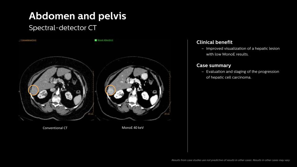
Clinical benefit– Improved visualization of a hepatic lesion
with low MonoE results.
Case summary– Evaluation and staging of the progression
of hepatic cell carcinoma.
Results from case studies are not predictive of results in other cases. Results in other cases may vary.
Abdomen and pelvis Spectral-detector CT

3
Clinical benefit – Multiple spectral results with every scan.
Case summary– History of renal cell carcinoma and a
sustained response to pazopanib therapy.
– Iodine no Water spectral result
demonstrated continued lack of iodine in
the majority of the lesion, confirming
response to therapy.
Conventional CT Iodine no WaterResults from case studies are not predictive of results in other cases. Results in other cases may vary.
Abdomen follow-upSpectral-detector CT

Neuro

3
Spectral-detector CTBrain
Clinical benefit – Spectral Magic Glass allows for the
simultaneous comparison of multiple
spectral results for a specific region of
interest.
Case summary– Patient presented to ED with symptoms of
an acute stroke, and was referred to CT.
– Low MonoE improved the visualization of
the perfusion defect in the pons.
Results from case studies are not predictive of results in other cases. Results in other cases may vary.

Spectral-detector CT
Indeterminate tracheal lesion
Clinical benefit– Spectral-detector CT can serve as a
problem-solving tool in onco-imaging and
help in reaching the diagnosis.
Case summary– Patient status post right-pneumonectomy
for small cell lung cancer.
– Two-year follow-up conventional CT images
(top left) shows a new hyper-attenuating
focus along the right anterolateral aspect of
the upper trachea, which was initially
thought to be mucous material.
– Combination of 40 keV images and
additional spectral reconstructions
demonstrate contrast enhancement within
the tracheal lesion consistent with tumor
recurrence.
– Confirmed by subsequent biopsy.
MonoE 40 keV
Results from case studies are not predictive of results in other cases. Results in other cases may vary.

MSK

MSKSpectral-detector CT
Virtual non-contrastConventional
Results from case studies are not predictive of results in other cases. Results in other cases may vary.
Clinical benefit– Improved visualization with Virtual non-
contrast spectral result.
Case summary– Conventional CT reconstruction demonstrates
comminuted left iliac fractures, and a 5 mm
hyperdense focus adjacent to the dominant
bone fragments (arrow).
– Unclear if this represented a shard of bone
versus active extravasation of contrast.
– Virtual non-contrast reconstruction
demonstrates suppression of this dense
focus, confirming the presence of iodine and
active hemorrhage.
– Acute fracture with active hemorrhage.
– Patient was brought to IR suite and this
finding was confirmed.

Cardiac/CTA
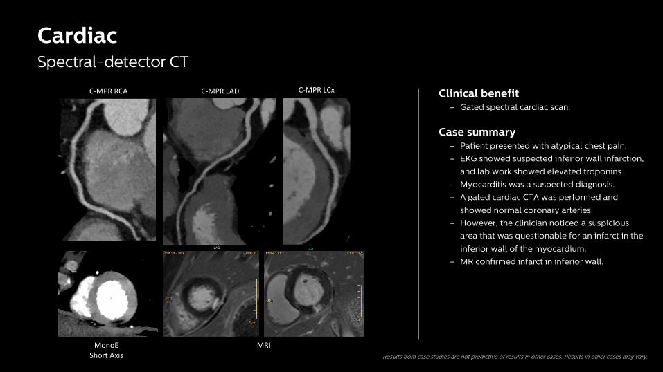
CardiacSpectral-detector CT
C-MPR LCxC-MPR LADC-MPR RCA
MonoEShort Axis
MRI
Clinical benefit– Gated spectral cardiac scan.
Case summary– Patient presented with atypical chest pain.
– EKG showed suspected inferior wall infarction,
and lab work showed elevated troponins.
– Myocarditis was a suspected diagnosis.
– A gated cardiac CTA was performed and
showed normal coronary arteries.
– However, the clinician noticed a suspicious
area that was questionable for an infarct in the
inferior wall of the myocardium.
– MR confirmed infarct in inferior wall.
Results from case studies are not predictive of results in other cases. Results in other cases may vary.

Spectral-detector CT
Chest for PE
Clinical benefit – Spectral-detector results aid when evaluating
a chest for a suspected PE and can provide
additional clinical benefits to make the right
diagnosis with the first scan.
Case summary– Patient admitted through the ED with
shortness of breath and chest discomfort.
– The injection timing was not optimal for a
PE study so fused spectral results were
reviewed.
– Radiologist identified the perfusion defect
in the left lower lung and a small PE using
Z effective fused.
Results from case studies are not predictive of results in other cases. Results in other cases may vary.