isolation, culture and identification of microcystis in
TRANSCRIPT
Yang, et al, Isolation, culture and identification of microcystis in source water
Isolation, culture and identification of microcystis in source water
Songqin Yang, Ling Dong, Chunxia Jiang, Xuemin Cheng, Huizhen Zhang, Liuxin Cui*
Department of Environmental Health, College of Public Health, Zhengzhou University, Zhengzhou, Henan 450052, China
Received February 12, 2007
AbstractObjective. To isolate microcystis in the main source water of Zhengzhou city and identify the toxicity. Methods. The
algae cell was isolated by 96-microbiological assay. The phycocyanin intergenic spacer region (PC-IGS) and the micro-cystin synthetase gene B (mcyB) were detected by whole-cell PCR. Toxicity of microcystin was identified by ELISA. Re-sults. Single cell clones were acquired successfully, and the whole-cell PCR results were positive of PC-IGS and mcyB. Compared to the reported mcyB of Microcystis aeruginosa in GenBank, the homology of gene sequence was 99%, which suggested that the three microcystis from the source water were cyanobacteria and toxigenic. The contents of the three microcystis powder were 1.07 µg/mg, 4.70 µg/mg, 0.47 µg/mg, respectively. Conclusion. 96-microbiological assay is a simple, fast and accurate method which can isolate algae cells from bloom water successfully. The microcystis isolated from the main source water in Zhengzhou city were blue-green algae which could produce microcystin. [Life Science Journal. 2007; 4(2): 28 – 32] (ISSN: 1097 – 8135).
Keywords: microcystis; isolation; culture; 96- microbiological assay; whole-cell PCR; ELISA
1 Introduction
With the aggravation of the source water pollution, a great quantity of industrial sewage and sanitary sewage has been discharged into the water, which has increased the content of the nitrogen and phosphorus in the water rapidly, and bloom is exploding frequently all over the world threatening human health[1,2]. The main pollutant of bloom is all kinds of toxin of algae, and microcystin which is released by Microcystin aeruginosa, Anabaena flosaquae, Oscillation aeruginosa, nostoc and so on is the most threatening. Microcystin is a kind of intracellu-lar toxin[3,4], which is investigated widely. In order to get the information of pollution state of the source water in Zhengzhou city, the author selected the Xiliu lake and the Huayuankou reservior which were the main source water of local area for sampling.
2 Materials and Methods
2.1 Collection of algae sample and cultureThe sampling sites were the Xiliu lake and the Hua-
yuankou reservior. The algae was collected by using the No.25 plankton net. The culture medium was BG-11, and illumination was 2500 LUX, the ratio of light and hazy was 12 hours vs. 12 hours, and the temperature was (25 ± 1)ºC.
2.2 Isolation and culture of the microcystisThe preincubated solution was diluted to 3 cells/ml,
and then 300 μl solution was delivered to 96-microbio-logical assay. The 96-microbiological assay was put into the illumination incubator until obvious color was seen. Then the plate was observed under inverted microscope and the monoplast solution was delivered to a test tube for growth. The passage should be made every 7 days.
2.3 Bionomics of the microcystis The morphology of microcystis was observed under
light microscope. 5 ml solution was verted to a cuvette and the optical density was determined with 680 nm
Supported by Medicine Innovation Talent Projecting Stake Item in Henan province (200311205); Department of Education Tackle Key Problems in Science and Technology (200633000).*Corresponding author. Email: [email protected]
∙ 28 ∙

Yang, et al, Isolation, culture and identification of microcystis in source water
∙ 29 ∙
wavelength. The optical density was determined at the same time of each day. The calculation formula of cell di-vision velocisity was k = 3.322/(n – 1)log(ODn/OD1), and n was the day number. The formula of the generation time of microsystis was Td = 24/k, and the unit was hour.
2.4 Identification of microcystis toxicity
2.4.1 Genes of PC-IGS and mcyB were detected by whole-cell PCR. The algae solution which was in expo-nential growth phase was 1×10⁶ cells/ml. And 1.5 ml of the solution was centrifuged for 5 minutes at 13,000 rpm. It was washed by sterile distilled water for 3 times and was diluted by 1.5 ml sterile water. The steps should be repeated for several times. The last supernatant was used as the template of whole cell PCR. The positive control was the No. 940 microcystis which was bought from the Chinese Academy of Science, Hydrobiont Research Cen-ter (Wuhan, China) and the negative control was the ster-ile double distilled water.
The primers of mcyB were 5'-AGGAACAAGTTG-CAC AGAATCCGCA-3' (p1), 5'-ACTAATCCCTATC-TAAAC ACAGTAACTCA-3' (p2 ); and primers of PC were 5'-GGC TGCTTGTTTACGCGACA-3' (p1), 5'-CCA GTA CCA CCA GCA ACT AA-3' (p2). Both of the primers were synthesized by Augct Biotechnology Co, Ltd (Beijing, China).
All PCRs were performed in a 30 μl reaction solution containing 10× buffer, 25 mmol/L MgCl2, 2.5 mmol/L dNTP, forward and reverse primers 7.5 pmol respective-ly, 3U Taq DNA polymerase, 10 μl template. The reac-tion procedure contained predenaturation for 6 minutes at 94ºC, followed by 35 cycles at 94ºC for 30 seconds, 55ºC for 1 minute, 72ºC for 1 minute and 72ºC for 8 min-utes. The equipment of PCR amplification was Biometra T-Gradient. All PCRs’ products were analyzed by elec-trophoresis in 1.2% agarose using TAE buffer.
2.4.2 The mcyB sequence of PCR amplifying production. The cloning and sequencing was finished by TaKaRa Bio-technology (Dalian, China) Co, Ltd after the PCR ampli-fication of mcyB was purified. The sequencing primer of mcyB were 5'-AGG AAC AAG TTG CAC AGA ATC CGC A-3' (p1), 5'-ACT AAT CCC TAT CTA AAC ACA GTA ACT CA-3' (p2 ); and the para-primers of PC were 5'-GGCTGCTTGTTTACGCGACA-3' (p1), 5'-CCAGTA CCACCAGCAACTAA-3' (p2). The sequence was made in the positive direction .
2.4.3 Extraction and determination by ELISA. 80 mg al-gae powder was disolved by 20 ml 5% acetic acid, and
was disintegrated for 30 minutes by ultrasonic, mixed for 30 minutes, centrifuged at 4ºC under 10,000 rpm for 10 minutes, and collected the supernatant named as No.1. The rest was mixed for 30 minutes after added into 20 ml 80% methanol, centrifuged for 10 minutes under the similarity condition, and collected the supernatant named as No.2. The No.3 supernatant was got by the same proce-dures. All of the supernatant was mixed together and rota-ry evaporation to near-dried at 70ºC under 100 rpm, then the microcystin was dissolved with 1 ml sodium chloride, centrifuged for 10 minutes at 10,000 rpm and filtrated by the 0.45 μm membrane in the end. The ultimate toxin was diluted by 80,000 and 100,000 times and determined by using Microcystin ELISA kit (Chinese Academy of Sci-ence, Wuhan).
3 Results
3.1 Morphology of microcystisThere were 3 types of microcystis that were isolated
successfully, containing 1 type from Xiliu lake, 2 types from the Huayuankou reservior, and were named as XLH, BM1, and BM2 respectively (Figure 1 A, B, C). XLH grew aggregating and the shape of the cells were globular or anomalistic. The volume was big with a gum theca and the distribution of the cytoplasm was uneven. The color of BM1 under the microscope was blue-green, and it had a gum theca. The distribution of the cytoplasm was uneven too, the volume of which was smallest of the three. The color of BM2 under microscope was green and BM2 had gum theca with the uneven distribution. The strains of the microcystis from the Huayuankou reservior were globular and grew monocellular.
3.2 Growth curve of microcystis From Figure 2 we can see that the cycle of the three
kinds of microcystis was about two weeks. The growth velocity of BM2 was higher than the others. Their growth curve was “S” shape and the highest velocity was from the eleventh day to the thirteenth day.
3.3 Cleavage velocity and the generation As we can see from Table 1, the mean cleaving ve-
locities of the three kinds of microcystis were 0.396, 0.199, 0.345 respectively. The geneation time of BM2 was 121 hours which was the longest of the three. The denominatort means the monitoring time and k means the cleaving velocity and Td means the generation time.

Life Science Journal, Vol 4, No 2, 2007 http://lsj.zzu.edu.cn
Figure 2. Growth curve of microcystis.
Table 1. Time of cleavage velocity and generationNumber OD0 ODn tn–1 (d) k Td (h)
BM1 0.182 1.635 12 0.396 61BM2 0.690 3.940 12 0.199 121XLH 0.100 1.388 11 0.345 70
3.4 Results of the amplification of whole cell PCRAs we can see from the Figure 3, the length of the prim-
er amplification products were 350 bp and 700 bp, which were as the same the length as the aim genes’ and we can see that all of the microcystises that researched could re-lease microcystin.
3.5 The mcyB sequence of PCR production 3.5.1 Sequence of XLH1 GGTATTTTTG AGGACAAAAT TAACCTATCA ACAGTTAAAT AACCGGGGCA ATCAGTTAGC61 TCACTGTTTA CGAGATAAGG GTGTAAATCC AGAAAGTTTA GTCGGGATTT TTATGGAGCG121 TTCCCTAGAG ATGGTCATCG GTTTATTAGG GATATTAAAA GCCGGGGGAG CTTATGTACC181 TTTAGATCCG GATTATCCTA CCGAGCGCTT GGGGGATATC CTCTCAGATT CGGGTGTTTC
241 TTTGGTGTTA ACTCAGGAAT CTTTAGGGGA TTTTCTTCCC CAAACTGGGG CTGAGTTACT301 GTGTTTAGAT
Results of sequencing indicated: length of the gene was 310 bp, and there was only one restriction enzymes of EcoRV at the 217th base. The sequence was submitted to GenBank using the BLAST, and the gene number given by GenBank was EF216874, and compared to the report-ed mcyB of Microcystis aeruginosa No.899316, 92109242, 14486356 in GenBank, the homology of gene sequence was 98%.
Figure 3. Spectrum of PCR amplification. M: Marker; Lane 1: negative control-mcyB; Lane 2: BM1-mcyB; Lane 3: BM2-mcyB; Lane 4: XLH-mcyB; Lane 5: positive control-mcyB; Lane 6: BM1-PC-IGS; Lane 7: BM2-PC-IGS; Lane 8: XLH-PC-IGS; Lane 9: positive control-PC-GS.
3.5.2 Sequence of BM11 GGGGAGAATC CCCTAAAGAT TCCTGAGTTA ACACCAAAGA AACACCCGAA TCTGAGAGGA61 TATCCCCCAA GCGCTCGGTA GGATAATCCG GATCTAAAGG TACATAAGCT CCCCCGGCTT121 TTAATATCCC TAATAAACCG ATGACCATCT CTAGGGAACG CTCCATAAAA ATCCCGACCA 181 AACTTTCTGG AACTACACCC TTATCTCGTA AACAGTGAGC TAACTGATTG CCCCGGTTAT 241 TTAACTGTTG ATAGGTTAAT TTTTGTCCTT CAAAAATAAC CGCTATTCCC TGCGGATTCT301 GTGCAACTTG TTCA
∙ 30 ∙
Figure 1. A: the microcystis of XLH; B: the microcystis of BM1; C: the microcystis of BM2.
Yang, et al, Isolation, culture and identification of microcystis in source water
The sequencing results indicated: the length of BM1 was 314 bp and the code-star was the second base. There was only one restriction enzymes of EcoRV in the 61th base. The sequence was submitted to GenBank using the BLAST, and the gene number given by GenBank was EF216872 with the protein number ABP04110. Com-pared to the reported mcyB of Microcystis aeruginosa No.92109242, 28804617, 28804615 in GenBank, the ho-mology of gene sequence was 99%.
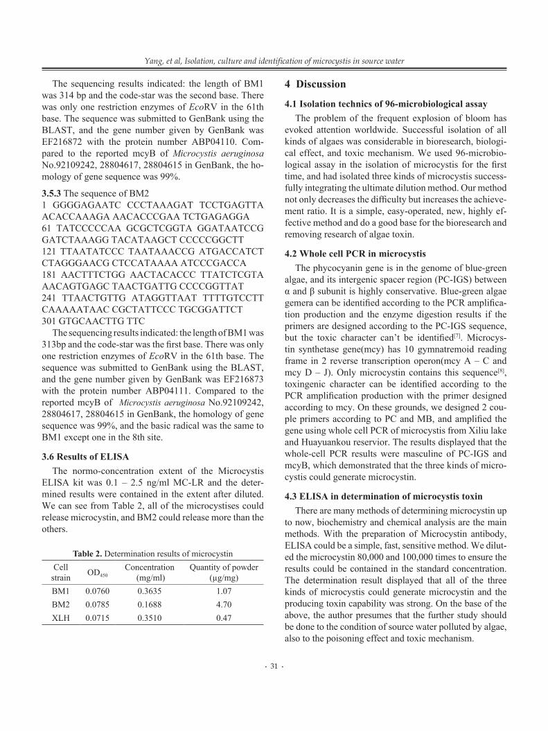
3.5.3 The sequence of BM21 GGGGAGAATC CCCTAAAGAT TCCTGAGTTA ACACCAAAGA AACACCCGAA TCTGAGAGGA61 TATCCCCCAA GCGCTCGGTA GGATAATCCG GATCTAAAGG TACATAAGCT CCCCCGGCTT121 TTAATATCCC TAATAAACCG ATGACCATCT CTAGGGAACG CTCCATAAAA ATCCCGACCA181 AACTTTCTGG AACTACACCC TTATCTCGTA AACAGTGAGC TAACTGATTG CCCCGGTTAT241 TTAACTGTTG ATAGGTTAAT TTTTGTCCTT CAAAAATAAC CGCTATTCCC TGCGGATTCT301 GTGCAACTTG TTC
The sequencing results indicated: the length of BM1 was 313bp and the code-star was the first base. There was only one restriction enzymes of EcoRV in the 61th base. The sequence was submitted to GenBank using the BLAST, and the gene number given by GenBank was EF216873 with the protein number ABP04111. Compared to the reported mcyB of Microcystis aeruginosa No.92109242, 28804617, 28804615 in GenBank, the homology of gene sequence was 99%, and the basic radical was the same to BM1 except one in the 8th site.
3.6 Results of ELISAThe normo-concentration extent of the Microcystis
ELISA kit was 0.1 – 2.5 ng/ml MC-LR and the deter-mined results were contained in the extent after diluted. We can see from Table 2, all of the microcystises could release microcystin, and BM2 could release more than the others.
Table 2. Determination results of microcystinCell
strain OD450Concentration
(mg/ml)Quantity of powder
(µg/mg)BM1 0.0760 0.3635 1.07BM2 0.0785 0.1688 4.70XLH 0.0715 0.3510 0.47
4 Discussion
4.1 Isolation technics of 96-microbiological assay The problem of the frequent explosion of bloom has
evoked attention worldwide. Successful isolation of all kinds of algaes was considerable in bioresearch, biologi-cal effect, and toxic mechanism. We used 96-microbio-logical assay in the isolation of microcystis for the first time, and had isolated three kinds of microcystis success-fully integrating the ultimate dilution method. Our method not only decreases the difficulty but increases the achieve-ment ratio. It is a simple, easy-operated, new, highly ef-fective method and do a good base for the bioresearch and removing research of algae toxin.
4.2 Whole cell PCR in microcystis The phycocyanin gene is in the genome of blue-green
algae, and its intergenic spacer region (PC-IGS) between α and β subunit is highly conservative. Blue-green algae gemera can be identified according to the PCR amplifica-tion production and the enzyme digestion results if the primers are designed according to the PC-IGS sequence, but the toxic character can’t be identified[7]. Microcys-tin synthetase gene(mcy) has 10 gymnatremoid reading frame in 2 reverse transcription operon(mcy A – C and mcy D – J). Only microcystin contains this sequence[8], toxingenic character can be identified according to the PCR amplification production with the primer designed according to mcy. On these grounds, we designed 2 cou-ple primers according to PC and MB, and amplified the gene using whole cell PCR of microcystis from Xiliu lake and Huayuankou reservior. The results displayed that the whole-cell PCR results were masculine of PC-IGS and mcyB, which demonstrated that the three kinds of micro-cystis could generate microcystin.
4.3 ELISA in determination of microcystis toxin There are many methods of determining microcystin up
to now, biochemistry and chemical analysis are the main methods. With the preparation of Microcystin antibody, ELISA could be a simple, fast, sensitive method. We dilut-ed the microcystin 80,000 and 100,000 times to ensure the results could be contained in the standard concentration. The determination result displayed that all of the three kinds of microcystis could generate microcystin and the producing toxin capability was strong. On the base of the above, the author presumes that the further study should be done to the condition of source water polluted by algae, also to the poisoning effect and toxic mechanism.
∙ 31 ∙

Life Science Journal, Vol 4, No 2, 2007 http://lsj.zzu.edu.cn
ReferencesAnna Lankoff, Lukasz Krzowski, Joanna Glab, et al. DNA damage and repair in human peripheral blood lymphocytes following treatment with microcystin-LR. Mutation Research Genetic Toxicology and Environ-mental Mutagenesis 2004; 559(1-2): 131 – 42.Zegura B, Sedmak B, Filipic M. Microcystin-lr induces oxidative DNA damage in human hepatoma cell line HepG2. Toxicon 2003; 41: 41 – 8.Chen Y, Yu SZ, Lin YD, et al. The epidemiologic survey of the relation between the students and the microcystin in Tai lake region. Medicine Transaction of Fudan university 2002; 29(6): 462 – 4.Ding WX, Shen HM, Ong CN. Critical role of reactive oxygen species and mitochondrial permeability transition in microcystin induced rapid apoptosis in rat hepatocytes. Hepatology 2000; 32(3): 547 – 55.
1.
2.
3.
4.
Neilan BA, Jacobs D, Goodman. Genetic diversity and phylogeny of toxic cyanobacteria determined by DNA polymorphisms within the phycocyanin locus. Appl Environ Microbiol 1995; 61(11): 3875 – 83.Kaebernick M, Neilan BA, Borner T, et al. Light and the transcriptional response of the microcystin biosynthesis gene cluster. Appl Environ Microbiol 2000; 66(8): 3387 – 92.Baker JA, Neilan BA, Barrie Entsch, et al. Identification of cyanobacte-ria and their toxigenicity in environmental samples by rapid molecular analysis. Environ Toxicol 2001; 16: 472 – 82.Tillett D, Dittmann E, Erhard M. Structural organization of microcystin biosynthesis in Microcystis aeruginosa PCC7806: an integrated pep-tide-polyketide synthetase system. Chemistry & Biology 2000; 7(10): 753 – 64.
5.
6.
7.
8.
∙ 32 ∙