isolation and identification of causal disease of ... and identification of causal disease of... ·...
TRANSCRIPT

ISOLATION AND IDENTIFICATION OF CAUSAL DISEASE OF EUCALYPTUS PELLITA
Muhammad Safrit Bin Abdul Rahim
SD 397 EI M952
Bachelor of Science with Honours 2011 (Plant Resource Science and Management)
20ll

I s
so 3Q't ~~ fi\ G\ 5":2.J..~ \').
~
P~hidmatMaklumat Akademik RSm MALAYSIA SARAWAK
ISOLATION AND IDENTIFICATION OF CAUSAL DISEASE OF
EUCALYPTUS PELLITA P.KHIDMAT MAKLUMAT AKADIMIK
111111111 r01ir11111i IIII 1000235553
MUHAMMAD SAFRIL BIN ABDUL RAHIM
This project report is submitted in partial fulfilment of the requirements for the Degree of Bachelor of Science with Honours
(Plant Resource Science and Management)
Department of Plant Science and Environmental Ecology
Faculty of Resource Science and Technology
UNIVERSITI MALAYSIA SARA W AK
June 2012

APPROVAL SHEET
Name of candidate: Muhammad Safril bin Abdul Rahim
Matric number: 24224
Title of dissertation: Isolation and Identification of Causal Disease of Eucalyptus pellita
(Prof Dr Sepiah binti Muid)
Supervisor
(Dr Siti Rubiah Zainudin)
Coordinator
Plant Resource Science and Management Programme
Department of Plant Science and Environmental Ecology
Faculty of Resource Science and Technology
Universiti Malaysia Sarawak
•

,.. ,
DECLARA'[ION
I hereby declare that the Final Year Project Report is based on my original work
except for quotations and citations, which have been duly acknowledged. I also
declare that it has not been previously or concurrently submitted for any degree at
UNIMAS or other institutions ofhigher learning.
\ .~ ~)r----__---,
(Muhammad Safril bin Abdul Rahim)
24224
Plant Resource Science and Management Programme
Department of Plant Science and Environmental Ecology
Faculty of Resource Science and Technology
Universiti Malaysia Sarawak
1

ACKNOWLEDGEMENT
In the name of Allah, the Most Beneficent, the Most Merciful.
Alhamdulillah, all praise is to Allah for all blessings and guidance for me.
Peace and blessings upon Prophet Muhammad.
First ofall I would like to express my deepest gratitude and thanks to Prof
Dr Sepiah binti Muid for all the guidance and advice throughout this study.
Thanks to all the lecturers especially Dr. Effendi Wasli for the guidance in the
statistics and also to Madam Jamliah binti Jamel for her guidance in the molecular
genetics techniques.
The most important people in my life, to both of my parents, Abdul Rahim
bin Abdul Rahman and Zainon binti Sidin and my lovely sister for their moral
support, love and financial support during this entire research.
I would also like to thank all of my coursemate, especially Zulaikha binti
Zainal, Mugunthan all Perumal and not to forget the postgraduate student Miss
Aishah and Miss Hafidah for all their help and kindness .
...

.
Pusat Khidmat Maklumat Akademik UNJVERSITI MALAYSIA SARAWAK
Table of content
Title
ACKNOWLEDGEMENT
TABLE OF CONTENT
LIST OF ABBREVIATIONS
LIST OF TABLES
LIST OF FIGURES
ABSTRACT / ABSTRAK
CHAPTER ONE: INTRODUCTION
1.1 Research background
1.2 Problem statement
1.3 Objectives
CHAPTER TWO: LITERATURE REVIEW
2.1 Taxonomy ofEucalyptus spp
2.2 Botanical description
2.3 Origin and distribution ofEucalyptus spp
2.4 Importance ofEucalyptus spp
2.5 Forest Plantation Disease
2.6 Pathogenic fungi
2.7 Forest plantation pathology principles
CHAPTER THREE: MATERIALS AND METHODS
3.1 Research materials
3.2 Disease description
3.3 Pathogens isolation of on PDA
3.4 Identification ofthe fungi by the morphological characteristic
3.5 Identification of fungi using the molecular approach
3.5.1 Spectrophotometer Quantification ofDNA Concentration
3.5.2 Gel electrophoresis
3.5.3 PCR based method
ii
Page No
11
IV
V
VI
2
4
5
6
6
7
7
8
9
10
11
11
11
13
13
14
15
14
I

3.5.4 DNA purification 15
3.5.5 DNA sequencing 16
3.6 Physiological test
3.6.1 Effect ofdifferent temperature on the fungi growth 17
3.6.2 Effect ofdifferent pH level on the fungi growth 17
3.6.3 Effect ofdifferent media on the fungi growth 17
3.7 Pathogenicity test 18
3.8 Data analysis 19
CHAPTER FOUR: RESULTS
4.1 Disease ofEucalyptus pel/ita 20
4.2 Fungi associated with the disease 23
4.3 Morphological description of the isolated fungi 25
4.4 Identification of fungi using molecular approach 32
4.4.1 DNA extraction of selected fungi species 32
4.4.2 DNA quantification using the spectrophotometer at 1..260/),280 34
4.4.3 Polymerase Chain Reaction and the Optimization of the DNA 35
4.5 Effect ofdifferent pH level on the fungi growth 37
4.6 Effect ofdifferent temperature on the fungi growth 38
4.7 Effect ofdifferent media on the fungi growth 39
4.8 Pathogenicity test 42
CHAPTER FIVE: DISCUSSION 45
CHAPTER SIX: CONCLUSION 48
CHAPTERSEVEN: REFERENCES 50
APPENDICES 54
iii
LIST OF ABBREVIATIONS
°C Celsius
% percentage
J.lg microgram
ml millilitre
giL gram per litre
w/v weight per volume
ANOVA Analysis ofV ariance
HCI Hydrochloric acid
KOH Sodium hydroxide
PDA Potato Dextrose Agar
PDB Potato Dextrose Broth
MEA Malt Extract Agar
CMA Corn Meal Agar
RAPD Random Ampiified Polymorphic DNA
iv

LIST OF TABLES
Table 1 Estimated areas of Eucalyptus plantations in the South-East 2
Asian region in 1995 until the year of2008.
Table 2 Percentage occurrence of fungi 24
Table 3 Optical density at A.2601A.280 of the genomic DNA from 6 33
different species of fungi
Table 4 A verage dry weight of mycelia (g) ofdifferent species at 37
different pH levels
Table 5 Average growth ofmycelia (cm) ofdifferent species at 38
different temperatures
Table 6 Average growth of mycelia (cm) of different speCIes at 39
different media
Table 7 Percentage of disease formed during the first week and second 42
week
v
,.'

LIST OF FIGURES
Figure 1 Plantation area of Eucalyptus spp. in Malaysia from the year
194]-2008
3
Figure 2 (a) E. pellita tree with disease infection (b) numerous spots
on leaves
20
Figure 3 The disease symptoms on the leaves of the E. pellita 22
Figure 4 (a) Cylindrocladium sp. colony on MEA (b) Reverse plate (c)
Conidia
25
Figure 5 (a) Pestalotiopsis sp. colony on MEA (b) Reverse plate (c)
Conidia
26
Figure 6 (a) Guignardia sp. colony on MEA (b) Reverse plate (c)
Conidia
27
Figure 7 (a) Aspergillus jlavus colony on MEA (b) Reverse plate (c)
Conidia
28
Figure 8 (a) Trichoderma sp. colony on MEA (b) Reverse plate (c)
Conidia
29
Figure 9 (a) Botryodiplodia theobromae colony on MEA (b) Reverse
plate (c) Conidia
30
Figure 10 (a) Aspergillus niger colony on MEA (b) Reverse plate (c)
Conidia
31
Figure 11 Genomic DNA extraction from six different species of fungi isolated from the leaf of the E. pellita in Sempadi Planted Forest: (Lane 1) Lambda Hind III ladder (Lane 2) Cylindrocladium sp. (Lane 3) Pestalotiopsis sp. (Lane 4) Botryodiplodia theobromae (Lane 5) Aspergillus Jlqvus
32
vi
,.'
•
I

(Lane 6) Aspergillus niger (Lane 7) Trichoderma sp.
Figure 12 Polymerase chain reaction of 6 different types of fungi using the primer of ITS4 and ITS5: (Lane I) 100 bp DNA ladder (Lane 2) Cylindrocladium sp. (Lane 3) Pestalotiopsis sp. (Lane 4) Botryodiplodia theobromae (Lane 5) Aspergillus fZavus (Lane 6) Aspergillus niger (Lane 7) Trichoderma sp.
35
Figure 13 Appearance of seven days old Trichoderma sp. on (a) MEA
(b) CMA (c) PDA
41
Figure 14 Appearance of seven days old Pestalotiopsis sp. on (a) MEA
(b) CMA (c) PDA
41
Figure 15 Appearance of seven days old Botryodiplodia theobromae on
(a) MEA (b) CMA (c) PDA
41
Figure 16 E. pellita leaf infected with the Cylindrocladium sp. on the
fIrst week of incubation. The black circle show the site
43
infected with distilled water. (a) Replicate 1 (b) Replicate 2
(c) Replicate 3
vii
.."

I Isolation of Fungi and Identification of Causal Disease of Eucalyptus pellita
Muhammad Safril bin Abdul Rahim
Plant Resource Science and Management Program Department of Plant Science and Environmental Ecology
Faculty of Resource Science and Technology Universiti Malaysia Sarawak
ABSTRACT
Eucalyptus plantation in Malaysia is still new and need a lot of attention and care especially on the prevention against the disease infection. Disease infection in the timber tree species of the Eucalyptus pellita negatively impacts the timber yield throughout the world and affecting the production. A study was conducted in Sempadi Forest Plantation in Bau area, the tissues from the Eucalyptus pel/ita were collected, the disease de cription were made based on the symptoms, and the pathogen were isolated from the leaves and identified for their species based on the morphology and the molecular approach. The common disease symptoms were brown leaf spots, yellow leaf spots, reddish brown leaf spots, yellowish spots, whitish grey spots and also some discolouration of the leaf tips. The fungi isolated were Cladosporium sp., Pestalotiopsis sp., Aspergillus niger, Aspergillus jlavus, Trichoderma sp., Botryodiplodia theobromae and Guignardia sp. Three physiological tests were conducted on the fungi isolated which were on different artificial media, temperature and pH. Three different media were used in the tests which were the MEA, CMA, and PDA. There were significant differences on the average growth of the fungi and the MEA was the best media for BOlryodiplodia theobromae and Trichoderma sp. while PDA was the best for Pestalotiopsis sp. The tested temperature was at 15, 20, 25, 30, 35, 40"C. The fungi isolated grew the best at the moderate temperature which was in the range of 25-30"C. The pH test were conducted with the tested pH were at pH 3,4,5,6,7,8 and the pH did not have a significant impact to the growth for most of the fungi. Pathogenicity test has been carried out on the detached leaf of the E. pellita by infecting it with Cladosporium sp. A serious study on the planted forest disease in Malaysia should be carried out on the wide range of timber species as a proper documentation on pathogen will resulting in the good prevention measures.
Keywords: timber, disease infection, pathogens, fungi , molecular approach
ABSTRAK
Per/adangan eucalyptus di Malaysia adalah masih baru dan memerlukan banyak perhatian dan penjagaan tenltamanya mengenai pencegahan terhadap jangkitan penyakit. Jangkitan penyakit dalam spesies pokok kayu Eucalyptus pellita negatif kesan hasil balak di selunlh dunia dan menjejaskan pengeluaran. Satu kajian telah dijalankan di Ladang HUlan Sempadi di kawasan Bat4 tisll dari Eucalyptus pellita telah dikumpulkan, kelerangan penyakit telah dibual berdasarkan gejala, dan patogen yang telah diasingkan daripada daun dan dikenal pasti untuk spesies mereka berdasarkan morfologi dan pendekatan molekul. Gejala-gejala penyakit bintik daun coklat, bintik daun kuning, bintik dalln coklat kemerahan, tompok kekuningan, bintik kelabu kepulihan dan juga beberapa perubahan warna hujung daun. Kulal yang lerpencil adalah Cladosporium sp., Peslalotiopsis sp., Aspergillus niger, Aspergillus jlavus, Trichoderma sp., Theobromae BOlryodiplodia dan Guignordia.. sp. Tiga £yian fisiologi telah diJf1lankan ke alas kulal lerpencil y.ang berada di media yang her/Clinan suhu tiruan, dan pH. Tiga media yang berbeza digunakan dalam lyian yang MEA, CMA, dan PDA. Terdapal perbezaan yang signifikan kepada perlumbuhan purala kulal dan MEA adalah media terbaik lin/uk BOlryodiplodia Iheobromae dan Trichoderma sp. manakala PDA adalah terbaik unluk Pestalotiopsis sp. Suhu yang diuji adalah pada /5, 20, 25, 30, 35, 40 • C. Kulal yang lerpencil berkembang terbaik pada suhu sederllanayang berada dalamjlllal 25-30· C. Ujian pH lelah dijalankan dengan pH yang dillji ada/ah pada pH 3, 4, 5, 6, 7, 8 dan pH lidak mempunyai impak yang besar kepada pertumbllhan kulat. Ujian patogenisiti lelah dija/ankan ke alas daun berkembar pellita E. dengan menjangkiti ia dengan Cladosporium sp. Satll kajiall serius lerhadap pellyakit hlltall yang ditanam di Malaysia perlll dijalankan dalam pelbagai spesies kayu sebagai dokumelliasi yang sepatlIInya pada patogen akan menyebabkan langkah-Iangkah pencegahan yang baik.
Kala kunci: kayu, jangkitan penyakit, patogen, kllia/, pendekatall molekul
1

CHAPTER ONE
INTRODUCTION
1.1 Research background
According to Old et al (2003), the Eucalyptus spp. is of second global importance
in the plantation tree programmes after the pines species. Data published by the
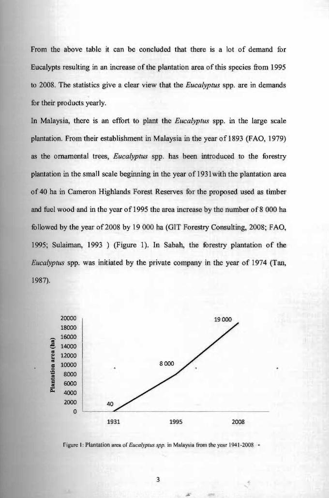
FAO (1995) and GIT Forestry Consulting (2008) stated that, in 1995 there were
683 000 ha of norma] Eucalypts plantation in the Southeast Asian region and that
was not including the equivalent of about 2.0 million ha as boundaries trees
around the field and also the scattered trees. In the year 2008, there is a huge
increment of the plantation area of the Eucalyptus spp. in those countries with the
total ofapproximately 1.5 million ha (Table 1).
Table 1. Estimated areas of Eucalyptus plantations in the South-East Asian region in 1995 until the year 0 f2008 S FAO andGITForestry C I'ources: onsu tmg.
Plantation area of the Eucalyptus by year (ha)
Country 1995 2008
I
Indonesia 80000 128000
Malaysia 0
8000 "
19000 "
Myanmar 40000 76189
Philippines 10000 189000
Thailand 195000 500000
Vietnam I 350000 586000
Total 683000 1 498 1&9
2
--
From the above table it can be concluded that there is a lot of demand for
Eucalypts resulting in an increase of the plantation area of this species from 1995
to 2008. The statistics give a clear view that the Eucalyptus spp. are in demands
for their products yearly.
In Malaysia, there is an effort to plant the Eucalyptus spp. in the large scale
plantation. From their establishment in Malaysia in the year of 1893 (F AO, 1979)
as the ornamental trees, Eucalyptus spp. has been introduced to the forestry
plantation in the small scale beginning in the year of 1931 with the plantation area
of 40 ha in Cameron Highlands Forest Reserves for the proposed used as timber
and fuel wood and in the year of 1995 the area increase by the number of8 000 ha
followed by the year of2008 by 19000 ha (GIT Forestry Consulting, 2008; FAO,
1995; Sulaiman, 1993 ) (Figure 1). In Sabah, the forestry plantation of the
Eucalyptus spp. was initiated by the private company in the year of 1974 (Tan,
1987).
20000
18000 16000
..c:: '-' " 14000 <II " 12000... " 10000 ¢ = ~ 8000a = 6000 s:" 4000
2000 40 a
1931 1995 2008
Figure I : Plantation area of Eucalyptus spp. in Malaysia from the year 1941-2008 .
19000
3

The statistical value shown by the above graph showed that there is an increase of
the planted Eucalyptus spp. area in Malaysia. It can be concluded that the
Eucalyptus spp. have a high demand in Malaysia and it can be made as one of the
timber sources.
Based on studies made by researchers in the other countries, the Eucalyptus spp. is
susceptible and vulnerable to the disease infection. Thus it can disrupt the growth
performance of the tree and reduce the quality and the productivity of the tree. As
an example, in many African countries, disease has negatively impacted the
plantations ofthe Eucalypts (Gezahgne, 2010).
This research is aims to identify all the possible pathogen that associated with the
Eucalyptus in Malaysia especially in Sarawak. Eucalyptus spp. plantation is still
new in Malaysia and need an extensive attention in the prevention of the disease.
1.2 Problem statement
The number of the Eucalyptus spp. plantations in the world especially in the Asian
region and Malaysia is increasing yearly. A high rate of plantation productivity is
needed in order to meet the demand of the worldwide population for the products
of the Eucalypts trees. The large-scale growth of the Eucalyptus spp. plantations
will cause the trees to be more vulnerable to disease and thus will reduce the
quantity and quality of the products. In Malaysia, Eucalyptus spp. is still in the
early stage of the plantation and there is still a lack of information on the disease
of the Eucalypts in the Malaysian environment and climate. Many new diseases
will develop in time and the inability to identify it will cause more problems in
. . handling the infection. So there is a need to rapidly isolate and identify the causal
4

Pusat Khidmat Maklumat Akademik UNlVERSm MALAYSIA SARAWAK
disease that mainly comes from an infection so that the first step for the
prevention can be taken. This step must be done in order to reduce the impact of
the infection, reducing the cost for the control and also to save this valuable
species from destruction. The morphological and molecular approach to fungal
identification and also the physiological characteristics of the pathogen will be
assessed in order to characterize the potential pathogen associated with diseases of
the Eucalypts species in Malaysia.
1.3 Objectives
The objectives of this research are:
a) To identify the causal disease of the Eucalyptus speCIes based on the
morphological characteristics of the fungi and also by using the molecular
technique.
b) To study the physiological characteristics of the fungi towards different
temperatures and pH level.
c) To detennine the pathogenicity of the fungi on the Eucalyptus species.
5

CHAPTER TWO
LITERATURE REVIEW
2.1 Taxonomy of eucalyptus
Eucalyptus is an enormous genus comprising of more than 800 species and two
third of them can be found in Australia (Barclay, 2004). The genus of Eucalypts
belongs to discrete groups which can be classify into five major subgenera which
are Blakella, Corymbia, Eudesmia, Gaubaea, Idiogenes, Monocalyptus and also
Symphyomyrtus as introduced by Pryor and Johnson (1971). The eucalyptus
comes from the family Myrtaceae which is commonly found in Australia, South
America and Asia and it is closely related to the syzygium genus (Lyne, 1996).
In Malaysia, there are two species of Eucalyptus that are commonly being planted
which are E. Deglupta and E. Grandis (Sabah Forestry Department, 2006). The
Eucalyptus is abundantly planted in the plantations in Sabah, Pahang and Negeri
Sembilan and some of them are planted as the ornamental plant (Sulaiman, 1993).
2.~ Botanical descriptio.n
Lyne (1 996) summarized the Eucalyptus as it exhibiting a variety of habits such as
it can be a shrub, mallees or trees and it mostly occur as forest and woodland
trees. The bark can be classified into two categories which are the smooth and
rough bark and in some species it is very hard. The flower is an umbellaster which
6
---------------.--~-.-~-~.. - .-.--- .-.

usually simple and axillary, compound and axillary and compound and terminal
whereas the fruit is a woody hypanthium that enclosed the base and sides of the
capsules and it may vary in shape according to their species.
2.3 Origin and distribution of eucalyptus
Species of the genus eucalyptus are commonly known as eucalypts throughout the
world, although in Australia they often called gum trees because ofthe gum (kino)
that exudes from the trunk of older trees (Zacharin, 1978). Eucalyptus originated
from Australia (Coppen, 2002). According to Turnbull (1999), the distribution of
natural eucalypts forest is widely spreading among the Australian country,
whereas there are a lot of countries in the world such as the Americas and South
Africa that plant the Eucalyptus in the large-scale that include the various of
species. In Sarawak, there are only 0.4 ha have been planted with Eucalyptus spp.
(Sulaiman, 1993).
2.4 Importance of eucalyptus
Young trees are a source of paper pulp, charcoal and fuel wood, poles, mining
timber, and fibreboard; mature trees within species provide strong and durable
timber, and all sizes are capable of use for other forest products such as volatile
oils for pharmaceutical and industrial uses, and honey (Penfold and Willis, 1961;
Jacobs, 1981; Hillis and Brown 1984; Boland et ai, 1991). Eucalypts provides
sawn timber, plywood, fibreboard, mine props, pulp for paper and rayon, poles,
7

ftrewood and charcoal, essential oils, honey, shade and shelter (Hillis and Brown,
1978). Less conventional uses include the production of plant growth regulator,
tannin extracts, industrial chemical additives, adhesives and fodder additives
(Song, 1992).
2.5 Forest plantation diseases
According to Callan (2001), tree diseases cover the wide range of pathogenic
infection, abnormalities, disturbance of the normal structure and growth of the tree
and he defmed tree disease as the deleterious effects resulting from injurious
agents other than fire and insect damage and it usually develop from the complex
interaction between the susceptible tree, predisposing environment condition or
infectious agents such as fungi. There are many diseases that infect the stem, root
and leaves. Cryphonectria canker caused by Cryphonectria cubensis, canker
and dieback is caused by Botryosphaeria spp., vascular wilt of Eucalyptus
caused by Ceratocystis fimbriata, pink disease caused by Erythricium
salmonicolor and Leaf blotch caused by Mycosphaerella spp. are examples
of diseases in commercial Eucalyptus plantations (Gezahgne, 2010). In
Malaysia, leaf fungus disease wiped out plantations ofEucalyptus
camaldulensis in Malaysia and the incidence of heart rot has resulted in a drastic,
temporary slowdown of plantation establishment in Peninsular Malaysia (Lee,
1993).
According to Davis (2002), the serious fungal infestation will occur when the
density of leafof the Eucalyptus is high and the damage due to the infestation can
spread very rapidly, destroying large areas of leaves within a few days if not
8
checked. Harrison et al (2003) stated that, the Ganoderma specIes has been
reported affecting the Eucalyptus spp. According to Philiips (1994), the most
common diseases for the Eucalypts are caused by the fungi of
I. Mycosphaerella spp. (Crinkle Leaf Disease)
2. Aulographina eucalypti (Corky Leaf Spot)
3. Pseudocercospora eucalyptorum
4. Septoria pulcherrima
5. Seimatosporium spp. (Angular Leaf Spot)
2.6 Pathogenic fungi
Pathogenic fungi can be described as the virulent fungi which produce the
inoculums which is the spores to infect the host. The classification and
identification of fungi, unlike other important pathogens such as bacteria or
viruses, relies mainly on morphological criteria. The identification of the fungi is a
complex process that usually requires microscopic examination and extensive
knowledge of the taxonomy of the fungus and most of the tree specialists and
arborist can recognize most fungus diseases by the appearance of the tree section
(the symptoms) (Tattar, 1989).
According to Callan (2001), fungi associated with the tree disease can be
classified into several classes which are:
1. Water molds: Oomycetes
2. Sac fungi and molds: Ascomycetes
3. Mushrooms and conks: Hymenomycetes
4. Rusts: Urediniomycetes (Basidiomycota)
9

3.
4.
2.7 Forest plantation pathology principles
Callan (200 I) stated that, there are several principles that is important in the forest
pathology research which are:
1. Culturing - the suspected pathogen should be obtained in a pure, artificial
culture, where possible
2. Pathogenicity study - according to Kochs' Postulates (see below), the
organism should be proven to be the cause ofan infectious disease.
Life cycle - all spore states should be identified and studied and related to
host range and phenology
Conditions for infection - the general physiology and requirements for
spore germination, penetration, and so on should be elucidated.
Koch Postulates:
I. Show constant association ofthe organism with the disease.
2. Isolate the organism in culture from the diseased plant.
3. Inoculate a healthy plant from the culture and produce the same disease.
4. Re-isolate the same organism from inoculated plants.
10

3.1
3.2
The descriptions
3.3
To
CHAPTER THREE
MATERIALS AND METHODS
Research material
Diseased tissues such as leaves, branches and roots from the Eucalyptus tree were
used in this research. Fresh infected samples of the Eucalyptus pellita species
were collected from the Sampadi Forest Plantation in Kuching, Sarawak.
Disease description
of the disease on the infected tissues were made. All the
symptoms such as the presents of the lesion on the leaves were identified. The
early identification of the disease was made based on the available references.
Pathogens isolation on the Potato Dextrose Agar
identify the potential pathogens that associated with the diseases, the
pathogens were isolated on the media. Potato dextrose agar (PDA) was as.a media
for the iso lation 0 f the pathogens.
The samples of the Eucalyptus leaves were cut into 100 small square segments
which have the size of 2mm x2mm. The tissues were cut at the margin of the
ion or between the healthy and the infected site by using the sterile &Calpel to
11

I prevent any other contamination. Then, the tissue segments were agitated in the
10% concentration of sodium hypochlorite, and the tissues segments were rinsed
into the sterilized distilled water, three times for five minutes in each session. The
segments were blotted dry using the sterilized filter paper.
The segments were put onto the media, with ten tissue segments for each Petri
dish The segments plated were incubated in the room temperature. The
observation was made daily until no new species of fungi can be found grow from
the plated plant tissues in the Petri dishes. The different types of the fungi were
observed and the potential pathogen was identified during the incubation period.
Percentage occurrence of the fungi associated with the tissue segments was
recorded.
A pure culture was prepared for the further studies. The pure cultures were
prepared by inoculating the morphologically different hyphal tips from the
isolated fungi into a new PDA media. The inoculated media were incubated at the
room temperature.
A stock culture was prepared. A small block of agar containing the mycelia from
the four to seven days culture were cut from the media and it were kept in a small
bottle containing the sterilized water and it were incubated at the temperature of
4°C.
The identification of the fungi was made based on the morphological
characteristics and also the molecular approach.
12

3.4 Identification of fungi using morphological characteristics
To detennine their morphological characteristics, the fungi were observed under
the compound microscope. In order to make it more visible under the microscope,
Acid fuchsin which is red in colour was used. The morphological characteristics
that was observed and identified are the vegetative hyphae and the shape of the
conidia. Picture ofa11 of these characteristics was capture using digital camera for
further record and identification. Reference literature used to aid the identification.
3.5 Identification of fungi using molecular approach
All selected fungal isolated were grown on the Malt Extract Agar (MEA) for four
to seven days. For a better DNA yield, the mycelial mat from the two weeks old
cultures in the Potato Dextrose Broth (PDB) also used for the DNA extraction. 0.3
g of the mycelium of the fungi was scraped off from the surface of the MEA. It
was grinded in liquid nitrogen using a mortar and pestle until the dry powdery
extract was formed. The mycelium powder was transferred into a sterile 1.5 ml
centrifuge tube. The 500 I.d DNA extraction buffer (100 mM Tris-HCl, pH 8.0, 10
mM EDTA; 3 M NaOAc; and 1 % SDS) was preheated in the 60' C water bath and
it was cooled down to the room temperature. Then, the extraction buffer was
added into the centrifuge tube and the mixture was incubated for 25 minutes at 55'
C. After the incubation, the mixture was vortexed to prevent the coagulation and
clumping of the sample. 500 J.ll of phen oIl chloroform! isoamylalcohol (25:24:1)
as added into the mixture and the centrifuge tube was centrifuged at 13 000 rpm
tbr five minutes. The top layer of supernatant resulting from the high speed
centrifugation was transferred into a new centrifuge tube and 500 J.ll of
13