ismb newsletter 32 ma y 2019 editor: sylvie ricard-blum · the ismb welcomes the new...
TRANSCRIPT

ISMB NEWSLETTER 32 May 2019 Editor: Sylvie Ricard-Blum
FROM THE PRESIDENT’S DESK
Dear ISMB members, The ISMB welcomes the new Vice-President, Suneel Apte, and new Council members, Alexandra Naba, Nikos Karamanos, Megan Lord, Alexander Nyström, Katia Schenke-Layland, Jean Schwarzbauer, and Anthony Weiss. I warmly congratulate them on behalf of the Society and I thank them for volunteering to contribute to the ISMB activities. They are from different parts of the world, works on different aspects of the extracellular matrix, and are at different stages of their scientific career, and these differences are an asset for the Council. The newsletter contains the usual items, including the meeting report of the annual meeting of the German society held in Regensburg last March, newly published articles, available positions and meeting announcements. "In Focus" (spotlights on methods that ISMB members may find useful) describes second harmonic generation imaging of fibrillar collagen for quantitative analyses. The "News from the labs” section is from Richard Farndale (Cambridge, UK), who made major contributions to our field. He retires in September but will continue to provide the libraries of synthetic triple-helical peptides (Collagen Toolkits) he developed. The creation of a new national society for Matrix Biology, Matrix Biology Israel with Irit Sagi (Weizman Institute of Science) as President, is announced in this issue of the newsletter. Last, I thank all the ISMB members, who contributed to this issue of the newsletter and more specifically Chloé Yeung in charge of In Focus, and Julia Etich for providing a report on the annual meeting of the German society, a list of articles published by members of the German society and information on some meetings.
A fibroblast within a tethered lattice, after 13 days, with a cavity surrounding the cell and areas of packed collagen fibrils (Porter et al. 1998 Wound Rep. Reg. 6: 157-166)
ISMB officers
President Sylvie Ricard-Blum Past-President Liliana Schaefer Vice-President Suneel Apte Secretary Jo Adams Treasurer Ruud Bank Council Members Anthony Day Julia Etich Nikos Karamanos Wei Kong Megan Lord Alexandra Naba Alexander Nyström Taina Pihlajaniemi Katia Schenke-Layland Jean Schwarzbauer Gerhard Sengle Hide Watanabe Anthony Weiss Chloé Yeung Ex officio Renato Iozzo Bjorn Olsen
ISMB NEWSLETTER May 2019
2
The ISMB has to increase its membership to continue to support young scientists in the Matrix Biology field, and to sponsor Matrix Biology meetings. Please free to contact me or any other member of the Council if you have ideas/suggestions to make this happen. Thank you very much in advance. Sylvie Ricard-Blum, ISMB President
COMPOSITION OF ISMB COUNCIL SUBCOMMITTEES
Communication Meetings and travel grants Jo Adams (UK) Anthony Day (UK) Julia Etich (Germany) Ruud Bank (The Netherlands, chair) Wei Kong (China) Gerhard Sengle (Germany) Sylvie Ricard-Blum (France, chair) Hide Watanabe (Japan) Chloé Yeung (Denmark) Membership Hide Watanabe (Japan) Suneel Apte (USA)
Like ISMB on Facebook and follow the ISMB on Twitter https://www.facebook.com/IntSocMatBio/ ISMB@IntSocMatBio https://twitter.com/intsocmatbio
ISMB correspondents of National Societies for Matrix Biology for Facebook & Twitter The American Society for Matrix Biology Alexandra Naba, University of Illinois, Chicago (USA) [email protected] The British Society for Matrix Biology Michal Dudek, University of Manchester (UK) [email protected] The Finnish Connective Tissue Society Piia Takabe, University of Eastern Finland, Kuopio (Finland) [email protected] The French Society for Matrix Biology Marion Marchand [email protected] The German Society for Matrix Biology Dominique Muschter, University of Regensburg (Germany)

ISMB NEWSLETTER May 2019
3
Matrix Biology Ireland. Alexandre Trotier, National University of Ireland, Galway (Ireland) [email protected] Matrix Biology Society of Australia and New Zealand (MBSANZ) Luise Kung, Murdoch Childrens Research Institute, Melbourne (Australia) [email protected] The Swiss Society for Matrix Biology (SSMB) Collin Ewald, ETH Zurich (Switzerland) [email protected] Twitter of SSMB: @SwissMatrixBio
NATIONAL SOCIETIES FOR MATRIX BIOLOGY
CREATION OF MATRIX BIOLOGY ISRAEL Dear ISMB members, We are delighted to inform you about the establishment of Matrix Biology Israel (MBIS) society. Extracellular matrix field is rapidly growing and is consisted by scientists coming from diverse backgrounds. The MBIS was formed to provide a forum for scientists with an interest in the extracellular matrix and to promote ECM research and education in Israel. MBIS is consisted by more than 25 research groups around the country that are currently conducting cutting edge work in diverse aspects of ECM field. Part of the activity of MBIS is to organize national and international meetings, to award young graduate students and postdocs with bursaries/fellowships and provide employment opportunities to the members. Members of MBIS are receiving regularly a newsletter with up-to-date information of the society. More details you can find in our website http://www.weizmann.ac.il/conferences/MBIS. Moreover, we intend to work closely with ISMB for making ECM a niche field and to further promote the journals of Matrix Biology and Matrix Biology Plus. On behalf of MBIS, Prof. Irit Sagi – President of MBIS Email: [email protected] Prof. Benjamin Geiger – Vice President of MBIS Email: [email protected] Dr. Nikolaos Afratis - Scientific advisor Email: [email protected] Dr. Inna Solomonov - Scientific advisor Email: [email protected]
ISMB NEWSLETTER May 2019
4
MEETING REPORT Annual Meeting of the German Society for Matrix Biology, March 28th - 30th 2019, Regensburg, Germany The annual meeting of the German Society for Matrix Biology (DGMB) took place in Regensburg from the 28th of March to the 30th, organized by Susanne Grässel as vice-president of the German Society for Matrix Biology and Attila Aszodi from the Ludwig-Maximilians-University Munich. 105 participants from seven countries discussed topics evolving from the field of extracellular matrix biology that ranged from ECM structure, cellular interactions and signaling to stem cell and tumor biology as well as tissue injury and engineering strategies. Danny Chan from the University of Hongkong gave an outstanding keynote lecture and was accompanied by session keynote speakers Carsten Grashoff1, Kim Midwood2, Brian Johnstone3, Yuval Rinkevich4, Michael Zeisberg5, Anna Mandinova6, Johanna Myllyharju7 and Michael Sixt8. As part of their final symposium, a special session was dedicated to research projects associated with the DFG funded Research Consortium FOR 2407 / ExCarBon, which was initiated by Susanne Grässel and Attila Aszodi in 2016 and embedded into this meeting. Established in 2017, the 2019 main conference was again preceded by a young scientists meeting from March 27th to 28th, offering a more casual platform for young scientists to share latest data with peers as well as numerous networking opportunities. Each year the German Society for Matrix Biology awards a Young Investigator Award to acknowledge outstanding work of scientists at an early career step. In a highly competitive field, Jana Riegger from the University of Ulm won this year´s prize with her work on chondrogenic stem/progenitor cells and their immunomodulatory and regenerative potential in cartilage injury. The award ceremony took place during the conference dinner in the historic Salzstadel located at the iconic Stone Bridge in the heart of the UNESCO World Heritage site of the old town of Regensburg. Furthermore, three poster prizes were awarded to Dominique Muschter9, Elke Pach10 and Heiko Rödig11. During the general assembly of the society, Susanne Grässel, vice-chair of the council, announced her retreat after 11 years of active participation and commitment. The council thanked her for her work as well as for her excellent organization of the conference. Frank Zaucke from the “Dr. Rolf M. Schwiete Research Unit for Osteoarthritis” of the Orthopaedic University Hospital Friedrichsheim, Frankfurt/Main was elected new vice-chair by the members. Furthermore, Jürgen Brinckmann12 (re-elected) and Uwe Hansen1 (newly elected) were confirmed as auditors for the financial business of the society. The German Society of Matrix Biology and the organizers thank all presenters and attendees for their passionate participation, inspiring discussions and thought-provoking impulse. With high anticipation, we are looking forward to the next conference 2020 to be held in Frankfurt/Main, Germany chaired by Frank Zaucke. 1University of Münster, 2University of Oxford, 3Oregon Health & Science University, 4Helmholtz Centre Munich, 5University Medicine Göttingen, 6Massachusetts General Hospital, 7University of Oulu, 8Institute of Science and Technology Austria, 9University Hospital Regensburg, 10University of Cologne, 11Goethe University Frankfurt am Main, 12University of Lübeck

ISMB NEWSLETTER May 2019
5
ISMB NEWSLETTER May 2019
6
POSITIONS AVAILABLE on ISMB website (http://ismb.org/career/)
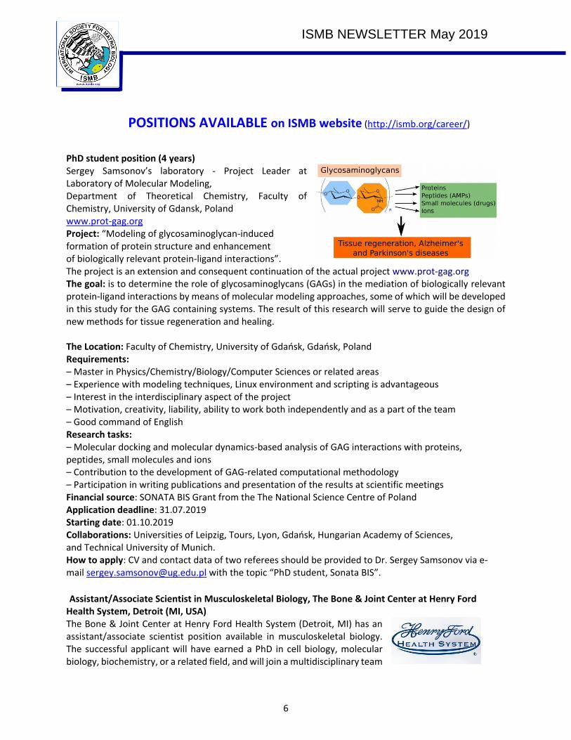
PhD student position (4 years) Sergey Samsonov’s laboratory - Project Leader at Laboratory of Molecular Modeling, Department of Theoretical Chemistry, Faculty of Chemistry, University of Gdansk, Poland www.prot-gag.org Project: “Modeling of glycosaminoglycan-induced formation of protein structure and enhancement of biologically relevant protein-ligand interactions”. The project is an extension and consequent continuation of the actual project www.prot-gag.org The goal: is to determine the role of glycosaminoglycans (GAGs) in the mediation of biologically relevant protein-ligand interactions by means of molecular modeling approaches, some of which will be developed in this study for the GAG containing systems. The result of this research will serve to guide the design of new methods for tissue regeneration and healing. The Location: Faculty of Chemistry, University of Gdańsk, Gdańsk, Poland Requirements: – Master in Physics/Chemistry/Biology/Computer Sciences or related areas – Experience with modeling techniques, Linux environment and scripting is advantageous – Interest in the interdisciplinary aspect of the project – Motivation, creativity, liability, ability to work both independently and as a part of the team – Good command of English Research tasks: – Molecular docking and molecular dynamics-based analysis of GAG interactions with proteins, peptides, small molecules and ions – Contribution to the development of GAG-related computational methodology – Participation in writing publications and presentation of the results at scientific meetings Financial source: SONATA BIS Grant from the The National Science Centre of Poland Application deadline: 31.07.2019 Starting date: 01.10.2019 Collaborations: Universities of Leipzig, Tours, Lyon, Gdańsk, Hungarian Academy of Sciences, and Technical University of Munich. How to apply: CV and contact data of two referees should be provided to Dr. Sergey Samsonov via e-mail [email protected] with the topic “PhD student, Sonata BIS”.
Assistant/Associate Scientist in Musculoskeletal Biology, The Bone & Joint Center at Henry Ford Health System, Detroit (MI, USA) The Bone & Joint Center at Henry Ford Health System (Detroit, MI) has an assistant/associate scientist position available in musculoskeletal biology. The successful applicant will have earned a PhD in cell biology, molecular biology, biochemistry, or a related field, and will join a multidisciplinary team

ISMB NEWSLETTER May 2019
7
of investigators with complementary research programs examining various aspects of the musculoskeletal system (https://www.hfhs-bjc.org/). The successful applicant will be expected to develop an externally funded research program and establish interdisciplinary research partnerships. The Bone & Joint Center is looking for exceptional individuals with research interests that may include osteoporosis, osteoarthritis, tendon/ligament disorders, or other areas of musculoskeletal biology. The Bone & Joint Center has expertise and well-equipped facilities for research in bone, cartilage, and skeletal muscle biology, histology, biochemistry, molecular biology (including CRISPR-based technologies), motion analysis, biomechanics, musculoskeletal modeling, computational mechanics, and 3D imaging. Scientists in the Bone & Joint Center have a strong history of collaboration with the Departments of Orthopaedic Surgery, Radiology, Public Health Science, Neurology, Endocrinology, and Rheumatology at Henry Ford Health System. The Bone & Joint Center occupies approximately 15,000 square feet in Wayne State University’s state-of-the-art Integrative Biosciences Center (www.ibio.wayne.edu), which affords additional opportunity for collaboration with the Wayne State University community. This position has strong institutional support and will include a generous startup package and salary that is commensurate with experience. Preference will be given to applicants with demonstrated research potential, a strong record of productivity, and a desire for multidisciplinary collaborations. Applicants should send their CV, a statement of their long-term research goals, and the names and contact information of at least three references to the e-mail address below. Consideration of applicants will continue until the position is filled. Michael J. Bey, PhD ([email protected])
NEWS FROM LABS The Toolkits Reloaded! Richard Farndale retires in September, having worked in the Department of Biochemistry, University of Cambridge (UK) since 1984. Richard’s triple-helical peptides have been distributed to over 100 laboratories worldwide, particularly for use in platelet research, as ligands for
Glycoprotein VI, integrin 21 and von Willebrand Factor (VWF), and there is increasing interest from pharma as well as academic laboratories in applying corresponding peptides to Discoidin Domain Receptors (DDRs), OSCAR and other matrix proteins. To continue to cater for the needs of researchers beyond his retirement, Richard has set up a company, CambCol Laboratories, to provide collagen peptides, offering initially the same materials as at present. Richard developed an interest in collagen and connective tissue during his PhD research at the Strangeways Research Laboratory (1978-84), directed by Sylvia Fitton Jackson, a colourful and generous supervisor. Moving to Biochemistry, Richard focussed largely on the activation of platelets by collagen, leading to a crucial collaboration with Michael Barnes, with whom specific synthetic collagen peptides
were developed to bind platelet Glycoprotein VI and integrin 21. This approach led to the Collagen Toolkits, libraries of synthetic triple-helical peptides embracing the collagenous domains of collagens II

ISMB NEWSLETTER May 2019
8
and III. The Toolkits were used to map binding sites for other collagen receptors such as OSCAR, Leukocyte-associated immunoglobulin-like receptor 1 (LAIR1), the DDRs and the remaining collagen-binding integrins. They were quickly applied to matrix proteins generally, such as VWF, matrix metalloproteinases, small-leucine-rich proteoglycans and thrombospondin-1. Most of the collagen-binding targets initially envisaged for the Toolkits and funded by the Wellcome Trust have been successfully addressed, but, through CambCol Laboratories, Richard will welcome new suggestions. Shortly, he expects to be able to offer a Collagen XIII Toolkit. For further information, he can be contacted at [email protected].
Spotlights on methods that other ISMB members may find useful
If you would like to feature in the next newsletter, please email [email protected]
Second harmonic generation (SHG) imaging of fibrillar collagen for quantitative analyses
Figure caption: SHG image of human breast tissue
Description: Second Harmonic Generation (SHG) imaging is a label-free microscopy technique that allows imaging of non-centrosymmetric entities such as collagen fibres. It is an important tool for evaluating the 3D spatial organisation of collagen fibres within tissues and organs without fluorescent staining, photodamage, or photobleaching1,2. Furthermore, because the typical wavelengths used are in the near-infrared spectral range (700−1000 nm), SHG microscopy can easily achieve high resolution imaging to depths of several
hundred microns2. The generation of an SHG signal occurs when two photons with the same wavelength fuse into a single photon with half of the original wavelength upon interaction with a

ISMB NEWSLETTER May 2019
9
non-centrosymmetric entity. This approach allows for high resolution imaging of native collagen fibres without the need for fixation or staining.
Useful for: Imaging collagen I organisation within the extracellular matrix in in vitro and in vivo settings. Particularly powerful for intravital imaging of native tissues, but can also be applied to unstained, as well as H&E FFPE sections. Once imaged, quantitative analysis of collagen parameters by SHG image segmentation can yield data on many aspects including orientation3–9, bundling and texture (using computational approaches such as grey-level co-occurrence matrix [GLCM10] analysis3–5,7,11,12), as well as spacing/porosity6,13,14, with free-to-use software such as Fiji15 or QuPath16.
Challenges/Tips: As with all microscopy, SHG is subject to aberrations induced by scattering in the specimen which decreases resolution. A major cause of this is due to the refractive index mismatch which decreases achievable contrast the deeper one images into specimens. To rectify this situation when imaging whole tissues or thick sections, a high refractive index, hyper-osmotic reagent (e.g., glycerol, sugars, or sugar alcohols) can be added to tissue to increase transparency and penetration depth by several fold.
Contact: Thomas R. Cox, Lab Head - Matrix and Metastasis, The Garvan Institute of Medical Research and The Kinghorn Cancer Centre, Sydney, Australia
Email: [email protected]
References:
1. Campagnola, P. J. & Loew, L. M. Second-harmonic imaging microscopy for visualizing biomolecular arrays in cells, tissues and organisms. Nat. Biotechnol. 21, 1356–1360 (2003).
2. Campagnola, P. Second harmonic generation imaging microscopy: applications to diseases diagnostics. Anal. Chem. 83, 3224–3231 (2011).
3. Conway, J. R. W. et al. Three-dimensional organotypic matrices from alternative collagen sources as pre-clinical models for cell biology. Sci. Rep. 7, 16887 (2017).
4. Vennin, C. et al. Transient tissue priming via ROCK inhibition uncouples pancreatic cancer progression, sensitivity to chemotherapy, and metastasis. Sci. Transl. Med. 9, (2017).
5. Chou, A. et al. Tailored first-line and second-line CDK4-targeting treatment combinations in mouse models of pancreatic cancer. Gut 67, 2142–2155 (2018).
6. Mayorca-Guiliani, A. E. et al. ISDoT: in situ decellularization of tissues for high-resolution imaging and proteomic analysis of native extracellular matrix. Nat. Med. 23, 890–898 (2017).
7. Cazet, A. S. et al. Targeting stromal remodeling and cancer stem cell plasticity overcomes chemoresistance in triple negative breast cancer. Nat. Commun. 9, 2897 (2018).
8. Provenzano, P. P. et al. Collagen density promotes mammary tumor initiation and progression. BMC Med. 6, 11 (2008).
9. Provenzano, P. P. et al. Collagen reorganization at the tumor-stromal interface facilitates local invasion. BMC Med. 4, 38 (2006).
10. Cicchi, R. et al. Scoring of collagen organization in healthy and diseased human dermis by multiphoton microscopy. J. Biophotonics 3, 34–43 (2010).
11. Huo, C. W. et al. High mammographic density is associated with an increase in stromal collagen and immune cells within the mammary epithelium. Breast Cancer Res. 17, 79 (2015).
12. Vennin, C., Pajic, M. & Timpson, P. Imaging fibrosis in pancreatic cancer using second harmonic generation. Pancreatology 15, 200–201 (2015).
ISMB NEWSLETTER May 2019
10
13. Acton, S. E. et al. Dendritic cells control fibroblastic reticular network tension and lymph node expansion. Nature 514, 498–502 (2014).
14. Tozluoğlu, M. et al. Matrix geometry determines optimal cancer cell migration strategy and modulates response to interventions. Nat. Cell Biol. 15, 751–762 (2013).
15. Schindelin, J. et al. Fiji: an open-source platform for biological-image analysis. Nat. Methods 9, 676–682 (2012).
16. Bankhead, P. et al. QuPath: Open source software for digital pathology image analysis. Sci. Rep. 7, 16878 (2017).
RECENT PAPERS
Assessing Collagen Deposition During Aging in Mammalian Tissue and in Caenorhabditis elegans. Methods Mol Biol. 2019 1944:169-188. Authors: Teuscher AC, Statzer C, Pantasis S, Bordoli MR, Ewald CY. Corresponding author: [email protected] Abstract: Proper collagen homeostasis is essential for development and aging of any multicellular organism. During aging, two extreme scenarios are commonly occurring: a local excess in collagen deposition, for instance during fibrosis, or a gradual overall reduction of collagen mass. Here, we describe a histological and a colorimetric method to assess collagen levels in mammalian tissues and in the nematode Caenorhabditis elegans. The first method is the polychrome Herovici staining to distinguish between young and mature collagen ratios. The second method is based on hydroxyproline measurements to estimate collagen protein levels. In addition, we show how to decellularize the multicellular organism C. elegans in order to harvest its cuticle, one of the two major extracellular matrices, mainly composed of collagen. These methods allow assessing collagen deposition during aging either in tissues or in whole organisms. Pedchenko V, Bauer R, Pokidysheva EN, Al-Shaer A, Forde NR, Fidler AL, Hudson BG, Boudko SP. A chloride ring is an ancient evolutionary innovation mediating the assembly of the collagen IV scaffold of basement membranes. J Biol Chem. May 17;294(20):7968-7981. Corresponding author: [email protected] Abstract: Collagen IV scaffold is a principal component of the basement membrane (BM), a specialized extracellular matrix that is essential for animal multicellularity and tissue evolution. Scaffold assembly begins with the trimerization of α-chains into protomers inside the cell, which then are secreted and undergo oligomerization outside the cell. For the ubiquitous scaffold composed of α1 and α2 chains, both intracellular and extracellular stages are mediated by the non-collagenous domain (NC1). The association of protomers is chloride-dependent, whereby chloride ions induce interactions of protomers’ trimeric NC1 domains leading to NC1 hexamer formation. Here, we investigated the mechanisms, kinetics, and functionality of the chloride ion-mediated protomer assembly by using a single-chain technology to produce a stable NC1 trimer comprising α1, α2, and α1 NC1 monomers. We observed that in the presence of chloride, the single-chain NC1 trimer self-assembles into a hexamer, for which the crystal structure was determined. We discovered that a chloride ring, comprising twelve ions, induces the assembly of and stabilizes the NC1 hexamer. Furthermore, we found that the chloride ring is evolutionarily conserved across all animals, first appearing in cnidarians. These findings reveal a fundamental role for the chloride ring in the assembly of collagen IV scaffolds of BMs, a critical event enabling tissue evolution and development. Moreover, the single-chain technology is foundational for generating trimeric NC1 domains of other α-chain compositions to investigate
ISMB NEWSLETTER May 2019
11
the α121, α345, and α565 collagen IV scaffolds and to develop therapies for managing Alport syndrome, Goodpasture’s disease, and cancerous tumor growth. Paik, Y.-K., Lane, L., and Overall, C.M. 2018. N. Launching the C-HPP neXt-CP50 Pilot Project for Functional Characterization of Identified Proteins with No Known Function. J Proteome Res. 2018 Dec 7;17(12):4042-4050 Corresponding author: [email protected] Abstract: An important goal of the Human Proteome Organization (HUPO) Chromosome-centric Human Proteome Project (C-HPP) is to correctly define the number of canonical proteins encoded by their cognate open reading frames on each chromosome in the human genome. When identified with high confidence of protein evidence (PE), such proteins are termed PE1 proteins in the online database resource, neXtProt. However, proteins that have not been identified unequivocally at the protein level but that have other evidence suggestive of their existence (PE2-4) are termed missing proteins (MPs). The number of MPs has been reduced from 5511 in 2012 to 2186 in 2018 (neXtProt 2018-01-17 release). Although the annotation of the human proteome has made significant progress, the "parts list" alone does not inform function. Indeed, 1937 proteins representing ∼10% of the human proteome have no function either annotated from experimental characterization or predicted by homology to other proteins. Specifically, these 1937 "dark proteins" of the so-called dark proteome are composed of 1260 functionally uncharacterized but identified PE1 proteins, designated as uPE1, plus 677 MPs from categories PE2-PE4, which also have no known or predicted function and are termed uMPs. At the HUPO-2017 Annual Meeting, the C-HPP officially adopted the uPE1 pilot initiative, with 14 participating international teams later committing to demonstrate the feasibility of the functional characterization of large numbers of dark proteins (CP), starting first with 50 uPE1 proteins, in a stepwise chromosome-centric organizational manner. The second aim of the feasibility phase to characterize protein (CP) functions of 50 uPE1 proteins, termed the neXt-CP50 initiative, is to utilize a variety of approaches and workflows according to individual team expertise, interest, and resources so as to enable the C-HPP to recommend experimentally proven workflows to the proteome community within 3 years. The results from this pilot will not only be the cornerstone of a larger characterization initiative but also enhance understanding of the human proteome and integrated cellular networks for the discovery of new mechanisms of pathology, mechanistically informative biomarkers, and rational drug targets. Tharmarajah, G., Eckhard, U., Jain, F., Marino, G., Prudova, A., Urtatiz, O., Fuchs, H., de Angelis, M.H., Overall, C.M., and Van Raamsdonk, C.D. 2018. Melanocyte Development in the Mouse Tail Epidermis Requires the Adamts9 Metalloproteinase. Pigment Cell Melanoma Res. 2018 Nov;31(6):693-707. Corresponding author: [email protected] Abstract: The mouse tail has an important role in the study of melanogenesis, because mouse tail skin can be used to model human skin pigmentation. To better understand the development of melanocytes in the mouse tail, we cloned two dominant ENU-generated mutations of the Adamts9 gene, Und3 and Und4, which cause an unpigmented ring of epidermis in the middle of the tail, but do not alter pigmentation in the rest of the mouse. Adamts9 encodes a widely expressed zinc metalloprotease with thrombospondin type 1 repeats with few known substrates. Melanocytes are lost in the Adamts9 mutant tail epidermis at a relatively late stage of development, around E18.5. Studies of our Adamts9 conditional allele suggest that there is a melanocyte cell-autonomous requirement for Adamts9. In addition, we used a proteomics approach, TAILS N-terminomics, to identify new Adamts9 candidate substrates in the extracellular matrix of the skin. The tail phenotype of Adamts9 mutants is strikingly similar to the unpigmented trunk belt in Adamts20 mutants,
ISMB NEWSLETTER May 2019
12
which suggests a particular requirement for Adamts family activity at certain positions along the anterior-posterior axis. Angelidis I, Simon LM, Fernandez IE, Strunz M, Mayr CH, Greiffo FR, Tsitsiridis G, Ansari M, Graf E, Strom TM, Nagendran M, Desai T, Eickelberg O, Mann M, Theis FJ, Schiller HB. An atlas of the aging lung mapped by single cell transcriptomics and deep tissue proteomics. Nat Commun. 2019 Feb 27;10(1):963. Corresponding authors: [email protected] and [email protected] Abstract: Aging promotes lung function decline and susceptibility to chronic lung diseases, which are the third leading cause of death worldwide. Here, we use single cell transcriptomics and mass spectrometry-based proteomics to quantify changes in cellular activity states across 30 cell types and chart the lung proteome of young and old mice. We show that aging leads to increased transcriptional noise, indicating deregulated epigenetic control. We observe cell type-specific effects of aging, uncovering increased cholesterol biosynthesis in type-2 pneumocytes and lipofibroblasts and altered relative frequency of airway epithelial cells as hallmarks of lung aging. Proteomic profiling reveals extracellular matrix remodeling in old mice, including increased collagen IV and XVI and decreased Fraser syndrome complex proteins and collagen XIV. Computational integration of the aging proteome with the single cell transcriptomes predicts the cellular source of regulated proteins and creates an unbiased reference map of the aging lung.
Bojarski KK, Sieradzan AK, Samsonov SA. Molecular dynamics insights into protein-glycosaminoglycan systems from microsecond-scale simulations. Biopolymers. 2019 Jan 22:e23252. Corresponding author: [email protected] Abstract: Heparin is a key player in cell signaling via its physical interactions with protein targets in the extracellular matrix. However, basic molecular level understanding of these highly biologically relevant intermolecular interactions is still incomplete. In this study, for the first time, microsecond-scale MD simulations are reported for a complex between fibroblast growth factor 1 and heparin. We rigorously analyze this molecular system in terms of the conformational space, structural, energetic, and dynamic characteristics. We reveal that the conformational selection mechanism of binding denotes a recognition specificity determinant. We conclude that the length of the simulation could be crucial for evaluation of some of the analyzed parameters. Our data provide novel significant insights into the interactions in the fibroblast growth factor 1 complex with heparin, in particular, and into the physical-chemical nature of protein-glycosaminoglycan systems in general, which have potential applicability for biomaterials development in the area of regenerative medicine. Samsonov SA, Zacharias M, Chauvot de Beauchene I. Modeling large protein–glycosaminoglycan complexes using a fragment‐based approach. J Comput Chem. 2019 May 30;40(14):1429-1439. Corresponding author: [email protected] and [email protected] Abstract: Glycosaminoglycans (GAGs), a major constituent of the extracellular matrix, participate in cell-signaling by binding specific proteins. Structural data on protein-GAG interactions are crucial to understand and modulate these signaling processes, with potential applications in regenerative medicine. However, experimental and theoretical approaches used to study GAG-protein systems are challenged by GAGs high flexibility limiting the conformational sampling above a certain size, and by the scarcity of GAG-specific docking tools compared to protein-protein or protein-drug docking approaches. We present for the first time an automated fragment-based method for docking GAGs on a protein binding site. In this approach, trimeric GAG fragments are flexibly docked to the protein, assembled based on their spacial overlap, and refined by
ISMB NEWSLETTER May 2019
13
molecular dynamics. The method appeared more successful than the classical full-ligand approach for most of 13 tested complexes with known structure. The approach is particularly promising for docking of long GAG chains, which represents a bottleneck for classical docking approaches applied to these systems. Vallet SD, Guéroult M, Belloy N, Dauchez M, Ricard-Blum S. A 3D model of human lysyl oxidase, a cross-linking enzyme. ACS Omega 2019 4:58495-8505 Corresponding author: [email protected] Abstract: Lysyl oxidase (LOX) is a cross-linking enzyme identified 50 years ago but its 3D structure is still unknown. We have thus built a 3D model of human LOX by homology modeling using the X-ray structure of human lysyl oxidase-like 2 (LOXL2) as a template. This model is the first one to recapitulate all known biochemical features of LOX, namely the coordination of the copper ion, the formation of the lysine tyrosylquinone (LTQ) cofactor and the disulfide bridges. Furthermore, this model is stable during a 1-µs molecular dynamics simulation. The catalytic site is located in a groove surrounded by two loops. The distance between these loops fluctuated during the simulations, which suggests that the groove forms a hinge with a variable opening, able to accommodate the various sizes of LOX substrates. This 3D model is a pre-requisite to perform docking experiments with LOX substrates and other partners in order to identify binding sites and to design new LOX inhibitors specific for therapeutic purpose. Poluzzi C, Nastase MV, Zeng-Brouwers J, Roedig H, Hsieh LTH, Michaelis JB, Buhl EM, Rezende F, Manavski Y, Bleich A, Boor P, Brandes RP, Pfeilschifter J, Stelzer EHK, Muench C, Dikic I, Brandts C, Iozzo RV, Wygrecka M, Schaefer L. Biglycan evokes autophagy in macrophages via a novel CD44/Toll-like receptor 4 signaling axis: Impact on renal ischemia/reperfusion injury. Kidney Int. 95: 540-562, 2019 This paper was highlighted in commentaries: DOI: https://doi.org/10.1016/j.kint.2018.12.007 Corresponding author: [email protected] Abstract: Biglycan, a small leucine-rich proteoglycan, acts as a danger signal promoting macrophage recruitment via TLR2/4- and CD14 co-receptors. We have now discovered that soluble biglycan triggered macrophage autophagy by promoting autophagosome formation in primary macrophages along with increased flux of autophagic markers Beclin-1, LC3-II, and p62. Mechanistically, biglycan evoked autophagy by acting as a novel, high-affinity ligand for CD44, a receptor involved in adhesion, migration, lymphocyte activation, homing, and angiogenesis. The biglycan-triggered pro-autophagic signal required TLR4/CD44 interaction. Moreover, we found a marked increase in the number of autophagic macrophages in various organs from mice stably overexpressing soluble biglycan. Notably, transient overexpression of circulating biglycan at the onset of renal ischemia/reperfusion injury (IRI) caused enhanced M1 macrophage recruitment into the kidneys of either Cd44+/+ or Cd44-/- mice but not in Cd14-/- mice. The interaction between biglycan and CD44 increased M1 autophagy and resulted in elevated number of renal M2 macrophages and reduced tubular damage during IRI regeneration. On the other hand, the biglycan-induced M2 polarization did not depend on the CD14 co-receptor. In addition, soluble biglycan evoked autophagy in human peripheral blood macrophages. Thus, we have discovered CD44 as a signaling co-receptor for biglycan, an interaction that is required for TLR4-CD44-dependent pro-autophagic activity in macrophages, and directly involved in preventing tubular damage in renal IRI. We hypothesize that interfering with the interaction between biglycan and its TLR co-receptors could represent a promising therapeutic intervention to curtail renal inflammation and damage. Shoemark, D.K, Ziegler, B., Watanabe, H., Strompen, J., Tucker, R.P., Özbek, S., Adams, J.C. (2019). Emergence of a Thrombospondin Superfamily at the Origin of Metazoans. Mol Biol Evol. 2019 Jun 1;36(6):1220-1238.

ISMB NEWSLETTER May 2019
14
Corresponding author: Jo Adams [email protected] Abstract: Extracellular matrix (ECM) is considered central to the evolution of metazoan multicellularity; however, the repertoire of ECM proteins in nonbilaterians remains unclear. Thrombospondins (TSPs) are known to be well conserved from cnidarians to vertebrates, yet to date have been considered a unique family, principally studied for matricellular functions in vertebrates. Through searches utilizing the highly conserved C-terminal region of TSPs, we identify undisclosed new families of TSP-related proteins in metazoans, designated mega-TSP, sushi-TSP, and poriferan-TSP, each with a distinctive phylogenetic distribution. These proteins share the TSP C-terminal region domain architecture, as determined by domain composition and analysis of molecular models against known structures. Mega-TSPs, the only form identified in ctenophores, are typically >2,700 aa and are also characterized by N-terminal leucine-rich repeats and central cadherin/immunoglobulin domains. In cnidarians, which have a well-defined ECM, Mega-TSP was expressed throughout embryogenesis in Nematostella vectensis, with dynamic endodermal expression in larvae and primary polyps and widespread ectodermal expression in adult Nematostella vectensis and Hydra magnipapillata polyps. Hydra Mega-TSP was also expressed during regeneration and siRNA-silencing of Mega-TSP in Hydra caused specific blockade of head regeneration. Molecular phylogenetic analyses based on the conserved TSP C-terminal region identified each of the TSP-related groups to form clades distinct from the canonical TSPs. We discuss models for the evolution of the newly defined TSP superfamily by gene duplications, radiation, and gene losses from a debut in the last metazoan common ancestor. Together, the data provide new insight into the evolution of ECM and tissue organization in metazoans. Teuscher AC, Jongsma E, Davis MN, Statzer C, Gebauer JM, Naba A, Ewald CY. The in-silico characterization of the Caenorhabditis elegans matrisome and proposal of a novel collagen classification. Matrix Biology Plus https://www.sciencedirect.com/science/article/pii/S2590028518300012 Corresponding authors: [email protected], [email protected], [email protected] Abstract: Proteins are the building blocks of life. While proteins and their localization within cells and sub-cellular compartments are well defined, the proteins predicted to be secreted to form the extracellular matrix - or matrisome - remain elusive in the model organism C. elegans. Here, we used a bioinformatic approach combining gene orthology and protein structure analysis and an extensive curation of the literature to define the C. elegans matrisome. Similar to the human genome, we found that 719 out of ~20,000 genes (~4%) of the C. elegans genome encodes matrisome proteins, including 181 collagens, 35 glycoproteins, 10 proteoglycans, and 493 matrisome-associated proteins. We report that 173 out of the 181 collagen genes are unique to nematodes and are predicted to encode cuticular collagens, which we are proposing to group into five clusters. To facilitate the use of our lists and classification by the scientific community, we developed an automated annotation tool to identify ECM components in large datasets. We also established a novel database of all C. elegans collagens (CeColDB). Last, we provide examples of how the newly defined C. elegans matrisome can be used for annotations and gene ontology analyses of transcriptomic, proteomic, and RNAi screening data. Because C. elegans is a widely used model organism for high throughput genetic and drug screens, and to study biological and pathological processes, the conserved matrisome genes may aid in identifying potential drug targets. In addition, the nematode-specific matrisome may be exploited for targeting parasitic infection of man and crops. Izzi V, Lakkala J, Devarajan R, Kaariainen A, Koivunen J, Heljasvaara R, Pilhajaniemi T. Pan-Cancer analysis of the expression and regulation of matrisome genes across 32 tumor types. Matrix Biology Plus. https://www.sciencedirect.com/science/article/pii/S2590028519300031 Corresponding author: [email protected]
ISMB NEWSLETTER May 2019
15
Abstract: The microenvironment plays a central role in cancer, and neoplastic cells actively shape it to their needs by complex arrays of extracellular matrix (ECM) proteins, enzymes, cytokines and growth factors collectively referred to as the matrisome. Studies on the cancer matrisome have been performed for single or few neoplasms, but a more systematic analysis is still missing. Here we present a Pan-Cancer study of matrisome gene expression in 10,487 patients across 32 tumor types, supplemented with transcription factors (TFs) and driver genes/pathways regulating each tumor's matrisome. We report on 919 TF-target pairs, either used specifically or shared across tumor types, and their prognostic significance, 40 master regulators, 31 overarching regulatory pathways and the potential for druggability with FDA-approved cancer drugs. These results provide a comprehensive transcriptional architecture of the cancer matrisome and suggest the need for development of specific matrisome-targeting approaches for future therapies. Chiarelli N, Carini G, Zoppi N, Ritelli M. Molecular insights in the pathogenesis of classical Ehlers-Danlos syndrome from transcriptome-wide expression profiling of patients’ skin fibroblasts. PLoS One. 2019 Feb 4;14(2): e0211647. Corresponding author: [email protected] Abstract: Classical Ehlers-Danlos syndrome (cEDS) is a dominant inherited connective tissue disorder mainly caused by mutations in the COL5A1 and COL5A2 genes encoding type V collagen (COLLV), which is a fibrillar COLL widely distributed in a variety of connective tissues. cEDS patients suffer from skin hyperextensibility, abnormal wound healing/atrophic scars, and joint hypermobility. Most of the causative variants result in a non-functional COL5A1 allele and COLLV haploinsufficiency, whilst COL5A2 mutations affect its structural integrity. To shed light into disease mechanisms involved in cEDS, we performed gene expression profiling in skin fibroblasts from four patients harboring haploinsufficient and structural mutations in both disease genes. Transcriptome profiling revealed significant changes in the expression levels of different extracellular matrix (ECM)-related genes, such as SPP1, POSTN, EDIL3, IGFBP2, and C3, which encode both matricellular and soluble proteins that are mainly involved in cell proliferation and migration, and cutaneous wound healing. These gene expression changes are consistent with our previous protein findings on in vitro fibroblasts from other cEDS patients, which exhibited reduced migration and poor wound repair owing to COLLV disorganization, altered deposition of fibronectin into ECM, and an abnormal integrin pattern. Microarray analysis also indicated the decreased expression of DNAJB7, VIPAS39, CCPG1, ATG10, SVIP, which encode molecular chaperones facilitating protein folding, enzymes regulating post-Golgi COLLs processing, and proteins acting as cargo receptors required for endoplasmic reticulum (ER) proteostasis and implicated in the autophagy process. Patients’ cells also showed altered mRNA levels of many cell cycle regulating genes including CCNE2, KIF4A, MKI67, DTL, and DDIAS. Protein studies showed that aberrant COLLV expression causes the disassembly of itself and many structural ECM constituents including COLLI, COLLIII, fibronectin, and fibrillins. Our findings provide the first molecular evidence of significant gene expression changes in cEDS skin fibroblasts highlighting that defective ECM remodeling, ER homeostasis and autophagy might play a role in the pathogenesis of this connective tissue disorder. Khan ES, Sankaran S, Paez JI, Muth C, Han MKL, del Campo A. Photoactivatable Hsp47: A Tool to Regulate Collagen Secretion and Assembly. Adv Sci (Weinh). 2019 Feb 28;6(9):1801982. Corresponding author: [email protected] Abstract: Collagen is the most abundant structural protein in mammals and is crucial for the mechanical integrity of tissues. Hsp47, an endoplasmic reticulum resident collagen‐specific chaperone, is involved in collagen biosynthesis and plays a fundamental role in the folding, stability, and intracellular transport of procollagen triple helices. This work reports on a photoactivatable derivative of Hsp47 that allows
ISMB NEWSLETTER May 2019
16
regulation of collagen biosynthesis within mammalian cells using light. Photoactivatable Hsp47 contains a non‐natural light‐responsive tyrosine (o‐nitro benzyl tyrosine (ONBY)) at Tyr383 position of the protein sequence. This mutation renders Hsp47 inactive toward collagen binding. The inactive, photoactivatable protein is easily uptaken by cells within a few minutes of incubation, and accumulated at the endoplasmic reticulum via retrograde KDEL receptor‐mediated uptake. Upon light exposure, the photoactivatable Hsp47 turns into functional Hsp47 in situ. The increased intracellular concentration of Hsp47 results in stimulated secretion of collagen. The ability to promote collagen synthesis on demand, with spatiotemporal resolution, and in diseased state cells is demonstrated in vitro. It is envisioned that photoactivatable Hsp47 allows unprecedented fundamental studies of collagen biosynthesis, matrix biology, and inspires new therapeutic concepts in biomedicine and tissue regeneration. Fitzgerald J, Endicott J, Hansen U, Janowitz C. Articular cartilage and sternal fibrocartilage respond differently to extended microgravity. NPJ Microgravity. 2019 Feb 18;5:3. Corresponding author: [email protected] Abstract: The effects of spaceflight on cartilaginous structure are largely unknown. To address this deficiency, articular cartilage (AC) and sternal cartilage (SC) from mice exposed to 30 days of microgravity on the BION-M1 craft were investigated for pathological changes. The flight AC showed some evidence of degradation at the tissue level with loss of proteoglycan staining and a reduction in mRNA expression of mechano-responsive and structural cartilage matrix proteins compared to non-flight controls. These data suggest that degradative changes are underway in the AC extracellular matrix exposed to microgravity. In contrast, there was no evidence of cartilage breakdown in SC flight samples and the gene expression profile was distinct from that of AC with a reduction in metalloproteinase gene transcription. Since the two cartilages respond differently to microgravity we propose that each is tuned to the biomechanical environments in which they are normally maintained. That is, the differences between magnitude of normal terrestrial loading and the unloading of microgravity dictates the tissue response. Weight-bearing articular cartilage, but not minimally loaded sternal fibrocartilage, is negatively affected by the unloading of microgravity. We speculate that the maintenance of physiological loading on AC during spaceflight will minimize AC damage. Alberton P, Dugonitsch HC, Hartmann B, Li P, Farkas Z, Saller MM, Clausen-Schaumann H, Aszodi A. Aggrecan Hypomorphism Compromises Articular Cartilage Biomechanical Properties and Is Associated with Increased Incidence of Spontaneous Osteoarthritis. Int J Mol Sci. 2019 Feb 26;20(5). Corresponding author: [email protected] Abstract: The gene encoding the proteoglycan aggrecan (Agc1) is abundantly expressed in cartilage during development and adulthood, and the loss or diminished deposition of the protein results in a wide range of skeletal malformations. Furthermore, aggrecan degradation is a hallmark of cartilage degeneration occurring in osteoarthritis. In the present study, we investigated the consequences of a partial loss of aggrecan in the postnatal skeleton and in the articular cartilage of adult mice. We took advantage of the previously described Agc1tm(IRES-CreERT2) mouse line, which allows for conditional and timely-regulated deletion of floxed, cartilage-expressed genes. As previously reported, the introduction of the CreERT2 cassette in the 3'UTR causes a disruption of the normal expression of Agc1 resulting in a hypomorphic deposition of the protein. In homozygous mice, we observed a dwarf phenotype, which persisted throughout adulthood supporting the evidence that reduced aggrecan amount impairs skeletal growth. Homozygous mice exhibited reduced proteoglycan staining of the articular cartilage at 6 and 12 months of age, increased stiffening of the extracellular matrix at six months, and developed severe cartilage erosion by 12 months.

ISMB NEWSLETTER May 2019
17
The osteoarthritis in the hypomorph mice was not accompanied by increased expression of catabolic enzymes and matrix degradation neoepitopes. These findings suggest that the degeneration found in homozygous mice is likely due to the compromised mechanical properties of the cartilage tissue upon aggrecan reduction. Ayachi O, Barlin M, Broxtermann PN, Kashkar H, Mauch C, Zigrino P. The X-linked inhibitor of apoptosis protein (XIAP) is involved in melanoma invasion by regulating cell migration and survival. Cell Oncol (Dordr). 2019 Jun;42(3):319-329. Corresponding author: [email protected] Abstract: BACKGROUND: The X-linked inhibitor of apoptosis (XIAP) is a potent cellular inhibitor of apoptosis, based on its unique capability to bind and to inhibit caspases. However, XIAP is also involved in a number of additional cellular activities independent of its caspase inhibitory function. The aim of this study was to investigate whether modulation of XIAP expression affects apoptosis-independent functions of XIAP in melanoma cells, restores their sensitivity to apoptosis and/or affects their invasive and metastatic capacities. METHODS: XIAP protein levels were analyzed by immunohistochemical staining of human tissues and by Western blotting of melanoma cell lysates. The effects of pharmacological inhibition or of XIAP down-regulation were investigated using ex-vivo and transwell invasion assays. The biological effects of XIAP down-regulation on melanoma cells were analyzed in vitro using BrdU/PI, nucleosome quantification, adhesion and migration assays. In addition, new XIAP binding partners were identified by co-immunoprecipitation followed by mass spectrometry. RESULTS: Here we found that the expression of XIAP is increased in metastatic melanomas and in invasive melanoma-derived cell lines. We also found that the bivalent IAP antagonist birinapant significantly reduced the invasive capability of melanoma cells. This reduction could be reproduced by downregulating XIAP in melanoma cells. Furthermore, we found that the migration of melanoma cells and the formation of focal adhesions at cellular borders on fibronectin-coated surfaces were significantly reduced upon XIAP knockdown. This reduction may depend on an altered vimentin-XIAP association, since we identified vimentin as a new binding partner of XIAP. As a corollary of these molecular alterations, we found that XIAP down-regulation in melanoma cells led to a significant decrease in invasion of dermal skin equivalents. CONCLUSION: From our data we conclude that XIAP acts as a multifunctional pro-metastatic protein in skin melanomas and, as a consequence, that XIAP may serve as a therapeutic target for these melanomas. Costa B, Eisemann T, Strelau J, Spaan I, Korshunov A, Liu HK, Bugert P, Angel P, Peterziel H. Intratumoral platelet aggregate formation in a murine preclinical glioma model depends on podoplanin expression on tumor cells. Blood Adv. 2019 Apr 9;3(7):1092-1102. Corresponding author: [email protected] Abstract: Binding of the sialomucin-like transmembrane glycoprotein podoplanin (PDPN) to the platelet receptor C-type lectin-like receptor 2 induces platelet activation and aggregation. In human high-grade gliomas, PDPN is highly expressed both in tumor cells and in tumor-associated astrocytes. In glioma patients, high expression of PDPN is associated with worse prognosis and has been shown to correlate with intratumoral platelet aggregation and an increased risk of venous thromboembolism (VTE). To functionally assess the role of PDPN in platelet aggregation in vivo, we established a syngeneic orthotopic murine glioma model in C57/Bl6 mice, based on transplantation of p53- and Pten-deficient neural stem cells. This model is characterized by the presence of intratumoral platelet aggregates and by the upregulation of PDPN both in glioma cells and in astrocytes, reflecting the characteristics of human gliomas. Deletion of PDPN either in tumor cells or in astrocytes resulted in glioma formation with similar penetrance and grade compared with
ISMB NEWSLETTER May 2019
18
control mice. Importantly, only the lack of PDPN in tumor cells, but not in astrocytes, caused a significant reduction in intratumoral platelet aggregates, whereas in vitro, both cell types have similar platelet aggregation-inducing capacities. Our results demonstrate a causative link between PDPN and platelet aggregation in gliomas and pinpoint the tumor cells as the major players in PDPN-induced platelet aggregation. Our data indicate that blocking PDPN specifically on tumor cells could represent a novel strategy to prevent platelet aggregation and thereby reduce the risk of VTE in glioma patients. Diederichs S, Tonnier V, März M, Dreher SI, Geisbüsch A, Richter W. Regulation of WNT5A and WNT11 during MSC in vitro chondrogenesis: WNT inhibition lowers BMP and hedgehog activity, and reduces hypertrophy. Cell Mol Life Sci. 2019 Apr 12. Corresponding author: [email protected] Abstract: Re-directing mesenchymal stromal cell (MSC) chondrogenesis towards a non-hypertrophic articular chondrocyte-(AC)-like phenotype is important for improving articular cartilage neogenesis to enhance clinical cartilage repair strategies. This study is the first to demonstrate that high levels of non-canonical WNT5A followed by WNT11 and LEF1 discriminated MSC chondrogenesis from AC re-differentiation. Moreover, β-catenin seemed incompletely silenced in differentiating MSCs, which altogether suggested a role for WNT signaling in hypertrophic MSC differentiation. WNT inhibition with the small molecule IWP-2 supported MSC chondrogenesis according to elevated proteoglycan deposition and reduced the characteristic upregulation of BMP4, BMP7 and their target ID1, as well as IHH and its target GLI1 observed during endochondral differentiation. Along with the pro-hypertrophic transcription factor MEF2C, multiple hypertrophic downstream targets including IBSP and alkaline phosphatase activity were reduced by IWP-2, demonstrating that WNT activity drives BMP and hedgehog upregulation, and MSC hypertrophy. WNT inhibition almost matched the strong anti-hypertrophic capacity of pulsed parathyroid hormone-related protein application, and both outperformed suppression of BMP signaling with dorsomorphin, which also reduced cartilage matrix deposition. Yet, hypertrophic marker expression under IWP-2 remained above AC level, and in vivo mineralization and ectopic bone formation were reduced but not eliminated. Overall, the strong anti-hypertrophic effects of IWP-2 involved inhibition but not silencing of pro-hypertrophic BMP and IHH pathways, and more advanced silencing of WNT activity as well as combined application of IHH or BMP antagonists should next be considered to install articular cartilage neogenesis from human MSCs. Eble JA, Niland S. The extracellular matrix in tumor progression and metastasis. Clin Exp Metastasis. 2019 Jun;36(3):171-198. Corresponding author: [email protected] Abstract: The extracellular matrix (ECM) constitutes the scaffold of tissues and organs. It is a complex network of extracellular proteins, proteoglycans and glycoproteins, which form supramolecular aggregates, such as fibrils and sheet-like networks. In addition to its biochemical composition, including the covalent intermolecular cross-linkages, the ECM is also characterized by its biophysical parameters, such as topography, molecular density, stiffness/rigidity and tension. Taking these biochemical and biophysical parameters into consideration, the ECM is very versatile and undergoes constant remodeling. This review focusses on this remodeling of the ECM under the influence of a primary solid tumor mass. Within this tumor stroma, not only the cancer cells but also the resident fibroblasts, which differentiate into cancer-associated fibroblasts (CAFs), modify the ECM. Growth factors and chemokines, which are tethered to and released from the ECM, as well as metabolic changes of the cells within the tumor bulk, add to the tumor-supporting tumor microenvironment. Metastasizing cancer cells from a primary tumor mass infiltrate into the ECM, which variably may facilitate cancer cell migration or act as barrier, which has to be proteolytically breached by

ISMB NEWSLETTER May 2019
19
the infiltrating tumor cell. The biochemical and biophysical properties therefore determine the rates and routes of metastatic dissemination. Moreover, primed by soluble factors of the primary tumor, the ECM of distant organs may be remodeled in a way to facilitate the engraftment of metastasizing cancer cells. Such premetastatic niches are responsible for the organotropic preference of certain cancer entities to colonize at certain sites in distant organs and to establish a metastasis. Translational application of our knowledge about the cancer-primed ECM is sparse with respect to therapeutic approaches, whereas tumor-induced ECM alterations such as increased tissue stiffness and desmoplasia, as well as breaching the basement membrane are hallmark of malignancy and diagnostically and histologically harnessed. Eble JA. Structurally Robust and Functionally Highly Versatile-C-Type Lectin (-Related) Proteins in Snake Venoms. Toxins (Basel). 2019 Mar 1;11(3). pii: E136. Corresponding author: [email protected] Abstract: Snake venoms contain an astounding variety of different proteins. Among them are numerous C-type lectin family members, which are grouped into classical Ca2+- and sugar-binding lectins and the non-sugar-binding snake venom C-type lectin-related proteins (SV-CLRPs), also called snaclecs. Both groups share the robust C-type lectin domain (CTLD) fold but differ in a long loop, which either contributes to a sugar-binding site or is expanded into a loop-swapping heterodimerization domain between two CLRP subunits. Most C-type lectin (-related) proteins assemble in ordered supramolecular complexes with a high versatility of subunit numbers and geometric arrays. Similarly versatile is their ability to inhibit or block their target molecules as well as to agonistically stimulate or antagonistically blunt a cellular reaction triggered by their target receptor. By utilizing distinct interaction sites differentially, SV-CLRPs target a plethora of molecules, such as distinct coagulation factors and receptors of platelets and endothelial cells that are involved in hemostasis, thrombus formation, inflammation and hematogenous metastasis. Because of their robust structure and their high affinity towards their clinically relevant targets, SV-CLRPs are and will potentially be valuable prototypes to develop new diagnostic and therapeutic tools in medicine, provided that the molecular mechanisms underlying their versatility are disclosed. Eisemann T, Costa B, Peterziel H, Angel P. Podoplanin Positive Myeloid Cells Promote Glioma Development by Immune Suppression. Front Oncol. 2019 Mar 26;9:187. Corresponding author: [email protected] Abstract: The dynamic and interactive tumor microenvironment is conceived as a considerable parameter in tumor development and therapy response. Implementing this knowledge in the development of future cancer treatments could provide novel options in the combat of highly aggressive and difficult-to-treat tumors such as gliomas. One compartment of the tumor microenvironment that has gained growing interest is the immune system. As endogenous defense machinery the immune system has the capacity to fight against cancer cells. This, however, is frequently circumvented by tumor cells engaging immune-regulatory mechanisms that disable tumor-directed immune responses. Thus, in order to unlock the immune system against cancer cells, it is crucial to characterize in great detail individual tumor-associated immune cell subpopulations and dissect whether and how they influence immune evasion. In this study we investigated the function of a tumor-associated myeloid cell subpopulation characterized by podoplanin expression on the development of high-grade glioma tumors. Here, we show that the deletion of podoplanin in myeloid cells results in increased (CD8+) T-cell infiltrates and significantly prolonged survival in an orthotopic transplantation model. In vitro co-cultivation experiments indicate a podoplanin-dependent transcriptional regulation of arginase-1, a well-known player in myeloid cell-mediated immune suppression. These findings identify podoplanin positive myeloid cells as one novel mediator of the glioma-induced immune suppression.

ISMB NEWSLETTER May 2019
20
Thus, the targeted ablation of podoplanin positive myeloid cells could be included in combinatorial cancer therapies to enhance immune-mediated tumor elimination. Holzer T, Probst K, Etich J, Auler M, Georgieva VS, Bluhm B, Frie C, Heilig J, Niehoff A, Nüchel J, Plomann M, Seeger JM, Kashkar H, Baris OR, Wiesner RJ, Brachvogel B. Respiratory chain inactivation links cartilage-mediated growth retardation to mitochondrial diseases. Corresponding author: [email protected] Abstract: In childhood, skeletal growth is driven by transient expansion of cartilage in the growth plate. The common belief is that energy production in this hypoxic tissue mainly relies on anaerobic glycolysis and not on mitochondrial respiratory chain (RC) activity. However, children with mitochondrial diseases causing RC dysfunction often present with short stature, which indicates that RC activity may be essential for cartilage-mediated skeletal growth. To elucidate the role of the mitochondrial RC in cartilage growth and pathology, we generated mice with impaired RC function in cartilage. These mice develop normally until birth, but their later growth is retarded. A detailed molecular analysis revealed that metabolic signaling and extracellular matrix formation is disturbed and induces cell death at the cartilage-bone junction to cause a chondrodysplasia-like phenotype. Hence, the results demonstrate the overall importance of the metabolic switch from fetal glycolysis to postnatal RC activation in growth plate cartilage and explain why RC dysfunction can cause short stature in children with mitochondrial diseases. Maly K, Schaible I, Riegger J, Brenner RE, Meurer A, Zaucke F. The Expression of Thrombospondin-4 Correlates with Disease Severity in Osteoarthritic Knee Cartilage. Int J Mol Sci. 2019 Jan 21;20(2). Corresponding author: [email protected] Abstract: Osteoarthritis (OA) is a progressive joint disease characterized by a continuous degradation of the cartilage extracellular matrix (ECM). The expression of the extracellular glycoprotein thrombospondin-4 (TSP-4) is known to be increased in injured tissues and involved in matrix remodeling, but its role in articular cartilage and, in particular, in OA remains elusive. In the present study, we analyzed the expression and localization of TSP-4 in healthy and OA knee cartilage by reverse transcription polymerase chain reaction (RT-PCR), immunohistochemistry, and immunoblot. We found that TSP-4 protein expression is increased in OA and that expression levels correlate with OA severity. TSP-4 was not regulated at the transcriptional level but we detected changes in the anchorage of TSP-4 in the altered ECM using sequential protein extraction. We were also able to detect pentameric and fragmented TSP-4 in the serum of both healthy controls and OA patients. Here, the total protein amount was not significantly different but we identified specific degradation products that were more abundant in sera of OA patients. Future studies will reveal if these fragments have the potential to serve as OA-specific biomarkers. Mamazhakypov A, Schermuly RT, Schaefer L, Wygrecka M. Lipids - two sides of the same coin in lung fibrosis. Cell Signal. 2019 Apr 15;60:65-80. Corresponding author: [email protected] Abstract: Idiopathic pulmonary fibrosis (IPF) is characterized by progressive extracellular matrix deposition in the lung parenchyma leading to the destruction of lung structure, respiratory failure and premature death. Recent studies revealed that the pathogenesis of IPF is associated with alterations in the synthesis and the activity of lipids, lipid regulating proteins and cell membrane lipid transporters and receptors in different lung cells. Furthermore, deregulated lipid metabolism was found to contribute to the profibrotic phenotypes of lung fibroblasts and alveolar epithelial cells. Consequently, several pharmacological agents, targeting lipids, lipid mediators, and lipoprotein receptors, was successfully tested in the animal models of
ISMB NEWSLETTER May 2019
21
lung fibrosis and entered early phase clinical trials. In this review, we highlight new therapeutic options to counteract disturbed lipid hemostasis in the maladaptive lung remodeling. Manikowski D, Jakobs P, Jboor H, Grobe K. Soluble Heparin and Heparan Sulfate Glycosaminoglycans Interfere with Sonic Hedgehog Solubilization and Receptor Binding. Molecules. 2019 Apr 23;24(8). Corresponding author: [email protected] Abstract: Sonic hedgehog (Shh) signaling plays a tumor-promoting role in many epithelial cancers. Cancer cells produce soluble a Shh that signals to distant stromal cells that express the receptor Patched (Ptc). These receiving cells respond by producing other soluble factors that promote cancer cell growth, generating a positive feedback loop. To interfere with reinforced Shh signaling, we examined the potential of defined heparin and heparan sulfate (HS) polysaccharides to block Shh solubilization and Ptc receptor binding. We confirm in vitro and in vivo that proteolytic cleavage of the N-terminal Cardin-Weintraub (CW) amino acid motif is a prerequisite for Shh solubilization and function. Consistent with the established binding of soluble heparin or HS to the Shh CW target motif, both polysaccharides impaired proteolytic Shh processing and release from source cells. We also show that HS and heparin bind to, and block, another set of basic amino acids required for unimpaired Shh binding to Ptc receptors on receiving cells. Both modes of Shh activity downregulation depend more on HS size and overall charge than on specific HS sulfation modifications. We conclude that heparin oligosaccharide interference in the physiological roles of HS in Shh release and reception may be used to expand the field of investigation to pharmaceutical intervention of tumor-promoting Shh functions. Marzi J, Brauchle EM, Schenke-Layland K, Rolle MW. Non-invasive functional molecular phenotyping of human smooth muscle cells utilized in cardiovascular tissue engineering. Acta Biomater. 2019 Apr 15;89:193-205. Corresponding author: [email protected] Abstract: Smooth muscle cell (SMC) diversity and plasticity are limiting factors in their characterization and application in cardiovascular tissue engineering. This work aimed to evaluate the potential of Raman microspectroscopy and Raman imaging to distinguish SMCs of different tissue origins and phenotypes. Cultured human SMCs isolated from different vascular and non-vascular tissues as well as fixed human SMC-containing tissues were analyzed. In addition, Raman spectra and images of tissue-engineered SMC constructs were acquired. Routine techniques such as qPCR, histochemistry, histological and immunocytological staining were performed for comparative gene and protein expression analysis. We identified that SMCs of different tissue origins exhibited unique spectral information that allowed a separation of all groups of origin by multivariate data analysis (MVA). We were further able to non-invasively monitor phenotypic switching in cultured SMCs and assess the impact of different culture conditions on extracellular matrix remodeling in the tissue-engineered ring constructs. Interestingly, we identified that the Raman signature of the human SMC-based ring constructs was similar to the one obtained from native aortic tissue. We conclude that Raman microspectroscopic methods are promising tools to characterize cells and define cellular and extracellular matrix components on a molecular level. In this study, in situ measurements were marker-independent, fast, and identified cellular differences that were not detectable by established routine techniques. Perspectively, Raman microspectroscopy and MVA in combination with artificial intelligence can be suitable for automated quality monitoring of (stem) cell and cell-based tissue engineering products. STATEMENT OF SIGNIFICANCE: The accessibility of autologous blood vessels for surgery is limited. Tissue engineering (TE) aims to develop functional vascular replacements; however, no commercially available TE vascular graft (TEVG) exists to date. One limiting factor is the availability of a well-characterized
ISMB NEWSLETTER May 2019
22
and safe cell source. Smooth muscle cells (SMCs) are generally used for TEVGs. To engineer a TEVG, proliferating SMCs of the synthesizing phenotype are essential, whereas functional, sustainable TEVGs require SMCs of the contractile phenotype. SMC diversity and plasticity are therefore limiting factors, also for their quality monitoring and application in TE. In this study, Raman microspectroscopy and imaging combined with machine learning tools allowed the non-destructive, marker-independent characterization of SMCs, smooth muscle tissues and TE SMC-constructs. The spectral information was specific enough to distinguish for the first time the phenotypic switching in SMCs in real-time, and monitor the impact of culture conditions on ECM remodeling in the TE SMC-constructs. McMaster R, Hoefner C, Hrynevich A, Blum C, Wiesner M, Wittmann K, Dargaville TR, Bauer-Kreisel P, Groll J, Dalton PD, Blunk T. Tailored Melt Electrowritten Scaffolds for the Generation of Sheet-Like Tissue Constructs from Multicellular Spheroids. Adv Healthc Mater. 2019 Apr;8(7):e1801326. Corresponding authors: [email protected] and [email protected] Abstract: Melt electrowriting (MEW) is an additive manufacturing technology that is recently used to fabricate voluminous scaffolds for biomedical applications. In this study, MEW is adapted for the seeding of multicellular spheroids, which permits the easy handling as a single sheet-like tissue-scaffold construct. Spheroids are made from adipose-derived stromal cells (ASCs). Poly(ε-caprolactone) is processed via MEW into scaffolds with box-structured pores, readily tailorable to spheroid size, using 13-15 µm diameter fibers. Two 7-8 µm diameter "catching fibers" near the bottom of the scaffold are threaded through each pore (360 and 380 µm) to prevent loss of spheroids during seeding. Cell viability remains high during the two week culture period, while the differentiation of ASCs into the adipogenic lineage is induced. Subsequent sectioning and staining of the spheroid-scaffold construct can be readily performed and accumulated lipid droplets are observed, while upregulation of molecular markers associated with successful differentiation is demonstrated. Tailoring MEW scaffolds with pores allows the simultaneous seeding of high numbers of spheroids at a time into a construct that can be handled in culture and may be readily transferred to other sites for use as implants or tissue models. Melnik S, Werth N, Boeuf S, Hahn EM, Gotterbarm T, Anton M, Richter W. Impact of c-MYC expression on proliferation, differentiation, and risk of neoplastic transformation of human mesenchymal stromal cells. Stem Cell Res Ther. 2019 Mar 5;10(1):73. Corresponding author: [email protected] Abstract: BACKGROUND: Mesenchymal stromal cells isolated from bone marrow (MSC) represent an attractive source of adult stem cells for regenerative medicine. However, thorough research is required into their clinical application safety issues concerning a risk of potential neoplastic degeneration in a process of MSC propagation in cell culture for therapeutic applications. Expansion protocols could preselect MSC with elevated levels of growth-promoting transcription factors with oncogenic potential, such as c-MYC. We addressed the question whether c-MYC expression affects the growth and differentiation potential of human MSC upon extensive passaging in cell culture and assessed a risk of tumorigenic transformation caused by MSC overexpressing c-MYC in vivo. METHODS: MSC were subjected to retroviral transduction to induce expression of c-MYC, or GFP, as a control. Cells were expanded, and effects of c-MYC overexpression on osteogenesis, adipogenesis, and chondrogenesis were monitored. Ectopic bone formation properties were tested in SCID mice. A potential risk of tumorigenesis imposed by MSC with c-MYC overexpression was evaluated. RESULTS: C-MYC levels accumulated during ex vivo passaging, and overexpression enabled the transformed MSC to significantly overgrow competing control cells in culture. C-MYC-MSC acquired enhanced biological functions of c-MYC: its increased DNA-binding activity, elevated expression of the c-
ISMB NEWSLETTER May 2019
23
MYC-binding partner MAX, and induction of antagonists P19ARF/P16INK4A. Overexpression of c-MYC stimulated MSC proliferation and reduced osteogenic, adipogenic, and chondrogenic differentiation. Surprisingly, c-MYC overexpression also caused an increased COL10A1/COL2A1 expression ratio upon chondrogenesis, suggesting a role in hypertrophic degeneration. However, the in vivo ectopic bone formation ability of c-MYC-transduced MSC remained comparable to control GFP-MSC. There was no indication of tumor growth in any tissue after transplantation of c-MYC-MSC in mice. CONCLUSIONS: C-MYC expression promoted high proliferation rates of MSC, attenuated but not abrogated their differentiation capacity, and did not immediately lead to tumor formation in the tested in vivo mouse model. However, upregulation of MYC antagonists P19ARF/P16INK4A promoting apoptosis and senescence, as well as an observed shift towards a hypertrophic collagen phenotype and cartilage degeneration, point to lack of safety for clinical application of MSC that were manipulated to overexpress c-MYC for their better expansion. Muschter D, Beiderbeck AS, Späth T, Kirschneck C, Schröder A, Grässel S. Sensory Neuropeptides and their Receptors Participate in Mechano-Regulation of Murine Macrophages. Int J Mol Sci. 2019 Jan 24;20(3). Corresponding author: [email protected] Abstract: This study aimed to analyze if the sensory neuropeptide SP (SP) and the neurokinin receptor 1 (NK1R) are involved in macrophage mechano-transduction, similar to chondrocytes, and if alpha-calcitonin gene-related peptide (αCGRP) and the CGRP receptor (CRLR/Ramp1) show comparable activity. Murine RAW264.7 macrophages were subjected to a cyclic stretch for 1⁻3 days and 4 h/day. Loading and neuropeptide effects were analyzed for gene and protein expression of neuropeptides and their receptors, adhesion, apoptosis, proliferation and ROS activity. Murine bone marrow-derived macrophages (BMM) were isolated after surgical osteoarthritis (OA) induction and proliferation, apoptosis and osteoclastogenesis were analyzed in response to loading. Loading induced NK1R and CRLR/Ramp1 gene expression and altered protein expression in RAW264.7 macrophages. SP protein and mRNA level decreased after loading whereas αCGRP mRNA expression was stabilized. SP reduced adhesion in loaded RAW264.7 macrophages and both neuropeptides initially increased the ROS activity followed by a time-dependent suppression. OA induction sensitized BMM to caspase 3/7 mediated apoptosis after loading. Both sensory neuropeptides, SP and αCGRP, and their receptors are involved in murine macrophage mechano-transduction affecting neuropeptide impact on adhesion and ROS activity. OA induction altered BMM apoptosis in response to loading indicate that OA-associated biomechanical alterations might affect the macrophage population. Niland S, Eble JA. Neuropilins in the Context of Tumor Vasculature. Int J Mol Sci. 2019 Feb 1;20(3). Corresponding author: [email protected] Abstract: Neuropilin-1 and Neuropilin-2 form a small family of plasma membrane spanning receptors originally identified by the binding of semaphorin and vascular endothelial growth factor. Having no cytosolic protein kinase domain, they function predominantly as co-receptors of other receptors for various ligands. As such, they critically modulate the signaling of various receptor tyrosine kinases, integrins, and other molecules involved in the regulation of physiological and pathological angiogenic processes. This review highlights the diverse neuropilin ligands and interacting partners on endothelial cells, which are relevant in the context of the tumor vasculature and the tumor microenvironment. In addition to tumor cells, the latter contains cancer-associated fibroblasts, immune cells, and endothelial cells. Based on the prevalent neuropilin-mediated interactions, the suitability of various neuropilin-targeted substances for influencing tumor angiogenesis as a possible building block of a tumor therapy is discussed.

ISMB NEWSLETTER May 2019
24
Rezaei M, Martins Cavaco AC, Seebach J, Niland S, Zimmermann J, Hanschmann EM, Hallmann R, Schillers H, Eble JA. Signals of the Neuropilin-1-MET Axis and Cues of Mechanical Force Exertion Converge to Elicit Inflammatory Activation in Coherent Endothelial Cells. J Immunol. 2019 Mar 1;202(5):1559-1572. Corresponding author: [email protected] Abstract: The neuropilin-1 (NRP1)-MET signaling axis regulates the motility of individual endothelial cells (ECs). It is unknown how this signaling pathway affects the endothelial barrier in coherent ECs forming a tight monolayer. We hypothesized that it is involved both in modulation of the endothelial barrier and in EC activation. To investigate the role of NRP1-MET signaling in inflammatory processes (e.g., systemic inflammatory response syndrome [SIRS] or snakebite-induced SIRS-like conditions), we employed the C-type lectin-related protein rhodocetin-αβ (RCαβ) as a specific trigger of this signal axis in ECs in vitro. In coherent HUVECs, RCαβ reinforced the actin cytoskeleton and increased cell stiffness, thus favoring vascular endothelial cadherin-mediated transmission of intercellular forces. Increased cell stiffness was associated with enhanced activation of RhoA and nuclear translocation of NF-κB. Simultaneously, RCαβ-triggered signaling via the NRP1-MET axis increased EC monolayer permeability, induced transcription of proinflammatory genes such as ICAM-1 and, consequently, leukocyte tethering. The RCαβ-induced transcriptome differed from that induced by hepatocyte growth factor, although in both cases the same tyrosine kinase, MET, was involved. This was due to RCαβ-mediated recruitment of the MET coreceptor NRP1 and additional Rho-mediated activation of the actomyosin system. RCαβ induced similar transcriptional and cellular changes if external shear forces were applied. These data highlight the modulatory role of NRP1 as MET coreceptor, and they explain how some snake venoms induce SIRS-like conditions. Additionally, this study demonstrates that inflammatory activation of coherent ECs is triggered by converging signals that are induced by NRP1-MET signaling and influenced by intercellular forces. Speichert S, Molotkov N, El Bagdadi K, Meurer A, Zaucke F, Jenei-Lanzl Z. Role of Norepinephrine in IL-1β-Induced Chondrocyte Dedifferentiation under Physioxia. Int J Mol Sci. 2019 Mar 11;20(5). Corresponding author: [email protected] Abstract: As part of the pathogenesis of osteoarthritis (OA), chondrocytes lose their phenotype and become hypertrophic, or dedifferentiate, mainly driven by interleukin-1β (IL-1β). The contribution of other factors to the dedifferentiation process is not completely understood. Recent studies suggested a dose-dependent role for the sympathetic neurotransmitter norepinephrine (NE) in OA chondrocyte metabolism. Therefore, the aim of this study was to analyze the contribution of NE (10-8 M, 10-6 M) to human articular OA chondrocyte dedifferentiation in the absence or presence of IL-1β (0.5 ng/mL). Here, we demonstrate that OA chondrocytes express α2A-, α2C- and β2-adrenoceptors (AR) and show the characteristic shift towards a fibroblast-like shape at day 7 in physioxic monolayer culture. NE alone did not affect morphology but, in combination with IL-1β, markedly accelerated this shift. Moderate glycosaminoglycan (GAG) staining was observed in untreated and NE-treated cells, while IL-1β strongly decreased GAG deposition. IL-1β alone or in combination with NE decreased SOX9, type II collagen, COMP, and aggrecan, and induced MMP13 and ADAMTS4 gene expression, indicating an accelerated dedifferentiation. NE alone did not influence gene expression and did not modulate IL-1β-mediated effects. In conclusion, these results indicate that low-grade inflammation exerts a dominant effect on chondrocyte dedifferentiation and should be targeted early in OA therapy. Vander Beken S, de Vries JC, Meier-Schiesser B, Meyer P, Jiang D, Sindrilaru A, Ferreira FF, Hainzl A, Schatz S, Muschhammer J, Scheurmann NJ, Kampilafkos P, Seitz AM, Dürselen L, Ignatius A, Kluth MA, Ganss C, Wlaschek M, Singh K, Maity P, Frank NY, Frank MH, Scharffetter-Kochanek K. Newly defined ABCB5+

ISMB NEWSLETTER May 2019
25
dermal mesenchymal stem cells promote healing of chronic iron overload wounds via secretion of interleukin-1 receptor antagonist. Stem Cells. 2019 Apr 19. doi: 10.1002/stem.3022. [Epub ahead of print]. Corresponding author: [email protected] Abstract: Here we report the beneficial effects of a newly identified dermal cell subpopulation expressing the ATP-binding cassette subfamily B member 5 (ABCB5) for the therapy of non-healing wounds. Local administration of dermal ABCB5+ -derived MSCs attenuated macrophage-dominated inflammation and thereby accelerated healing of full-thickness excisional wounds in the iron overload mouse model mimicking the non-healing state of human venous leg ulcers. The observed beneficial effects were due to interleukin-1 receptor antagonist (IL-1RA) secreted by ABCB5+ -derived MSCs, which dampened inflammation and shifted the prevalence of unrestrained pro-inflammatory M1 macrophages toward repair promoting anti-inflammatory M2 macrophages at the wound site. The beneficial anti-inflammatory effect of IL-1RA released from ABCB5+ -derived MSCs on human wound macrophages was conserved in humanized NOD-scid IL2rγnull mice. In conclusion, human dermal ABCB5+ cells represent a novel, easy accessible and marker-enriched source of MSCs which holds substantial promise to successfully treat chronic non-healing wounds in humans. SIGNIFICANCE STATEMENT: The ATP-binding cassette protein ABCB5, a single molecular marker, can be used to isolate dermal cell subpopulation of the skin with multipotent mesenchymal stromal cell (MSC) characteristics from its endogenous niche. The ABCB5+ MSCs maintain most of their stemness and mesenchymal marker during large in vitro expansion cultures as well as the capacity for clonal self-renewal and, importantly, promote healing of non-healing iron-overload wounds in a murine model, which may be exploited as a potential regenerative therapy for chronic venous leg ulcers in human patients. Roedig H, Nastase MV, Wygrecka M, Schaefer L. Breaking down chronic inflammatory diseases: the role of biglycan in promoting a switch between inflammation and autophagy. FEBS J. 2019 Feb 18. doi: 10.1111/febs.14791. [Epub ahead of print] Review. Corresponding author: [email protected] Abstract: It is well established that biglycan, a small leucine-rich proteoglycan, acts as an extracellular matrix-derived danger signal in its soluble form. By binding to innate immunity Toll-like receptors (TLR) 2 and 4, biglycan initiates and perpetuates the inflammatory response. Previous work has conveyed that biglycan's role in inflammation extends far beyond its function as a canonical danger signal. It has been shown that biglycan acts in an anti-inflammatory capacity, wherein it tightly regulates the inflammatory response. In this review, we will discuss a paradigm shift to our understanding of biglycan signaling in inflammation. Mounting evidence suggests that the selective interactions between biglycan, TLRs, and their adapter proteins critically regulate downstream signaling and disease outcome. Biglycan can act as a high-affinity ligand for TLR coreceptors CD14 and CD44, further providing an additional layer of complexity. We propose a novel concept, that biglycan steers signaling toward inflammation by interacting with CD14, whereas it can trigger autophagy by binding to CD44. Thus, biglycan, and perhaps others soluble proteoglycans, could function as molecular switches which could either propagate the signaling of chronic inflammation or promote the resolution of inflammatory processes. Obviously, these new functions have broad implications in the regulation of various inflammatory diseases and could provide the basis for developing novel therapeutic regimens that would selectively target the interactions between biglycan, TLRs, coreceptors, and adapter molecules. Pudełko A, Wisowski G, Olczyk K, Koźma EM. The dual role of the glycosaminoglycan chondroitin-6-sulfate in the development, progression and metastasis of cancer. FEBS J. 2019 May;286(10):1815-1837. Authors: Pudełko A, Wisowski G, Olczyk K, and Kozma EM
ISMB NEWSLETTER May 2019
26
Corresponding author: [email protected] Abstract: The remarkable structural heterogeneity of chondroitin sulfate (CS) and dermatan sulfate (DS) generates biological information that can be unique to each of these glycosaminoglycans (GAGs), and changes in their composition are translated into alterations in the binding profiles of these molecules. CS/DS can bind to various cytokines and growth factors, cell surface receptors, adhesion molecules, enzymes and fibrillar glycoproteins of the extracellular matrix, thereby influencing both cell behavior and the biomechanical and biochemical properties of the matrix. In this review, we summarize the current knowledge concerning CS/DS metabolism in the human cancer stroma. The remodeling of the GAG profile in the tumor niche is manifested as a substantial increase in the CS content and a gradual decrease in the proportion between DS and CS. Furthermore, the composition of CS and DS is also affected, which results in a substantial increase in the 6-O-sulfated and/or unsulfated disaccharide content, which is concomitant with a decrease in the 4-O-sulfation level. Here, we discuss the possible impact of alterations in the CS/DS sulfation pattern on the binding capacity and specificity of these GAGs. Moreover, we propose potential consequences of the stromal accumulation of chondroitin-6-sulfate for the progression and metastasis of cancer. Gondelaud F, Ricard-Blum S. Structures and interactions of syndecans. FEBS J. 2019 Apr 1. doi: 10.1111/febs.14828. [Epub ahead of print] Corresponding author: [email protected] Abstract: The four syndecans identified in mammals are membrane proteoglycans that play major roles in regulating cell behavior, cell signaling, and cell-matrix interactions. The membrane forms of these syndecans function as receptors and co-receptors. Their ectodomains, which are proteolytically released in the extracellular matrix by shedding, also regulate various biological processes. Apart from the cytoplasmic domain of syndecan-4, the 3D structures of syndecans are poorly characterized, which hinders our understanding of the molecular mechanisms underlying syndecan functions that are mediated by numerous interactions. This mini-review summarizes the structural data that are available for syndecans and provides a comprehensive syndecan interactome, which comprises three hundred and fifty-one partners, including those identified by the high-throughput method affinity purification-mass spectrometry. It also gives a perspective on future studies of syndecan structures and interactions, which are required to further elucidate the molecular recognition processes that mediate the biological roles of the membrane and shed forms of syndecans.
ISMB MEMBERSHIP: BECOME A MEMBER OF ISMB!
ISMB is dedicated to promoting matrix biology research on a global scale and to facilitating communication among matrix-related organizations and researchers from different countries. Members are eligible for discounted registration fees at matrix meetings supported by ISMB. The Society sends out newsletters highlighting recent research advances, descriptions of matrix biology resources, new appointments and awards, together with announcements of relevant meetings. Every two years, the Society presents the Rupert Timpl Award to a young scientist (<40 years old) for the best paper related to matrix biology published in the previous two years and gives the Distinguished Investigator Award for lifetime achievement in the field of matrix biology. ISMB sponsors travel grants for young scientists to attend international matrix meetings. If you work in the matrix biology area, consider becoming a member of ISMB to support the international matrix community and give your input on ways to
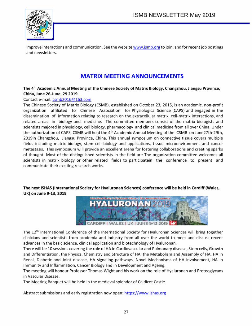
ISMB NEWSLETTER May 2019
27
improve interactions and communication. See the website www.ismb.org to join, and for recent job postings and newsletters.
MATRIX MEETING ANNOUNCEMENTS
The 4th Academic Annual Meeting of the Chinese Society of Matrix Biology, Changzhou, Jiangsu Province, China, June 26-June, 29 2019 Contact e-mail: [email protected] The Chinese Society of Matrix Biology (CSMB), established on October 23, 2015, is an academic, non-profit organization affiliated to Chinese Association for Physiological Science (CAPS) and engaged in the dissemination of information relating to research on the extracellular matrix, cell-matrix interactions, and related areas in biology and medicine. The committee members consist of the matrix biologists and scientists majored in physiology, cell biology, pharmacology and clinical medicine from all over China. Under the authorization of CAPS, CSMB will hold the 4th Academic Annual Meeting of the CSMB on June27th-29th, 2019in Changzhou, Jiangsu Province, China. This annual symposium on connective tissue covers multiple fields including matrix biology, stem cell biology and applications, tissue microenvironment and cancer metastasis. This symposium will provide an excellent arena for fostering collaborations and creating sparks of thought. Most of the distinguished scientists in the field are The organization committee welcomes all scientists in matrix biology or other related fields to participatein the conference to present and communicate their exciting research works.
The next ISHAS (International Society for Hyaluronan Sciences) conference will be held in Cardiff (Wales, UK) on June 9-13, 2019 The 12th International Conference of the International Society for Hyaluronan Sciences will bring together clinicians and scientists from academia and industry from all over the world to meet and discuss recent advances in the basic science, clinical application and biotechnology of Hyaluronan. There will be 10 sessions covering the role of HA in Cardiovascular and Pulmonary disease, Stem cells, Growth and Differentiation, the Physics, Chemistry and Structure of HA, the Metabolism and Assembly of HA, HA in Renal, Diabetic and Joint disease, HA signaling pathways, Novel Mechanisms of HA involvement, HA in Immunity and Inflammation, Cancer Biology and in Development and Ageing. The meeting will honour Professor Thomas Wight and his work on the role of Hyaluronan and Proteoglycans in Vascular Disease. The Meeting Banquet will be held in the medieval splendor of Caldicot Castle. Abstract submissions and early registration now open: https://www.ishas.org

ISMB NEWSLETTER May 2019
28
ASMB 2019 Summer Workshop co-organized by Tom Barker (University of Virginia, USA, ([email protected]) and Merry Lindsey (University of Nebraska Medical Center, USA, [email protected])
ASMB biennial meeting 2019 on Basement Membranes Organizers: - Roy Zent, Vanderbilt (University Medical - Jeffrey Miner (Washington University St. Louis) https://www.asmb.net/basement-membrane-workshop
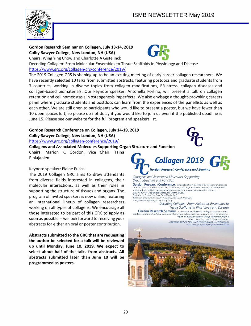
ISMB NEWSLETTER May 2019
29
Gordon Research Seminar on Collagen, July 13-14, 2019 Colby-Sawyer College, New London, NH (USA) Chairs: Wing Ying Chow and Charlotte A Gistelinck Decoding Collagen: From Molecular Ensembles to Tissue Scaffolds in Physiology and Disease https://www.grc.org/collagen-grs-conference/2019/ The 2019 Collagen GRS is shaping up to be an exciting meeting of early career collagen researchers. We have recently selected 10 talks from submitted abstracts, featuring postdocs and graduate students from 7 countries, working in diverse topics from collagen modifications, ER stress, collagen diseases and collagen-based biomaterials. Our keynote speaker, Antonella Forlino, will present a talk on collagen retention and cell homeostasis in osteogenesis imperfecta. We also envisage a thought-provoking careers panel where graduate students and postdocs can learn from the experiences of the panellists as well as each other. We are still open to participants who would like to present a poster, but we have fewer than 10 open spaces left, so please do not delay if you would like to join us even if the published deadline is June 15. Please see our website for the full program and speakers list. Gordon Research Conference on Collagen, July 14-19, 2019 Colby-Sawyer College, New London, NH (USA) https://www.grc.org/collagen-conference/2019/ Collagens and Associated Molecules Supporting Organ Structure and Function Chairs: Marion K. Gordon, Vice Chair: Taina Pihlajaniemi Keynote speaker: Elaine Fuchs The 2019 Collagen GRC aims to draw attendants from diverse fields interested in collagens, their molecular interactions, as well as their roles in supporting the structure of tissues and organs. The program of invited speakers is now online, featuring an international lineup of collagen researchers working on all types of collagens. We encourage all those interested to be part of this GRC to apply as soon as possible -- we look forward to receiving your abstracts for either an oral or poster contribution. Abstracts submitted to the GRC that are requesting the author be selected for a talk will be reviewed up until Monday, June 10, 2019. We expect to select about half of the talks from abstracts. All abstracts submitted later than June 10 will be programmed as posters.
ISMB NEWSLETTER May 2019
30
FASEB Science Research Conference The Matricellular Proteins in Tissue Remodeling and Inflammation July 14 - 19, 2019, Lisbon (Portugal) Conference organizers: - Olga Stenina-Adognravi, Cleveland Clinic (USA) - Joanne Murphy-Ullrich, University of Alabama at Birmingham (USA) Register Now: FASEB Science Research Conference – The Matricellular Proteins in Tissue Remodeling and Inflammation Conference Dear Colleague, Join a community of science professionals to explore emerging research and expand your expertise at The Matricellular Proteins in Tissue Remodeling and Inflammation Conference, taking place July 14-19, 2019, in Lisbon, Portugal. Organized by Chairs Olga Stenina-Adognravi, PhD, Cleveland Clinic, and Joanne Murphy-Ullrich, PhD, University of Alabama at Birmingham, with support from the Federation of American Societies for Experimental Biology (FASEB), this conference is highly interactive and brings together an interdisciplinary group of scientists from academic, clinical, and industry backgrounds, who study and conduct research on matricellular proteins. As a part of FASEB’s Science Research Conference Series, it is designed as a multi-day learning experience to foster engagement between biomedical researchers at every level of their career. Featured conference events include networking opportunities, a career development workshop, selected abstract presentations, and more. Learn more and register by visiting https://src.faseb.org/matricellular-proteins Advance registration ends May 30, 2019. Questions about conference programming, possible travel awards, and other logistical details can be directed to [email protected]. Gordon Research Seminar on Elastin, Elastic Fibers and Microfibrils Elastic Tissues and Regulation of Growth Factor Signaling in Development, Homeostasis and Disease July 20 - 21, 2019 Chairs Anna Tarakanova and Mukti Singh Southern New Hampshire University, NH, USA The Gordon Research Seminar on Elastin, Elastic Fibers and Microfibrils is a dynamic research forum for graduate students, post-docs and early career scientists at the forefront of elastic fiber-
ISMB NEWSLETTER May 2019
31
related research to present their own cutting-edge findings and exchange ideas amongst their peers, as well as established faculty in the field. This meeting encourages young scientists from a broad range of backgrounds and interests to lead discussions on topics including the structure and assembly of elastin, elastic fibers and microfibrils, elastic tissue mechanics and mechanisms of development and aging, as well as matrix regulation of cell signaling. An important focus will also be novel approaches and analytical tools for investigating elastin and associated proteins in health and disease, and translational approaches for tissue engineering and regenerative medicine. This meeting provides an exciting opportunity for early career scientists to engage in a professional development session with their peers and a panel of established scientists focusing on mentorship and career development. Gordon Research Conference “Elastin, Elastic Fibres and Microfibrils” Biochemical, Biomechanical and Biological Functions of Elastin and Related Fibers in Homeostasis and Disease, July 21 - 26, 2019 Chair: Daniel B. Rifkin, Vice Chair: Dianna Milewicz Southern New Hampshire University, NH, USA The Gordon Research Conference (GRC) and the Gordon Research Seminar (GRS) on Elastin, Elastic Fibers and Microfibrils will focus on elastic fiber molecules and the roles that elastic fibers and related fiber systems exert in human disease, vertebrate development, tissue homeostasis and aging. Elastin, elastic fibers and microfibrils are critical components of the skin, bone, vasculature, lung, tendons, eye and other tissues. Elastin fibers are unique because they provide elasticity to many dynamic tissues, i.e., deforms under physiological forces and subsequently releases stored energy to drive passive recoil. This conference will focus on elastic tissue biology and topics include: molecular properties of elastin, fibrillin and other proteins essential for the assembly and function of elastic fibers; elastic tissue matrix regulation of cell signaling; mechanosensing of the elastin-smooth muscle cell complex; aging and stiffening of elastin-containing tissues; technologies for elastic fiber system analysis; genetics and pathobiology of elastic fiber associated molecules in inherited and acquired disorders; tissue regeneration, bioengineering of elastic tissues; and application to translational medicine. The conference will provide new insights into the mechanisms underlying the normal function of elastic fiber-rich tissues and explore new therapeutic avenues for genetic and acquired disorders originating from understanding the detailed functions of the elastic fiber system. The elastic fiber-rich musculoskeletal, cardiovascular, and pulmonary tissues are a major focus. Disorders discussed during the conference that are directly relevant to these systems include but are not limited to aneurysms and dissections, aortic stiffening, aortic calcification, heritable thoracic aortic disease, Marfan syndrome, Marfanoid-progeroid-lipodystrophy, Loeys-Dietz syndrome, supravalvular aortic stenosis, Williams-Beuren syndrome, arterial tortuosity syndrome, chronic obstructive pulmonary disease (COPD), acromicric dysplasia and geleophysic dysplasia, Weill-Marchesani syndrome, Urban-Rifkin-Davis syndrome, cutis laxa, pseudoxanthoma elasticum, glaucoma, and loss of elasticity in skin during normal and UV-induced photo-aging. These acquired and inherited diseases affect millions of Americans and hundreds of millions worldwide.
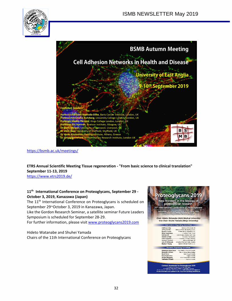
ISMB NEWSLETTER May 2019
32
https://bsmb.ac.uk/meetings/ ETRS Annual Scientific Meeting Tissue regeneration - "From basic science to clinical translation" September 11-13, 2019 https://www.etrs2019.de/ 11th International Conference on Proteoglycans, September 29 - October 3, 2019, Kanazawa (Japan) The 11th International Conference on Proteoglycans is scheduled on September 29~October 3, 2019 in Kanazawa, Japan. Like the Gordon Research Seminar, a satellite seminar Future Leaders Symposium is scheduled for September 28-29. For further information, please visit www.proteoglycans2019.com Hideto Watanabe and Shuhei Yamada Chairs of the 11th International Conference on Proteoglycans

ISMB NEWSLETTER May 2019
33
ASMB 2020 Biennial meeting, November 8-11, 2020, St Louis, MO (USA)