introduction - universiteit utrecht
TRANSCRIPT

Chapter 1
Introduction


Introduction
7
Introduction
The skull is one of the latest inventions of vertebrate evolution. It serves many very vital
functions, like eating and defence. Moreover, it harbours the brain and many sense organs that
are required for perception of the environment, e.g. for sight, smell, hearing and taste. In
primates it has equipped the animal with a unique tool to express emotions. Unfortunately, the
skull appears to be very susceptible to malformations. Many babies are born each year with
craniofacial birth defects due to e.g. inappropriate development of the craniofacial primordia or
due to abnormalities in neural tube development. Recent studies show that mutations in
developmental genes appear to be the underlying cause of many of these birth defects.
In this introduction I will discuss the embryonic origin of most of the skull bones and give an
overview of craniofacial morphogenesis. Moreover, I will discuss the roles of a number of
genes important in craniofacial development. The chapter will conclude with the discussion of
a number of well-known craniofacial defects.
The skull and its embryonic origins
The skull is one of the most complicated parts of the vertebrate body. It has long stimulated
questions as to how it is constructed and how it develops during ontogeny. Anatomically the
skull can be divided into brain case and the facial skeleton. The braincase consists of the
frontal, parietal and supraoccipital bones, which overlie and protect the brain. The skull base
supports the brain and consists of exo- and basioccipital and sphenoid bones and the nasal
capsule (=ethmoid bone) (see Fig. 1.1). The facial skeleton is constituted by the nasal bones,
the premaxillary, maxillary, zygomatic and squamosal bones and the mandible. The nasal and
otic capsules are sensory organs that develop from the otic and nasal placodes, respectively
(see Fig. 1.1).
A subdivision of the skull bones can also be made based upon their embryonic origin (Couly et
al., 1993). The chordal skeleton consists of bones that are derived from cephalic and somitic
mesoderm. The cephalic mesoderm surrounds the primitive brain vesicles and it condensates to
form the supraoccipital bone, part of the otic capsule and the basisphenoid bones. On the other
hand, mesoderm derived from the first five occipital somites forms the basi- and exooccipital
bones and also some structures of the otic capsule (Couly et al., 1993).

Chapter 1
8
The prechordal (or achordal) skeleton consists of bones that are derived from the cranial neural
crest. The neural crest is a population of cells that detaches from the neural folds during neural
tube closure and undergoes an epithelial to mesenchymal transition. The cells migrate away
from the neural tube and populate many regions in the embryo to give rise to various tissues.
The cranial neural crest is derived from fore- mid- and hindbrain regions. It contributes to
formation of many structures like connective tissue, odontoblasts and neurons and glia of
cranial ganglia. Moreover, it forms a significant part of the skull. The basipresphenoid and
more anterior bones comprising the entire facial skeleton are entirely neural crest derived, but
also a part of the otic capsule (Couly et al., 1993).
The embryonic origin of the calvaria remains uncertain. Studies carried out by Couly et al.
(1993) show that the frontal and parietal bones are neural crest derived, whereas other studies
conclude that they are derived from cephalic mesoderm (Goodrich, 1930; LeLievre, 1978;
Jarvik, 1980; de Beer, 1985; Noden, 1986). It should be kept in mind that all these studies were
carried out using chick embryos, which are well accessible to tissue transplantation studies.
However, the avian skull is considerably different from the mammalian skull and many
structures along the neural crest:mesoderm interface are anatomically very different (e.g.
vaults, palatine, sphenoid regions). Therefore, it is not clear if all avian data on the embryonic
origins of certain skull bones can easily be extrapolated to the mammalian situation. A recently
generated two component transgenic mouse line system, in which LacZ is expressed in the
Wnt1 domain may shed more light on this issue (Chai et al., 2000).
basipresphenoid
Fig. 1.1: Anatomy of the mouse skull. Lateral view on the left. Dorsal view on cranial base with vaults and mandible removed on the right.
frontal parietal
interparietal
supraoccipital
exooccipital
otocyst
basioccipital
tympanic ring squamosal basisphenoid
alisphenoid
mandible
maxilla nasal capsule
nasal bone
premaxilla
zygomatic arch
supra- occipital
exo- occipital
basi- occipital
otic capsule
tympanic ring
squamosal
alisphenoid
basisphenoid
maxilla + palatal shelves
zygomatic arch
nasal septum
nasal capsule

Introduction
9
Craniofacial morphogenesis
The face develops on the rostral end of the embryonic axis from the facial primordia that are
arranged around the primitive mouth, the stomodeum. The primordia include the frontonasal
process and the first branchial arch derived paired mandibular and maxillary processes (see Fig.
1.4). The second, third, fourth and sixth arch will contribute to more posterior structures of the
larynx and some thoracic blood vessels. Early during craniofacial development, the frontonasal
process and the branchial arches are populated with paraxial mesoderm and cranial neural crest
cells (reviewed by Noden, 1988; Köntges and Lumsden, 1996; Couly et al., 1996).
The branchial arches
The branchial arches develop as bulges surrounding the pharynx (see Fig. 1.2). Within the
pharynx, they are lined with endoderm and their outer surface is covered with ectoderm.
Branchial pouches and branchial grooves demarcate their borders on the inner and outer
surface of the pharynx, respectively. In between the arches the branchial membranes are
formed at the boundary between endo- and ectoderm. The branchial arches are populated by
paraxial mesoderm and neural crest cells (Couly et al., 1993). The paraxial mesoderm forms
the craniofacial muscles, some skeletal elements and vascular tissues, while cranial neural crest
cells contributes to the peripheral nervous system, connective tissues and cartilage (Le
Douarin, 1982; Noden, 1988; Kimmel et al., 1991; see Fig. 1.2).
Fig. 1.2: Schematic view of pharynx and branchial arches. Every branchial gives rise to a basic set of structures: a cartilage component, a nervous component, a vascular component and a muscular component.
1st arch
4th arch
3rd arch
2nd arch
1st groove
2nd groove
3rd groove Nervous component Vascular component Cartilage component
1st pouch
2nd pouch
3rd pouch
Muscular component
Anterior
Posterior
Lingual swelling
Chapter 1
10
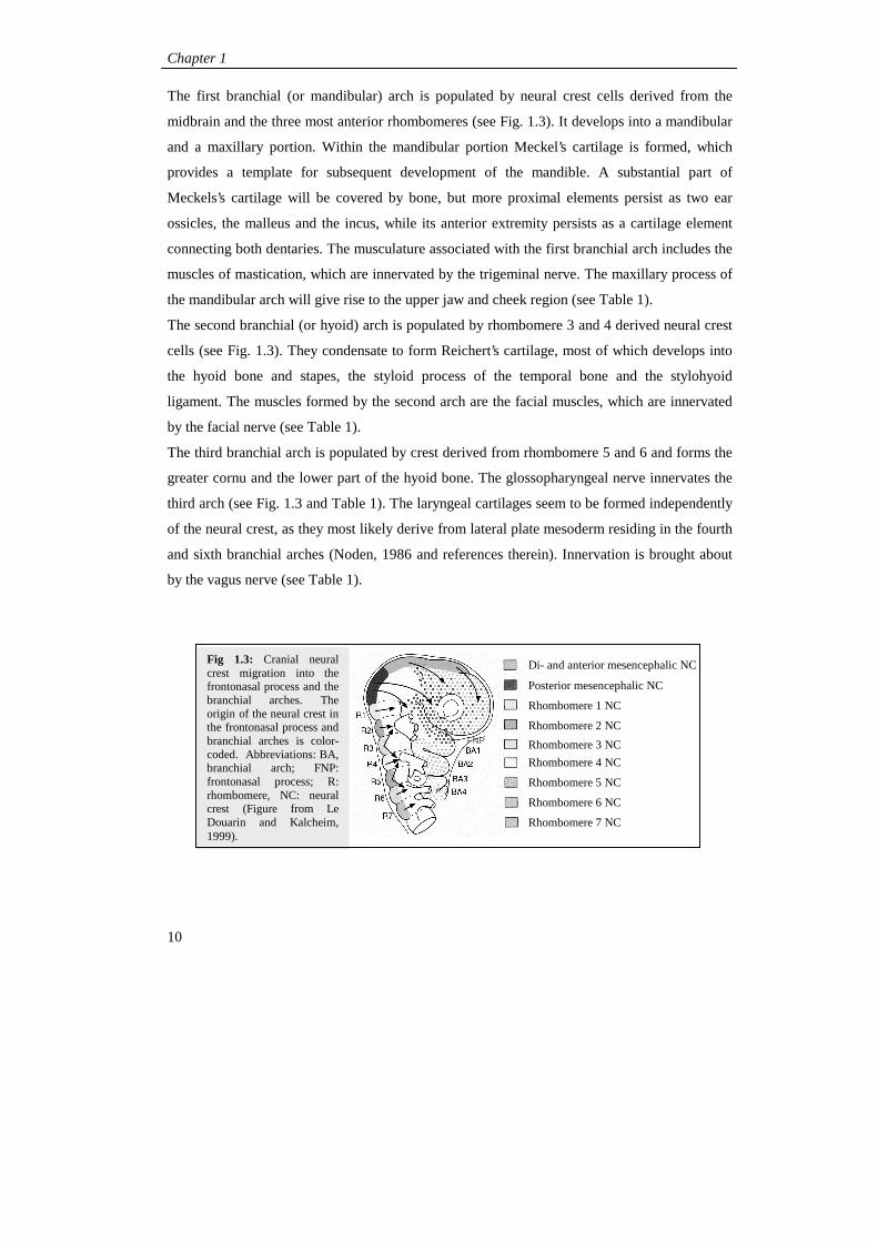
The first branchial (or mandibular) arch is populated by neural crest cells derived from the
midbrain and the three most anterior rhombomeres (see Fig. 1.3). It develops into a mandibular
and a maxillary portion. Within the mandibular portion Meckel’s cartilage is formed, which
provides a template for subsequent development of the mandible. A substantial part of
Meckels’s cartilage will be covered by bone, but more proximal elements persist as two ear
ossicles, the malleus and the incus, while its anterior extremity persists as a cartilage element
connecting both dentaries. The musculature associated with the first branchial arch includes the
muscles of mastication, which are innervated by the trigeminal nerve. The maxillary process of
the mandibular arch will give rise to the upper jaw and cheek region (see Table 1).
The second branchial (or hyoid) arch is populated by rhombomere 3 and 4 derived neural crest
cells (see Fig. 1.3). They condensate to form Reichert’s cartilage, most of which develops into
the hyoid bone and stapes, the styloid process of the temporal bone and the stylohyoid
ligament. The muscles formed by the second arch are the facial muscles, which are innervated
by the facial nerve (see Table 1).
The third branchial arch is populated by crest derived from rhombomere 5 and 6 and forms the
greater cornu and the lower part of the hyoid bone. The glossopharyngeal nerve innervates the
third arch (see Fig. 1.3 and Table 1). The laryngeal cartilages seem to be formed independently
of the neural crest, as they most likely derive from lateral plate mesoderm residing in the fourth
and sixth branchial arches (Noden, 1986 and references therein). Innervation is brought about
by the vagus nerve (see Table 1).
Di- and anterior mesencephalic NC
Posterior mesencephalic NC
Rhombomere 1 NC
Rhombomere 2 NC
Rhombomere 3 NC
Rhombomere 4 NC
Rhombomere 5 NC
Rhombomere 6 NC
Rhombomere 7 NC
Fig 1.3: Cranial neural crest migration into the frontonasal process and the branchial arches. The origin of the neural crest in the frontonasal process and branchial arches is color-coded. Abbreviations: BA, branchial arch; FNP: frontonasal process; R: rhombomere, NC: neural crest (Figure from Le Douarin and Kalcheim, 1999).
FNP

Introduction
11
The frontonasal process
The frontonasal process is populated by neural crest cells derived from fore- and midbrain
regions (Osumi-Yamashita et al., 1994; see Fig. 1.3). They give rise to the trabecular cartilage,
which originally arises as a pair of bar-like cartilages. Later it develops into the premaxillary
bones, the ethmoid (comprising the nasal capsule and the nasal septum) and the presphenoid
bones. Also the frontal and nasal bones develop from the frontonasal process (Rathke, 1839;
reviewed by de Beer, 1931, 1937; see Table 1). There are a few reasons that suggest that the
trabecula may represent a "premandibular" component of the facial skeleton and belong to the
same segment as the ophtalmic nerve. These are its neural crest origin, its topographical
location, its morphology and the metamerical organization of cranial nerves (Huxley, 1874;
reviewed by Goodrich, 1930; de Beer, 1931, 1937; Stadmüller, 1936; Kuratani, 1997).

Chapter 1
12
Mus
cula
ture
Mus
cles
for
mas
ticat
ion
Myl
ohyo
id
Asn
teri
or b
elly
of
the
diga
stri
c T
enso
r ty
mpa
ni
Ten
sor
pala
tini
Mus
cles
for
fac
ial e
xpre
ssio
n Po
ster
ior
belly
of
diga
stri
c St
aped
ius
Styl
ohyo
id
Intr
insi
c m
uscl
es o
f th
e la
rynx
Vas
cula
ture
Max
illar
y ar
teri
es
Stap
edia
l art
erie
s (i
n em
bryo
on
ly)
Com
mon
and
inte
rnal
car
otid
ar
teri
es
Part
of
adul
t arc
h of
aor
ta
Part
of
subc
lavi
an a
rter
y Pa
rt o
f le
ft p
ulm
onar
y ar
tery
D
uctu
s ar
teri
osus
Pa
rt o
f ri
ght p
ulm
onar
y ar
tery
Ner
ve
Oph
thal
mic
bra
nch
of
trig
emin
al n
erve
(V
)
Tri
gem
inal
(V
)
Faci
al
(VII
)
Glo
sso-
Ph
aryn
geal
(I
X)
Vag
us
(X)
Skel
etal
ele
men
ts
Tra
becu
lar
cart
ilage
s (t
rans
ient
) E
thm
oid
(=na
sal c
apsu
le)
N
asal
bon
e
Fron
tal b
one
Lac
rym
al b
one
Bas
ipre
sphe
noid
bon
e Pr
emax
illa
Mec
kels
’ car
tilag
e (t
rans
ient
) M
alle
us
Incu
s M
andi
ble
Max
illa
Zyg
omat
ic a
rch
Pala
tine
Alis
phen
oid
Squa
mos
al
Rei
cher
t’s c
artil
age
(tra
nsie
nt)
Stap
es
Styl
oid
proc
ess
Les
ser
corn
u of
hyo
id
Upp
er p
art o
f bo
dy h
yoid
bon
e
Gre
ater
cor
nu o
f hy
oid
Low
er p
art o
f bo
dy o
f hy
oid
bone
Thy
roid
, C
rico
id,
Ary
then
oid,
C
orni
cula
te a
nd C
unef
orm
car
tilag
es
Tab
le 1
: St
ruct
ures
dev
elop
ing
in t
he b
ranc
hial
arc
hes
Arc
h
(0)
Pre
man
dibu
lar
arch
1 (M
andi
bula
r ar
ch)
2 (H
yoid
arc
h)
3 4+6

Introduction
13
Development of the face
Facial development starts with the emergence of the facial primordia, the branchial arches and
the frontonasal process. They grow out by controlled proliferation of neural crest derived
mesenchymal cells, which are dependent on signals from the overlying ectoderm (Wedden,
1987; Richman and Tickle, 1992). The primordia undergo complex morphogenetic interactions
and ultimately they a complete face.
Development of lower and upper jaw and nose region
The mandibular and maxillary processes of the first arch grow out and develop in a
ventromedial direction (see Fig. 1.4A-D and Table 1). The mandibular processes fuse in the
midline and eventually form the lower jaw. The maxillary processes of the first arch give rise
to the upper jaw and cheek regions (see Fig. 1.4A-D and Table 1). Early during facial
development the surface ectoderm on lateral regions of the frontonasal process thickens and
forms the nasal placodes (see Fig 1.4A). Due to outgrowth of the frontonasal process the
placodes deepen, resulting in formation of the lateral and medial nasal processes (see Fig. 1.4B,
C). The nasal processes fuse inferiorly with each other and with the outgrowing maxillary
processes to form the intermaxillary segment (see Fig. 1.4C). The intermaxillary segment will
form the primary palate, premaxilla and philtrum (see Fig. 1.4C and D, Fig. 1.5A, B, C and
Table 1). The nasolacrimal groove is the cleft, which separates the lateral nasal and maxillary
processes (see Fig. 1.4C and D).
.
Fig. 1.4: Development of the lower and upper jaw and nose region. Frontal views on cranial regions of developing embryos. Abbreviations: 2nd: 2nd branchial arch, 3rd: 3rd branchial arch, FNP: frontonasal process, FP: frontal process, He: heart bulge, IMS, intermaxillary segment; LNP: lateral nasal process, Mn: Mandibular process, MNP: medial nasal process, Mx: maxillary process; NLG: nasolacrymal groove, Npi: nasal pit, NPl: nasal placode; Ph: philtrum, St: stomodeum.
A B C D
FNP NPl
Mx Mx
NPi
Mn Mn 2nd
3rd He
St
St NLG
Eye
FP
MNP
LNP
Mx Eye
Ph
NPi
Mn
IMS

Chapter 1
14
Development of secondary palate and internal nose structures
From the inner walls of the maxillary arches palatal shelves grow out (see Fig. 1.5A). First they
project vertically (Fig. 1.5D), but later they extend upward and fuse in the midline with each
other. Anteriorly they fuse with the primary palate to form the secondary or definitive palate
(see Fig. 1.5C, F). The nasal septum grows downwards from the frontal process and fuses with
the palate (see Fig. 1.5D, E, F). The definitive palate physically separates nasal and oral
cavities and the nasal septum separates both nasal cavities. The nasopalatine canal (incisive
foramen) persists in the palatine midline between the premaxillary portion of the maxilla and
the palatine processes of the maxilla (see Fig. 1.5C). Within the lateral walls of the nasal cavity
the nasal conchae are formed and develop into the nasal labyrinth.
Genes involved in craniofacial development
Whilst a lot is known about origins of skull bones and craniofacial morphogenesis, relatively
little is known about molecular mechanisms regulating craniofacial development. Craniofacial
development is a multi-stage process. It involves the formation and migration of cranial neural
crest cells, followed by the correct outgrowth of the facial primordia and the morphogenesis
Fig. 1.5: Formation of definitive palate. A, B, C: ventral view maxilla. D, E, F: frontal section at the level of the nasal and oral cavities. Abbreviations: DP: definitive palate, FP: frontal process, IF: incisive foramen, Mx: maxilla, NC: nasal cavity, Nco: nasal conchae, NS: nasal septum, OC: oral cavity, Ph: philtrum, PP: primary palate, PS: palatine shelf, T: tongue.
A
PP
NS
PS PS
NC
Eye
PP
IF
DP
NS
T
NS
T T
NS
NC NC
OC
OC
DP
PS PS
NCo
B C
D E F
Ph Ph Ph
Mx
Mx
Mx
FP FP

Introduction
15
and differentiation of the skeletal elements. During these stages, members of many gene
families are expressed and have functions in the (presumptive) neural crest cells and in the
development of the craniofacial primordia (review by Francis-West, 1998).
Genes involved in prepatterning of the cranial neural crest
Before craniofacial development starts, members of different gene families regionalize the
neural tube. They are responsible for the identity of neural crest cells that originate from these
regions. However, prepatterning of the neural tube alone does not bring about specification of
neural crest identity. Environmental signals, such as signals emitted from the branchial arches
or the somites, are also important (Grapin-Botton et al., 1995; Itasaki et al., 1996; Grapin-
Botton et al., 1997; Hunt et al., 1998, Trainor and Krumlauf, 2000).
Hox genes
Hox genes are homeobox genes expressed from caudal neural tube regions up to a specific
anterior border. The ’Hox-code’, which is the combination of Hox genes expressed within a
certain body segment, determines its AP identity. The most anterior Hox genes are expressed
up to hindbrain regions. Their overexpression or loss-of-function mutations cause craniofacial
abnormalities (see Fig. 1.6). Hoxa1 and Hoxb1 are transiently expressed in rhombomeres 4 to
6. Hoxa1 and Hoxb1 mutant mice display abnormalities in second branchial arch derivatives
and the VIIth to XIth cranial nerves (Lufkin et al., 1991; Chisaka et al., 1992; Carpenter et al.,
1993; Dollé et al., 1993; Mark et al., 1993; Goddard et al., 1996; Studer et al., 1996, 1998;
Gavalas et al., 1998). Hoxa2 is expressed in the developing hindbrain. In Hoxa2 mutant mice,
the identity of cranial neural crest cells in the second branchial arch seems to have changed into
a first branchial arch identity (Gendron-Maguire et al., 1993; Rijli et al., 1993). Hoxa3 is
expressed up to the 5th rhombomere and mouse mutants display abnormalities in cartilages and
bones of the jaw (Lufkin et al., 1991; Chisaka and Capecchi, 1991).
Otx1, Otx2, Emx1, Emx2 and Gbx2
The homeobox genes Otx1 and -2 and Emx1 and -2 are homologs of the Drosophila genes
orthodenticle and empty spiracles, respectively. They are expressed in nested patterns in the
fore- and midbrain territories indicative of the existence of a genetic code responsible for AP

Chapter 1
16
patterning of these brain regions (see Fig. 1.6). Indeed, genetic analysis reveals a role for these
genes in patterning these regions and craniofacial primordia.
Otx2 is expressed in fore- and midbrain (see Fig. 1.6, Simeone et al., 1993; Ang et al., 1994).
Inactivation or overexpression of Otx2 causes abnormalities of brain development (Matsuo et
al., 1995; Acampora et al., 1995; Pannese et al., 1995; Ang et al., 1996). Otx2 heterozygous
mutant mice display multiple skeletal defects in the prechordal skull. Homozygous Otx2
mutant embryos die before E10.0 and lack structures corresponding to the rostral head (Matsuo
et al., 1995).
Otx1 is expressed in the more posterior forebrain regions and in the midbrain. Otx1 mutants
display morphological transformation of fore- and midbrain regions into hindbrain (see Fig.
1.6, Acampora et al., 1996). Moreover, Otx1 is capable to take over functions of Otx2 in
patterning of the neural crest (Suda et al., 1999). Emx1 and Emx2 are expressed in overlapping
patterns in the forebrain. Emx1 and Emx2 mutants display abnormalities in the telencephalic
cortex (see Fig. 1.6, Qiu et al., 1996; Pellegrini et al., 1996; Yoshida et al., 1997).
Gbx2 is expressed in regions of the anterior hindbrain with a sharp boundary at the mid-
/hindbrain border, marking the isthmic organizer (see Fig. 1.6). Gbx2 mutant mice display
abnormal development of the anterior hindbrain and genetic markers of the isthmic organiser
Gbx-2 Hox-a2 Hox-b1 Hox-a3 Hox-a1
BA1
BA2 BA3
BA4-BA5
Fig. 1.6: Genes that are involved in regionalization of the fore-, hind- and midbrain regions and determination of neural crest identity.
R1 R2 R3 R4 R5 R6 R7 R8
Otx2
Emx2 Otx1
Emx1

Introduction
17
region have shifted. These genetic studies suggest a role for Gbx2 in positioning the mid-
/hindbrain boundary (Wassarman et al., 1997; Millet et al., 1999; Simeone, 2000).
Genes involved in neural crest migration
Neural crest cells migrate away in streams from the neural tube to reach their destination in the
facial primordia and branchial arches (see Fig. 1.2). The process of migration is regulated by a
several genes, two of which will be discussed below.
AP-2
The transcription factor AP-2 is a retinoic acid responsive gene that is first expressed during
neural crest determination, migration and in the facial primordia (Mitchell et al., 1991; Shen et
al., 1997). In AP-2 mutant mice, the cranial neural tube fails to close., Moreover, abnormalities
are found in the prechordal craniofacial skeleton (Schorle et al., 1996; Zhang et al., 1996).
TUNEL analysis revealed that neuroepithelial cells and late migrating cranial neural crest cells
are apoptotic (Schorle et al., 1996). Apparently AP-2 is involved with survival of premigratory
and migratory cranial neural crest cells.
PDGFDR
Platelet derived growth factors (PDGFs) have been shown to regulate cell growth and survival,
but also cell morphology and movement (for review see Cleasson-Welsh, 1994; Kazlauskas,
1994). Two PDGF receptors exist, PDGFαR and PDGFβR (Seifert et al., 1989). Mice with
mutations in PDGFαR, among which the Patch mouse (Ph), show a number of defects
comprising cleft faces and spina bifida (Grüneberg and Truslove, 1960; Soriano, 1997). The
cleft face phenotype has been associated with a defect in the migration and with apoptosis of
migrating neural crest cells (Morrison-Graham et al., 1992; Soriano, 1997).
Genes involved in patterning of the craniofacial primordia
Members of many gene families are expressed in the developing craniofacial primordia, both in
ectoderm and mesenchyme. Their outgrowth is dependent on proliferation of mesenchymal
cells, which is tightly controlled by signals from the overlying epithelium and branchial pouch
endoderm (Wedden, 1987; Richman and Tickle, 1992).

Chapter 1
18
Fibroblast growth factor family (Fgf)
At least 15 members of the Fgf family and the Fgf receptors-1, -2 and -3 are expressed in the
developing craniofacial primordia. The Fgf ligands are expressed in the epithelium, while their
receptors are expressed by the underlying mesenchyme (Drucker and Goldfarb, 1993; Wall and
Hogan, 1995; Hartung et al., 1997; Bachler and Neubüser, 2001). Fgf2 and Fgf4 coated beads
were shown to support outgrowth of the frontonasal process and mandibular arch mesenchyme
in chick embryos (Richman et al., 1997). Fgf8 is expressed by the epithelium of the nasal pits
and the first branchial arches. Hypomorphic Fgf8 mutant and conditional Fgf8 mutant mouse
embryos have craniofacial and branchial arch abnormalities (Meyers et al., 1998; Trumpp et
al., 1999). In humans several craniofacial syndromes are known to be due to mutations in FGF
receptors, such as many forms of craniosynostosis (reviewed by Muenke and Schell, 1995;
Wilkie et al., 1995; Wilkie et al., 1997).
TGF-E superfamily
TGF-β molecules, bone morphogenetic proteins (Bmps) and activins belong to the superfamily
of transforming growth factor β (TGF-β)-signalling proteins. Many members and their
receptors are expressed by the craniofacial primordia (Pelton et al., 1989, Pelton et al., 1991;
Millan et al., 1991). Mutations in some TGF-β isoforms result in craniofacial abnormalities in
upper and lower jaws and palatal shelves (Sanford et al., 1997; Kaartinen et al., 1995; Proetzel
et al., 1995). Bmp2 and -4 null mutants die before onset of craniofacial development.
Haploinsufficient Bmp4 mutants are viable and have abnormalities in frontal and nasal bones
(Winnier et al., 1995; Zhang and Bradley, 1996; Dunn et al., 1997). Bmp7 mutants have
abnormalities in cranial bones and in the skull base (Dudley et al., 1995; Luo et al., 1995). The
short ear mutant was shown to carry mutations in the Bmp5 gene (Kingsley et al., 1992).
Sonic hedgehog
Shh is a signaling protein that is related to the Drosophila gene hedgehog (hh). Mouse Shh is
expressed in the craniofacial primordia. It is expressed by the ectoderm and endoderm of the
first branchial arch and in the ectoderm of the nasal processes. Shh mutants have severe
craniofacial defects. Most defects are secondary to the defective splitting of the eye field, but
some are directly caused by mutations in Shh. In Shh mutants the first branchial arch
degenerates after E9.5, resulting in malformation or absence of most first branchial arch

Introduction
19
derivatives at birth (Chiang et al., 1996). In chick, overexpression and inhibition of Shh
signalling also results in severe craniofacial abnormalities (Hu and Helms, 1999). Evidence
suggests that Shh plays a role in regulation of cranial neural crest cell survival (Ahlgren et al.,
1999). In humans, mutations in SHH are implicated in the etiology of holoprosencephaly
(Belloni et al., 1996; Roessler et al., 1996).
Retinoic acid receptor-D and -
Several types of retinoic acid receptors (RARs) have been identified. RAR-α, -β and - �DQG�WKH�
retinoid X receptors, RXR-α, -β� DQG� �� RAR-α and RAR- are strongly expressed by the
frontonasal process and branchial arches during and following neural crest migration (Ruberte
et al., 1990). In RAR-α/RAR- double mutant mouse skulls, all the structures derived from the
frontonasal process were partially or completely absent (Lohnes et al., 1994; Mendelsohn et al.,
1994). The frontal, nasal, premaxillary, ethmoid and presphenoid bones were largely missing.
In contrast, the derivatives of the mandibular process of the first arch were present and only
little affected. These abnormalities probably result from increased cell death in the frontonasal
mesenchyme at E10.5. Therefore retinoic acid may be required for survival of post-migratory
neural crest cells within the frontonasal process (Lohnes et al., 1994).
Gli genes
Members of the Gli family are zinc finger containing proteins that show significant sequence
similarity to the product of the Drosophila segment polarity gene cubitus interuptus (ci)
(Ruppert et al., 1988; Orenic et al., 1990; Hui et al., 1994). They are mediators of Shh
signaling (Marigo et al., 1996; Lee et al., 1997). In both human and mouse, three closely
related Gli genes exist, Gli1-3. During craniofacial development the Gli genes are expressed in
the neural crest derived mesenchyme of the frontonasal process and branchial arches. Gli2 and
Gli3 are also expressed in the migrating neural crest (Walterhouse et al., 1993; Hui et al.,
1994). Mutation of Gli2 results in truncation of the distal part of the maxilla and mandible.
Moreover, loss of the presphenoid, maxillary bone and palatine shelves causes clefting in the
skull of these mice. In contrast, mutations in Gli3 result in an enlargement of the maxillary
region. Furthermore, the nasal processes are smaller and some clefting occurs. In Gli2/Gli3
double mutants the abnormalities in the craniofacial region are enhanced (Mo et al., 1997).
Mutation in Gli3 results in, amongst others, craniofacial abnormalities in the human syndrome

Chapter 1
20
Greig’s cephalopolysyndactyly (GCPS) and the extra toes (Xt) mouse (Johnson, 1967; Gollop
and Fontes, 1985; Vortkamp et al., 1991, 1992; Schimmang et al., 1992; Hui and Joyner,
1993).
Msx genes
The homeobox containing Msx genes are expressed in migratory and cranial neural crest
derived mesenchyme. They have important functions in epithelio-mesenchymal interactions
(Hill et al., 1991; Robert et al., 1989; Mackenzie et al., 1991a,b; Suzuki et al., 1991; Mina et
al., 1995). In Msx1 mutant mice all facial structures fail to develop normally and they lack
teeth (Satokata and Maas, 1994). Human syndromes exist that are associated with mutations in
Msx1 and Msx2. MSX1 haploinsufficiency results in loss of premolar and molar teeth, but the
facial skeleton develops normally (Vastardis et al., 1996). A mutation in one copy of human
MSX2, causes craniosynostosis (Liu et al., 1996; Ma et al., 1996).
Dlx genes
Dlx genes are homeobox genes that are related to the Drosophila gene Distaless (Dll). In
mouse 6 Dll-related genes exist. They are expressed in migrating cranial neural crest and in the
cranial neural crest derived mesenchyme or ectoderm overlying the nasal pits (Qiu et al, 1995;
Qiu et al., 1997; Acampora et al., 1999; Depew et al, 1999; Thomas et al., 2000). Dlx1, -2 and -
5 mutant mice have severe craniofacial abnormalities. Dlx2 mutants have abnormalities in
proximal first and second arch derived structures, whereas Dlx1 mutants only have
abnormalities in first arch derivatives (Qiu et al., 1995; Qiu et al., 1997). Dlx1/Dlx2 double
mutants have similar abnormalities in both first and second arch derivatives as found in the
single mutants. In addition, they lack the upper molars. Dlx5 mutants have abnormalities in
ears, noses, lower jaw and calvaria (Depew et al., 1999; Acampora et al., 1999).
aristaless-related genes
aristaless (al) is a Drosophila gene that is expressed in central regions of the leg, eye and wing
imaginal discs that grow out and become the most distal tips of the appendages (Schneitz et al.,
1993). It controls growth and differentiation of the tip of the leg (Campbell and Tomlinson,
1998). In vertebrates, a large group of genes related to aristaless exists. They can be
categorized in three classes of which class I genes are predominantly expressed in

Introduction
21
mesenchymal structures of limb and craniofacial regions as described in Chapter 2. Among
them are Alx4, the gene that is defective in the Strong’s Luxoid mutant, Prx1, Cart1, Prx2,
Prx3 and Alx3 (Meijlink et al, 1999).
Alx3, Alx4 and Cart1 are expressed by the frontonasal process and nasal processes, distally in
the branchial arches and anteriorly in the limbs as described in Chapter 2. Alx4 and Cart1
single and double mutant mice were generated in other labs. In Chapter 3 the generation of the
Alx3 single and Alx3/Alx4 double mutants is described. Alx3 mutant mice do not have an
obvious phenotype. In contrast, Alx4 mutants have preaxial polydactyly and mild craniofacial
abnormalities. The human syndrome symmetric parietal foramina (PFM) is also caused by
mutations in the ALX4 gene (Mavrogiannis et al., 2001; Wuyts et al., 2000). Cart1 mutant
mice lack all skull vaults and have abnormalities in their skull base and facial skeleton (Zhao et
al., 1995; Qu et al., 1997; Chapter 4 of this thesis). Alx4/Cart1 and Alx3/Alx4 double mutants
have cleft nose regions and limb abnormalities (Qu et al., 1999; Beverdam et al., in press).
These data, together with the results presented in Chapter 4 suggest that these three genes have
overlapping functions in patterning the nasal processes and distal regions of the mandibular
arch.
Prx1 and Prx2 are also expressed in overlapping patterns by the frontonasal process, nasal
processes and in the branchial arches as described in Chapter 2. Prx1 mutant mice were
generated in E.N. Olson’s lab and Prx2 single and Prx1/Prx2 double mutant mice were
generated both in our lab and by Lu and colleagues (ten Berge et al., 1998; Lu et al., 1999).
Prx2 mutant mice do not have abnormalities. In contrast, Prx1 mutants display complex
craniofacial phenotype including malformation of bones of the facial skeleton, the skull base,
the otic capsule and second branchial arch derived structures (Martin et al., 1995). In
Prx1/Prx2 double mutants, the Prx1 phenotype is enhanced suggesting overlapping roles for
both genes in patterning of the mandibular and hyoid arch structures (ten Berge et al., 1998; Lu
et al., 1999).
Prx3 is expressed in foetal and adult brain, initially in broad areas that develop in the dorsal
thalamus, pretectum and tectum. In the adult, the gene is most prominently expressed in nuclei
that are part of the subcortical visual system (Van Schaick et al., 1997). In addition, it is

Chapter 1
22
expressed by the craniofacial primordia and proximally in the limb buds (Chapter 2 of this
thesis). Human Prx3 is a candidate gene for the Cornelia de Lange syndrome (De Lange, 1933;
Blaschke et al., 1998; Semina et al., 1998). This syndrome is characterized by a combination of
mental retardation, craniofacial features, eye defects and limb defects.
SHOX is highly related to Prx3. The gene gives rise to at least two splice variants, SHOXA and
SHOXB. Both are expressed in skeletal muscle and bone marrow fibroblasts. SHOXA is also
expressed in placenta, heart and pancreas and SHOXB in the foetal kidney (Rao et al., 1997).
SHOX is a pseudoautosomal gene that has been linked to idiopathic short stature and Turner
syndrome (Rao et al., 1997; Shanske et al., 1999; Clement-Jones et al., 2000; Blaschke and
Rappold, 2000, 2001). In addition, Prx3 is deleted in the similar Leri-Weill dysochondrosteosis
syndrome (Belin et al., 1998; Shears et al., 1998; Blaschke and Rappold, 2000, 2001; Huber et
al., 2001).
Craniofacial birth defects
Many babies are born each year with birth defects. The parents of one out of every 28 babies
receive the frightening news that their baby has a birth defect. A birth defect is an abnormality
of structure, function or body metabolism (inborn error of body chemistry) present at birth that
results in physical or mental disability, or is fatal. A number of well known craniofacial
disorders will be discussed below.
Orofacial clefting
Orofacial clefting is among the most frequently occurring cranial defect. Cleft lip with or
without cleft palate occurs in 1 out of 350 births and cleft palate alone occurs less frequently.
Cleft lip may be unilateral or bilateral. Unilateral cleft lip results from failure of one maxillary
prominence to merge with the nasal processes, while bilateral cleft lips are due to the failure of
both maxillary processes to fuse (see Fig. 1.7). Cleft palate, with or without cleft lip, is caused
by a failure of the palatal shelves to meet and fuse with each other, with the nasal septum,
and/or with the posterior margin of the primary palate (see Fig. 1.7). The causes of orofacial
clefting are not fully understood. Studies suggest that a number of genes, as well as
environmental factors, such as drugs (including antiseizure drugs), infections, maternal
illnesses, maternal alcohol use and, possibly, deficiency of B vitamin folic acid may be
involved (Thorogood, 1997).

Introduction
23
Neural tube defects
Neural tube defects (NTDs) occur in 1 of 1000 life births (Edmonds and James, 1990). Spina
bifida, meningocele and meningomyelocele are caused by incomplete closure of the neural
tube at spinal cord levels, whereas anencephaly and encephalocele are caused by neural tube
closure defects at the more anterior brain levels (Campbell et al., 1986; Copp et al., 1990). The
causes of the neural tube closure defects are in most cases unknown, but results from many
studies show that there is a link between neural tube closure defects and decreased folate
levels. Therefore treatment of pregnant mothers with folic acid at about 29 days of pregnancy
strongly reduces the risk on neural tube closure defects (Smithells et al., 1989). Cart1 mutant
mice have acrania and meroanencephaly at birth, but prenatal treatment with folic acid strongly
reduces the incidence of neural tube closure defects (Zhao et al., 1996).
Craniosynostosis
Craniosynostosis is a birth defect that occurs in 1 out of 1600 and often occurs as a part of
other malformation syndromes of which the best known are Apert, Pfeiffer, Saethre Chotzen
and Crouzon syndromes. It is characterized by premature closing of one or more cranial sutures
and it leads to malformation of the cranial cavity and other craniofacial features (Thorogood,
1997). Dominant missense mutations in the gene encoding fibroblast growth factor receptors
(FGFRs) 1-3 cause the development of many craniosynostosis syndromes (Muenke and Schell,
1995; Wilkie et al., 1995; Naski and Ornitz, 1998). Recently a mouse mutant was generated
A B
Fig. 1.7: Cleft lip and cleft palate. Frontal views on developing embryos. Black lines in A indicate positions where failure of fusion results in uni- or bilateral cleft lip. Black line in B indicates position where failure of fusion results in cleft palate.

Chapter 1
24
with gain-of-function mutations in the Fgfr2 gene. This mouse has a phenotype with strong
parallels to some Apert’s and Pfeiffer’s syndrome patients (Hajihosseini et al., 2001).
Holoprosencephaly
Holoprosencephaly (HPE) is a disorder with an incidence of 1 of 16.000 life births and 1 of
250 induced abortions that results from a failure of the embryonic forebrain to separate into
cerebral and lateral hemispheres, coupled with an abnormal development of the frontonasal
process (Cohen, 1989a, 1989b, 1992). A progressive range of craniofacial malformations can
be recognized. A mild phenotype involves absence of the intermaxillary process resulting in
midfacial clefting. Further deficiency, coupled with a narrower midbrain and a suspected
failure of midline definition of the bilateral olfactory placodes produces faces with reduced
nasal structures, sometimes manifest as a small nose with a single nostril (cebocephaly). Even
further reduction results in complete absence of nose and olfactory structures, with eyes
developing much closer to the midline (hypotelorism). In most severe cases the defects results
in a single eye present in the midline (cyclopia; Thorogood, 1997). Mutations in SHH were
shown to cause HPE, making it an important candidate gene for the etiology of the syndrome
(Belloni et al., 1996; Roessler et al., 1996).
Treacher-Collins syndrome
Treacher-Collins (TCS) is a rare autosomal dominant disorder that occurs in 1 of 50.000 live
births (Gorlin et al., 1990). It affects the entire face: the lower jaw is underdeveloped
(microagnathia), the palatine is cleft and the external ears are malformed, which often goes
together with conductive deafness due to absent or dysmorphic ossicles (Thorogood, 1997).
Mutations in the TCOF1 gene, mostly leading to premature stop codons, cause development of
TCS (Dixon et al., 1997; Wise et al., 1997). Tcof1 haploinsufficiency in mice causes massive
increase in the levels of apoptosis in the prefusion neural folds, which likely underlies the
development of the craniofacial malformations (Dixon et al., 2000).
DiGeorge syndrome
DiGeorge syndrome (DGS) occurs with an estimated frequency of 1 in 4000 live births
(Emanuel et al., 1999). Patients have the craniofacial defects as described for Treacher-Collins
syndrome, but additionally both the thymus and parathyroids are either absent or reduced and

Introduction
25
there are cardiovascular anomalies including persistent truncus arteriosus and dysmorphic
aortic vessels. The parathyroid and thymic anomalies are likely consequences of phenotypes in
third and fourth branchial pouches associated with defects in neural crest development and arch
morphogenesis. The cardiovascular problems are thought to result from disruption of the
cardiac neural crest, which emerges from the posterior hindbrain (Thorogood, 1997). DGS is
usually associated with deletions of chromosome 22q11 (Scambler, 2000). Recent studies
implicate the transcription factor TBX1 as a key candidate gene for the malformations seen in
DGS (Jerome and Papaioannou, 2001; Lindsay et al., 2001). However, other genes within a
closely linked region of 22q11, like CRKL, might affect the same developmental pathway
(Guris et al., 2001).
Aim of the Ph. D. project and this thesis
At the start of this project our group had just begun to recognize that the aristaless-related
genes have similar functions during embryonic development. We had cloned mouse Prx1 and
Prx2, studied their expression patterns during development and suggested the current
nomenclature (Leussink et al., 1995). The phenotype of the Prx1 mutant mouse had also been
published (Martin et al., 1995). Derk ten Berge had generated the Prx2 mutant mouse and
analysed the mouse Alx3 gene during his Ph. D. project (ten Berge et al., 1998a,b). In the Alx3
expression paper we report the existence of a group of aristaless-related genes. The same year
the phenotype of the Cart1 mutant mice was published. The description of the Alx4 mutant
mouse phenotype followed soon after (Zhao et al., 1996; Qu et al., 1997).
The aim of my project was to generate Alx3 mutant mice and to study its function and the
functions of the highly related Alx4 and Cart1 genes during craniofacial development. In
Chapter 2 the expression patterns of Alx3, Alx4, Cart1 and Prx1-3 in the craniofacial primordia
and the outgrowing limbs are described. I suggest that a further categorization of group I
aristaless-related genes in three subgroups could be made based upon protein structure,
expression patterns and functional data. In Chapter 3, the generation of the Alx3 mutant mouse,
the analysis of the Alx3/Alx4 double mutant embryos, the etiology of the phenotype and a
probable cellular mechanism leading to the phenotype are described. Chapter 4 describes the
skeletal phenotype of Alx3/Cart1 double and Alx3/Alx4/Cart1 triple mutant mice.

26