introduction main organ eye accessory organs eyelids lacrimal apparatus extrinsic muscles
TRANSCRIPT
SENSE OF VISION
Introduction
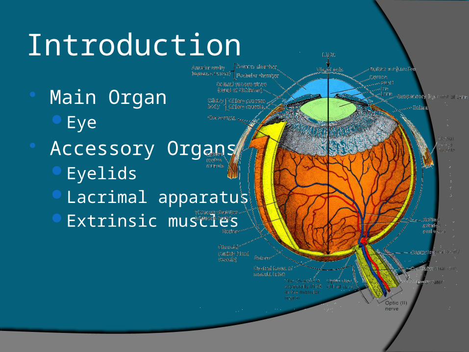
Main OrganEye
Accessory OrgansEyelidsLacrimal apparatusExtrinsic muscles
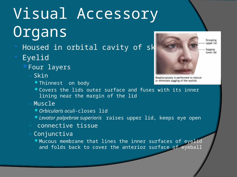
Visual Accessory Organs Housed in orbital cavity of skull Eyelid
Four layers○ Skin
Thinnest on body Covers the lids outer surface and fuses with its inner lining near
the margin of the lid
○ Muscle Orbicularis oculi-closes lid Levator palpebrae superioris raises upper lid, keeps eye open
○ connective tissue○ Conjunctiva
Mucous membrane that lines the inner surfaces of eyelid and folds back to cover the anterior surface of eyeball
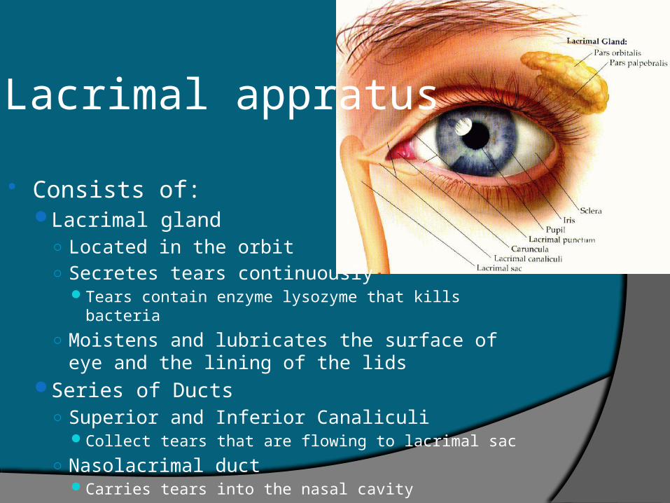
Lacrimal appratus
Consists of:Lacrimal gland
○ Located in the orbit○ Secretes tears continuously
Tears contain enzyme lysozyme that kills bacteria
○ Moistens and lubricates the surface of eye and the lining of the lids
Series of Ducts○ Superior and Inferior Canaliculi
Collect tears that are flowing to lacrimal sac
○ Nasolacrimal ductCarries tears into the nasal cavity
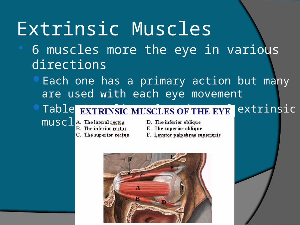
Extrinsic Muscles 6 muscles more the eye in various directions
Each one has a primary action but many are used with each eye movement
Table 10.2 lists functions of extrinsic muscles
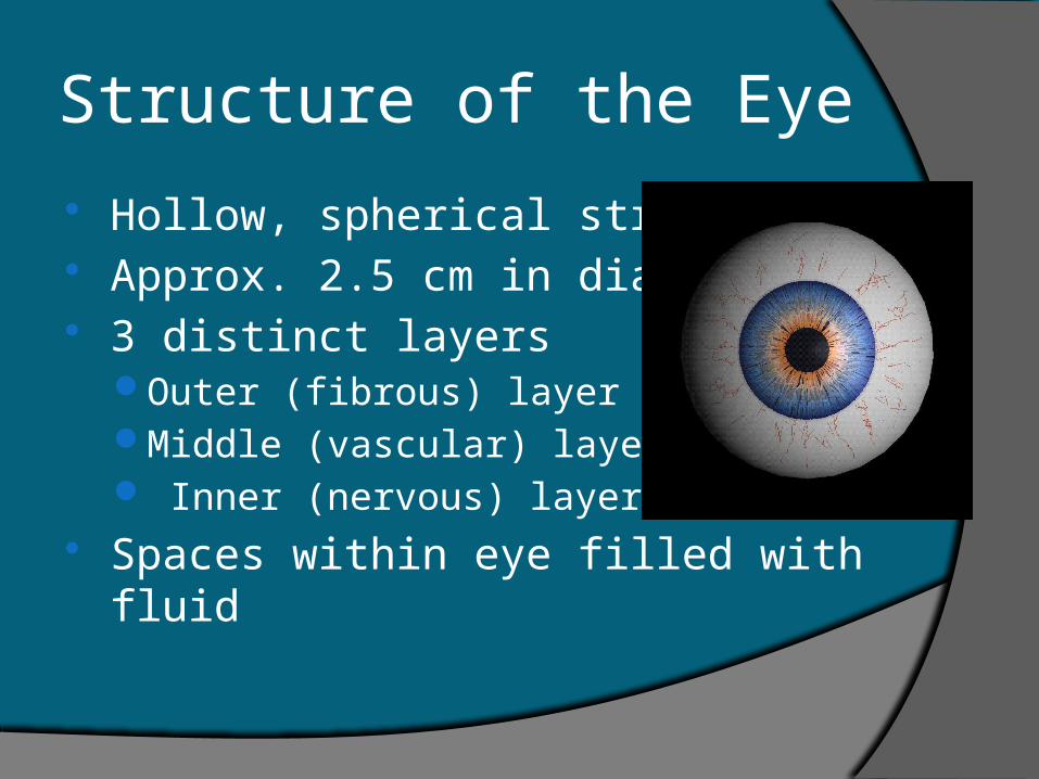
Structure of the Eye
Hollow, spherical structure Approx. 2.5 cm in diameter 3 distinct layers
Outer (fibrous) layerMiddle (vascular) layer Inner (nervous) layer
Spaces within eye filled with fluid
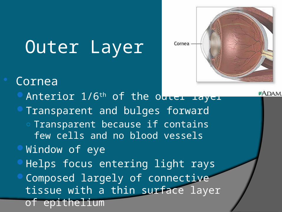
Outer Layer
CorneaAnterior 1/6th of the outer layerTransparent and bulges forward
○ Transparent because if contains few cells and no blood vessels
Window of eyeHelps focus entering light raysComposed largely of connective tissue with
a thin surface layer of epithelium
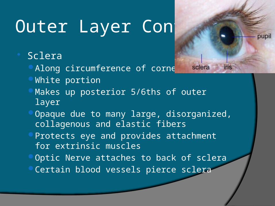
Outer Layer Cont.
ScleraAlong circumference of corneaWhite portionMakes up posterior 5/6ths of outer layerOpaque due to many large, disorganized,
collagenous and elastic fibersProtects eye and provides attachment for
extrinsic musclesOptic Nerve attaches to back of scleraCertain blood vessels pierce sclera
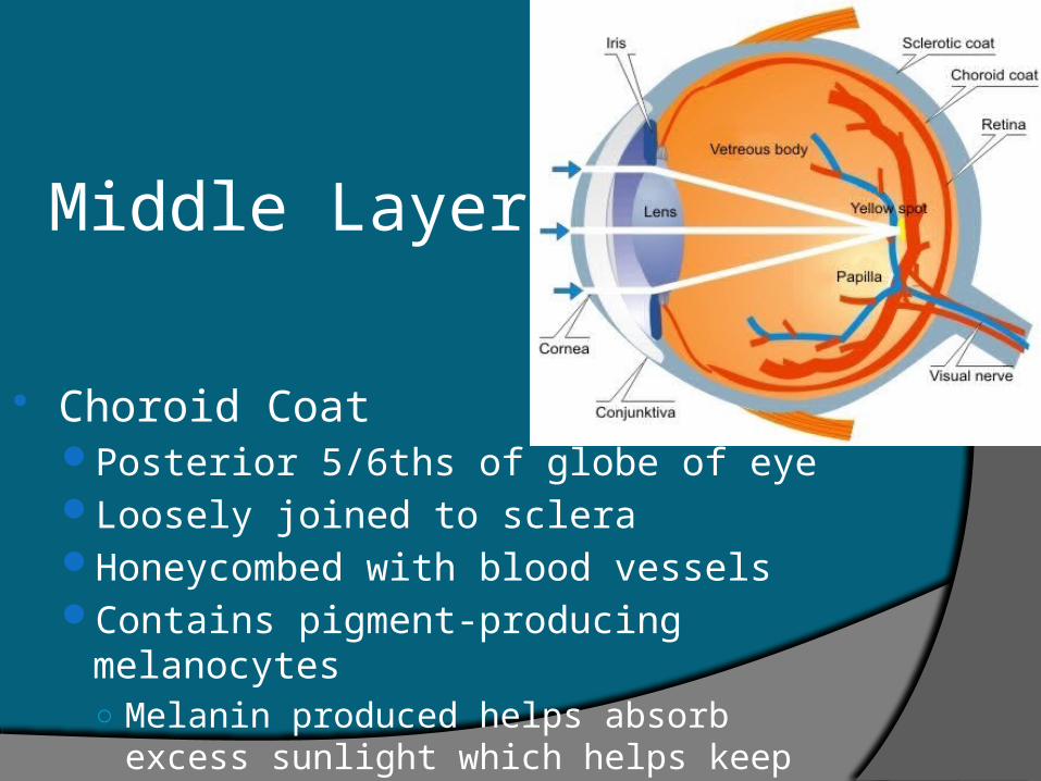
Middle Layer
Choroid CoatPosterior 5/6ths of globe of eyeLoosely joined to scleraHoneycombed with blood vesselsContains pigment-producing melanocytes
○ Melanin produced helps absorb excess sunlight which helps keep inside of eye dark
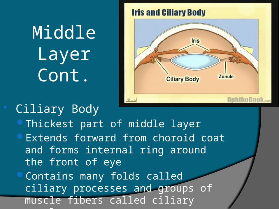
Middle Layer Cont.
Ciliary BodyThickest part of middle layerExtends forward from choroid coat and
forms internal ring around the front of eyeContains many folds called ciliary processes
and groups of muscle fibers called ciliary muscles
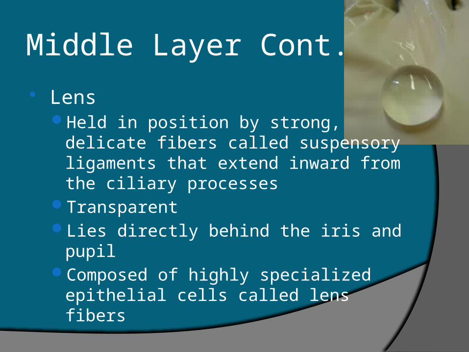
Middle Layer Cont.
LensHeld in position by strong, delicate fibers
called suspensory ligaments that extend inward from the ciliary processes
TransparentLies directly behind the iris and pupilComposed of highly specialized epithelial
cells called lens fibers
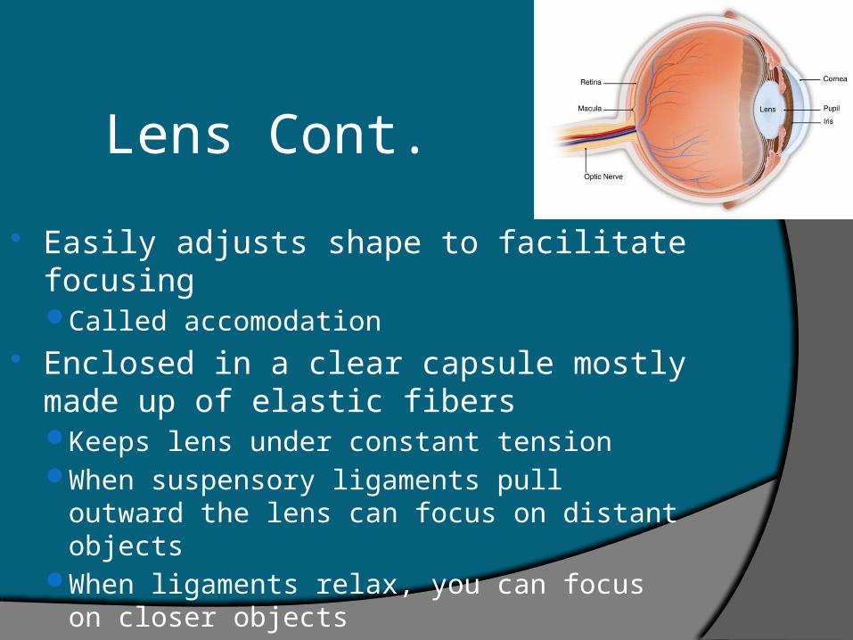
Lens Cont.
Easily adjusts shape to facilitate focusingCalled accomodation
Enclosed in a clear capsule mostly made up of elastic fibersKeeps lens under constant tensionWhen suspensory ligaments pull outward
the lens can focus on distant objectsWhen ligaments relax, you can focus on
closer objects
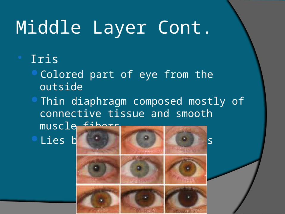
Middle Layer Cont.
IrisColored part of eye from the outsideThin diaphragm composed mostly of
connective tissue and smooth muscle fibersLies between cornea and lens
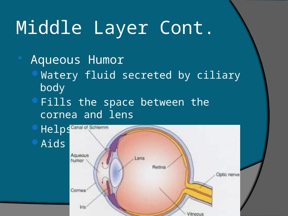
Middle Layer Cont.
Aqueous HumorWatery fluid secreted by ciliary bodyFills the space between the cornea and lensHelps nourish cornea and lensAids in maintaining eye shape
Middle Layer Cont.
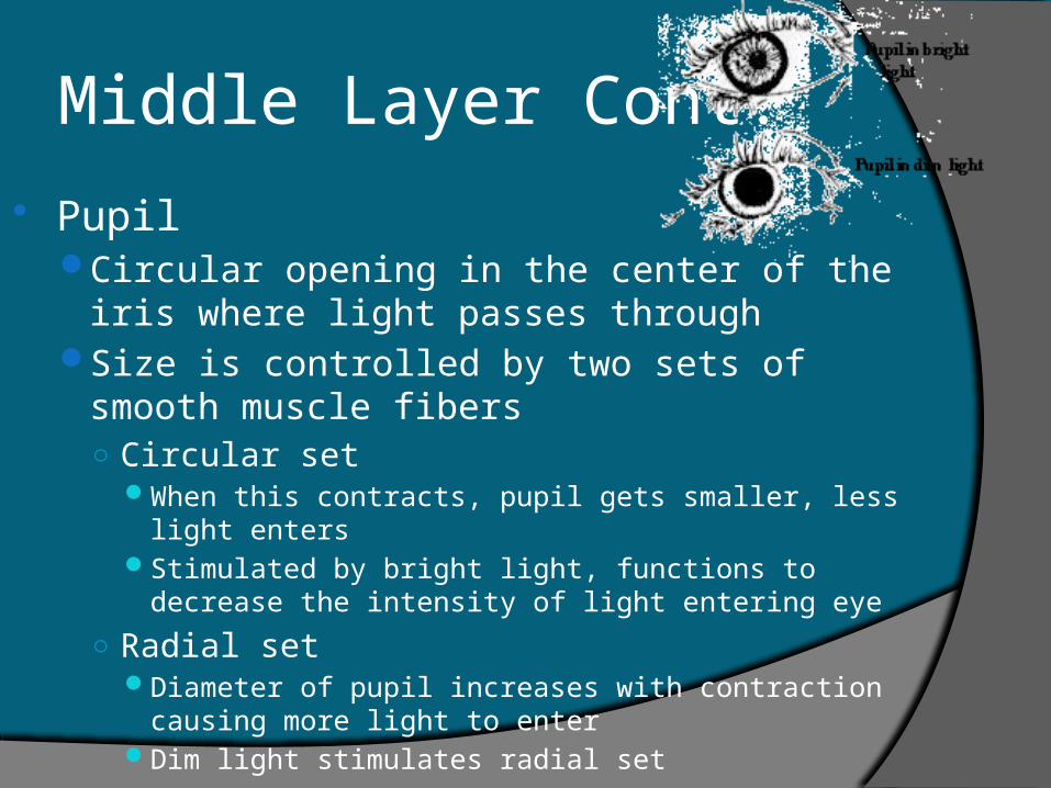
PupilCircular opening in the center of the iris where
light passes throughSize is controlled by two sets of smooth muscle
fibers○ Circular set
When this contracts, pupil gets smaller, less light entersStimulated by bright light, functions to decrease the
intensity of light entering eye
○ Radial setDiameter of pupil increases with contraction causing
more light to enterDim light stimulates radial set
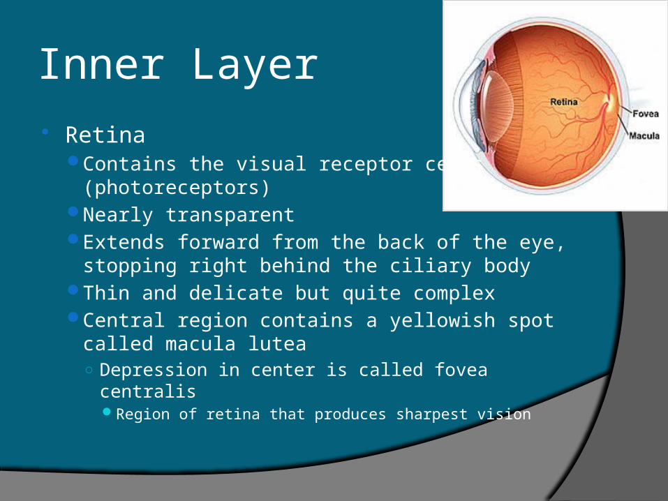
Inner Layer
RetinaContains the visual receptor cells
(photoreceptors)Nearly transparentExtends forward from the back of the eye,
stopping right behind the ciliary bodyThin and delicate but quite complexCentral region contains a yellowish spot called
macula lutea○ Depression in center is called fovea centralis
Region of retina that produces sharpest vision
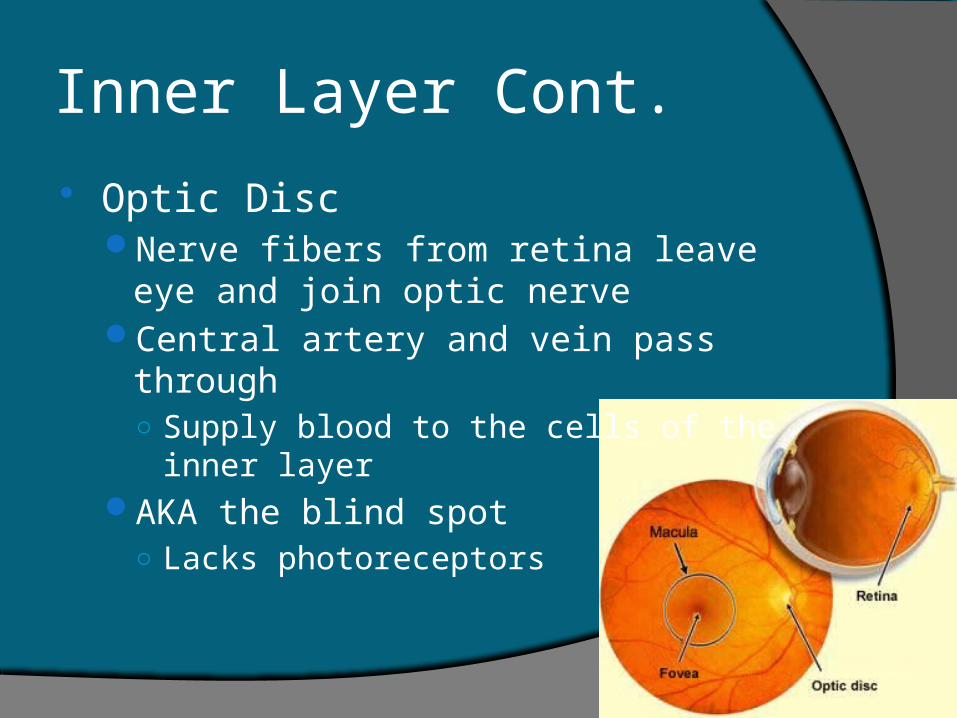
Inner Layer Cont.
Optic DiscNerve fibers from retina leave eye and join
optic nerveCentral artery and vein pass through
○ Supply blood to the cells of the inner layerAKA the blind spot
○ Lacks photoreceptors
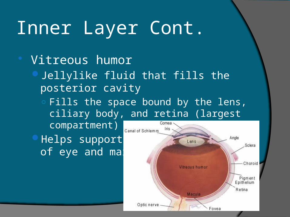
Inner Layer Cont.
Vitreous humorJellylike fluid that fills the posterior cavity
○ Fills the space bound by the lens, ciliary body, and retina (largest compartment)
Helps support the internal parts of eye and maintain shape
Light Refraction As light waves enter the eye the image is
focused on the retina Focusing bends the light waves
Refraction As light enters, the convex surface of lens
refracts the image and then passes it on to the retina where it gets refracted again.Works much like a movie screen in the back of the
eye with the exception that the image that forms on the retina is upside down and reversed from left to right. Visual cortex must interpret the image in proper position.
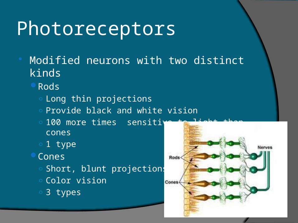
Photoreceptors
Modified neurons with two distinct kindsRods
○ Long thin projections○ Provide black and white vision○ 100 more times sensitive to light than cones○ 1 type
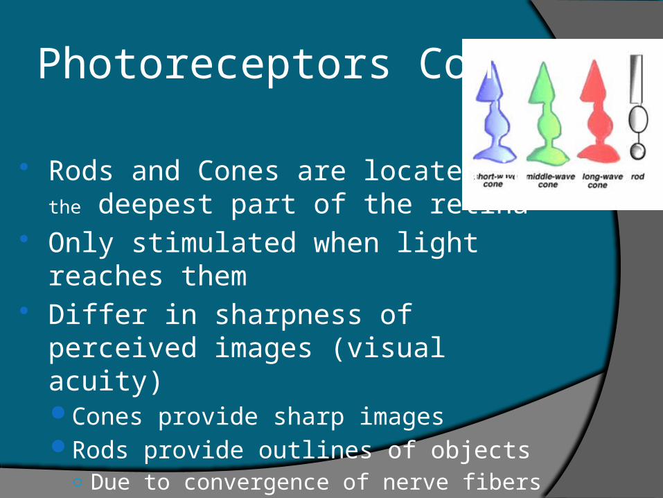
Cones○ Short, blunt projections○ Color vision○ 3 types
Photoreceptors Cont.
Rods and Cones are located in the deepest part of the retina
Only stimulated when light reaches them Differ in sharpness of perceived images
(visual acuity)Cones provide sharp imagesRods provide outlines of objects
○ Due to convergence of nerve fibers (too much traveling on same road)
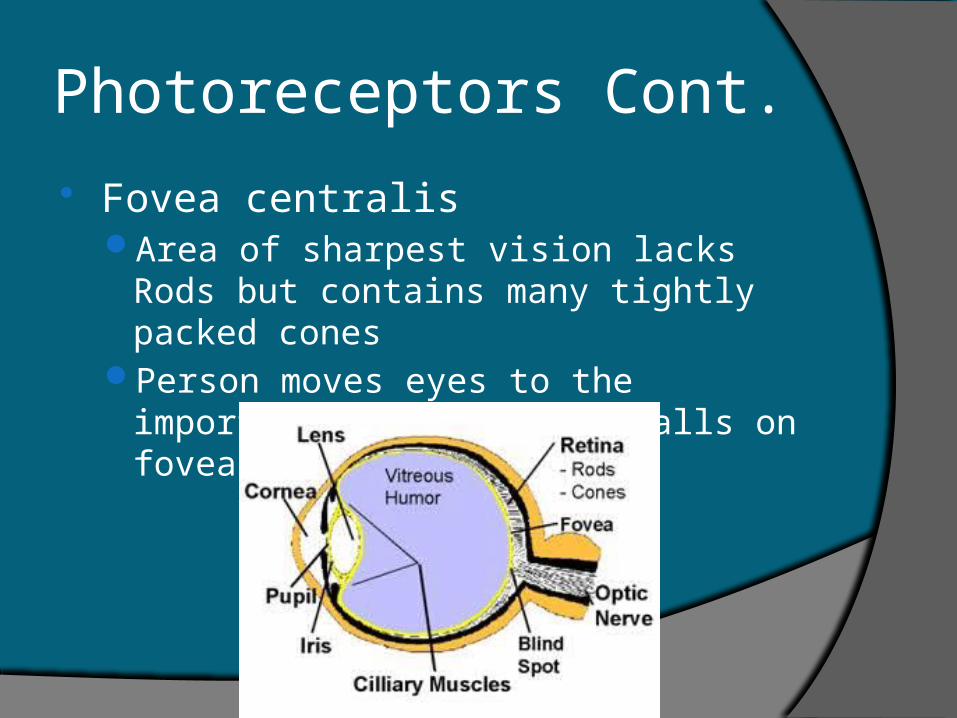
Photoreceptors Cont.
Fovea centralisArea of sharpest vision lacks Rods but
contains many tightly packed conesPerson moves eyes to the important part of
image falls on fovea centralis
Photopigments
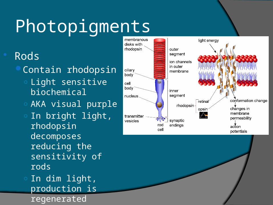
RodsContain rhodopsin
○ Light sensitive biochemical
○ AKA visual purple○ In bright light,
rhodopsin decomposes reducing the sensitivity of rods
○ In dim light, production is regenerated
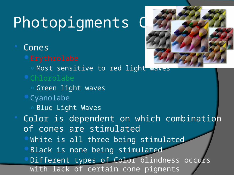
Photopigments Cont. Cones
Erythrolabe○ Most sensitive to red light waves
Chlorolabe○ Green light waves
Cyanolabe○ Blue Light Waves
Color is dependent on which combination of cones are stimulatedWhite is all three being stimulatedBlack is none being stimulatedDifferent types of Color blindness occurs with lack of
certain cone pigments
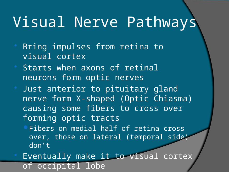
Visual Nerve Pathways
Bring impulses from retina to visual cortex Starts when axons of retinal neurons form
optic nerves Just anterior to pituitary gland nerve form
X-shaped (Optic Chiasma) causing some fibers to cross over forming optic tractsFibers on medial half of retina cross over,
those on lateral (temporal side) don’t Eventually make it to visual cortex of
occipital lobe
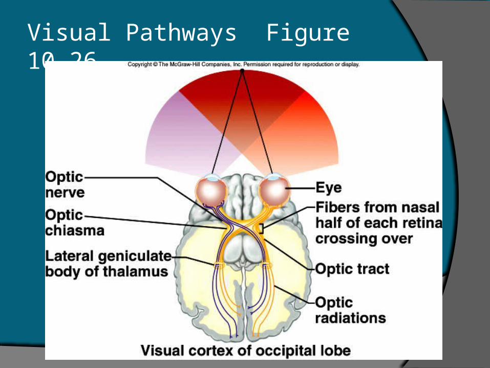
Visual Pathways Figure 10.26