intestinal goblet cell carcinoid presenting with recurrent ...intestinal goblet cell carcinoid...
TRANSCRIPT
CASE REPORT Open Access
Intestinal goblet cell carcinoid presentingwith recurrent sterile peritonitis in a patienton peritoneal dialysis: a case reportChih-Wei Chen1, Jan-Show Chu2, Li-Chun Hsieh3,4, Chih-Chin Kao1,5, Yen-Chung Lin1,5 and Hsi-Hsien Chen1,5*
Abstract
Background: Goblet cell carcinoid is a rare variant of appendiceal carcinoid with mixed endocrine and exocrine features.The most common symptom and signs are abdominal pain, acute appendicitis and palpable mass. Additionally, abdominalpain is common in patient on peritoneal dialysis, which may confound the diagnosis in such patient.
Case presentation:We report a 71- years- old woman on peritoneal dialysis that experienced several episodes ofabdominal cramping pain and sterile peritonitis. She had one episode of severe pain and underwent an appendectomy forsuspicion of appendicitis. Goblet cell carcinoid was diagnosed. She had no further abdominal pain after she receivedappendectomy.
Conclusions: Malignant dialysate was rarely reported in patient with peritoneal dialysis. However, goblet cell carcinoid caninitially present with acute appendicitis, chronic intermittent abdominal pain and mimicking peritonitis. In systemicallyreviewing the literature, this is the first case report of sterile peritonitis with peritoneal dialysis caused by goblet cellcarcinoid.
Keywords: Case report, Goblet cell carcinoid, Sterile peritonitis, Peritoneal dialysis
BackgroundGoblet cell carcinoid is a rare variant of appendicealcarcinoid with mixed endocrine and exocrine features.The most common clinical presentation is acute appen-dicitis [1, 2]. Typically, GCC tumors are infiltrative andinvolve the entire appendix circumferentially [3]. Never-theless, Tang et al. [1] reported that abdominal pain andpalpable mass are also reported presented in 50% ofGCC patients. Additionally, abdominal pain is commonin patients on peritoneal dialysis, which may confoundthe diagnosis in such patient. For these patients, righthemicolectomy has been traditionally suggested byphysician [2]. We performed systematic review ofPubMed and Cochrane Library databases and noted nosimilar cases of goblet cell carcinoid causing abdominalpain which mimics peritoneal dialysis related peritonitis.
We report this unique case of GCC presenting with ster-ile peritonitis in a patient on peritoneal dialysis toprompt clinicians to consider GCC in the differentialdiagnosis of similar symptoms in such patients.
Case presentationA 71- years- old woman with underlying hypertensionand end- stage renal disease on peritoneal dialysis, expe-rienced several episodes of abdominal cramping painwith mild diarrhea 3 months after starting peritonealdialysis. She had chills but no fever. A physical examin-ation showed lower abdominal tenderness, but norebounding pain. Initial complete blood count showedno leukocytosis (8400/μL). However, the initial dialysatestudies showed increasing leukocytes with a predomin-ance of neutrophils in first few sample, shifting tolymphocyte predominance in later sample (Table 1). Di-alysate and blood cultures from 15 sets over 4 monthssince initial abdominal pain showed no growth. Sinceempirical antibiotic treatment did not improve the pa-tient’s condition of recurrent cramping pain, abdominalcomputerized tomography (CT) was performed which
* Correspondence: [email protected] of Nephrology, Department of Internal Medicine, Taipei MedicalUniversity Hospital, Taipei, Taiwan5Division of Nephrology, Department of Internal Medicine, School ofMedicine, College of Medicine, Taipei Medical University, Taipei, TaiwanFull list of author information is available at the end of the article
© The Author(s). 2017 Open Access This article is distributed under the terms of the Creative Commons Attribution 4.0International License (http://creativecommons.org/licenses/by/4.0/), which permits unrestricted use, distribution, andreproduction in any medium, provided you give appropriate credit to the original author(s) and the source, provide a link tothe Creative Commons license, and indicate if changes were made. The Creative Commons Public Domain Dedication waiver(http://creativecommons.org/publicdomain/zero/1.0/) applies to the data made available in this article, unless otherwise stated.
Chen et al. BMC Nephrology (2017) 18:62 DOI 10.1186/s12882-017-0477-x

revealed no abnormality. The patient still complainedintermittent abdominal pain after the CT, so pain con-trol medication (including acetaminophen and tramadol)was prescribed for the patient. The Tenckhoff catheterwas removed 2 months after the onset of abdominalpain, and then hemodialysis was started. Laparotomyand biopsy of peritoneal were performed during removalof Tenckoff catheter. Staining for acid-fast bacilli wasnegative. She continued to complain of intermittentabdominal cramping pain. One and half year after herinitial abdominal pain, abdominal CT was again per-formed due to an episode of particularly severe abdom-inal pain. The scan showed fluid collection in the rightpelvic cavity extending to the distal part of the appendix(Fig. 1). An appendectomy was immediately performed,and goblet cell carcinoid (GCC) with suppurative appen-dicitis was diagnosed. The CT scan before patient re-ceived appendectomy show the swollen appendixwithout obvious extraappendiceal lesion. On the otherhand, according to the surgical and histopathologicalfindings, the appendix was perforated with adhesion to
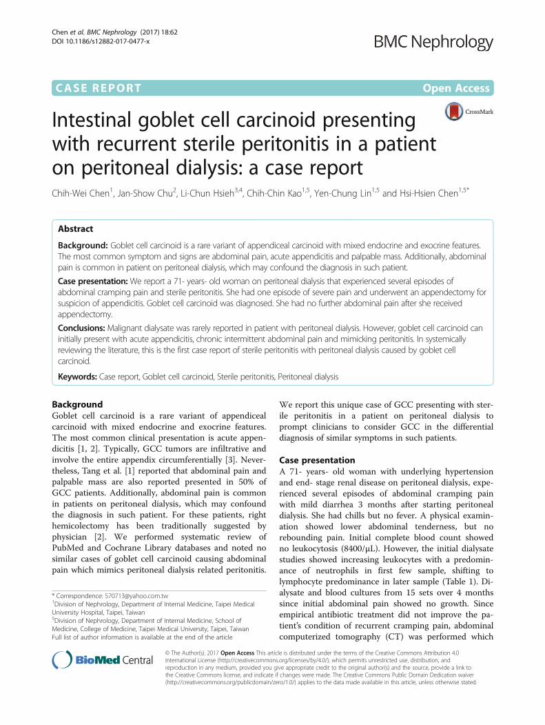
ileum, accompanied by regional inflammatory changesin pelvic cavity. Microscopically, the infiltrating neoplas-tic cells were found in the appendiceal wall, includingmucosa, submucosal, muscular propria, and subserosallayers. The infiltrated wall also showed fibrotic change.No evidence extra-appendiceal invasion of the tumorcould be found (Fig. 2, the pathology specimen of thispatient). Right hemicolectomy and adjuvant chemother-apy were suggested for complete treatment of GCC butthe suggestions were refused by the patient. No furtherabdominal pain has been noted since appendectomy,and the patient still visits oncologist outpatient depart-ment for regular follow up without evident of recurrenttumor.
Discussion and ConclusionsPeritonitis in during peritoneal dialysis is a significantcomplication in clinically which can cause death orstructural changes of the peritoneum [4]. Peritoneal in-fection is also a common cause of dialysis patientsswitching the therapy from peritoneal dialysis tohemodialysis [5]. On the other hand, sterile peritonitis isa common issue in the patients receiving peritonealdialysis. Sterile peritonitis, culture-negative peritonitis, isoften caused by antecedent empirical antibiotic treat-ment before dialysate culture are collected for culture.Additionally, infection caused by atypical organisms ornon-infectious causes, can also lead to sterile peritonitis[6]. Peritoneal dialysate cultures prove to be negative inup to 22% of case [6]. Patients using icodextrin forperitoneal dialysis often develop peritonitis with anassociated non-neutrophil predominant increasing indialysate white blood cells [7]. Tuberculous (TB) peri-tonitis and malignancy related peritonitis also causesimilar changes [6]. However, non-neutrophil-dominantincreases in dialysate white blood cells in associationwith malignancy dialysate are rare [8, 9]. Lymphomasometimes mimics peritonitis in patients on ambulatoryperitoneal dialysis [8]. Peritoneal metastases can alsomimic peritonitis [9].Goblet cell carcinoid (GCC), an unique and enigmatic
tumor involving the appendix almost exclusively, is arare variant of appendiceal carcinoid [10]. Carcinoid tu-mors account for up to 85% of all appendiceal tumors,white, GCC accounts for only 6% of all appendiceal
Table 1 Serial dialysate analysis of patient during episodes of abdominal pain in 2012
Date 09/13 09/16 09/19 09/21 09/25 10/02 10/12 11/19
Specific gravity 1.010 1.018 1.010 1.010 1.014 1.014 1.010 1.010
Red blood cells/mm3 6 180 63 297 126 81 75 9
White blood cells/mm3 136 648 243 90 144 222 252 494
Neutrophil:Lymphocyte:H:M 24:21:48:2:5(E) 80:20 48:16:28:8 24:27:48:1 35:32:22:4:7 10:81:1:8(E) 4:92:4 44:53:1:2
H macrophage, M mesothelial, E Eosinophil
Fig. 1 Contrast-enhanced pelvic computed tomography. The scanreveals one 2.1 × 1.6 cm irregular-shaped cystic mass (arrow) withmarginal enhancement in the right pelvic cavity, abutting the swollenappendix (arrowhead). The tumor is located at the tip of appendix withpossible subserosa invasion and attaches to the right pelvic side wall.The scan also shows inflammatory change with regional ileal bowelloop adhesion to the mass. Some fluid collection around the tumor-bowel complex is noted
Chen et al. BMC Nephrology (2017) 18:62 Page 2 of 4
carcinoid tumors [11]. The term GCC was firstly intro-duced by Subbuswamy et al. in 1974 [12]. GCC derive froma pluripotent intestinal stem cell that differentiates into bothmucinous and neuroendocrine cells [13]. Thus, it shareshistologic features of both adenocarcinomas and carcinoidtumors. Additionally, GCC is reportedly more aggressiveand unpredictable in nature than other carcinoid tumors.The most common clinical presentation is acute ap-
pendicitis [1]. Abdominal pain and a palpable mass arealso reported by 50% of GCC patients [2]. Pham et al.[1]. also reported other symptoms including bowel ob-struction, intussusception, gastrointestinal bleeding, andchronic intermittent lower abdominal pain. Comparedwith other carcinoid tumors which are usually asymp-tomatic, GCC often presents with clinical symptoms.Thus, the incidental diagnostic rate is only about 3% [2].
Despite serotonin can be detected immunohistochemi-cally in GCC tumor [14], there is no carcinoid syndromehas been reported in GCC patients and urinary 5-hydroxyindoleacetic acid (5-HIAA) levels in these pa-tients are usually within normal limits, as opposed tocarcinoid tumors, which are commonly with elevatedurinary 5-HIAA and serum chromogranin A [15].The most common route of metastases is trans-
coelomic/peritoneal invasion, and the most commonsites involved are ovaries, and the peritoneal surfaces ofthe pelvis and abdominal cavity [10].The natural history of patients with GCC is intermedi-
ate in aggressiveness between adenocarcinomas andclassical carcinoids [10]. Based on its natural history andmalignant nature, optimal therapeutic startegies are ingeneral similar to adenocarcinomas rather than classicalcarcinoids [10]. Thus, surgical management with righthemicolectomy is recommended after appendectomy formost cases, particularly those with an adenocarcinomacomponents [2]. Nevertheless, right hemicolectomy is asignificant abdominal procedure with an associated risk,especially in the infirm or elderly [15]. Peritoneal carcin-omatosis is the most common cause of death [10]. Gob-let cell carcinooids are associated with a 60% 10-yearsurvival and is similar to survival after treatment of alow-risk adenocarcinoma [15]. The prognosis of GCC isintermediate between appendiceal carcinoids and adeno-carcinomas [10].Our patient, who experienced abdominal pain inter-
mittently for months had been diagnosed with periton-itis related to peritoneal dialysis. Given a clinical historyof recurrent sterile peritonitis with a predominance oflymphocytes in the dialysate, an atypical infection (in-cluding tuberculosis) and intra-abdominal malignancymust be considered in the differential diagnosis. How-ever, no dialysate or ascites was sent to cytology analysisin our patient. We also considered intermittent bowelobstruction as the cause of abdominal pain, which wasthe common presentation of GCC [1], but there were nosufficient evidence to support the possibility.Of all the carcinoid tumors, GCC, in particular, can
initially present with acute appendicitis, chronic inter-mittent abdominal pain, and malignant ascites mim-icking peritonitis related to peritoneal dialysis in suchpatient. Through systematic review of PubMed andCochrane Library databases, there were no similarcases reported. We report this unique case of GCCpresenting with sterile peritonitis in a patient on peri-toneal dialysis to prompt clinicians to consider GCCin the differential diagnosis of similar symptoms insuch patients.
Abbreviations5-HIAA: 5-hydroxyindoleacetic acid; CT: Computerized tomography;GCC: Goblet cell carcinoid
Fig. 2 Peritoneum obtained by laparotomy. (200X) a H & E stainingshows typical goblet cell carcinoid. The photomicrography showsgoblet cell carcinoid composed of concentric infiltrating neoplasticcells in the appendiceal wall, including the mucosa, submucosa,muscular propria, and subserosal layers. The infiltrated wall showsfibrotic change. The tumor includes small solid or tubule-like tumorclusters, displaying monotonous nuclei with fine chromatin andeosinophilic cytoplasm, and occasional mucin-producing goblet cellsarranged singly or small nests. b The Immunohistochemical study ispositive for chromogranin A Brown color (arrow)
Chen et al. BMC Nephrology (2017) 18:62 Page 3 of 4
AcknowledgementsNone.
FundingThis study was not supported by any funding.
Availability of data and materialsThe data supporting the conclusions of this article is included within the article.
Authors’ contributionsCWC wrote the manuscript and contributed to acquisition of data. HHC wasthe treating physician of the patient and contributed to revising criticallyimportant intellectual content of the manuscript. JCS performed pathologicanalysis and interpretation. LCH performed interpretation of CT image. CCKand YCL analyzed the clinical course and contributed to conception ofconclusion. All authors read and approved the final manuscript.
Competing interestsThe authors declare that they have no competing interests.
Consent for publicationWritten informed consent was obtained from the patient for publication ofthis case report and any accompanying images. The authors adhered toCARE guidelines/methodology.
Ethics approval and consent to participateNot applicable.
Author details1Division of Nephrology, Department of Internal Medicine, Taipei MedicalUniversity Hospital, Taipei, Taiwan. 2Department of Pathology, School ofMedicine, College of Medicine, Taipei Medical University, Taipei, Taiwan.3Department of Medical Imaging, Taipei Medical University Hospital, Taipei,Taiwan. 4Translational Imaging Research Center, College of Medicine, TaipeiMedical University, Taipei, Taiwan. 5Division of Nephrology, Department ofInternal Medicine, School of Medicine, College of Medicine, Taipei MedicalUniversity, Taipei, Taiwan.
Received: 21 March 2016 Accepted: 8 February 2017
References1. Pham TH, Wolff B, Abraham SC, Drelichman E. Surgical and chemotherapy
treatment outcomes of goblet cell carcinoid: a tertiary cancer centerexperience. Ann Surg Oncol. 2006;13(3):370–6.
2. Tang LH, Shia J, Soslow RA, Dhall D, Wong WD, O’Reilly E, Qin J, Paty P,Weiser MR, Guillem J, et al. Pathologic classification and clinical behavior ofthe spectrum of goblet cell carcinoid tumors of the appendix. Am J SurgPathol. 2008;32(10):1429–43.
3. Pickhardt PJ, Levy AD, Rohrmann Jr CA, Kende AI. Primary neoplasms of theappendix: radiologic spectrum of disease with pathologic correlation.Radiographics. 2003;23(3):645–62.
4. Akoh JA. Peritoneal dialysis associated infections: an update on diagnosisand management. World J Nephrol. 2012;1(4):106–22.
5. Jaar BG, Plantinga LC, Crews DC, Fink NE, Hebah N, Coresh J, Kliger AS, PoweNR. Timing, causes, predictors and prognosis of switching from peritonealdialysis to hemodialysis: a prospective study. BMC Nephrol. 2009;10:3.
6. de Freitas DG, Gokal R. Sterile peritonitis in the peritoneal dialysis patient.Perit Dial Int. 2005;25(2):146–51.
7. Martin J, Sansone G, Cirugeda A, Sanchez-Tomero JA, Munoz C, Selgas R.Severe peritoneal mononucleosis associated with icodextrin use incontinuous ambulatory peritoneal dialysis. Adv Perit Dial. 2003;19:191–4.
8. Vlahakos D, Rudders R, Simon G, Canzanello VJ. Lymphoma-mimickingperitonitis in a patient on continuous ambulatory peritoneal dialysis (CAPD).Perit Dial Int. 1990;10(2):165–7.
9. Bagnis C, Gabella P, Bruno M, Cosseddu D, Marangella M, Yacha GM, LinariF. Cloudy dialysate due to adenocarcinoma cells in a CAPD patient. PeritDial Int. 1993;13(4):322–3.
10. Roy P, Chetty R. Goblet cell carcinoid tumors of the appendix: an overview.World J Gastrointest Oncol. 2010;2(6):251–8.
11. Gupta A, Patel T, Dargar P, Shah M. Metastatic appendiceal goblet cellcarcinoid masquerading as mucinous adenocarcinoma in effusion cytology:a diagnostic pitfall. J Cytol. 2013;30(2):136–8.
12. Subbuswamy SG, Gibbs NM, Ross CF, Morson BC. Goblet cell carcinoid ofthe appendix. Cancer. 1974;34(2):338–44.
13. Alsaad KO, Serra S, Chetty R. Combined goblet cell carcinoid and mucinouscystadenoma of the vermiform appendix. World J Gastroenterol. 2009;15(27):3431–3.
14. Anderson NH, Somerville JE, Johnston CF, Hayes DM, Buchanan KD, SloanJM. Appendiceal goblet cell carcinoids: a clinicopathological andimmunohistochemical study. Histopathology. 1991;18(1):61–5.
15. Goede AC, Caplin ME, Winslet MC. Carcinoid tumour of the appendix. Br JSurg. 2003;90(11):1317–22.
• We accept pre-submission inquiries
• Our selector tool helps you to find the most relevant journal
• We provide round the clock customer support
• Convenient online submission
• Thorough peer review
• Inclusion in PubMed and all major indexing services
• Maximum visibility for your research
Submit your manuscript atwww.biomedcentral.com/submit
Submit your next manuscript to BioMed Central and we will help you at every step:
Chen et al. BMC Nephrology (2017) 18:62 Page 4 of 4