metastatic intestinal carcinoid - department of surgery at suny
TRANSCRIPT

Johanna Basa M.D.
SUNY Downstate- Brooklyn VA
March 14, 2013
METASTATIC INTESTINAL CARCINOID
www.downstatesurgery.org
CASE • 63 yr old male with PMH of prostate cancer, HTN, obesity, presented with
complaints of abdominal pain, nausea and vomiting, no flatus or bowel movements for one day. He denied palpitations, hot flashes, diarrhea and wheezing.
• PSH: prostatectomy • PE afebrile, VSS • Gen: NAD • CV:+s1, s2 no murmurs • Abd: low infraumbilcal scar well healed, obese, no hernias, soft tender,
distended • Rectal: no masses, skin tags, no gross blood, guiac negative
www.downstatesurgery.org
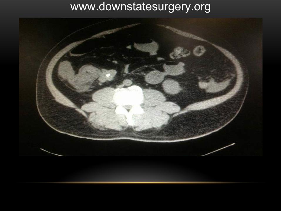
CASE • November 2012 admitted for small bowel obstruction. Ct showed stellate
mesenteric lesion with calcification
• December 3.2012 Somatostatin PET scan which showed 3 lesions, anterior surface of liver, segment 5, lower pole of right kidney
• December 8, 2012 EGD- 3 lesions in ileum and one dominant lesion in terminal ileum extending into the Cecum
• December 14, 2012 MRI showed multiple liver lesions bilobar mostly on the right. Pericecal mass, mesenteric mass.
www.downstatesurgery.org
www.downstatesurgery.org

CASE • December 23,2012 Tumor Board discussion:Heme/Onc, GI, Rad Onc,
Surgery, Pulmonary. Plan for exploratory laparotomy • December 30, 2012 Colonoscopy- Left sided diverticulosis and large cecal
mass, biospy pathology: Carcinoid
• Urine 5 HIAA: 25, elevated • Chromogranin A: 6.5 normal
• Pt was started on Octreotide 100mg sub cutaneous Q8hr 2 weeks pre-
operatively, changed to Octreotide infusion 12 hours pre op.
www.downstatesurgery.org
CASE • Pt was taken to the OR on 1/2/13 for Exploratory laparotomy, right hemicolectomy,
right mesenteric mass excision, cholecystectomy, segement V liver resection, EGD, Intra operative US, cystoscopy and ureteral stent placement
• POD#1-8 Pt remained NPO awaiting bowel function, started on TPN
• POD#8-18 Pt started on diet, JP removed, treated for UTI, octreotide discontinued
• POD# 18-20 Pt still had intermittent fevers with leukocytosis, found to have hepatic abscess drained by IR
• POD# 27 Discharged home
• Repeat Octreoscan was negative for residual tumor, CT abdomen and pelvis showed resolution of hepatic abscess
www.downstatesurgery.org

HISTORY • 1867 Theodor Langhans was the first to describe the histology of carcinoid
tumor, Otto Lubrarsh is credited with the first report of two patients with ileal carcinoid
• 1907 Siegfried Obendorfer coined the term Karzinoide “carcinoma like” • 1948 Rappaport discovered serotonin as the vasoactive substance • 1952 the origin of the amine 5HIAA was the Kulchitsky cell • 1968 Williams and Sandler proposed the classification into foregut, midgut
and hindgut.
www.downstatesurgery.org

• GLANDULAR
• Pituitary
• Parathyroids
• Paraganglia
• Adrenal Medulla
• DIFFUSE
• Skin
• Thyroid
• Lung
• Thymus
• Pancreas,
• Gastrointestiinal **
• Biliary tree
• Urogenital
NEUROENDOCRINE CELLS
www.downstatesurgery.org

WHAT IS IN A NAME? • Carcinoid
• Neuroendocrine
• Enteroendocrine
• APUD- amine precursor uptake and decarboxylation
• Gastorenteropancreatic neuroendocrine tumors (GEP-NETs)
• Seritoninomas
www.downstatesurgery.org

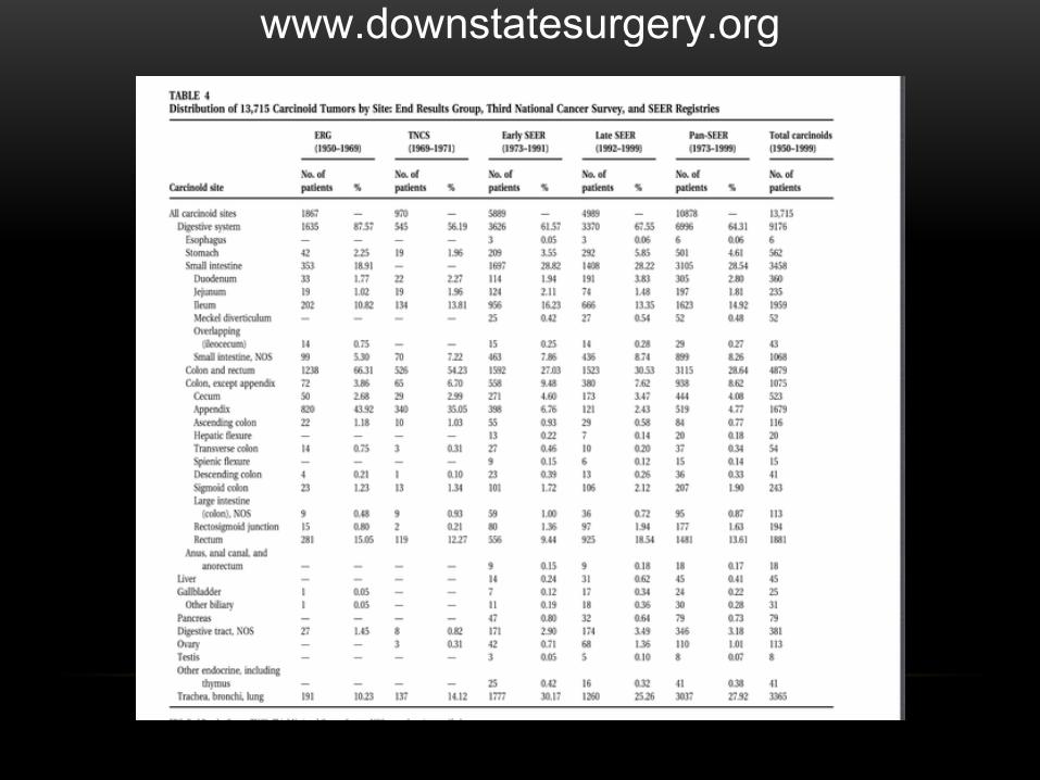
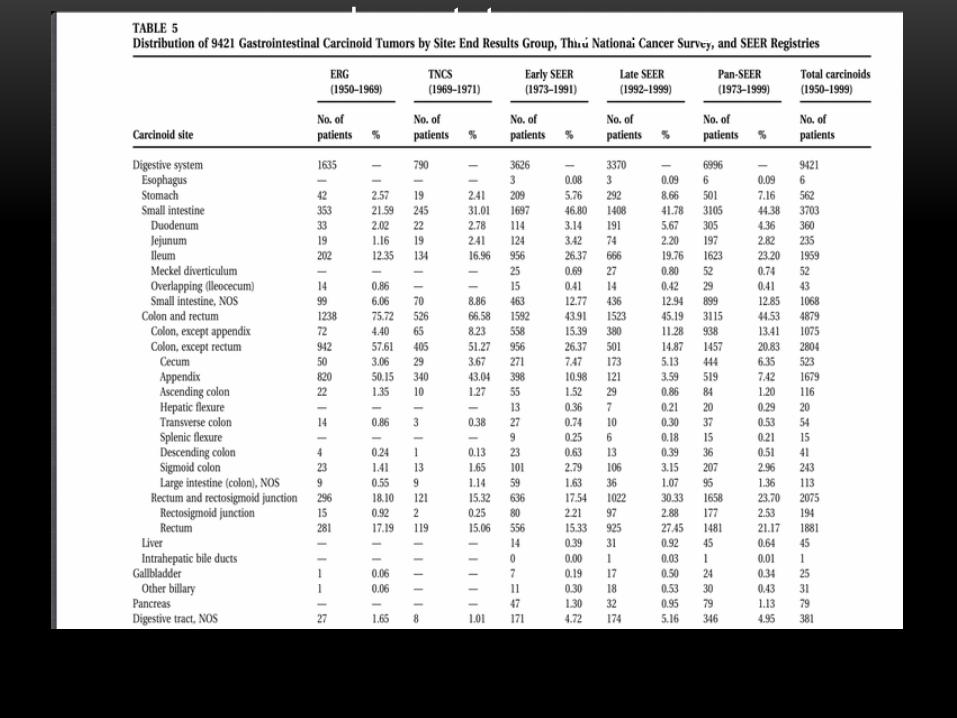
INCIDENCE • 1.3 per 100,000 from 36 yr study in England
• Ranges 2.47-2.58 per 100,000 from 2008 SEER data depending on site
• 3-10% increase in incidence of neuroendocrine tumors over the past 30 yrs
• Jejunum, ileum, and cecum, stomach, and rectum have increasing
incidence,while appendiceal has decreasing incidence
• Median age for midgut NET is 64, appendiceal subgroup was 47
www.downstatesurgery.org
www.downstatesurgery.org
www.downstatesurgery.org
RISK FACTORS • Parent or sibling with history of carcinoid tumor • Parent with history of brain, endocrine, breast, liver and urinary tract cancer • Genetic disorders with pancreatic neuroendocrine tumors (PNET)
• MEN I • von Hipple Lindau • von Recklinhausens • Tuberous sclerosis
www.downstatesurgery.org

• LABORATORY WORK UP • 24 hour urine 5HIAA • Serum Chromogranin A
levels • Serum Serotonin levels
• SYMPTOMS • Flushing***Most common sx • Burning sensation of skin • Secretory diarrhea • Bronchospasm • Cramping abdominal pain
DIAGNOSIS
www.downstatesurgery.org

GASTRIC CARCINOID • 4% of all GI NET, 1% of gastric neoplasms • 4 subtypes • Types 1-3 originate from enterochromaffin cells in the gastric mucosa. • Types 1, 2 are gastrin dependent, multifocal, small (<2cm) • Type 3 not associated with hypergastrinemia, usually solitary and large
(>2cm) • Type 4 is poorely differentiated, usually large (>5cm) ulcerated and
unresectable, poor prognosis
www.downstatesurgery.org

SMALL INTESTINE • Most common location of carcinoid tumor, usually in terminal ileum
• Multiple tumors in 1/3 of patients
• Carcinoid syndrome is common with liver metastasis
• Metastasis to liver in 50%, nodal spread in 70%
• Fibrosis around nodal metastases causes contraction of mesenteryocclusion of mesenteric vessels and ischemia chronic ischemia on antimesenteric border
• Size does NOT predict metastatic potential
• Small tumors less than 1cm can be segmentally resected, wide excision for tumors greater than 1cm. Right hemicolectomy for lesions of the terminal ileum
• 5 yr survival is fair (60%)
www.downstatesurgery.org

TREATMENT ALGORITHM
www.downstatesurgery.org
APPENDIX • No longer the most common site of carcinoid tumor based on newer SEER data • Is the most common neoplasm of the appendix • Incidence is decreasing • Most found incidentally • Indications for right hemicolectomy for appendiceal carcinoids 1-2cm in size
• Invasion into the mesoappendix • Lymphovascular invasion • Serosal involvement • Positive margins, positive LN on appendectomy specimen • HIGH KI67 Index • Goblet cell variant
www.downstatesurgery.org

TREATMENT ALGORITHM
www.downstatesurgery.org
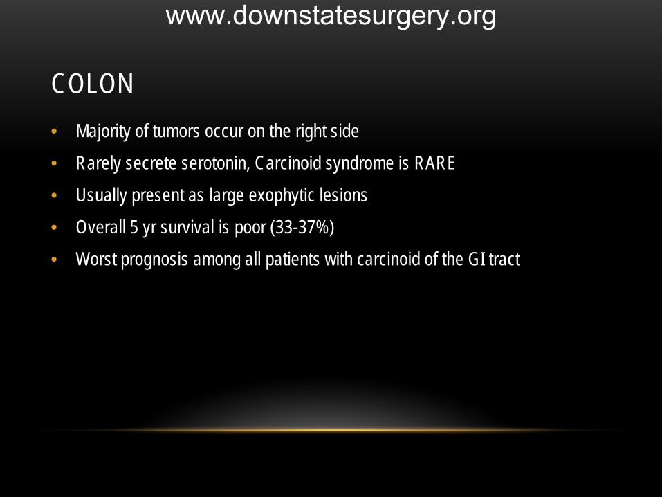
COLON • Majority of tumors occur on the right side • Rarely secrete serotonin, Carcinoid syndrome is RARE • Usually present as large exophytic lesions • Overall 5 yr survival is poor (33-37%) • Worst prognosis among all patients with carcinoid of the GI tract
www.downstatesurgery.org
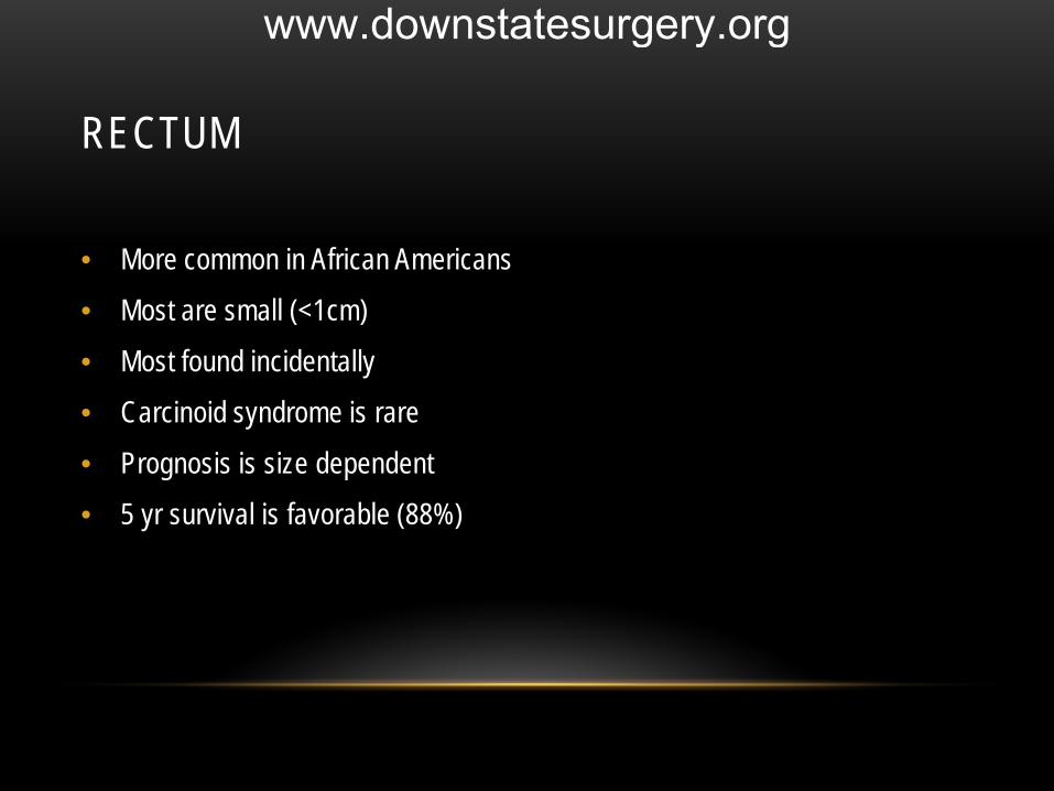
RECTUM • More common in African Americans • Most are small (<1cm) • Most found incidentally • Carcinoid syndrome is rare • Prognosis is size dependent • 5 yr survival is favorable (88%)
www.downstatesurgery.org

TREATMENT ALGORITHM
www.downstatesurgery.org
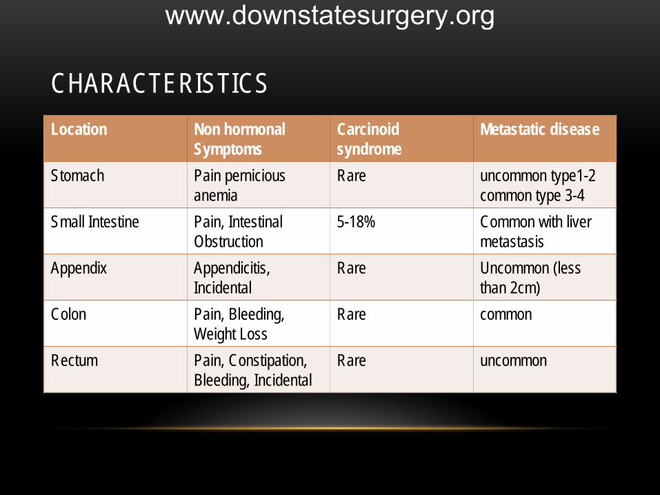
CHARACTERISTICS Location Non hormonal
Symptoms Carcinoid syndrome
Metastatic disease
Stomach Pain pernicious anemia
Rare uncommon type1-2 common type 3-4
Small Intestine Pain, Intestinal Obstruction
5-18% Common with liver metastasis
Appendix Appendicitis, Incidental
Rare Uncommon (less than 2cm)
Colon Pain, Bleeding, Weight Loss
Rare common
Rectum Pain, Constipation, Bleeding, Incidental
Rare uncommon
www.downstatesurgery.org

DIAGNOSIS-IMAGING • Plain radiograph • Cross sectional CT of the abdomen and pelvis • Octreotide scintigraphy
• Imaging should be performed at the end of dosing interval (3-6 wks after last dose) those on infusion pump should have stopped for 48 hrs.
• Localizes primary, recurrent tumor and staging
www.downstatesurgery.org
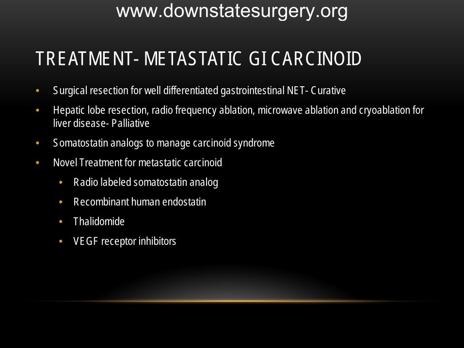
TREATMENT- METASTATIC GI CARCINOID • Surgical resection for well differentiated gastrointestinal NET- Curative
• Hepatic lobe resection, radio frequency ablation, microwave ablation and cryoablation for liver disease- Palliative
• Somatostatin analogs to manage carcinoid syndrome
• Novel Treatment for metastatic carcinoid
• Radio labeled somatostatin analog
• Recombinant human endostatin
• Thalidomide
• VEGF receptor inhibitors
www.downstatesurgery.org

CONCLUSIONS • Symptom control
• Somatostatin analogues control hypersecretion of neuropeptides in foregut and midgut carcinoids that express somatostatin receptors
• Biochemical control • Systemic chemotherapeutics like interferon alpha upregulate
somatostatin receptors to act synergistcally • Tumor control
• Cytoreductive surgery is the mainstay of treatment and includes resection of primary tumor, ablative therapy or resection of hepatic metastasis
www.downstatesurgery.org

QUESTIONS • A 54 yr old male reports a 2 month history of abdominal pain and significant weight loss.
He had undergone upper endoscopy, lower endoscopy, and CT, all of which is normal. On a barium upper GI study with small bowel follow through, he was noted to have a mass in his mid ileum. A surgical exploration he is found to have carcinoid on frozen section. Which of the following is true?
• A. The prognosis is related to tumor size, location and histologic pattern
• B. The cell of origin is the Kupffer cell
• C. The rectum is the most common site of origin
• D. Carcinoid tumors are usually easily palpable on external physical examination of the bowel
• E. Resection is not indicated in patients with metastatic disease.
www.downstatesurgery.org

QUESTIONS • On abdominal exploration for a suspected carcinoid tumor a 2cm mass is found at the
terminal ileum. No liver lesions were detected on preoperative imagining or with intraoperative palpation. What is the best treatment option for this patient?
• A. Segmental resection
• B. Medical therapy with octreotide
• C. Resection of the terminal ileum with preservation of the ileocecal valve
• D Right hemicolectomy with wide resection of the terminal ileum
• E. Neoadjuvant therapy with streptozotocin and 5 FU.
www.downstatesurgery.org
REFERENCES • Feldman : Sleisenger and Fordtran’s Gastrointestinal and Liver disease 9th ed. Chapter 31
Gastrointestinal Carcinoid Tumors
• Fazio: Current Therapy in Colon and Rectal Surgery 12th ed. Chapter 79 Carcinoid tumors of the large and small bowel
• Pinchot,S, Holen, K, Sippel, R, Chen, H. Carcinoid Tumors. The Oncologist 2008; 13:1255-1269
• Tsikitis, V, Wertheim, B, Guerrero, M. Trends of Incidence and Survival of Gastrointestinal Neuroendocrine Tumors in the United States a SEER analysis. JCancer. 2012;3:292-302
• Bourdeaux, JP, et al Surgical Treatment of Advanced Stage Carcinoid Tumors. Ann of Surg.2005;241(6)845-846
• Bourdeax, JP et al. The NANETS Consensus Guidelines for the Diagnosis and Management of Neuroendocrine Tumors.Pancreas.2010;39: 753-766
www.downstatesurgery.org