interesting case
DESCRIPTION
Chiombon. Chua. Corpuz. Cua. David. De Vera. Detera. Diaz. Din. Dizon. Eugenio. Evangelista. Interesting case. General Data. CT 29/Female DOA: 03 August 2010. Chief Complaint. Blurring of Vision. History of Present Illness. History of Present Illness. History of Present Illness. - PowerPoint PPT PresentationTRANSCRIPT

Chiombon. Chua. Corpuz. Cua. David. De Vera. Detera. Diaz. Din. Dizon. Eugenio. Evangelista.
1

General Data
CT 29/Female DOA: 03 August 2010
2

Chief Complaint
Blurring of Vision
3

History of Present Illness
4

History of Present Illness
5

History of Present Illness
6
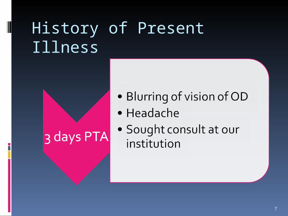
History of Present Illness
7
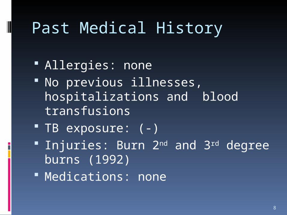
Past Medical History
Allergies: none No previous illnesses,
hospitalizations and blood transfusions
TB exposure: (-) Injuries: Burn 2nd and 3rd degree
burns (1992) Medications: none
8

Personal and Social History Diet: Mixed Smoking: 3.5 pack years Alcohol: 2 bottles of beer/day from
23-27 years Substance abuse: Denies illicit drug
use
9

Family History
Diabetes Mellitus- Grandmother Glaucoma- (-) Hypertension – (-) Cancer – (-)
10

Review of Systems
(-) Weight change , (-) fever & chills (-) rashes; (-)pruritus;(-) bruising (-) dizziness; syncope (-) blurring of vision, eye discharge (-) hearing changes; pain; discharge; vertigo; (-) epistaxis; obstruction; nasal discharge, gum bleeding; oral
ulcers (-) cough, (-) dyspnea, (-) night sweats (-) chest pain; (-)dyspnea on exertion; (-)PND; (-)palpitations (-) abdominal pain; (-)dysphagia, (-)nausea, (-)vomiting,(-)
Dyspepsia (-)diarrhea, (-) constipation (-) indigestion, (- flatulence (-) frequency; urgency; dysuria; nocturia; dribbling (-) arthralgia/arthritis (-) trauma; (-)back pain
11

Physical Examination • Conscious, Coherent, Ambulatory, not in Cardiorespiratory distress
• Vital signs: 110/80, PR 74 Regular, RR: 20, Temp 36.6 C
• Warm, moist skin, no active dermatoses (+) Scars, both upper extremeties
• Pink palpebral conjunctiva, anicteric sclerae, pupils L= 3-4mm, RTL, R= 2-3 mm RTL anisocoric, slightly hyperemic conjunctiva
• Septum midline, turbinates not congested, no nasoaural discharge, impacted cerumen
• Moist buccal mucosa, no oral and palatal lesions, nonhyperemic posterior pharyngeal wall, tonsils not enlarged
• No limitation of neck movement, no palpable cervical lymphadenopathy
• (-) thyroid gland enlargement
• No breast mass palpated, no discharge
• Thorax: no deformities, no intercostal and subcostal retractions, symmetrical chest expansion, equal tactile fremiti, resonant, clear and equal breath sounds
12

Physical Examination •Adynamic precordium, AB at the 5th LICS MCL, no LV heave, thrills, no lifts, s1 louder than s2 at the apex, s2 louder than s1 at the base, no murmurs
13

Physical Examination
Globular abdomen, Normoactive bowel sounds, (-) bruits, (-) tenderness, guarding, masses, (-) Murphy’s sign, Nonpalpable gallbladder, Traube’s space not obliterated
Pulses full and equal, (-) cyanosis, edema, clubbing
14

Eye Examination
Visual Acuity Right Eye Left Eye
Without Correction 20/50, J12 20/200, J12
Pin hole 20/30 20/50
Amsler Grid
(+) Scotoma on OU(+) Metamorphopsia
15

Eye Examination
External eye examination: Eyelids: non tender Lashes: not matted Conjunctiva: Hyperemic Sclera: anicteric Cornea: Clear Anterior Chamber: deep Lens: Clear Pupils: Anisocoric L= 3-4mm, RTL, R= 2-3 mm
RTL Iris: Pigmented
16

Eye Examination
EOM: Full and equal Fundoscopy:
(+) ROR
(+) Blurred disc Margins , OU (+) Serous Retinal Detachment, OU (+) Hyperemia of choroid
Tonometry: 18mmHg OU Fluorescein angiography:
hyperfluoresence of optic disc
17

NeuroExam
Conscious, coherent, oriented to 3 spheres, able to follow commands, GCS 15 E4V5M6
(-) Anosmia, anisocoric pupils L= 3-4mm, RTL, R= 2-3 mm RTL; Can smile, frown, raise eyebrows, uvula midline, can shrug shoulders, turn head from side to side against resistance tongue deviated to right. Motor exam: RUE: 5/5, RLE:5/5, LUE and LLE 5/5.
Reflexes: ++ Cerebellar- can do APST, FTNT with ease, No tremors No sensory deficit (-) Babinski, Nuchal rigidity, Brudzinski, Kernig’s sign
18

Initial Assessment
Incomplete Vogt-Koyanagi-Harada Disease, Acute uveitic stage
r/o TB Uveitis
19
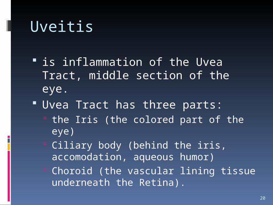
Uveitis
is inflammation of the Uvea Tract, middle section of the eye.
Uvea Tract has three parts: the Iris (the colored part of the eye) Ciliary body (behind the iris,
accomodation, aqueous humor) Choroid (the vascular lining tissue
underneath the Retina).
20

DIFFERENTIAL DIAGNOSIS
21
Our Patient Sympathetic Ophthalmia
Tuberculosis
(-) History of Trauma (-) Previous TB exposure
Previous History of Trauma , perforating eye injury
Previous Infection of Tuberculosis
Sees “Flashes of light” RednessBlurred Vision
PhotophobiaRedness Blurred visionFloaters
Unilateral Cases of ocular involvement
(+) ROR (+) Blurred disc Margins , OU (+) Serous Retinal Detachment, OU(+) Hyperemia of choroid(+) Hypreflouresence of optic disc
Soft yellow white exudates in the deep layer of the Retina
Granulomatous keratic precipitates or choroidal granulomas are present

Incomplete Vogt-Koyanagi-Harada Disease, Acute uveitic stager/o TB uveitis
22

DIAGNOSTICS
23
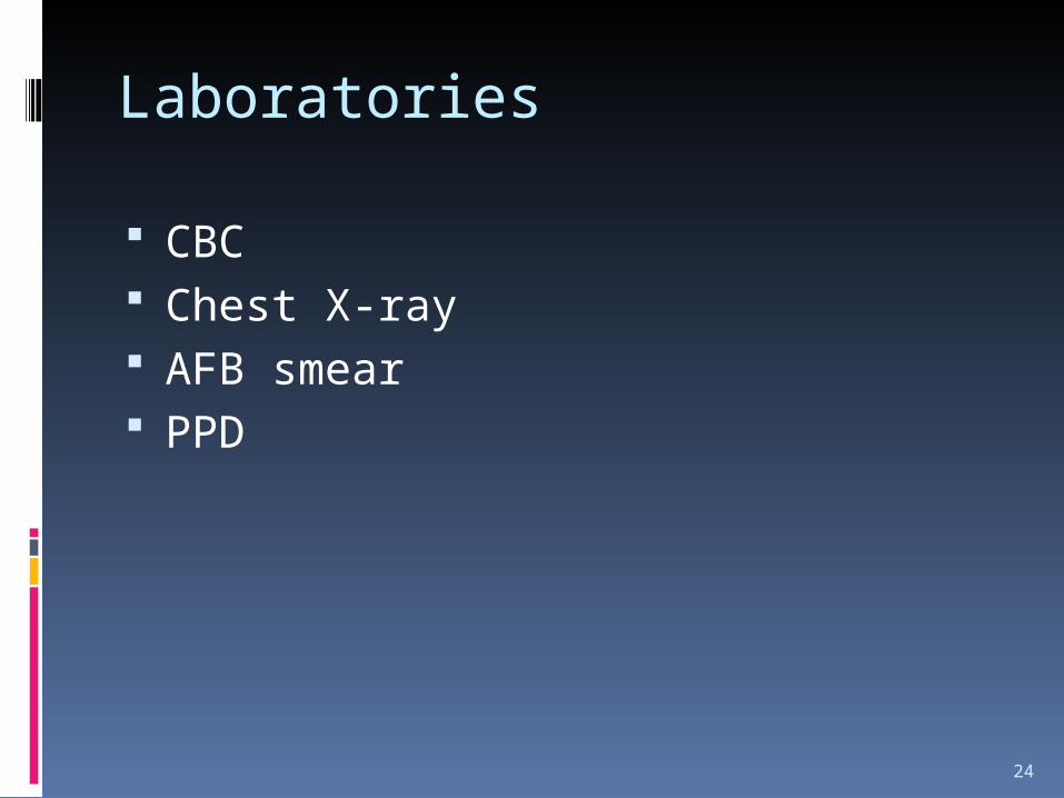
Laboratories
CBC Chest X-ray AFB smear PPD
24

CBC
HGB 126
RBC 4.03
HCT 0.38
MCV 94
MCH 31.3
MCHC 33.3
RDW 12.30
MPV 7.40
PLT 255
WBC 7.90
Neut 0.52
Seg 0.52
Lymph 0.44
Eosin 0.04
ESR 8.0
25

Chest X-ray
Lung fields are clear The heart is not enlarged Both hemidiaphragm and
costophrenic sulci are intact Impression: No significant chest
findings
26

AFB Stain
No Acid Fast Bacilli seen in 300 oil immersion fields on both routine and concentration methods
27

Therapeutics
Methylprednisolone 1mg/kg/day per IV for 3 days
Ranitidine 150mg/tab, 1 tab OD Tropicamide eyedrops 1gtt BID CaC03 + Vit D 1 tab OD
28

Course in the wards
On admission: CBC with Platelet, PPD, Chest X-ray, and
ESR were requested. Tropicamide eyedrops OU was also
started. On the 2nd hospital day
Referred to Rheumatology Plans for induction of high-dose steroids
29

Course in the Wards
On the 3rd Hospital day: PPD test was started
On the 4th Hospital day Methylprednisolone 1g/kg to run for 1
hour for 3 days.
30
Visual Acuity Right Eye Left Eye
Without Correction 20/50, J12 20/200, J12

Course in the wards
On the 5th hospital day, visual acuity of the patient improved:
(-) Hyperemia of the Conjunctiva (-) PPD test Patient was given 2nd dosage of
Methylprednisolone 1mg/kg/IV to run for 1 hour
CaC03 + Vit D 1 tab OD was started
Visual Acuity Right Eye Left Eye
Without Correction
20/40 20/70
31

Course in the wards
On the 6th hospital day
Patient received last dose of Methylprednisolone 1mg/kg/IV to run for 1 hour.
Visual Acuity Right Eye Left Eye
Without Correction
20/40 20/40
32

Course in the wards
7th hospital day Prednisone 20mg/tab 1 tab OD was
started
Visual Acuity Right Eye Left Eye
Without Correction 20/40 20/40
33

Course in the wards
8th hospital day
Indirect Fundoscopy: Decrease in macular edema, decrease in vitreous cell and optic nerve hyperemia
Patient was discharged
Visual Acuity Right Eye Left Eye
Without Correction 20/40 20/40
34

VOGT-KOYANAGI-HARADA DISEASE
DISCUSSION
35

Vogt-Koyanagi-Harada disease Inflammatory condition of autoimmune
nature in which cytotoxic T cell target melanocytes (eyes, inner ears, skin)
Described by Persian Physician (Ali-ibn-Isa 940-1010 AD) –Poliosis + eye inflammation
1932- Combined disorders described by Vogt, Koyanagi and Harada manifestations were under the same disease process
New insights into Vogt-Koyanagi-Harada disease. Arq Bras Oftalmol. 2009;72(3):413-2036

Epidemiology
Predilection for more darkly pigmented races: Asians, Hispanics, American Indians
6.8-9.2% of all Uveitis referrals in Japan
37

Vogt-Koyanagi-Harada diseaseClassification International Nomenclature
Committee Revised Diagnostic Criteria
Classification: Complete VKH disease Incomplete VKH disease Probable VKH disease
New insights into Vogt-Koyanagi-Harada disease. Arq Bras Oftalmol. 2009;72(3):413-2038

Applicability of the 2001 revised diagnostic criteria in Brazilian Vogt-Koyanagi-Harada disease patients Arq Bras Oftalmol. 2008;71(1):67-7039

Vogt-Koyanagi-Harada diseaseStages Prodromal stage Acute uveitic stage Convalescent stage Chronic recurrent stage
40

Stages Prodromal Acute Uveitic Stage Convalsecent
stage Chronic Recurrent stage
Mimics viral Infection
Bilateral blurring of vision
Ocular pain secondary to Ciliary spasm
VitiligoAlopeciaPoliosis
43% in 1st three months52% in 1st six months
Fever
Neurological Symptoms
Multifocal Choroidtis Multifocal detachment of the sensory retina Exudative retinal detachment
Uveal depigmentationSunset glow
Foci of hyperpigmentation of RPE
GlaucomaCataractSubretinal Fibrosis
41

Vogt-Koyonagi-Harada Disease
Pathophysiology
42

Vogt-Koyanagi-Harada diseaseAutoimmunity Against Melanocytes
Clinical features of choroidal and skin depigmentation
Transmission electron microscopy (early stage): close contact between melanocytes and lymphocytes in the uvea
Histopathology (end stage): disappearance of choroidal melanocytes, and
Immunohistochemistry (end stage): T and B lymphocytes in the choroid
New insights into Vogt-Koyanagi-Harada disease. Arq Bras Oftalmol. 2009;72(3):413-2043

Vogt-Koyanagi-Harada diseaseAutoimmunity Against Melanocytes Role of CD4+ T cells
Cytotoxic leukocytes against melanoma cells in peripheral blood and CSF
Cytotoxic CD4 and CD8 T lymphocytes against human melanocytes are present in the peripheral blood.
Activated CD4+ T cells in depigmented skin and melanin-laden macrophages d in CSF
New insights into Vogt-Koyanagi-Harada disease. Arq Bras Oftalmol. 2009;72(3):413-2044

Vogt-Koyanagi-Harada diseaseAutoimmunity Against Melanocytes
Immunogenetics HLA-DR4/DR53 secondary association with HLA-DR1
involving a shared sequence linked to susceptibility to rheumatoid arthritis.
HLA-DRB1*0405
New insights into Vogt-Koyanagi-Harada disease. Arq Bras Oftalmol. 2009;72(3):413-2045

Pathophysiology
46

Clinical findings in acute phase of VKHFigure 1 - A & B: Fundus pictures of both eyes show disc hyperemia, white-yellowish choroidal lesions,
and localized exudative retinal detachments; C & D: Fluorescein angiographies of both eyes show pin-point hyperfluorescence and dye pooling corresponding to areas of retinal detachments; E & F: Indocyanine green angiographies show areas of diffuse hyperfluorescence, dark spot, and “hot-spots”
New insights into Vogt-Koyanagi-Harada disease. Arq Bras Oftalmol. 2009;72(3):413-20
47

Clinical findings in chronic phase of VKHFigure 2 – A & B: Fundus pictures of both eyes show diffuse retinal depigmentation and peripapillary fibrosis;
C & D: Fluorescein angiographies of both eyes show diffuse window retinal pigment epithelium defects; E & F: Indocyanine green angiographies
show dark spots and diffuse late hyperfluorescence suggestive of disease activity
New insights into Vogt-Koyanagi-Harada disease. Arq Bras Oftalmol. 2009;72(3):413-20
48
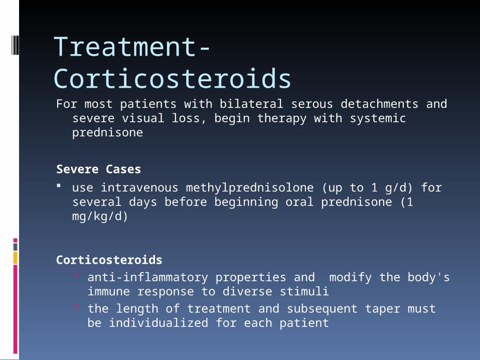
Treatment- CorticosteroidsFor most patients with bilateral serous detachments and
severe visual loss, begin therapy with systemic prednisone
Severe Cases use intravenous methylprednisolone (up to 1 g/d) for
several days before beginning oral prednisone (1 mg/kg/d)
Corticosteroids anti-inflammatory properties and modify the body's
immune response to diverse stimuli the length of treatment and subsequent taper must be
individualized for each patient

Treatment- Systemic Corticosteroids
Prednisone• Decrease inflammation
– reversing increased capillary permeability and suppressing PMN activity
• DOSE– 1-1.5 mg/kg/d PO qd initially– Severe cases with profound loss of vision and
bilateral serous detachments may require up to 2 mg/kg/d
– length of treatment and tapering individualized for each patient • not be less than 3 mo to avoid recurrence
• CI: hypersensitivity; viral infection; peptic ulcer disease; hepatic dysfunction; connective tissue infections; fungal or tubercular skin infections; GI disease
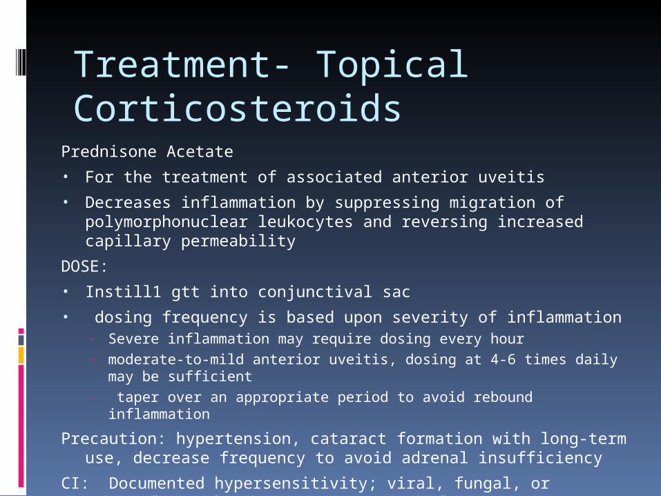
Treatment- Topical Corticosteroids
Prednisone Acetate
• For the treatment of associated anterior uveitis
• Decreases inflammation by suppressing migration of polymorphonuclear leukocytes and reversing increased capillary permeability
DOSE:
• Instill1 gtt into conjunctival sac
• dosing frequency is based upon severity of inflammation – Severe inflammation may require dosing every hour– moderate-to-mild anterior uveitis, dosing at 4-6 times daily may be
sufficient– taper over an appropriate period to avoid rebound inflammation
Precaution: hypertension, cataract formation with long-term use, decrease frequency to avoid adrenal insufficiency
CI: Documented hypersensitivity; viral, fungal, or tubercular infections; cataract and glaucoma

Treatment- CycloplegicsTropicamide parasympatholytic that produces short acting mydriasis and
cycloplegia Instillation of a long-acting cycloplegic agent can relax any ciliary
muscle spasm that can cause a deep aching pain and photophobia used to treat anterior uveitis, decreasing risk of posterior
synechiae and decreasing inflammation in the anterior chamber of the eye
Side Effects transient stinging and a slight and transient rise in intraocular
pressure cause redness or conjunctivitis (inflammation) and also blurs vision
for a short while after instillation
Tropicamide is preferred over Atropine Atropine has a longer half-life, causing prolonged dilation and blurry vision for
up to a week

Treatment- Homatropine
Homatropine• Blocks responses of sphincter muscle of iris and
muscle of ciliary body to cholinergic stimulation, inducing mydriasis in 10-30 min and cycloplegia in 30-90 min
• last up to 48 h• Individuals with heavily pigmented irides may
require larger dosesDOSE: 1-2 gtt of 2% or 1 gtt of 5% solution up to qid
to induce cycloplegia and relieve ciliary spasmCI: Documented hypersensitivity; narrow-angle
glaucomaPrecaution: elderly persons w/ increased intraocular
pressure; toxic anticholinergic systemic adverse effects can occur but are rare when used sparingly

Treatment- ImmunosuppressivesFor those patients who fail to respond to high-
dose systemic corticosteroids or develop intolerable adverse effects, immunodulatory therapy
Cyclosporine Mycophenolate mofetil Azathioprine Tacrolimus Cyclophosphamide

55