interactions of bacteriophage tccoded gene 32...
TRANSCRIPT

J . Mol. Riol. (1981) 145, 75-104
Interactions of Bacteriophage TCcoded Gene 32 Protein with Nucleic Acids
I. Characterization of the Binding Interactions
STEPHEN C. KOWALCZYKOWSKI, NILS LONBERG?, JOHN W. NEWPORT~ AND PETER H. VON HIPPEL
Institute of Molecular Biology and Bepartmen,t of Chemistry
University of Oregon Eugene, Ore. 97403, U.S .A .
(Received 8 April 1980, and in revised form 2 August 1980)
In this paper we examine molecular details of the interaction of bacteriophage T4- coded gene 32 protein with oligo and polynucleotides. I t is shown that the binding affinity (KO,,,,) of oligonucleotides of length ( I ) from two to eight nucleotide residues for gene 32 protein is essentially independent of base composition or sugar type. This binding also shows little dependence on salt concentration and on oligonu- cleotide length; even the expected statistical length factor in KO,,,, is not observed, suggesting that binding occurs a t the end of the oligonucleotide lattice and that the oligonucleotide is not free to move across the binding site. Co-operative (contiguous) or isolated binding of gene 32 protein to polynucleotides is very different; here binding i s highly salt dependent (a log Kwld log [NaCl] 2. - 7) and essentially stoichiometric a t salt concentrations less than -0.2 M (for poly(rA)). Binding becomes much weaker and the binding isotherms appear typically co-operative (sigmoid) in protein concentration a t higher salt concentrations. We demonstrate, by fitting the co-operative binding isotherms to theoretical plots a t various salt concentrations and also by measuring binding a t very low protein binding density ( v ) , that the entire salt dependence of Kw is in the intrinsic binding constant (K) ; the co-operativity parameter ( w ) is essentially independent of salt concentration. Furthermore, by determining titration curves in the presence of salts containing a series of different anions and cations, it is shown that the major part of the salt dependence of the gene 32 protein-polynucleotide interaction is due to anion (rather than to cation) displacement effects. Binding parameters of oligonucleotides of length sufficient to bind two or more gene 32 protein monomers show behavior intermediate between the oligonucleotide and the polynucleotide binding modes. These different binding modes probably reflect different conformations of the protein; the results are analyzed to produce a preliminary molecular model of the interactions of gene 32 protein with nucleic acids in its different binding modes.
t Present address: Department of Biochemistry and Molecular Biology, Harvard University, Cambridge, MA 021 38, U.S.A. 1 Present address: Department of Biochemistry and Biophysics, University of' California, San
Franrisco, CA 94143, T1.S.A.
0 1981 Academic Press Inc. (London) Ltd.

76 S . C . K O W A L C Z Y K O W S K I ET A L
1. Introduction The protein encoded by gene 32 of the bacteriophage T4 plays an essential role in the life-cycle of this phage; amber or temperature-sensitive mutations in gene 32 severely inhibit, or prevent altogether, the successful completion of DNA repli- cation, recombination and repair processes (Epstein et al., 1963; Tomizawa et al., 1966; Bernstein, 1968; Berger et al., 1969; Riva et al., 1970: Wu & Yeh, 1973). The function of the protein depends on its tight and co-operative binding to single- stranded DNA sequences formed as intermediates in the above processes. Binding appears to be stoichiometric and complete to these transient single-stranded regions, and is thought to serve (at a minimum) to : (1) protect these sequences from the single-strand-specific nucleases of the cell; (2) remove (weak) double-stranded hairpin loops from these structures; (3) facilitate the utilization of these sequences by T4-specific enzymes and enzyme complexes in carrying out the orderly progression of molecular events involved in phage development.
Recently this situation has become more complex. I t has now been demonstrated unambiguously (Krisch et al., 1974; Gold et al., 1976; Russel et a1.,1976; Lemaire et al., 1978), using approaches both in vivo and in vitro, that gene 32 protein titrates the single-stranded DNA structures present in the cell a t various parts of the phage life- cycle, and autogenously regulates its ow11 (free) intracellular concentration via translational control of synthesis. These workers have shown that this process involves titration of the available single-stranded DNA sites, followed by binding of the protein to its own (stable) messenger RNA in a way that shuts down the translation of this message, but does not affect the utilization of other T4 rnRNAs. Furthermore, this shutdown is reversible and appears to be a function of the iiitracellular gene 32 protein concentration. As required to establish the reality of such an autogenous control system, Lemaire et al. (1978) have shown by means of elegant translation studies in vitro that when the concentration of single-stranded DNA rises to a critical level (presumably thereby drawing off the excess free protein) the system is derepressed and more gene 32 protein is synthesized. Clearly, a complex balance of net affinities of the protein for DNA and for a t least some sequences of WNA is involved, and in this and the accompanying papers (Newport et al., 1980; Lonberg et al., 1980) we report measurements designed to provide further molecular (and functional) insight into the nature of the various gene 32 pro- tein-nucleic acid complexes involved
In earlier studies from this laboratory it was shown that the binding of gene 32 protein to nucleic acids results in a partial quenching of the intrinsic tryptophan fluorescence of the protein (Kelly & von Hippel, 1976; see also Hi.l&ne et al., 1976). This quenching was used to measure the binding constants of oligonucleotides of varying sugar and base composition and residue length to gene 32 protein (Kelly et al., 1976). Binding parameters were also measured by studying changes due to gene 32 protein binding in the optical and melting properties of certain single and double- stranded polynucleotides (Jensen et a l , 1976; McGhee, 1976). These studies established the following facts relevant to the nucleic acid binding affinity and specificity of gene 32 protein. (1) The protein binds various short oligonucleotides (1 to 8 residues in length) with an affinity that can be measured by intrinsic tryptophan fluorescence quenching (2) The maximum quenching obtained d e p m d ~ on the

B I N D I N G I N T E R A C T I O N S O F G E N E 32 P R O T E I N 77
properties of the oligonucleotides, varying from 2 to 3% for mononucleotides up to - 30 to 40% for large oligo- and polynucleotides. (3) The magnitude of the observed quenching also depends on the presence or absence of terminal phosphate groups; short oligonucleotides quench very differently, depending on whether they carry 3' or 5'-phosphate termini. This demonstrated that binding of the protein to the nucleic acid lattice is polar. (4) The apparent binding constants for oligonucleotides, ranging from two to eight nucleotide residues in length, are approximately constant at a value of K,,,,,E lo5 M - ' in -- 0.1 M - N ~ C ~ . This suggested that the primary binding site on gene 32 protein is two nucleotides in length, but showed also that the statistical effect on the apparent binding constant (i.e. a trinucIeotide should be able to bind in two different ways, a tetranucleotide in three different ways, etc., and thus should increase K,,,,, proportionately) appeared to be absent, or to be offset by unfavourable binding interactions that increase in magnitude with increasing oligonucleotide chain length. ( 5 ) Within the limits of error of the absolute measurements made, the binding affinity to short oligonucleotides was estimated to be virtually independent of sugar or base composition. (6) Binding measurements made with polynucleotides showed that the site size for binding of gene 32 protein to nucleic acids is - 7 + 1 nucleotide residues, that binding is co-operative in protein concentration ( W E lo3), and that Kw = 10' to lo9 M-' (in -- 0.01 M-NaCl). Crude estimates from titration curves suggested the co-operative binding affinities of gene 32 protein for poly(dA) and poly(rA) to be of comparable magnitude, in apparent accord with point ( 5 ) above; thus it appeared that OJ and K might have similar values for the binding of the protein to DNA and to RNA. (7) The observation reported by Alberts & Frey (1970), that native double-strained DNA could not be melted by gene 32 protein, was confirmed. Since under the same conditions poly[d(A-T)] could be melted to equilibrium, we concluded that the block to native DNA melting is kinetic, rather than thermodynamic.
Some of these results seemed inconsistent with the findings of Gold and co- workers on the autogenous regulation of this protein, since their findings appeared to require that the physiologically effective binding affinities of the protein decrease in the following order: single-stranded DNA > gene 32 protein mRNA > other mRNAs (Lemaire et al., 1978). Bobst & Pan (1975), via competition measurements with spin- labelled polynucleotides, had also shown some apparent binding specificity a t the polynucleotide level. We therefore undertook a further examination of the system. The results presented here permit the development of a much more detailed molecular picture of the interactions of this genome regulatory protein, and also show that the autogenous regulation of gene 32 protein synthesis can be completely explained and interpreted on the basis of measured parameters and simple physico- chemical principles. In this paper we consider the interaction of the protein with oligo- and polynucleotides, in order to further elucidate the nature of the binding interaction, the binding site(s) on the protein, and,the molecular properties of the binding co-operativity. In the second paper (Newport et al., 1980), we examine the specificity of the binding of gene 32 protein to various synthetic and natural polynucleotides, and show how the very small differences in absolute binding affinity described in this paper are increased and modulated in the polynucleotide binding mode, and then furbher a,mplificd intto rt control syntc,m of rem~rkn,blc

specificity as a consequence of binding co-operativity. I n t h e th i rd paper (Lonberg et al., 1980), we present d a t a o n nucleic acid binding t o specific gene 32 protein fragments produced by controlled proteolytic degradation. These results permit u s t o begin t o define t h e roles played b y certain domains of t h e protein in binding affinity, co-operativity and specificity.
2. Materials and Methods (a) Ohpmicals and buffers
A11 chemicals used were reagent grade, and all solutions were made with 2 x glass-distilled water. Three general classes of buffers were used: buffer A (50 ~ M - N ~ ~ H P O , , 1 mM- Na,EDTA, 1 m~-p-mercaptoethanol, pH 7.7); buffer B ( 1 ~ M - N ~ ~ H P O , , 0.1 mM- Na2EDTA, pH 7.7) ; buffer C (1 0 m ~ - H E P E S . 1 0 mM-NaC1, 0.1 ~ M - N ~ ~ E D T A . pH 7.7). NaCl (and other salts) were added to these buffers as required to adjust the salt concentrations.
(b) Oligonuclrotides
All oligonucleotides were purchased from either P-L Biochemicals or Collaborative Research, and were used interchangeably with no detectable differences. When necessary, the commercial preparations were checked for purity and/or size heterogeneity by thin-layer chromatography. Concentrations were determined spectrophotometrically, using the extinc- tion coefficients tabulated by Kelly & von Hippel (1976). Extinction coefficients for other oligonucleotides were obtained using these data and the "additive" method of calculation described by Fasman (1976).
(c) Polynucleotide~
All polynucleotides (except poly(rsA)), were purchased from Miles Biochemicals, and were used without further purification. Poly-1,~~-ethanoadenylic acid (poly(reA)) was obtained from P-L Biochemicals and fractionated on a Sepharose CIA-4B-200 (Sigma) column before use. Concentrations were determined using the following extinction coefficierlts (per mol phosphate) a t -25°C in -0.1 M-N~CI: poly(dA), 10.0 x lo3 M-' em-' a t 257 nm; poly(rA), 10.3 x lo3 M-' cm-' a t 260 nm; poly(dT), 8.1 x lo3 M - ' em-' a t 260 nm; poly(dC), 7.2 x lo3 M-' cm-I a t 270 nm; poly(rC), 6.5 x lo3 M- ' cm-' at 267 nm; poly(rU) (and poly(dU)), 9.2 x lo3 M- ' cm-I a t 260 nm; poly(dI), 9.4 x lo3 M - ' em-' a t 260 nm; poly[d(A-T)], 6.65 x 1 o3 M - ' cm-' at 262 nm; and poly(reA), 5.11 x 1 o3 M - I cm-I a t 260 nm.
( d ) Preparation of gene 32 protein,
Gene 32 protein was prepared from Escherichia coli B grown in a 23-1 fermenter a t 37°C to a final density of -4 x lo9 cells/ml. In order to grow log phase cells to this density in a 23-1 fermenter it was necessary to use very rich growth medium (Super Luria broth), and to aerate the fermenter with pure O2 at - 12 llmin. These cells were then infected (at a multiplicity of infection of 5) with a T4 mutant phage which overprodl~ces gene 32 protein (T4 33-, 55!, 61-). The cells were harvested by centrifugation 1 h after infection. These conditions tmically yielded 500 g infected cells, from which we isolated -500 mg of pure gene 32 protein. Protein purification was carried out according to the procedure of Alberts & Frey (1970), as modified by Jenserl rt al. (1976). Protein prepared by these procedures was >95% pure by polyacrylamide gel electrophoretic analysis, and w-as free of ribonucleases. These preparations did occasionally contain a DNase contaminant which, however, was inactive in buAsual EDTA-containing (no divalent cations) buffers. When necessary, this contaminant was removed by the procedure of Bittner et al. (1979). We are very grateful to Dr Larry Gold and his co-workers for providing both the cell growth procedure and the T4 phage mutants used in this protein preparation.

B I S D I S G I S T E R . 1 C T I O S S O F G E S E 3 2 P R O T E I N 79
(e) Fluore.scence spectra and titrations
Fluorescence spectra and fluorescence titration curves were obtained using the computer- controlled Schoeffel Instrument Corp. spectrofluorimeter previously described by Kelly & von Hippel (1976). For some titrations the fluorimeter-computer system was modified by the addition of a computer-controlled syringe apparatus. With this modification, titration data were collected automatically by programming the computer to deliver a prescribed volume of titrant (as small as 1 stir the solution, stop, wait a fixed tinie for equilibrium, average the readings of the sample and reference photometers 1000 to 10,000 times to minimize noise, and then repeat the sequence until 20 to 50 data points had been obtained. These data were typically accurate to - 0.5%. Sample volumes were generally 1.2 to 1.6 ml. For studies utilizing the quenching of intrinsic protein fluorescence, an excitation wavelength of 290 nm was used with an emission wavelength of 340 nm. Titrations in which we monitored the enhancement of the fluorescence of poly(reA) as a consequence of gene 32 protein binding were conducted similarly, except that an excitation wavelength of 309 nm was used and emission was monitored a t 406 nm. (A 405 nm band pass filter was added to the emission monochrometer in these latter experiments.)
Titrations with oligonucleotides were typically performed by adding the nucleic acid component (via a syringe) to a fixed amount of protein in the cuvette. Inner filter effects were avoided by working a t total final n~cleot~ide concentrations designed to maintain the AZg0 of the system below 0.05. Poly(r~A) experiments were conducted by using the syringe system to add portions of protein to a fixed amount of nucleic acid.
Some polynucleotide binding determinations were also carried out by means of a "salt titration" procedure, in which measured portions of concentrated salt solutions were added to bring about progressive salt-induced dissociation of a preformed gene 32 protein-nucleic acid complex. The experimental procedures were similar to those described above, with binding monitored by following the increase in intrinsic protein fluorescence accompanying dissociation of complex (see Fig. 5).
( f ) Ultraviolet absorbancr titrations
Titrations of gene 32 protein with nucleic acids, in which reactions were followed via changes in the apparent optical density of the system as a consequence of protein-nucleic acid complex formation, were performed using either a Cary 16 or a Cary 14 u.v. t spectrophoto- meter interfaced to a Varian 620i computer.
(g) Determination of binding parameters by computer-$tting of theoretical birrding isotherms to thr rxperimental binding data
The protein binding (titration) data obtained by monitoring either fluorescence or absorbance changes have been analyzed by fitting them to theoretical curves generated using eqn (15) of McGhee & von Hippel (1974) (see also correction, McGhee & von Hippel, 1976). This equation (which applies to nucleic acid lattices of infinite length) takes into account both overlap of potential protein binding sites and protein binding co-operativity. Best fits were obtained by matching the theoretical curves to the experimental data, though some deviations in the fit to the high binding density (v) part of the titration curves have to be taken into account when using data obtained with shorter polynucleotide lattices for which the infinite lattice approximation starts to break down (Newport rt al., 1980, and urlpublished results). In these calculations the raw data were corrected for dilution and for the contribution of the protein to the observed fluorescence or optical density; generally titration curves were plotted as fraction lattice covered (0) versus total protein concentration added.
Theoretical titration curves were generated by inserting thc experimentally determined values of n (protein binding site size; -7 rlucleotide residues/gene 32 protein monomer; Jensen et al., 1976), and test values of K (the intrinsic association constant, in units of M- ' )
and w (the protein co-operativity parameter, unitless; see McGhee & von Hippel, 1974) into
t Ahhrrviatior~ used: u.v., ultraviolet light

80 S . C . K O W A L C Z Y K O W S K I ET dl,
the above equation with the constraint that the product ( K w ) be held equal to the experimentally observed value. Values of L, (the free ligand concentration) were then calculated at constant increments of v (binding density, in mol protein bound/mol nucleotide phosphate) ranging from 0 to l / n . The amount of protein bound to the polynucleotide (L,) lattice is added to these calculated values of L, to obtain the total ligand concentration (L,). These calculations were performed on a Varian 620i computer, and the results plotted using a Tektronix 4662 digital plotter. Theoretical curves were plotted directly over the experimental data using this arrangement, and best fits were determined by visual inspection.
(h) Measur~mrnts of protein binding constants at lou, protein binding density using DNA-cellulose columns
deHaseth et al. (1977a) devised a procedure for measuring protein-nucleic acid association constants by measuring the retardation of the protein in a DNA-cellulose oolumn. This procedure was modified to measure binding constants at very low binding densities (v), in order to permit the determination of K for this co-operatively binding protein system under conditions where virtually all the protein is bound a t isolated sites on the DNA lattice.
In these experiments we used small columns containing single-stranded calf thymus DNA- cellulose prepared as described by Jensen et al. (1976). Experiments were performed by loading 0.5 ml portions (1.6 x M in gene 32 protein in buffer C plus various levels of added NaCl) onto 0-6 ml columns of single-stranded DNA-cellulose previously equilibrated with the same buffer. The columil was eluted with the same buffer in each case, and 0.5 ml fractions were collected. The free protein concentration in the first few fractions was assayed on the Hitachi spectrofluorimeter (Jensen & von Hippel, 1976) using the intrinsic protein fluorescence a t 340 nm (excitation wavelength=290 nm) calibrated relative to portions containing 1.6 x protein concentration in the same buffer and salt concentration. After each set of experiments had been completed, the concentration of (protein-accessible) DNA on the column was determined spectrophotometrically by digesting the column packing with DNase I a t 37°C for 3 h in 10 ml of 10 mM-MgCl,, 20 miv-Tris, 100 mM-NaC1 (pH 8.1).
3. Results ( a ) Binding of short oligonucleotides to gene 32 protein
Titrations were carried ou t using automatic procedures a n d computer averaging a s described in Materials a n d Methods. A typical t i t rat ion curve (for d(pT), in 0.1 M-
NaCI-) is shown in Figure l ( a ) , a n d t h e Scatchard plot for this t i trat ion is shown in Figure l ( b ) . A t these protein concentrations (-0.1 p ~ ) t h e protein exists almost entirely a s monomers (see Carroll et al., 1975; Jensen et al., 1976). Binding constants (K) a n d maximum quenching parameters (Q,; defined a s t h e maximum quenching, a s a percentage, for t h e system extrapolated t o infinite oligonucleotide concen- trat ion) were obtained a s follows.
W e initially assumed t h a t each protein binds only one oligonucleotide, a n d t h a t t h e to ta l ligand concentration can be taken a s approximately equal t o t h e free ligand concentration ( a valid assumption for K < 1 o6 M - I a t these protein concentrations). The d a t a were then fit t o a linear Scatchard plot (e.g. see Fig. 1 (b)) using initially a n unweighted linear least-squares procedure, a n d then a procedure t h a t weights each point in proportion t o t h e amount of quenching obtained. The values of K a n d Q, obtained from t h e slopes and intercepts of these plots b y th is procedure were then used t o calculate a new free ligand concentration which, in turn , was used t o recalculate ( b y means of a least-squares fit) new values of K a n d Q,, etc. This iteration procedure was repeated until t h e difference between successive values of K

B I N D I N G I N T E R A C T I O N S O F G E N E 32 P R O T E I N
FIG. 1. (a) Fluorescence titration curve of gene 32 protein with d(pT), in buffer A a t 25°C; protein concentration = 2.6 x lo-' M.
(b) Scatchard plot for the titration data of (a) . Analysis as described in the text yielded a value of Q,=53% and K O l i g o = 2 . 6 ~ 0 . 1 x I O 5 M I , with the first data point discarded (see the text).
was less than 1%. Typically, only two such iterations were required after the initial determination of K.
To minimize the effects of instrumental noise on the initial data points (which typically represent extents of quenching comparable to the average noise level), these points were eliminated from the fit one-by-one, until a minimum in the standard deviation was obtained. (Thereafter the standard deviation began to increase as "good" data points were eliminated.) The final ("best") values of K and Q, were t;ypically derived from data sets from whicli several ( - 5 ) of the initial data points had been eliminated. The validity of this method of analysis was checked using both artificial (con~puter-generated) "data", and experimental data for oligonucleotide-gene 32 protein complexes for which binding constarlts had been

82 S C. K O W A L C Z Y K O W S K I ET A L
established by other procedures. The results of such analyses for the various oligonucleotide-protein complexes investigated are presented in Table 1 .
Table 1 shows that, for most oligonucleotides, Q, increases somewhat with chain length. For oligonucleotides longer than l = 6 this increase is dramatic; thus the 1 = 8 oligonucleotides of Table 1, as well as l ong~r oligonucleotides and polynucleotides (see below), quench much more than the short oligomers. This suggests that interactions not available to the short oligonucleotides become important in fluorescence quenching when the entire site size of the protein can be spanned (e.g. for I = 8), or when binding becomes co-operative (for oligonucleotides with 12 10; see below).
TABLE 1
Association constants and quenching parameters for the binding of short ( 1 = 2 to 8 ) oligonucleotides to gene 32 protein
Oligonuc~leotid~ Q, ( % ) I (M-')I
~ ( P T ) , 9 2.3( 10 .6 ) x 1 o5 c l ( ~ ' u 3 15 1.8(f0.5) x 10' d(P?')4 12 4.5(*1.5) x l o5 ~ ( P T ) , 18 3-5( f 0.5) x 1 O5 ~ ( P T ) , 57 2.5( + 0.2) x 10'
d(PA)2 13 1,5(f 1.0) x lo5 4 ~ 4 % 5 3..5( ~ 2 . 0 ) x I o5 d(pA)s 28 1.7(f 0.1) x l o5
d(pG)z 12 2.3(f 0.4) x lo5 d(p(:)4 28 1.3( + 0.3) x 105 ~ ( P G ) , 44 2.0( f 0.3) 1 o5 d(PC)2 8 2.0( + 0.3) lo5 d(pC)4 18 2.0( f 0.5) x 10' d(pC)s 33 1.8(f 0.4) x lo5
rc(PC)3 40 5.7(*2.0) x lo4 rA(PA)3 20 4.0( f 2.0) x lo4 rA(pA), 19 .5.5(f 2.0) x lo4
Titrations were performed. i11 buffer A (Materials and Methods). + The values reported for KO,,,, and Q, are average values based on 1 to 7 replicate titrations with each
oligonucleotide. The standard errors (in parentheses) for KO,,,, represent weighted averages. including both replicate titrations and thc scatter of'the points around the "best fit" Scatchard plots in individual titrations. Percentage standard errors in Q, are comparable to, or slightly larger than, those listed for the associated value of K,,,,,. The riho-oligonucleotide values were obtained by interpolation of the data for the salt dependence of K,,,,, (and Q,) to 0.1 N-NaC1.
The results summarized in Table 1 show that there is very little base specificity in the binding of the oligonucleotides by gene 32 protein; certainly the differences are less than a factor of 2 in K. Perhaps, considering particularly the data of Table 1 for the 8-mers (for which Q, is largest and the standard errors therefore smallest) a marginal decrease in K for the oligodeoxyribonucleotides in the order ~ ( P T ) ~ >d(pG), PA)^ =d(pC), can be detected. However, these differences are

B I N D I N G I N T E R A C T I O K S O F G E N E 3 2 P R O T E I N 83
very small, and do not approach those seen for binding to polynucleotides in the co- operative binding mode (see Newport et al., 1980). The results with the oligoribonu- cleotides suggest that binding to gene 32 protein for these entities, as a group, is somewhat (3 to 6-fold) weaker tha.n for the deoxyribose-containing analogues-t.
The invariance of the apparent binding constant (Table 1) of the various oligonucleotides as a function of chain length confirms the previous finding (Kelly et al., 1976) that a dinucleotide (actually a dinucleoside monophosphate) achieves the full level (in terms of free energy) of interaction attained by an oligonucleotide with a single gene 32 protein molecule, suggesting that the interaction site for binding in the oligonucleotide binding mode is quite short. In addition, the fact that the apparent binding constant does not even show the increase with chain length expected from purely statistical considerations suggests that the oligonucleotide lattice cannot hind equally to the protein via any dinucleotide sequence along its length. If the lattice were free to "shuffle" into any of the presumably equivalent binding positions, and if, as indicated above, the number of residues (m) that actually interact with the lattice is 2, then for a trinucleotide the apparent binding constant (KOli,,) should be twice that for a dinucleotide (K,) and, in general: KO,,,, = (1 -m + 1) K, (see Kelly et al., 1976). Table I shows quite clearly that KO,,,, does not increase with 1, indicating that the protein is not free to bind ~tatist~ically to the oligonucleotide lattice.
We have also characterized the binding of these short oligonlxcleotides to gene 32 protein as a function of salt concentration: again using quenching of intrinsic protein fluorescence to monitor binding. (Q, for any particular oligonucleotide lattice appears to be approximately independent of salt concentration.) The results of these measurements are presented in Figure 2 as plots of log Koligo versus log [NaCl] (Record et al., 1976); the slopes obtained from these plots are summarized in Table 2. Figure 2 shows that the various oligonucleotides tested exhibit a small but measurable linear dependence of log Koligo on log [NaCI]; the binding constant increases -30% per tenfold decrease in salt concentration.
The slope of plots such as those of Figure 2 can be interpreted, using the approach of Record et al. (1976), to obtain an estimate of the number of ionic interactions involved in the binding process$. Assuming no binding of oligonucleotide- displaceable anions to the protein, and no change in effective water activity (and
t We note that these oligoribonucleotides are not entirely analogous to the oligodeoxyribonucleotides tested, since the former lack a 6'-terminal phosphate. On the other hand (see Kelly et al., 1976) we have previously shown that fbr 1 2 2 , the presence of a terminal phosphate does not appear to affect Koligo.
$ I t should be noted that this analysis is based on the interpretation of salt effects on protein-nuclei? acid interactions as outlined by Record et al. (1976,1978), and that the assumptions underlying this approach underlie the interpretations made here as well. Most of these assumptions have been supported by calibration studies for the interaction of clo?cbl~-stranded nucleic acids with proteins and model ligands (MgZ+, oligolysines, lac repressor; see ltecortl et al., 1976,3978; deHaseth et al., 19776; Revzin $
vo11 Hippel, 1977), and we assume here that this approach is equally applicable to single-stranded nucleic acid-protein interactions (using, of course, parameters such as i,b (see below) applicable to this system). As shown in this and the accompanying papers, the results obtained are often supported by (and consistent with) observations on the system that tlo not depend on the interpretation of salt effects. In addition, current studies on the salt dependence of binding of'T4 DNA polymerase to single-stranded DNA appear to serve as partial "calibrating" support for such interpretations (Newport & von Hippel, unpublished results).

4.0 1 I I I 1 - 2 -I 0
Log [N~CL]
FIG. 2 . Log KOli,, versus log [NaCl] for gene 32 protein binding to various oligonucleotides in buffer B plus added R'aC1. The lines represent the linear least-squares best fit to the indicated experinlentalpoints (data obtained at [NaC1]<50 mnl were not used in the fit). (0) d(pG),; (0 ) d(pA),; (A) rA(pA),; (A) d(pT),; (0) rC(pC),; (a) d(pT),.
Effect of varyir~g salt concentration on the binding afJinity of short oligonucleotides to gene 32 protein
Oligonucleotide (a 1% K,I,,,/~ lo$? LxaC11)t f $ c o ~ § m'
t Based on least-squares fit to data at salt concentrations 20 .05 M-NaC1 (in buffer A) f Taken from Record et al. (1976); see the text. §Corrected as per Record & Lohman (1978): see the text.
thus protein water of hydration) over the salt concentration range tested (see Record et al., 1978), we may write (Record et al., 1976) :
log Ko,igo - - - mt+,
d log [NaCl]
where the left-hand side of the equation is the slope of the log-log plot (Fig. 2), m' is the number of ionic interactions formed between nucleotide phosphates and basic

RI_\JTIIKG I K T E K A C T I O N S O F G E N E 3 2 P R O T E I N 85
protein residues as a consequence of the interaction, and i,h is the (thermodynamic) fraction of the phosphate charge neutralized in the free nucleic acid by counterion condensation. The assumption of no anion effects for oligonucleotide binding is supported by experiments in which sodium salts with other anions were used in place of NaCl as the prime source of solvent counterions ; in marked contrast to the polynucleotide binding situation (see below), changing anions had no effect on the slope of the log-log plots.
The (uncorrected) values of i,h listed for polynucleotides in Table 2 were obtained from Record et al. (1976); the poly(rA) value was used for all the purine-containing oligonucleotides and that for poly(rU) for all the pyrimidine-containing moiet'ies. Corrections for the decreased co~nt~erion binding (relative to an infinite poly- nucleotide lattice of the same base composition) per unit lattice due to the decreased electrostatic potential a t the ends of the oligonucleotide were made by the semi- empirical procedure of Record & Lohman (1978). The resulting (average) values of p" are also listed in Table 2, and were used to calculate m' i .
Only data obtained a t salt concentrations greater than 50 m ~ - N a C l were used to calculate m'; a t lower salt concentration (see especially Fig. 12) we find that starts to decrease with decreasing salt concentration. This may reflect a salt- dependent conformational change to a non-nucleic acid binding form of the protein ; similar observations involving decreased polynucleotide binding at low salt have been made by Jensen et al. (1976).
Table 2 shows that for short oligonucleotides m' falls between zero and unity. Since anion binding does not appear to contribute to the slope, we conclude t'hat, a t most, one ionic interaction is involved in complex formation between gene 32 protein and short oligonucleotides. This result, of course, is in good accord with reasoning based on the chain length dependence of the binding constant (see also Kelly et al., 1976).
(b ) Binding of gene 32 protein to polynucleotides
Previous studies of the co-operative binding of gene 32 protein to polynucleotides, primarily based on analysis of the perturbation of poly[d(A-T)] melting curves by this helix-destabilizing protein, had shown that the binding is co-operative in protein concentration, and is characterized by a co-operativity parameter (w) of - lo3. Under the conditions of the poly[d(A-T)] melting analyses ( - 0.01 M-Na+), the effective co-operative binding constant (Kw) for the protein to this nucleic acid lattice was found to be - 10' M - ' . suggesting that the polynucleotide binding interaction might, as a first approximation, be viewed simply as the product of the binding constant for a dinucleotide (such as d(ApA); KO,,,, 1 . 1 O 5 M - l ) and the co- operativity parameter ( w 1. lo3) . The subsequent discovery of the very different salt
t The values of m'obtained by this mcthod may represent an underestimate of the true value, since the oligonucleotide values of'~/P'" used in Table 2 were obtained by Record & Lohman (1978) as average values for the entire oligonucleotide chain. The ends of'the oligonucleotide lattice may bind less counterions than assumed here. This would result in a lower value for than that used here anti, consequently, would result in a greater value of'm' than reported. However, this should not be a large effect, and fbr most of'the oligonucleotides listed in 'I'able 2 should not i~lvalidat~e our conclusion that m' falls hetween zero and unity.
dependencies of short oligonucleotide and polynucleotide binding to gene 32 protein (this paper), together with the enhanced dependence of the binding on the base and sugar-composition of the polynucleotide (Newport et al., 1980), have shown that the co-operative polynucleotide binding interaction of this protein must differ ap- preciably from its mode of binding to short oligonucleotides, and has led us to examine the polyilucleotide binding interaction in detail. In this paper the general features of co-operative binding to polynucleotides will be elucidated; in the following paper (Newport pt al., 1980) the nature and consequences of the different binding specificities to various polynucleotides will be considered.
Four different procedures were used to monitor binding; two (the u.v. and poly(rsA) fluorescence titrations) involve the monitoring of changes in nucleic acid spectra that arise as a consequence of the base unstacking that accompanies binding; and two (the protein fluorescence and salt titrations) follow the quenching of the intrinsic fluorescence of the protein as a consequence of nucleic acid binding. Each method has some advantages, and all have been used in a concerted way to measure binding parameters and the perturbation of these parameters by variatioi~s in salt concentration.
(c) TJltraviobet light titrations
In low salt (e.g. buffer B containing 10 m ~ - N a + ) , u.v. absorbaiice titrations of polynucleotides with gene 32 protein are essentially stoichiometric (i.e. virtually all the protein added is bound). Such experiments are useful for establishing site size (n), and confirmed for several polynucleotides (see Newport et al., 1980) that n for gene 32 protein binding co-operatively is - 7 -t 1 nucleotide residues. (A typical example of such a "stoichiometric" titra,tion is shown at the left in Fig. 3.) Because binding under these conditions is very tight, such titration curves can only establish minimum values of Kw as calculated from component concentrations. Since these solutions contained - 10 p~-polynucleotide binding sites, such experiments established only that the overall binding constnat ( K w ) in 0.01 M-N~ ' , is > lo7 M I .
Figure 3 shows, however, that this binding (for poly(rA)) is very salt sensitive, and that at higher salt concentrations binding becomes less tight so that binding parameters can, in principle, be measured. We note the typical "co-operative" (sigmoid) shape of the binding isotherms at higher salt concentrations (compare Fig. 7 , McGhee & von Hippel, 1974), a lag phase a t low protein concentration over which no binding is seen, followed by a typically abrupt rise in binding as the protein concentration exceeds the co-operative "threshold" value. Figure 3 shows that this threshold protein concentration increases markedly with increasing salt concen- tration; from -- 0.5 @-gene 32 protein a,t 0.35 M - N ~ C ~ , to -- 6 p~-prote in a t 0.50 M-
NaCl. It has been shown elsewhere (e.g. see McGhee & von Hippel, 1974) that for co-
operatively binding systems for which w % n>, the value of the free ligand (protein) concentration at the midpoint of the titration curve is:
L,, 0.5 = l /Kw. ( 2 )
L,,,., is calculated by subtracting the amount of bound protein (based on n = 7 nucleotide residues) from the total added (remember the abscissa in plots such as

B I N D I N G I S T E K A C T I O N S O F G E N E 3 2 P R O T E I S
0 5 0 15 20
[Gene 32 prote~n] ( p ~ i
FIG. 3. Salt concentration dependence of the binding of gene 32 protein to poly(rA) in buffer B plus added NaC1, 25"C, measured by change in u.v. absorbance. The concentrations (mol phosphate) of poly(rA) were (from left to right) 2.46, 1.97, 3.58, 3.73 and 3.62 x R.I. The unbroken lines (except fbr that labelled 0.01 wNaC1, where binding was stoichiometric) are calculated theoretical curves using the following best fit parameters: n = 7 nucleotide residues, Kw =experimental values (see text) and w =d-0 , 2.0, 2.5 and 2.0 x lo3 for the curves, from left to right, respectively.
Fig. 3 corresponds to total protein added). Values of Kw for poly(rA) calculated in this way using the data of Figure 3 are I .7 x lo6 M-' , 8.2 x lo5 M-l , 2.5 x lo5 M - I
and 1.6 x lo5 M - I , for 0.35 M, 0.40 M. 0.45 M and 0.50 M - N ~ C ~ , respectively. This represents a substantial dependence of Kw on salt; it will be shown below (and by Newport et al.. 1980) that for this co operative system a plot of log (Kw) versus
log [NaCl] is linear with a slope of -- - 6.6 The curves connecting the data points of Figure 3 (and Fig 4) represent best fit values of K and w, calculated with the constraint that the product Kw is held constant a t the value established a t the midpoint of each titration (eqn (2)). The values of K and w used in constructing the theoretical curves shown are indicated in the legend to each Figure.
Figure 4 shows data for the binding of gene 32 protein to poly(rA) in 0.5 M-NaCl for three different polynucleotide concentrations. This plot confirms that the binding co-operativity is indeed a function of protein, rather than polynucleotide concentration, since the protein concentration required to reach the co-operative threshold concentration is virtually independent of polynucleotide concentration. (The slopes of the rising parts of the titration curves do, of course, depend on polynucleotide concentration, since in this phase more total protein is required to reach the various titration levels because more is hound a t the higher polynucleotide concentrations. As expected, the samp curve is obtained for all three polynucleotide concentrations when the data are replotted as a function of free protein concen- trat,ion; Fig. 4.)

S . C . KOM'ALCZYKOWSKI E T A L
1 0 5 10 15 20
[Gene 32 prote~n] ( p ~ )
FIG. 4. Effect of increasing poly(rA) concentrat,ion on the binding of gene 32 protein in buffer B plus 0.5 &I-NaCl, 25°C. The unbroken lines represent the calculated theoretical curves for n=7 nucleotide residues, Kw= 1.6 x lo5 M - I , and w = 1.5,1.5,2.0and2.0 x lo3 (left to right). Theleftmost curve (withno data points) applies to [poly(rA)] = 0 ; i.e. the expected curve if the x-axis were plotted as concentration of f ree, rather than total, protein for all three sets of data.
(d) Salt titrations
The u.v. absorbance titration procedure outlined above is optimally useful for titrations with polynucleotides (such as poly(rA)) for which base stacking in the uncomplexed form is appreciable. With many other polynucleotides the changes in u.v. absorbance spectra of the system are small, or virtually absent. A convenient way to explore binding (and its salt dependence) for such systems is by monitoring quenching of the intrinsic protein fluorescence as a function of salt concentration. We call this a "salt back-titration" procedure. The principle of this procedure is shown in Figure 5, using the poly(rU)-gene 32 protein interaction as an illustratioii. Here (extreme left of the Figure) a portion of gene 32 protein is added to a cuvette at low salt (buffer C), the initial (uncomplexed) intrinsic fluorescence of the protein is measured, and portions of poly(rU) are then added (under these low salt-tight binding conditions) until quenching of the protein fluorescence is complete. At this point we begin adding portions of concentrated (5 M) NaCI, resulting eventually in the restoration of the original free protein fluorescence after all of the complex has been dissociated. The salt concentration of the system a t which one-halfthe original (uncomplexed) protein fluorescence has been restored represents the point at which one-half the protein is free (L,, o.,), and therefore (eqn (2)) provides a measure of Kw a t this salt concentration.
Data such as those shown schematically for a single salt back-titration in Figure 5 can be represented as conventional binding isotherms as a function of salt concentration. A series of such isotherms, representing increasing concentrations of gene 32 protein (and therefore of poly(rU)), are shown in Figure 6(a). The midpoints

B I X D I N G I K T E R A C T I O N S O F G E S E 3 2 P R O T E I S
FIG. 5. Forward and reverse salt (fluorescence) titrations of gene 32 protein binding to poly(rC) (prU) : initial ~ ~ o l u m e of 1.2 ml containing 10 m&r-HEPES (pH 7.81, 0.1 mivi-EDTA, 50 mni-NaC1 and 9.4 x lo-' or-gene 32 protein. The first half of the plot shows the decrease in gene 32 protein fluorescence due to the additon of 2-pl portions of poly(rU) (stock concn=9.9 &I) ; the second half sholr-s the increase in fluorescence due to the addition of 10-p1 portions of 5 ivi-XaC1. All measurements hare been corrected for dilution effects.
of the binding isotherms fall a t progressively higher values of KaC1 concentrations. corresponding to higher values of L,, ,.,, and thus to lower values of Kw.
Values of Kw were determined from the midpoints of the titrations of Figure 6(a). and are plotted as log Kw versus log [NaCl] in Figure 6(b). As expected, a linear plot is obtained, again (as for the poly(rA) data cited above) showing a very large dependence of Kw on salt concentration. For poly(rUj (Fig. 6(b)) the slope (8 log Kwld log [NaCl]) is - 7.4 f 0.5. Thus these data show a very large decrease in Kw with increasing salt concentration, in marked contrast to the small decrease seen with short oligonucleotides (Table 2). The molecular bases of this salt dependence are considered further below.
(e) Fluorescent titrations of polyriho (1,S6-ethanoadenzjlic acid)
Ribo(l,N6-ethanoadenylic acid) ( r d ) was first synthesized by Secrist et al . (1972), and first incorporated into a polynucleotide (po ly ( r~d) ) by Steiner et a l . (1973) and Janik et al . (1973). The extended ring system of the monomer results in a marked fluorescence of this compound in the near ultraviolet: this monomer fluorescence is largely quenched ( - 10 to 25-fold) and slightly blue-shifted in poly(reA), presumably as a consequence of base-stacking. Since binding of gene 32 protein to single-stranded polynucleotides extends the backbones and unstacks the bases (Delius et al., 1972: Jensen et a1 1976), 11-e have investigated the fluorescent properties of poly(rsAj in the presence and absence of gene 32 protein.
Figure 7(a) shows that when saturating amounts of gene 52 protein are added to

S . C . KOWALCZYKOWSKI ET A L
5." 0!55 I I I
-0-50 -045 -0.40
Log [ N ~ C I ]
FIG. 6. (a) Poly(rC)-gene 32 protein salt back titrations a t various gene 32 protein concentrations under the same conditions as for Fig. 5. The initial gene 32 protein concentrations were: curve 1, 9.3 x lo-' ; curve 2, 1.8 x curve 3, 4.4 x ; curve 4, 9.1 x M . The poly(rU) to gene 32 protein ratio equals 7 nucleotides/protein monomer.
(b) Effect of NaCl concentration on Kw for gene 32 protein binding to poly(rU), based on the data of'(a). Each point was obtained by determining the amount of gene 32 protein free a t the midpoint of the relevant salt back titration (see t,he text).
poly(rsA), the fluorescence of the polynucleotide is enhanced approximately twofold; in addition (Fig. 7(b)), the difference spectrum shows that the fluorescence maximum shifts from -- 400 to -- 405 nm. This increase in fluorescence of the analogue is similar to that induced by thermal or solvent denaturation of the polynucleotide (Steiner et al., 1973 ; Lehrach & Scheit, 1973), indicating that gene 32 protein does bind to and unstack this polynucleotide.

B I N D I N G I N T E R A C T I O S S O F G E N E 32 P R O T E I K
Wavelength (nm)
FK:. 7. (a) Fluorescence emission spectra of polg(reA) alone (------); gene 32 protcin alone (- ), and thegene32protein-poly(reA) co~nplex (-.-.-).The excitation wavelength was 308 nm and the concentrations of poly(reA) and buffer A, 25°C. The wavelengths of' maximum intensity are indicated on each spectrum. (See also Toulme & HBlBne, 1980.)
(b) Difference spectrum, calc~lat~ed from the spectra of (a).
This change in polynucleotide fluorescence a t 405 nm has been used to examine further and to quantitate the interaction of gene 32 protein with polynucleotides. Titrations were carried out a t low salt (0.10 M) , where binding is essentially stoichiometric ; a fluorescence enhancement a t 405 nm of -twofold was obtained, and a site size ( 8 ) = 6 + 1 nucleotide residues established (in good agreement with values of n obtained by other procedures; data not shown) At higher salt concentrations the titration curves of poly(reA) with gene 32 protein have the same form as those obtained ui th other polynucleotides; i.e. there is a pronounced lag region, the length of which is very salt dependent, followed by a sharp increase in amount bound (e.g. see Figs 3 and 4).
Such poly(reA) titration curves were used to determine the salt dependence of Kw for this polynucleotide. As pointed out above, the free protein concentration a t the midpoint of the titration is equal to 1/Kw. Values of Kw determined by this means for gene 32 protein binding to poly(reA) as a function of salt concentration are summarized in Table 3. Plotting these values as a log-log plot versus [NaClj, a straight line is obtained with a slope of - 6.5 f 0.5; very close to the measured value of ( a log Kw/d log [NaCIj) determined with poly(rA) ( - 6.6) and poly(rU) ( - 7.4). Such data were also used to determine the temperature dependence (between 18 and 38°C) of the binding of gene 32 protein to poly(reA) a t three different salt
concentrations; the standard enthalpy values calculated from these data at each salt concentration are also summarized in Table 3 .
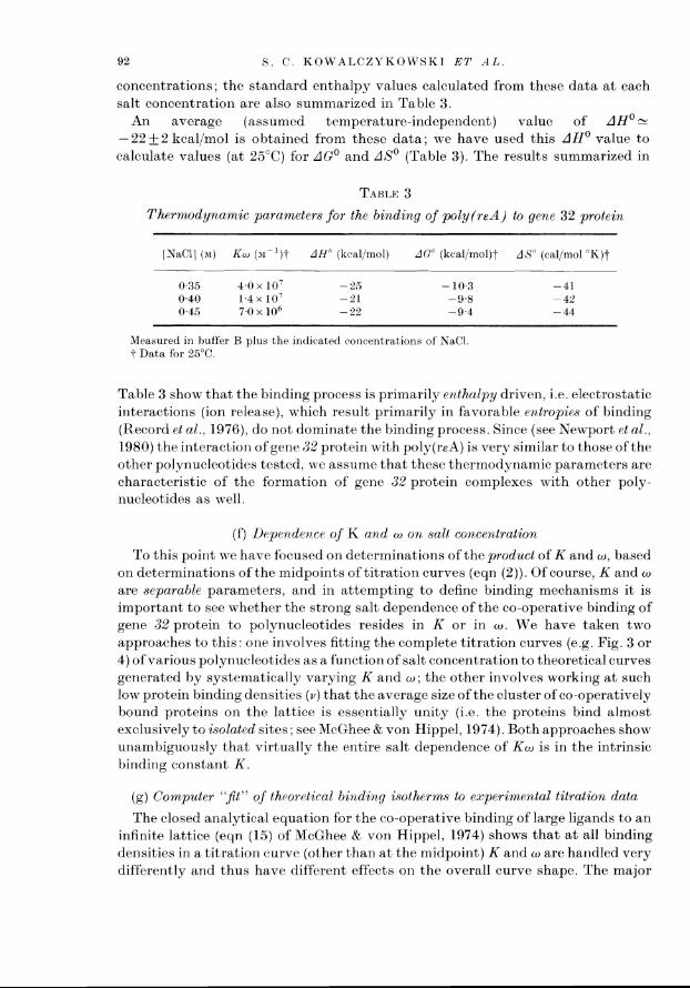
An average (assumed trmperature-independent) value of AH0 - -22+2 kcal/mol is obtained from these data; we have used this AH0 value to calculate values (at 25°C) for AGO and AS0 (Table 3). The results summarized in
TABLE 3
Thermodynamic parameters for the binding of poly(reA) to gene 32 protein
INaCl] (w) Kw (31-')-I AH" (Itcal/mol) A0" (kcal/rnol)t AS" (cal/mol "Kj t
Measured in buffer B plus the indicatrtl concentrations of' NaCl t Data for 25°C.
Table 3 show that the binding process is primarily er~thalpy driven, i.e. electrostatic interactions (ion release), which result primarily in favorable entropies of binding (Record et al., 1976), do not dominate the binding process. Since (see Newport et al., 1980) the interaction of gene 32 protein with poly(reA) is very similar to those of the other polynucleotides tested, we assume that these thermodynamic parameters are characteristic of the formation of genc 32 protein complexes with other poly- nucleotides as well
(f) Drpc.ndence of K and w orb salt concentration
To this point we have focused on determinations of the product of K and W , based on determinations of the midpoints of titration curves (eqn (2)). Of course, K and w
are separable parameters, and in attempting to define binding mechanisms it is important to see whether the strong salt dependence of the co-operative binding of gene 32 protein to polynucleotides resides in K or in w . We have taken two approaches to this one involves fitting the complete titration curves (e.g. Fig. 3 or 4) of various polynucleotides as a function of salt concentration to theoretical curves generated hy systematically varying K and w ; the other involves working a t such low protein binding densities (v ) that the average size of the cluster of co-operatively bound proteins on the lattice is essentially unity (i.e the proteins bind almost exclusively to isolated sites; see McGhee & von Hippel, 1974). Both approaches show unarrtbiguously that virtually the entire salt dependence of K w is in the intrinsic binding constant K .
(g) Comput~r " j t " of throrrtical bi71ding isotherm to experimental titration data
The closed analytical equation for the co-operative binding of large ligands to an infinite lattice (eqn (15) of McGhee & von Hippel, 1974) shows that a t all binding densities in a titration curve (other than a t the midpoint) K and w are handled very differently and thus have different effects on the overall curve shape. The major

BINDING INTERACTIONS O F G E N E 32 P R O T E I X 93
effect of increasing w is to make the curves "sharper" ; a value of w = ar, transforms the titration into a step function. The major effect of increasing K is to shift the entire curve toward lower protein concentrations; i.e. to reduce the length of the lag phase of the titration (e.g. see McGhee & von Hippel, 1974; Fig. 7).
The procedure we have used for fitting theoretical titration curves to experi- mental data is outlined in Materials and Methods ; a typical set of trial theoretical fits to a poly(reA) titration are shown in Figure 8. Clearly, the most discriminating
I I I 1 0 0.10 0.20 0.30 0-40
[ ~ e n e 32 protein] ( p ~ l
FIG. 8. Titration of poly(reA) (1.90 x M ) with gene 32 protein in buffer B with 0.45 m-NaCI at 25°C. The unbroken lines are theoretical curves generated for various values of w (with K w = 7.0 x lo6 M - I and n = 7).
parts of the fit are in the curved regions at the foot and a t the top of the steeply rising part of the curve. Since the fact that thenurleic acid lattices used are not of infinite length perturbs the fit most a t the top of the titration curves (see Newport et al., 1980, and unpublished calculations), we emphasize the fit to the foot region here. A typical set of best-fit parameters of w and K as a function of salt concentration are collected in Table 4 for poly(reA) ; and more extensive data for other polynucleotides are summarized in Table 3 of the following paper (Newport et al., 1980). The results are clear-cut; within the limits of error the salt dependence of Kw is all in K , and w is essentially salt independent.
(h) Salt dependenc~ of protein binding to isolated sites
By definition (McGhee & von Hippel, 1974) w is the ratio of the probability that a ligand will bind to a contiguous site (i.e. extend a previously initiated protein cluster

Temperature dependence of K w , K , and w for poly(reA)
LNaCl] 2' Kw w range Best fit w K ( M ) ( O C ) (Y- ' 1 0 - 1 ) ( x 1 0 - 3 ) ( x ( M - I x
0.45 25 0.7 4 . 5 1 0 5 1.4 37 0.2 0.5-2.5 1 1.7
Titrat,ions in huf'fer 13. plus added NaCl.
on the lattice) to the probability that a ligand will bind to an isolated site (i.e. initiate a new protein cluster). At relatively high binding densities ( v ) , binding is predominantly in clusters; as v is decreased the average cluster size will decrease, approaching unity. Using equation (15) of McGhee & von Hippel (1974), it can be shown that the limiting value of the free protein concentration (LF) as V + 0 is v / K ; for practical purposes this limit is attained at v w 5 0 .1 . Thus a t v > O. l /w , LF will be a function of both K and w : a t v < O.l/w. LF will be a function of K only. Therefore, in order to determine which component of K w is changing as the salt concentration is being varied, one need only to monitor L, (while keeping v constant), as the salt concentration is changed. In the limit of v + 0, one would expect LF to vary linearly with K, if K is being affected by changes in salt concentration; a t the other extreme, if only w is being affected by alterat~ons in salt concentrations, then no change in L , would be observed as the salt concentration is varied.
The low values of v required to perform such experiments with a protein character~zed by a value of w 1. l o 3 preclude the use of spectroscopic methods. On the other hand, chromatographic methods can be applied. To this end we have used small. single-strand DNA-cellulose columns under conditions where the concentration of DNA binding sites far exceeds the amount of bound protein, to maximize binding to Isolated sites.
To carry out the necessary measurements, solutions of known total protein concentration a t varying salt concentrations were added to the column, and after equilibration the protein was eluted by buffer of the same salt concentration in each case. Portions of approximately the same volume as the original column volume were collected and analyzed for protein as described in Materials and Methods. By extrapolation of these data the free protein concentration LF of each original sample equilibrated with the column could be determined.
Values of L, measured a t various salt concentrations in a set of such experiments are presented in Figure 9 From previous data we know that either K or w (or both in concert) decrease by six to seven orders of magnitude per tenfold increase in NaCl concentration The theoretical lines in Figure 9 were obtained by fixing either K or w at the value shown, and allowing the other variable to decrease by 6.3 orders of

B I N D I N G I N T E R A C T I O N S O F G E S E 32 P R O T E I S
I I I -0.1 - 0.2 - 0.3
Log [NOC!]
FIG. 9. Plot of "single-plate" DNA-cellulose column determinations of log L, as a function of log [NaCI], -23°C. The points represent experimental measurements made a t v = 1.3 x the size of the points indicates the magnit,ude of the standard error. The 2 sets of'theoretical lines represent values of L, (i.e. l/Kw) for fixed values of K = 100,200,500 and 740 x-' with w varying with salt (lines 1 through 4), and for fixed values of'w =500,750, 1000,1500 and 2000 with K varying with salt (lines 5 through 9). Experimental values of Kw were obtained from measurements made on gene 32 protein binding to 4x174 DNA (see Fig. 4, Newport et al., 1980).
magnitude per log [NaCl] over the indicated range of salt concentration. LF for each salt concentration was then calculated from equation (15) of McGhee & von Hippel (1974) for the values of the total protein concentration used in the experiment; the lines for constant K and varying w shorn- some curvature because at the experimental protein concentration some protein molecules still bind at contiguous sites. Clearly, the measured values of log L, show a straight-line dependence on log [NaCI] with a good fit to the theoretical line corresponding to a constant value of w of -- lo3, while no fit is possible to the theoretical lines generated by varying w a t constant K. Thus these experiments also demonstrate that it is the intrinsic binding constant of the protein monomer to the polynucleotide lattice, and not the protein co-operativity parameter, which varies with salt concentration.
(i) The salt dependenc~ of K for the binding of gene 32 protein to polynucleotid~s depends on both cations and anions
To this point we have used equation (1) to represent the salt dependence of the binding of gene 32 protein to nucleic acid lattices. This equation attributes the entire salt effect upon K to cation displacement; it is assumed that no anions subject to displacement are bound to the free protein. This assumption has been justified experimentally for the binding of Mg2+, oligolysines and lac repressor to D N A (see Rrrord et al., 1976)

96 S . C . K O W A L C Z Y K O W S K I ET A L .
Based on a slope (8 log Kwla log [NaCl]) of - - 7 in the log-log plots (see above and Newport et al., 1980), and assuming equation (l), we calculate a value of m' = 10 to 11 ionic interactions for each molecule ofgene 32 protein bound to single-stranded polynucleotides. Since the binding site size (n) for gene 32 prohein is only six to seven nucleotide residues in length, the above value of m' is obviously far too large, suggesting that anion binding (or perhaps changes in protein hydration) must also be involved. To determine what proportion of the salt dependence of gene 32 protein binding to polynucleotide is due to anion displacement, a series of titrations was carried out using sodium salts with different anions. In Figure 10 it can be seen that changing the anion has an effect on both the slope and position of
Log [NO+]
FIG. 10. Effects of various anions on Kw of gene 32 protein binding to poly(rA), determined by titrations contlucted identically to those of Fig. 6(a) and (b). OAc, acetate.
the line defining Kw as a function of [Na']. This strongly suggests that anions are released from the protein when it binds to a polynucleotide.
Anions that bind with relatively higher affinity to proteins, as predicted by the Hofmeister series (see von Hippel & Schleich, 1969), have a larger effect on the binding of gene 32 protein to poly(rA) than do anions that are expected to be less tightly bound. Titrations with poly(rC) (not shown) a t low salt concentration show the same change in slope that is observed with poly(rA). For salt concentrations 2 150 mM, the log Kw versus log [salt] plots are linear for all polynucleotides. At constant pH (follou7ing Record et al., 1976), we may write:
- a log Kw
= m'# + a KArX-1 a log [ M X ] 1 + K,[X-] '
where a is the number of independent anion binding sites per protein, K , is the binding constant for anions to these sites, and [X-] is the concentration of anions.
The lack of curvature in the plots of Figure 10 suggests that K,[X-] 9 1, and therefore equation (3) reduces to :
- a log KW
= ml#+a a log [MX]

B I N D I N G I N T E R A C T I O N S O F G E N E 32 P R O T E I X 97
Thus the value of a log Kwld log [MX] must be attributed to both cation (mlz,h) and to anion ( a ) displacement. The slopes obtained from log-log plots for the various sodium salts tested (Fig. 10) are summarized in Table 5 . For these salts (mlz,h+a) is smallest for NaF which, based on the Hofmeister series, would be expected to contain the weakest-binding anion tested (F-). Thus m' can be no more than 4.5 for Na' and may be less if F- does bind to the protein.
TART,E 5
Salt dependence of the binding of gene 32 protein to polynucleotides
Polynucleotide Salt
Poly(rA) NaCl Na,SO, Na(OAc) NaF
Poly(rU) NaCl MpC1, Mg(OAc),
Based on data obtained by the salt back-titration method (see Figs 10 and I I ) . All titrations were carried out in buffer C plus added salt as indicated.
t See eqn (4); all slopes show a stantlard error of' i 0.6. 1 Na,SO, may be incompletely ionized at these concentrations (Robinson & Stokes, 1959). 5 Single-ion values estimated from the slopes of'the Na+ and Mg2+containing salts, based on eqn (4)
and the constraint that (ml$),,+ = 2 (vz'$),,~+. 1 1 Cation contribution to the overall slope; this term =m'$. 7 Anion contribution to the overall slope. OAr. acetate.
( j ) M g 2 + binding
To determine the fraction of the salt dependence of the binding of gene 32 protein to polynucleotides that is due to anions, a series of titrations similar to those of Figure 10 was carried out using Mg2+ salts (Fig. 11). Mg2+ binds much more tightly to polynucleotides than does Naf (deHaseth et al., 19773). Furthermore, because it is divalent, that portion of the slope (d log Kwld log [MgCl,]) defining the gene 32 prot,ein salt dependent binding that is due to cation competition should be one- half of that observed when Na' is used as the cation. As can be seen in Figure 11, Mg2+ shifts the position of line defining the salt dependence of gene 32 protein binding to poly(rU) to much lower salt concentrations, indicating that Mg2' binding and displacement do occur. However, there is only a small effect on the slopes of these lines relative to those obtained using NaC with the same anions (All titrations in Mg2+ were carried out a t MgZ+ concentrations low enough to avoid any salt dependent changes in polynucleotide conformation.)
Both of these observations are consistent with the notion that m'4 for Na+ (and Mg2+) is small, and that the slope is dominated by anion displacement effects. The change in slope is consistent with the single ion values assigned in Table 5 , which

S C K O W A L C Z B K O W S K I ET A L
5.0 I I I I 1 \ I
-1.1 - 1.0 - 0.9 - 0.8 -0.7 - 0-6 - 0.5
Log [catton or an~on]
FK:. 11. Effects of MgZt concentration on Kw for gene 32 protein binding to poly(rU). The unbroken lines corresponti to log K w plotted against the logarithm of thc cation concentration; the broken lines represent the same data plotted against the logarithm of the anion concentration. OAc, acetate.
were calculated based on equation (4), with the additional constraint that (mli,b),,+ must be twice (mli,b),,z+. We note that there is no requirement that m'$ be aninteger; the estimates of a able 5 simply represent first approximation values of these parameters, consistent with the accuracy to which the differences in the slopes of' the log K w versus log /MX] plots can be estimated. We conclude from these results that m' for Na' is between two and four; i.e. two to four ionic interactions involving nucleic acid phosphates and basic residues of the protein are formed on complex formation between a gene 32 protein monomer and a nucleic acid lattice. This is larger than the value of m' measured for short oligonucleotide binding to gene 32 protein (we note also that oligonucleotide binding showed no anion dependence). However, the results of the salt dependence for polynucleotide binding suggest that up to four C1 ions may be bound to t>he protein and displaced by nucleic acid binding in the polynucleotide m o d ~ (see Discussion).
( k ) Binding of gene 32 protein to oligonucleotides of intermediate length (1=10 to 12)
A nurnber of semiquantitative experiments wab performed to examine the binding of gene 32 protein to oligonucleotides s~xfficiently long to be capable of binding tu~o protein monomers, to obtain some insight into the transition region between short oligonucleotide binding to a single protein monomer, and co operative protein bindirlg to a polynucleotide. Titrations of 10 and 12-mers were carried out as described for the short oligonucleotides, monitoring the quenching of intrinsic protein fluorescence as oligonucleotides were added to a fixed concentration of gene 32 protein The protein caoncentrations used were -2 x 1 0 ' to 3 x lo-' M, and titrations were performed in buffer B containing appropriate concentrations of added Na('1.

R I N D I K G I?rTTEKACTIOSS O F G E N E 3 2 P R O T E I N 99
All 10-mers and 12-mers examined (d(pT),,, d(pT),, and d(pA),,) bound two protein monomers per oligonucleotide. This conclusion is based on the fact that a t conditions close to binding saturation (at both high and low salt concentrations), the ratio of bound protein to oligonucleotide always fell above one (and, of course, below 2 ) . Values of binding constants (K,,,) were estimated as the reciprocal of the values of LF that apply a t the midpoints of the titrations. Clearly, the constants obtained represent "hybrid" values; if we assume that one protein binds essentially in the oligonucleotide binding mode (KO,,,,) and the other binds (co-operatively) in the polynucleotide mode (Kpolyw), then K,,, should be of the order of magnitude of the sum of KOli,, and (KpOlyw) under these conditions (see derivation by Draper & von Hippel, 1979).
The salt dependence of K,,, (measured as above) is shown in Figure 12, and also summarized in Table 6. Figure 12 shows that log K,,, is an approximately linear
Log [N~CI]
FIG. 12. Salt dependence of K,,, for various intermediate sized oligonucleoticles in buffer B plus added KaCI a t 25°C. The numbers in parentheses are the values of Q, obtained in that particular titration; note that Q, increases at low salt concentrations. (m) d(pT),,,; (a) d(pT), , ; (A) d(pA),,.
function of log [NaCl] for salt concentrations greater than - 0.1 M. The slopes (d log Kapp/d log LNaCI]) listed in Table 6 are obtained fiom these data. Figure 12 also shows that oligonucleotide binding becomes weaker a t low salt concentrations; KO,,,, dropping precipitously with decreasing salt concentrations below - 0.01 M-
NaC1. This is in keeping with earlier observations indicating that gene 32 protein

Salt dependence of the binding of intermediate length oligonucleotides to gene 32 protein
Oligonucleotitle (? log K,,,/a 1% 1NaCll)t o r m'S
K,,, determined fiwm tit,rat,iorls as clescrihed in the text. t Determin~d using only the data points of Fig. 12 obtained a t [NaCl]>O.l M.
1 I'olynucleotitle values of' # (Recorti et al., 1976) for poly(rU) were used fbr the T-containing oligonuc~leotides, and $ values fbr poly(rA) were used for the A-containing oligonucleotitles. End effect corrections to these poly~lucleotide values of 4 were made as tlescribed by Recortl & Lohman (1978): see the text.
5 Values of m' are determined nsst~ming no anion binding effects for gene 32 protein binding to these oligonucleotides. This is probably rrot entirely correct; see the preceding sections.
becomes denatured a t low salt (Jensen et al . , 1976), and suggests that the protein undergoes a (reversible) change to a non-binding conformation at low salt concentration. We note that Q , also changes with salt concentration a t low salt (Fig. 12), which is consistent with a conformational change in the protein under these conditions. This non-binding (low salt) form of the protein should not affect binding data obtained a t 0.1 M - N ~ C ~ or above.
The salt dependencies of the oligonucleotides listed in Table 6 show a marked increase of the salt dependence of K,,, on oligonucleotide length. (Data for d(pT), and d(pA),, taken from Table 2, are included for comparison.) Clearly, as l increases the salt dependence changes toward that characteristic of the polynucleotide binding mode, in keeping with the interpretation that one protein is binding in the short (low-salt dependence) oligonucleotide mode, and the other (co-operatively) in the (high-salt dependence) polynucleotide mode.
4. Discussion
In this paper we have established the following facts concerning the interaction of gene 32 protein with nucleic acids :
(1) Oligonucleotides of length shorter than, or approximately equal to, the pro- tein monomer site size (n =7 f 1 nucleotide residues) bind with a binding con- stant (K,,,,,) -. lo5 M - ' . Within the limits of error of our (absolute) measurements, K,,,,, is virtually independent of base composition and chain length (for 2 2 1 2 8), though RNA oligomers may bind somewhat more weakly (3 to 6-fold) than the homologous DNA oligomers. KO,,,, is also relatively salt insensitive (a log K,,,,,/d log (NaCl] 5 - 1 for this binding mode), and the binding is anion independent.
(2) Gene 32 protein binds co-operatively to polynucleotides, with an apparent

B I N D I N G I N T E R A C T I O N S O F G E N E 32 P R O T E I N 101
affinity (Kw) that varies significantly with base composition and sugar type (see Newport et al . , 1980). Binding is also strongly dependent on salt concentration (a log Kwld log /NaCl] 2. - 7 for this binding mode), and a large part of this effect is due to the displacement of bound anions from the protein as a consequence of complex formation. The experiments in which K and w have been measured separately also show that the intrinsic binding constant (K) for a protein molecule bound to a polynucleotide lattice a t either an isolated or a contiguously bond site is the same, and that the salt dependence of the binding is virtually all in K. The co- operativity parameter ( w ) appears to be invariant with salt concentration a t a value of - lo3.
These results suggest that there are two modes of interaction of gene 32 protein with nucleic acid, and that these two modes show quite different binding characteristics to nucleie acid lattices and probably involve different protein conformations. Figure I3 presents models for the binding interactions that are consistent with all these facts.
In Figure 13(a) we depict the interaction of gene 32 protein with short oligonucleotides. To account fbr the absence of the statistical ("shuffling") factor in the binding constant, we surmise that binding can take place only a t the end of the
( a ) Ol~gonucleotide binding mode ( b ) Polynucleotide b~nding mode
Anlon blnding site &
2 residues 7 residues
( c Co-operative polynucleotide b~nding mode
= 103
FIG. 13. Schematic models o f 3 modes of' gene 32 protein binding to nucleic acid lattices. (a) Binding in the oligonucleotide binding mode; note the presence of the block to statistical shuffling of
the oligonucleotide in the binding site, as well as the presence of the anion binding site (see the text). (b) Isolated binding in the polynurleotide mode: note that the shuffling block has been moved away,
exposing the positively charged binding sub-site, and the anion binding site has been disrupted. Also the nucleic acid lattice between the 2 binding sull-sites is some~i~hat stretched, and the nail-electrostatic (XpX) binding sub-site is somewhat altered, indicating that gene 3% protein binding in this mode (confbrmation) shows somewhat enhanceti base compositiorial specificity (see the text; and h'ewport et al., 1980).
( c ) Contiguous binding in the polyrrucleotitle mode: the gene 32 protein binding conformation is unchanged from that of (b) . except for protein-protein interactions and co-operative extension of the nucleic acid lattice between a11d through contiguously bound protein monomers (see the text).

102 8 . C . K O W A L C Z Y K O W S K I ET A L
oligonucleotide lattice (we have no basis, a t presel~t,, to choose between the 5' and the 3'-end), and involves primarily two nucleoside units and the intervening phosphate. This is consistent with the fact that the full binding affinity is attained with d(XpX) (or r(XpX)) and that KO,,,, is not increased over the XpX value for pXpX or XpXp (Kelly et al., 1976). It is also consistent with the salt dependence of binding, which suggests the involvement of a t most one ionic interaction between a phosphate and a basic amino acid residue. The longer oligonucleotide lattices do bring about appreciable additional quenching of the intrinsic (primarily tryptophan; see Kelly & von Hippel, 1976) fluorescence of the protein, suggesting that the chain makes contact with the protein surface, though not in a way that contributes significantly to the binding free energy. The easiest way to think about this binding interaction is to imagine that there is a "block" ("flap"; "lump"; see Fig. 13(a)) in the binding site that prevents statistical binding (by a simple steric effect?) of oligonucleotides longer than 1 = 2 when the protein is in its oligoiiucleotide binding conformation.
In Figure 13(b) we model the int,eraction of a gene 32 protein monomer bound to a polynucleotide lattice in the "isolated" binding mode. In order to "span" the polynucleotide (i.e. to let it bind i11 such a way that the lattice runs "through" the protein), the lump blocking lattice shuffling in Figure 13(a) has been removed; permitting binding across the entire protein molecule. Concomitant with the removal of this block, a positively charged site on the protein is "disrupted", with the accompanying displacement of several ( -4, if C1-) anions from the protein (see also Lonberg et al., 1980). The nature of this anion binding sife and its disruption is not clear; obviously it cannot involve simple replacement of the Cl ions bound by additional nucleotide phosphates, since then additonal Na' (or other cationic) counterions bound to the DNA would have to be displaced from these. Presently, the simplest view is to imagine that the adoption by the protein of the poly- nucleotide binding mode requires a conformational change that results in the pulling apart of a cluster of p~si t~ively charged residues on the protein that had previously comprised the tight anion binding siteis); (note that the schemat,ic "depression" in Fig. 13(a), corresponding to the supposed anion binding site, has been removed in Fig. 13(b) and (c)). Two to four basic amino acid residues (three are shown in Fig. 13(b)) that had been shielded (perhaps by the block to oligoniicleotide shuffling, see above) from interacting with the nucleic acid simultaneously became available for DNA phosphate binding.
This interpretation of the differences between the short oligonucleotide and polynucleot'ide binding modes is consistent with the binding properties of inter mediate length oligonucleotides t,hat can bind two protein molecules. The length dependence of the binding interaction, the magnit,ude of the apparent binding constant, and the salt dependence of the binding are all consistent with two proteins binding co-operatively; one in the short olignonucleotide and one in the poly- nucleotide binding mode.
We may ask why the sites of interactions involved in polynucleotide binding are not availabe to oligonucleotide lattices of sufficient length (1 = 6 to 8) ? Presumably the free energy cost of the anion displacement, toget>her with that of the conformational change in the protein (including the physical displacement of the lump of Fig. l3(a)) is such as to make isolated binding a t moderate salt

B I N D I N G I N T E R A C T I O N S O F G E N E 3 2 P R O T E I N 103
concentrations less favorable than oligonucleotide-type end binding (see also discussion by Newport et al., 1980). For a long polynucleotide lattice there are so few ends that effectively only isolated binding in the polynucleotide mode is available to the system, even at very low binding density.
Figure 13(c) shows several gene 32 protein monomers binding contiguously (co- operatively) to the polynucleotide lattice. The major interactions between the protein and the polynucleotide are identical in both isolated and contiguous binding (see above), but co-operativity further stabilizes the interaction by increasing the effective binding affinity by a factor of -- 1 o3 ( w ) per protein monomer. At this point we know relatively little about the molecular interactions corresponding to this co- operativity. They may involve favorable protein-protein contacts (see Fig. 13(c); also Lonberg et al., 1980); though these interactions cannot be electrostatic in nature, since OJ shows virtually no salt dependence. Another possibility is that co- operativity of binding may arise in part from the relative placement of'the two nucleic acid binding sub-sites (Fig. 13(b)).
One way of imagining this latter possibility is to place the XpX and the salt dependent nucleic acid binding sub-sites oil the protein monomer in such a way as to require the lattice to "stretch" (be deformed) in order to bind to both sub-sites simultaneously. This stretched configuration of the backbone might then be stabilized by the next contiguously binding protein monomer, which is shown in Figure 13(c) as being "in register" with the stretched configuration required to span the two sites in the initial protein monomer. This "lattice distortion" model is consistent with the preferential binding of gene 32 protein clusters to DNA relative to RNA (see Newport et al., 1980), though results obtained with specifically proteolytically cleaved fragments of gene 32 protein suggest that direct pro- tein-protein interactions may indeed be responsible for most of the observed co- operativity (see Lonberg et al., 1980).
This research was supported in part by United States Public Health Service research grant GM-15792 and by American Cancer Society post-doctoral fellowship PF-1301 (to S. C. K.). One of us ( J . W. N.) was a pre-doctoral trainee on United States Public Health Service training grants GM-00715 and GM-07759; a portion of this work has been submitted (by J. W. N.) to the Graduate School of the University of Oregon in partial fulfillment of the requirements for the Ph.D. degree in Chemistry. We are grateful to Dr Larry Gold of the University of Colorado for helpful discussions throughout this work, and for providing the gene 32 protein-overproducing T4 strains. A preliminary account of some of this work was presented a t the FASEB meeting in Dallas, Texas, April 1979 ; as well as a t the FEBS meeting on Protein-Nucleic Acid Interactions in Prague, Czechoslovakia, September 1979.
REFERENCES
Alberts, B. M. & Frey, L. (1970). Nature (London), 227, 1313-1318. Berger, H., Warren, A. J . & Frey, K. E . (1969). .J. Virol. 3, 171-175. Bernstein, H. (1968). Cold Spring Harbor S y m p . Quant. Biol. 33, 325-331. Bittner, M., Burke, R . L. & Alberts, B. M. (1979). J. Biol. Chem. 254, 95659572. Bobst, A. M. & Pan, Y.-C. E. (1975). Biochem. Riophys. Res. Commun. 67, 562-570. Carroll, R . B., Neet, K. & Goldthwait, D. A. (1975). J. Mol. Biol. 91, 27*%291. deHaseth, P. L., Gross, C. A., Burgess, R . R . & Record, M. T. J r (1977a). Biochemistry, 16,
4777-4783.

104 S . C . K O W A L C Z Y K O W S K I ET A L .
deHaseth, P. L., Lohman, T. M. & Record, M. T. J r (19776). Biochemistry, 16, 47834790. Delius, H., Mantell, N. J . & Alberts, B. (1972). J. ,Wol. Biol. 67, 341-350. Draper, D. E. & von Hippel, P. H. (1979). Biochemistry, 18, 753-760. Epstein, R . H., Bolle, A, , Steinberg, C. M., Kellenberger, E., Boy de la Tour, E. , Chevally, R.,
Edgar, R . S., Susman, M., Denhardt,, G. H. & Lielausis, A. (1963). Cold Spring Harbor Symp. Quar~t. Biol. 28, 375394.
Fasman, G. D. (1975). Editor of Handbook of Biochemistry and Molecular Biology, 3rd edit. vol. 1, p. 589, CRC Press, Cleveland.
Gold, L., O'Farrell, P. Z. & Russel, M. (1976). J. Biol. Chem. 251, 7251-7262. HelBne, C., ToulmB, F . , Charlier, M. & Yaniv, M. (1976). Biochem. Biophys. Res. Commun. 71,
91-98. Janik, B., Sommer, R . G., Kotiek, M. P., Wilson, D. P. & Euckson, R. J. (1973). Physiol.
Chem. Phys. 5, 27-36. Jensen, D. E. & von Hippel, P. H. (1976). J. Biol. Chem. 251, 7198-7214. Jensen, D. E., Kelly, R . C. & von Hippel, P. H. (1976). J. Biol. Chem. 251, 721,57228. Kelly, R . C. & von Hippel, P. H. (1976). J. Biol. Chem. 251, 7229-7239. Kelly, R . C., Jensen, D. E. & von Hippel, P. H. (1976). J. Biol. Chern. 251, 7240-7250. Krisch, H. M., Bolle, A. & Epstein, R . H. (1974). J. Mol. Biol. 188, 89-104. Lehrach, H. & Scheit, K. H. (1973). Biochim. Biophys. Acta, 308, 28-34. Lemaire, G., Gold, L. & Yarus, M. (1978). J. Mol. Riol. 126, 73-90. Lonberg, N., Ko~a lcz~kowsk i , S. C., Paul, L. S. &: von Hippel, P. H. (1980). J. Mol. Biol.
145, 123-138. McGhee, J. D. (1976). Biopolymers, 15, 1345-1375. McGhee, J. D. & von Hippel, P . H. (1974). J. Mol. Biol. 86, 469-489. McGhee, J . D. & von Hippel, P. H. (1976). J. Mol. Biol. 103, 679, (correction for McGhee &
von Hippel (1974)). Newport, J. W., Lonberg, N., Kowalczykowski, S. C. & von Hippel, P. H. (1980). J. Mol. Biol.
145, 105-121. Record, M. T. J r & Lohman, T. M. (1978). Biopolymers, 17, 159-166. Record, M. T. J r , Lohman, T. M. & deHaseth, P. (1976). J. Mol. Biol. 107, 14r-158. Record, M. T. J r , Anderson, C. F. & Lohman, T. M. (1978). Quart. Rev. Biophys. 11, 103-178 Revzin, A. & von Hippel, P. H. (1977). Biochemistry, 16, 4769-4776. Riva, S., Cascino, A. & Geiduschek, E . 1'. (1 970). J. Mol. Biol. 54, 85-1 02. Robinson, R . A. & Stokes, R. H . (1959). In Electrolyte Solutions, p. 416, But,terworths,
London. Russel, M., Gold, L., Morrisett, H. & O'Farrell, P. Z. (1976). J. Biol. Chem. 251, 7263-7270. Secrist, J. A,, Barrio, R . J., Leonard, N. J. & Weber, G. (1972). Biochemistry, 11, 3499-3506. Steiner, R . G., Kinnier, W., Lunacin, A. & Delac, J . (1973). Biochim. Biophys. Acta, 294,
24-37. Tomizawa, J., Anraku, h'. & Iwama, Y. (1966). J. Mol. Biol. 21, 274-283. ToulmB, J. J. & Helkne, C. (1980). Biochim. Biophys. Acta, 606, 95-104. von Hippel, P. H. & Schleich, T. (1969). Acc. Chem. Res. 2, 257-265. Wu, J . & Yeh, Y . (1973). J. Virol. 12, 758-765.