inspirational evolution: the avian lower respiratory tract · and birds exhibit similar anatomy and...
TRANSCRIPT
www.aavac.com.au© 1
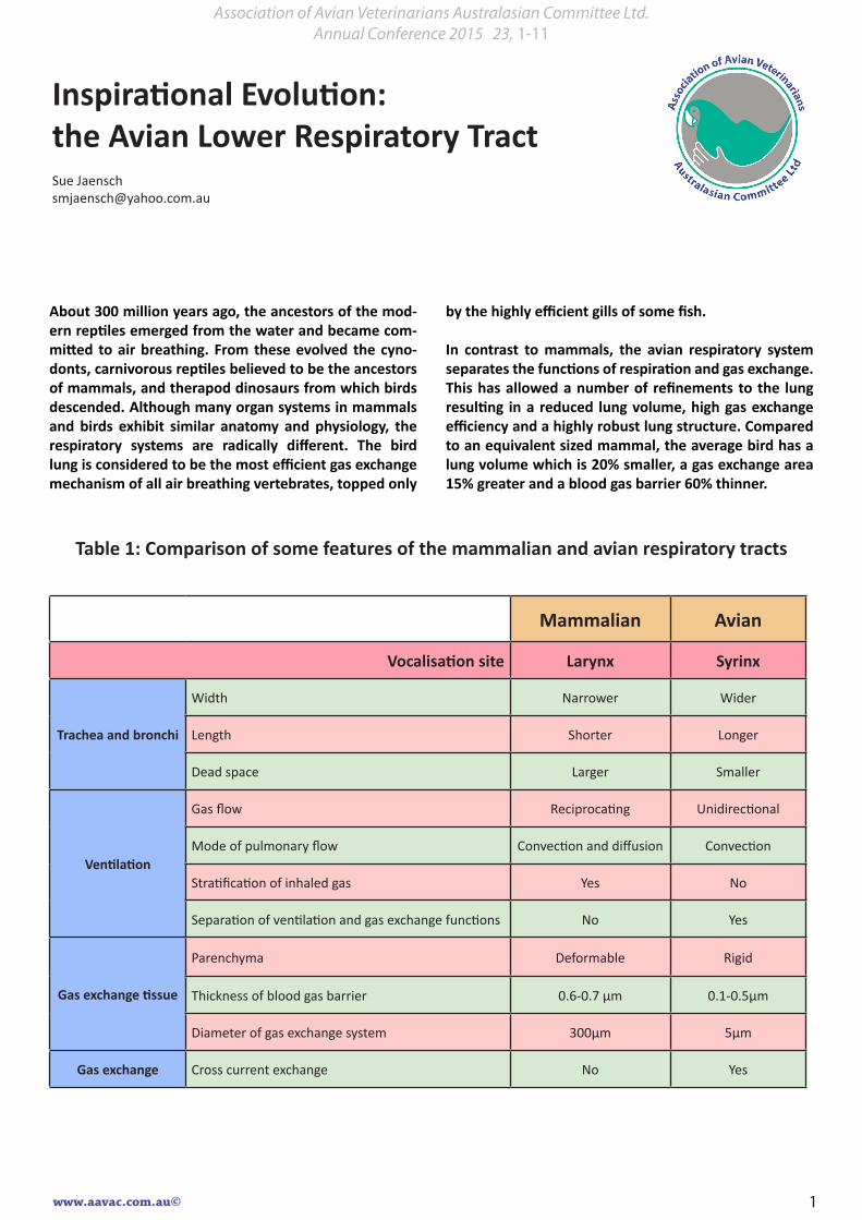
About 300 million years ago, the ancestors of the mod-ern reptiles emerged from the water and became com-mitted to air breathing. From these evolved the cyno-donts, carnivorous reptiles believed to be the ancestors of mammals, and therapod dinosaurs from which birds descended. Although many organ systems in mammals and birds exhibit similar anatomy and physiology, the respiratory systems are radically different. The bird lung is considered to be the most efficient gas exchange mechanism of all air breathing vertebrates, topped only
by the highly efficient gills of some fish.
In contrast to mammals, the avian respiratory system separates the functions of respiration and gas exchange. This has allowed a number of refinements to the lung resulting in a reduced lung volume, high gas exchange efficiency and a highly robust lung structure. Compared to an equivalent sized mammal, the average bird has a lung volume which is 20% smaller, a gas exchange area 15% greater and a blood gas barrier 60% thinner.
Inspirational Evolution: the Avian Lower Respiratory TractSue [email protected]
Association of Avian Veterinarians Australasian Committee Ltd. Annual Conference 2015 23, 1-11
Mammalian Avian
Vocalisation site Larynx Syrinx
Trachea and bronchi
Width Narrower Wider
Length Shorter Longer
Dead space Larger Smaller
Ventilation
Gas flow Reciprocating Unidirectional
Mode of pulmonary flow Convection and diffusion Convection
Stratification of inhaled gas Yes No
Separation of ventilation and gas exchange functions No Yes
Gas exchange tissue
Parenchyma Deformable Rigid
Thickness of blood gas barrier 0.6-0.7 µm 0.1-0.5µm
Diameter of gas exchange system 300µm 5µm
Gas exchange Cross current exchange No Yes
Table 1: Comparison of some features of the mammalian and avian respiratory tracts

www.aavac.com.au© 2
Respiratory cycle
Gas flow directions in the avian respiratory tract has been the subject of many studies published over the last 40+ years, and the theories of gas movement have changed dramatically over this time. Some of the early models de-clared that there was no gas flow through the lungs at all! Under the currently accepted model, it takes two respi-ratory cycles to move gas through the entire respiratory tract. This initially appears inefficient, but in fact is an important physiological process behind respiratory effi-ciency in birds, as it allows continuous and unidirectional gas flow through the lung both on inspiration and expi-ration. Control of the direction of airflow though the re-spiratory tract remains poorly understood, with physical valving, speed of gas flow and arteria PCO2 all likely play-
ing a role. CO2 receptors are the principle driving force behind respiration in birds. These are directly inhibited by halothane, explaining the ready induction of apnoea when using this anaesthetic agent.
We are all familiar with diagrams depicting gas flow though the respiratory tract (Figure 1). In reality, gas flow is more complex, with approximately 50% of the inspired air passing into the lungs and 50% into the caudal air sacs. The inspired air that directly enters the lungs moves though both neobronchi (in species in which they is pres-ent) and parabronchi, and contributes significantly to gas exchange. On the first expiration, the gas from the caudal air sacs passes into the lungs. On the second expiration, the gases move from the lungs into the cranial airs sacs. On the second expiration, these gases are finally exhaled.
Figure 1: Avian respiratory cycle
http://www.peteducation.com/article.cfm?c=15+1829&aid=2721

www.aavac.com.au© 3
Gas in the caudal air sac has a very slightly lower PO2 and slightly higher PCO2 than room air largely due to dead space of the trachea plus some minor gas exchange as some inspired air passes through the lungs on the way to the caudal air sacs. Gas in the cranial air sacs has a PO2 significantly lower and PCO2 significantly higher than room air (see Table 2).
Trachea
Inhaled air passes down the trachea, through the syrinx,
then down the bronchi. This seems like a simple enough process, but marked species variation in anatomy of the trachea and syrinx are present. Some species have remarkably elongated and convoluted tracheas (Figure 2). These are thought to have evolved to allow loud and resonate vocalisation, and are variably theorised to allow a trombone like effect or a harmonic vibrating effect (in species with tracheas within the keel). However, not all species with such trachea make loud vocalisations, and some birds produce similarly loud and resonant vocalisa-tions without an elongated trachea.
Figure 2b. Elongated tracheae of the trumpet manu-code (Phonygammus keraudrenii), family Paradisaei-dae.
Three whole adult males, American Museum of Natural History Alcohol Col-lection nos. 17, 19 and 18. Specimen 18 (C,C’) was at least 13 years old when it died - its trachea was overlapped in two places, and was approxi-mately 75 cm long.Figure 2a. Examples of tracheal loops
Figure 2: Variations in avian tracheal anatomy
Both images: http://scienceblogs.com/tetrapodzoology/2009/04/29/elongate-avian-trachea/
From a medical point of view, these convoluted tracheas may reflect in an increased risk of foreign body deposition, an increased risk of spread of re-spiratory disease to surrounding structures, and due to their dead space, potential complications with anaesthesia. Radiology and endoscopy can be used to evalate tracheal disease. Endoscopy may allow foreign body removal and biopsy of lesions for his-tology and culture. Laser ablation and baloon dila-tation of tracheal obstructions are described in the literature. Surgical excision of tracheal lesions may be complicated by the complete cartilagenous rings, minimal ability to elongate the trachea to achieve closure and rapid reduction in diameter of the tra-cheal below the larynx in some species, resulting in mis-match of sizes when anastomising.
Syrinx
The syrinx is located at the end of the trachea and is variably fused to tracheal and / or bronchial cartilag-es. There is considerable variation in the anatomy of the syrinx between species, with some birds hav-ing extensive out-pouching structures which may be asymmetrical (left Figure 3). The syrinx undergoes significant alternations in internal structure during vocalisation with rapid contraction of the syringeal muscles either unilaterally or bilaterally (right Figure 3).
The convoluted structure of the syrinx provides the first major site of change of air flow speed and direc-tion within the respiratory tract. Thus it is a common site for deposition of inhaled particles. As a result, obstruction by foreign bodies or granulomas is rel-atively common. The syrinx can also be affected by

www.aavac.com.au© 4
nutritional deficiency resulting in squamous meta-plasia, parasites and neoplastic obstruction.
The location of the syrinx complicates therapy. Simi-lar disease entities occurring in a mammal, although problematic, are more amenable to surgical evalua-
tion and treatment. Endoscopy can be used to eval-uate disease in the syrinx, and may be curative when an inhaled foreign body or inspissated mucous plugs can be removed. Endoscopically sourced biopsy of granulomas or masses can provide diagnostic sam-ples for histology and culture.
Figure 3: Syrinx functional anatomy
Left: http://www.ejpau.media.pl/volume13/issue4/art-22.htmlRight: http://www.pnas.org/content/94/26/14787/F2.expansion.html
Air sacs
The air sacs are capacious, transparent structures which communicate with the lungs via the ostia. The air sacs contribute significantly to the higher re-spiratory volume of birds (100-200 ml/kg bw) com-pared to mammals (35-70 ml/kg bw). Air sacs are avascular and play no direct role in gas exchange. Their interconnections, extensions and sizes vary between species, however a common pattern in do-mestic species is paired cranial thoracic, caudal tho-racic and abdominal air sacs, with single (or fused paired) cervical and clavicular air sacs (Figures 4 and 5). Extensions into and around bones as well as into perirenal and subcutaneous spaces are common in flying birds and less common in walking and diving birds, leading to a hypothesis that pneumaticity like-ly contributes to reduction in tissue mass and densi-ty. There is also a cervicocephalic air sac which arises from the infraorbital sinuses but is not attached to the lungs. This is variably developed between spe-cies, being most prominent in strongly flying birds.
The air sac walls predominantly consist of simple squamous epithelium but near the ostia patches of ciliated columnar cells occur. Diving species may
have slightly thicker walled air sacs with columnar type epithelial cells. In the absence of a diaphragm, air movement through the air sacs is driven by move-ment of the ribs and sternum. In running or flying birds, the respiratory cycle is coupled to the wing beat or foot movements that increase thoracic vol-ume during respiration. The uncinate processes on the ribs are important in cranially rotating the ribs (and hence ventrally rotating the sternum) during in-spiration, and are particularly active when birds are resting in a sternal position. Minor tail movements can be seen with inspiration in normal birds. Exag-gerated tail bobbing is often identified in birds with significant lower respiratory disease and reflects in-creased respiratory effort. Tail bobbing may also be identified in birds in sternal recumbency reflecting reduced mobility of the sternum in this position.
The air sacs are particularly helpful to veterinari-ans by providing contrast during radiography and allowing excellent visualisation of internal organs during endoscopy. They also allow for an alternative route of gaseous anaesthesia administration in cas-es where tracheal administration is inconvenient or impossible (see discussion below).

www.aavac.com.au© 5

www.aavac.com.au© 6
The air sacs most commonly come to veterinary attention when infectious agents take root or with rupture of the air sacs causing subcutaneous em-physema. Clinical signs of air sac disease may in-clude altered respiratory pattern (tail bobbing etc) or feather picking over areas of air sac disease. Lo-calised lesions which do not disrupt air flow may be clinically occult. Infectious lesions are most com-monly identified in the caudal air sacs, reflecting the pathway of air flow through the respiratory system. Mass lesions in the air sacs may be identified on ra-diography. Diagnostic samples are best retrieved via endoscopy. Options include cytology and culture us-ing cytology brushes or lavage to collect samples, or biopsy of lesions for histology and culture. Foreign bodies can be removed by endoscopy.
As normal air sacs are poorly vascularised, systemic therapy may not result in adequate drug concen-trations in the air sacs. With inflammation, vascu-larisation of the lesion may increase the delivery of therapeutic drugs. Nebulisation (but not vapori-sation) can also be used to deliver drugs to the air sacs. Particle size is important and should ideally be no larger than 0.5 micron, but particles up to 3 micron may make it to the air sacs. Particles larger than this will be deposited in the upper airway and trachea. Nebulisation can only be expected to result in therapeutic levels of drugs in the caudal air sacs and lungs. Cranial air sacs cannot be reliably treated by this route. Systemic uptake of drugs from the air sacs is minimal.
Lungs
Advantages of the unidirectional gas flow in the lung of birds:
1. Even ventilation of the gas exchange tissue: In mammals, the lung remains partially inflated at the end of expiration, thus the inspiratory volume does not match the lung volume. As a result, proximal alveoli are preferentially venti-lated by the inspired air. More peripheral alveoli may only receive ventilation by diffusion, which is slow and inefficient. The unilateral air flow in birds results in minimal stratification of inspired air, instead all air capillaries are evenly ventilat-ed by convection.
2. Higher oxygen tension of alveolar gas: A flow on effect of the mixing of fresh and existing gas in the mammalian lung is that alveolar PO2 is considerably lower than the inspired air. The flow through system in bird lungs minimises stratification and allows higher air capillary and arterial oxygen tension (Table 2).
3. Cross-current gas exchange: Cross current gas exchange allows a highly efficient exchange of gases between the air and blood in the lungs. As blood travels towards the parabronchial lumen and air from the parabronchial lumen, there is continuous exchange of oxygen to the blood and carbon dioxide from the blood. The oxygen con-centration of the blood is lower than the oxygen concentration of the air along the full length of contact. As a result, unlike in mammals, arterial PO2 can be higher than expired air PO2.
Mammal Bird
P02 PCO2 PO2 PCO2
Inspired air 150 0 150 0
Caudal air sac 120 20
Alveoli /Air capillary 100 40 120 20
Cranial air sac 90 40
Arterial 75-90 35-45 90-100 25-30

www.aavac.com.au© 7
Figure 5: Comparison of concurrent flow and counter current flow
https://en.wikipedia.org/wiki/Countercurrent_exchange
lungs are minimally deformable, with studies in penguins showing minimal change in lung volume regardless of the positioning of recum-bency (note however that air sac volumes can be significantly reduced by different positions of recumbency). This robust nature has been char-acterised for both the air capillaries and blood capillaries in birds. In birds, pulmonary blood capillaries are supported by a three dimensional honeycomb of air capillaries providing rigidity. In contrast, mammalian pulmonary blood capil-laries have little support at right angles to their axis, increasing their risk of damage. The sequel-la of this is an increased risk of pulmonary hae-morrhage with exercise - a common outcome in racing thoroughbreds and elite human athletes.
4. Small terminal air exchange units: In mammals, air flow to the peripheral alveolar requires a mix of convection and diffusion, resulting in a need for low resistance and hence large volume alve-oli. The larger diameter alveoli in mammals re-quires greater support (including collagen) and reduces the integrity of the alveoli walls under stress. Small diameter air capillaries allow even-ly thin walled, and hence high efficiency gas dif-fusion surfaces with minimal support structures.
Advantages of separation of gas exchange and ven-tilation functions:
1. Lack of repetitive distortion of the air exchange tissue: In mammals, there in inherent tension between the drive for thin and efficient gas ex-change surfaces, and the need for these surfaces to be robust enough to withstand repetitive dis-tortion with every breath. In humans, this repet-itive distortion is thought to result in the “nor-mal” degree of emphysema identified in normal aging lungs. This requirement to tolerate distor-tion has resulted in a need for thicker walls in alveoli compared to air capillaries, and the pres-ence of fibroblasts and collagen in the alveolar walls. As discussed above, both factors reduce efficacy of gas exchange in comparison to birds.
2. Robust lungs: Mammalian lung tissue is deform-able. Compression of the parenchyma results in cessation of ventilation. In contrast, avian

www.aavac.com.au© 8
Figure 6: Electron micrographs of pulmonary capillaries in chicken (A) and dog lung (B). Note that the blood-gas barrier in the bird is much thinner and much more uniform in thickness than in the mammal. EPI, epithe-lium; F, fibrils of type I collagen; FB, fibroblasts.
http://ajpregu.physiology.org/content/297/6/R1625
Disease susceptibility
Respiratory disease is a relatively common clinical presentation in birds. One histopathological survey of chickens reported 10% of cases were respiratory (excluding poultry viral respiratory diseases).
The local immunity of the avian respiratory system remains relatively poorly understood. Substantial populations of lymphocytes are present in the na-sal mucosa and bronchus. The trachea is lined by ciliated epithelial cells, with good efficacy in clear-ing inhaled small particles by mucociliary transport. Coughing occurs in birds, but has reduced clearing of mucous from the upper airways in comparison to mammals. Birds have complete cartilaginous rings in the trachea. The lack of partial collapse of the tra-chea during coughing in birds reduces the velocity of
air flow up the trachea in comparison to mammals, and reduces the efficacy of tracheal clearing.
In the air sacs, there are small islands of lymphoid tissue and ciliated cells predominantly near the openings to the lungs, but function of these is poor-ly understood. There are few resident macrophages or lymphocytes in the air sacs, making them appear to be perfectly adapted for culture of infectious re-spiratory disease. Additionally, there is poor vascu-larisation of the air sacs, thus parenteral treatment of air sac disease is problematic. Pneumatisation of bones, between muscle bodies and subcutaneous-ly may allow the spread of respiratory diseases to abdominal organs, bone or soft tissues. Common diseases localised in the air sacs include aspergillus, bacterial infections (especially coliforms) and chla-mydia.

www.aavac.com.au© 9
Macrophages and heterophils are poorly represent-ed in the avian lung system in comparison to mam-mals. Their resident numbers in bird lungs are only 5% that in mammalian lungs however recruitment into the lungs can be rapid.
Inhalation of foreign material, whether inert or in-fectious, is a common cause of respiratory disease in birds. The size of an inhaled foreign body strongly influences the predicted site of deposition. Larger bodies will tend to be deposited by inertial impac-tion in the upper respiratory system, most common-ly in sites where the air flow is slowed, including tra-cheal bends, the syrinx and the caudal end of the primary bronchi. Smaller bodies may be deposited in the same sites, but may also accumulate in the caudal air sacs and parabronchi by gravitational sed-imentation. Understanding of species difference is important in predicting where such foreign bodies may initiate disease. Birds with more complex syr-inxes or convoluted and / or elongated tracheas may be at higher risk for foreign body associated disease in these areas. Considerations before inhalant anaesthesia
Inhalant general anaesthesia is a commonly used tool in avian medicine. Rapid improvements in tech-niques and agents have occurred over the past 20 years, with a subsequent significant drop in associ-ated morbidity and mortality. Isoflurane or sevoflu-rane administered though low dead space, non-re-breathing systems has become routine. Positioning of patients and the use of masks or intubation re-main more difficult decisions.
The ideal positioning of the patient during anaes-thesia remains difficult, with recently published papers providing conflicting findings from different species. Given the need for the keel to move for-wards and upwards to provide negative pressure during inspiration, ventral recumbency is generally considered contra-indicated. However, studies of air sac and lung volumes in anesthetised penguins and hawks found that ventral recumbency caused less reduction in air sac volume than dorsal recumben-cy. Right lateral recumbency was found to provide the least alteration in air sac volume. Lung volume was not significantly affected by positioning. Blood gas analysis was not performed in the penguins. In hawks, dorsal recumbency maintained higher PO2 than right lateral recumbency despite smaller air sac volumes. Blood gases were not assessed in ventral
recumbency. Clearly, more work needs to be per-formed to determine ideal positioning during anaes-thesia, and if this is species dependant.
The choice to intubate or not, tube design and depth of placement is something which needs to be assessed on an individual case basis. Intubation has the advantages of securing the airway (pro-tecting against regurgitation / inhalation etc) and allowing IPPV. However, there is a risk of inducing post intubation tracheal obstruction (see below) and the small tube diameter needed in many spe-cies can risk obstruction of the tube with mucous. The most common alternative is to use face mask administration. Face mask administration does not provide a secure airway but may allow some IPPV. A newer alternative is the use of laryngeal mask ad-ministration. Laryngeal masks allow the use of IPPV, but do not protect completely against regurgitation / inhalation. Careful stabilisation and monitoring of the contact of the mask with the larynx is required. Laryngeal masks are not available for smaller avian species at this time, but new products are being re-leased for small exotics, and this is an area worth watching.
Post intubation tracheal obstruction appears to be a relatively uncommon sequella of intubation, but carries significant morbidity and mortality. In one zoological setting study, between 2 and 8% of in-tubations resulted in tracheal obstruction of vari-able severity, with an overall mortality rate of 70%. Clinical presentation was 7-21 days post intubation. Potential contributors to post intubation tracheal obstruction include mechanical trauma, chemical ir-ritation (including from sterilisation liquids and gas-es), duration of intubation, dry gases, high gas flows rates and the use of positive pressure ventilation. Possible variation in risk between species has been suggested by one author with Gruiformes, Anseri-formes Galliformes and Passeriformes at higher risk and Falconiformes, Coraciformes and Psittaciformes at low risk. This may reflect anatomical differences, with a more dramatic narrowing of the trachea be-tween the glottis and the syrinx in some species, as well as an increased risk of mechanical trauma in long-necked birds.
When tracheal access is difficult during anaesthesia, due to obstruction of the surgical field or obstruc-tion of the trachea or syrinx, air sac cannulation can be utilised to provide anaesthesia. In the case of tracheal or syrinx obstruction, air sac cannulation

www.aavac.com.au© 10
Figure 7: Neonatal laryngeal mask
http://resus.me/lma-for-newborn-resuscitation/
can also be used for emergency stabilisation of pa-tients, by bypassing the obstructed area and allow-ing adequate ventilation. Low flow rates (0.3 ml/kg/min) +/- oxygen and nitrous oxide mixtures are rec-ommended for air sac anaesthesia to minimise the decrease in arterial PCO2 and induction of alkalosis, the first of which can lead to apnoea and the latter cardiac arrhythmias. Despite low flow rates, apnoea may occur if the trachea is patent. Pulse oximetry
is important in monitoring anaesthesia in apnoeic patients. Isoflurane concentrations may need to be higher than when using tracheal administration es-pecially if the trachea is patent. Exhaled gases re-quire scavenging to minimise human health risks. Air sac administration of anaesthesia results in low-er arterial and venous PO2 than routine ET tube ad-ministration, but excellent saturation is maintained.
Figure 8a: Radiograph of a duck with an air sac cannular placedCourtesy Professor Bob Doneley, Universtity of Queensdland

www.aavac.com.au© 11
Figure 8b: Radiograph of a duck with an air sac cannular placedCourtesy Professor Bob Doneley, Universtity of Queensdland
Further reading
Fedde MR. 1988. Relationship of structure and func-tion of the avian respiratory system to disease sus-ceptibility. Poultry Science 77, 1130-1138.
Maina JN. 2006. Development, structure, and func-tion of a novel respiratory organ, the lung-air sac system of birds: to go where no other vertebrate has gone. Biological Research 81, 545-579.
Maina JN, West JB, Orgeig S et al. 2010. Recent advances into understanding some aspects of the structure and function of mammalian and avian lungs, Physiolical and Biochemical Zoology 2010, 83, 792-807.
Malka S, Hawkins M, Jones J et al. 2009. Effect of body position on respiratory system volumes in anesthetized red-tailed hawks (Buteo jamaicensis) as measured by computerised tomography. Ameri-can Journal of Veterinary Research 70, 1155-1160.
Nevitt BN, Langan JN, Adkesson MJ et al. 2014. Com-parison of air sac volume, lung volume and lung den-
sities determined by use of computed tomography in conscious and anesthetized Humbolt penguins (Spheniscus humboldti) positioned in ventral, dorsal, and right lateral recumbency. American Journal of Veterinary Research 75, 739-745.
Smialek M, Tykalowski B, Stenzel T, Konciki A. 2011. Local immunity of the respiratory mucosal system in chickens and turkeys. Polish Journal of Veterinary Sciences 14, 291-297.
Suthers RA and Zollinger SA. 2004. Producing song: the vocal apparatus. Annals of the New York Acade-my of Sciences 1016: 109-129.
Sykes JM, Neiffer D, Terrell S et al. 2013. Review of 23 cases of postintubation tracheal obstructions in birds, Journal; of Zoo and Wildlife Medicine 44, 700-713.
West JB. 2009. Comparative physiology of the pul-monary blood-gas barrier: the unique avian solu-tion. American Journal of Physiology. Regulato-ry, Integrative and Comparative Physiology 297, R1825-R1634.