inorganica chimica acta - utc.edu · interaction with calf-thymus dna and photoinduced cleavage of...
TRANSCRIPT

Inorganica Chimica Acta 469 (2018) 484–494
Contents lists available at ScienceDirect
Inorganica Chimica Acta
journal homepage: www.elsevier .com/locate / ica
Research paper
Interaction with calf-thymus DNA and photoinduced cleavage of pBR322by rhodium(III) and iridium(III) complexes containing crown thioetherligands
https://doi.org/10.1016/j.ica.2017.10.0050020-1693/� 2017 Elsevier B.V. All rights reserved.
⇑ Corresponding author.E-mail address: [email protected] (J. Kim).
Jisook Kim ⇑, Ashley D. Cardenal, Hendrik J. Greve, Weinan Chen, Hitesh Vashi, Gregory Grant,Titus V. AlbuDepartment of Chemistry and Physics, University of Tennessee at Chattanooga, Chattanooga, TN 37403, USA
a r t i c l e i n f o
Article history:Received 15 August 2017Received in revised form 3 October 2017Accepted 5 October 2017Available online 10 October 2017
Keywords:PhotoactivationPhotonucleaseCisplatin9S3RhodiumIridium
a b s t r a c t
In this report, we present our investigation on the photoinduced cleavage of plasmid pBR322 and thebinding interactions with calf-thymus (CT) DNA by a series of thioether metal complexes. The complexesof interest are rhodium and iridium complexes containing thiacrown ligands 1,4,7-trithiacyclononane(9S3) and 1-oxa-4,7-dithiacyclononane (9S2O), and the complexes are abbreviated as [Rh(9S3)Cl3], [Rh(9S2O)Cl3], and [Ir(9S3)Cl3]. In the nicking assay, pBR322 was treated with each complex and irradiatedat 254 and 350 nm, respectively, in concentration- and time-dependent studies. The nicking assayrevealed that, under exposure to 254-nm radiation, [Rh(9S3)Cl3] and [Rh(9S2O)Cl3] cleaved pBR322 effi-ciently forming a nicked form, while [Ir(9S3)Cl3] was least efficient. For the 350-nm irradiation, a similartrend was observed, with [Rh(9S3)Cl3] being the most efficient one, however with a lower efficiency thanat 254 nm. An ethidium bromide displacement assay was also carried out to evaluate the binding inter-action of each compound with CT-DNA by titrating the pre-equilibrated complex of CT-DNA and EB withthe investigated complexes. The efficient concentration to achieve a 50% loss in fluorescent emission wasfound to be 24 lM for [Rh(9S3)Cl3] and 35 lM for [Rh(9S2O)Cl3], while [Ir(9S3)Cl3] was an ineffectiveDNA binder.
� 2017 Elsevier B.V. All rights reserved.
1. Introduction
Cisplatin has been a popular anticancer drug and is commonlyused for treating various types of cancers such as ovarian/cervicalcancer, bladder cancer, and/or melanoma [1–3]. The mechanism ofits action involves several steps such as cellular delivery of cis-platin, dissociation of Cl� inside the cells, binding of cisplatin tonitrogen (N-7 position) in guanines, formation of intra-strandcross-linkage to the target DNA, and apoptosis triggered by DNAdamage [1,3,4]. In spite of its efficiency as an anticancer agent,the usage of cisplatin comes with issues such as potential deactiva-tion in cells, cytotoxicity, and drug resistance [1–5]. In an effort toovercome drawbacks of cisplatin, there have been great efforts infinding new anticancer inorganic compounds. Several studiesspearheaded by photo-activation approaches showed promisingresults, focusing mainly on Ru complexes and also including Fe,Cu, Rh, and Ir complexes [2,6–28]. The idea of utilizing light ason/off switch to activate a drug for targeting DNA in cancer cells
is attractive since it is possible to design a drug which can be acti-vated only in the presence of light, while remaining inert or lessactive without irradiation. Furthermore, it is possible to tune theeffectiveness of a photoactivated inorganic compound by control-ling a time lapse for light exposure to the localized target tissueor by choosing an effective irradiating wavelength selectively.
Whether photoactivated or not, the mechanism of DNA modifi-cations caused by inorganic compounds can be complex, and itinvolves binding interactions with DNA that can be covalent,non-covalent, tight binding (via intercalation or groove binding),loose binding in a nonspecific manner, oxygen-mediated, or oxy-gen-independent [2,23,28,29]. The most likely results of interac-tions between inorganic complexes and DNA are either adductformation or DNA strand cleavage. The adduct formation withDNA was observed for platinum containing complexes such as cis-platin, carboplatin, and satraplatin [1,3,23,30], while the DNAcleavage was observed mostly in ruthenium complexes. The corestructure of the Ru complexes is [Ru(bpy)nL3-n]2+, where bpy =2,20-bipyridine and L can be ferrocene/non-ferrocene conjugatedimidazole phenol ligand [6], a series of o-, m-, or p-(nitrophenyl)imidazo[4,5-f] [1,10]phenanthroline) [7], 1,12-diazaperylene (i.e.
J. Kim et al. / Inorganica Chimica Acta 469 (2018) 484–494 485
DAP) [2,8], dipyrido[3,2-a:20,30-c]phenazine (i.e. dppz) [9,10], or4,5,9,16-tetraaza-dibenzo[a,c]naphthacene (i.e. dppn) [11,12].
In addition to extensively studied Pt- and Ru-containing com-plexes, a number of studies were focused on the biological activityof complexes with other metal centers such as Fe [19,21], Cu[15,19,31], Rh [16,20], or Ir [17,18]. Rh(III) and Ir(III) complexesdid not receive as much attention until recently since theses com-plexes were perceived to be relatively less reactive due to theirchemical stability and slow solvent exchange rate [18,32,33]. Thestability of Rh(III) and Ir(III) are in part due to the low spin state,with a d6 electron configuration, and an octahedral geometry[18,32,33]. Interestingly, some studies focus on activating Rh(III)and Ir(III) complexes by adopting suitable ligands upon irradiation.Examples are [Rh(bpy)2Cl2]+ [34], [Rh(phen)2Cl2]+ (phen, 1,10-phenanthroline) [33,34], [RhCl(bpy)9S3]2+ (9S3 = 1,4,7-trithiacy-clononane) [35,36], [IrLn]3+ where L is a terpyridyl-like ligand[17], and [(g5-Cp⁄)Ir(phen)Cl] (Cp⁄ = tetramethyl(phenyl)cy-clopentadiene) [18]. The presence of one or more chloride ionscoordinated to Rh(III) or Ir(III) metal center appears to be essentialin photoactivation of the complexes in these cases. Importantly,many of the complexes above were also shown to be active asDNA cleaving agents upon irradiation, and this finding is promisingin utilizing phototherapy for selective and efficient cancertreatment.
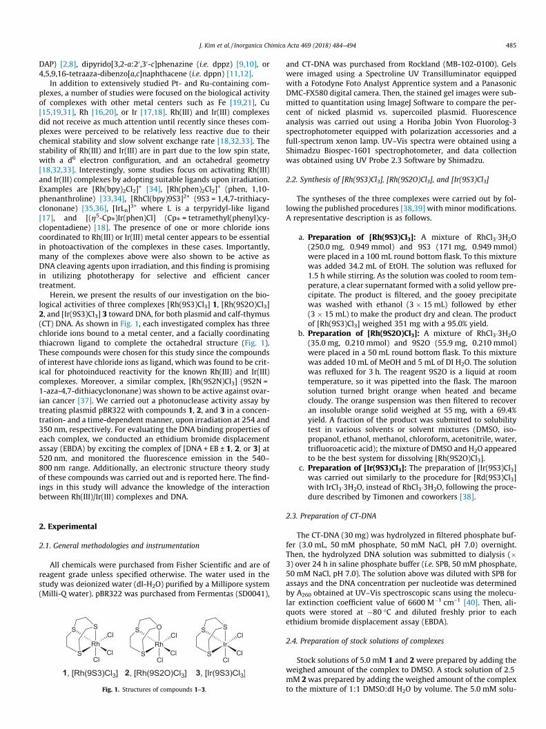
Herein, we present the results of our investigation on the bio-logical activities of three complexes [Rh(9S3)Cl3] 1, [Rh(9S2O)Cl3]2, and [Ir(9S3)Cl3] 3 toward DNA, for both plasmid and calf-thymus(CT) DNA. As shown in Fig. 1, each investigated complex has threechloride ions bound to a metal center, and a facially coordinatingthiacrown ligand to complete the octahedral structure (Fig. 1).These compounds were chosen for this study since the compoundsof interest have chloride ions as ligand, which was found to be crit-ical for photoinduced reactivity for the known Rh(III) and Ir(III)complexes. Moreover, a similar complex, [Rh(9S2N)Cl3] (9S2N =1-aza-4,7-dithiacyclononane) was shown to be active against ovar-ian cancer [37]. We carried out a photonuclease activity assay bytreating plasmid pBR322 with compounds 1, 2, and 3 in a concen-tration- and a time-dependent manner, upon irradiation at 254 and350 nm, respectively. For evaluating the DNA binding properties ofeach complex, we conducted an ethidium bromide displacementassay (EBDA) by exciting the complex of [DNA + EB ± 1, 2, or 3] at520 nm, and monitored the fluorescence emission in the 540–800 nm range. Additionally, an electronic structure theory studyof these compounds was carried out and is reported here. The find-ings in this study will advance the knowledge of the interactionbetween Rh(III)/Ir(III) complexes and DNA.
2. Experimental
2.1. General methodologies and instrumentation
All chemicals were purchased from Fisher Scientific and are ofreagent grade unless specified otherwise. The water used in thestudy was deionized water (dI-H2O) purified by a Millipore system(Milli-Q water). pBR322 was purchased from Fermentas (SD0041),
Fig. 1. Structures of compounds 1–3.
and CT-DNA was purchased from Rockland (MB-102-0100). Gelswere imaged using a Spectroline UV Transilluminator equippedwith a Fotodyne Foto Analyst Apprentice system and a PanasonicDMC-FX580 digital camera. Then, the stained gel images were sub-mitted to quantitation using ImageJ Software to compare the per-cent of nicked plasmid vs. supercoiled plasmid. Fluorescenceanalysis was carried out using a Horiba Jobin Yvon Fluorolog-3spectrophotometer equipped with polarization accessories and afull-spectrum xenon lamp. UV–Vis spectra were obtained using aShimadzu Biospec-1601 spectrophotometer, and data collectionwas obtained using UV Probe 2.3 Software by Shimadzu.
2.2. Synthesis of [Rh(9S3)Cl3], [Rh(9S2O)Cl3], and [Ir(9S3)Cl3]
The syntheses of the three complexes were carried out by fol-lowing the published procedures [38,39] with minor modifications.A representative description is as follows.
a. Preparation of [Rh(9S3)Cl3]: A mixture of RhCl3�3H2O(250.0 mg, 0.949 mmol) and 9S3 (171 mg, 0.949 mmol)were placed in a 100 mL round bottom flask. To this mixturewas added 34.2 mL of EtOH. The solution was refluxed for1.5 h while stirring. As the solution was cooled to room tem-perature, a clear supernatant formed with a solid yellow pre-cipitate. The product is filtered, and the gooey precipitatewas washed with ethanol (3 � 15 mL) followed by ether(3 � 15 mL) to make the product dry and clean. The productof [Rh(9S3)Cl3] weighed 351 mg with a 95.0% yield.
b. Preparation of [Rh(9S2O)Cl3]: A mixture of RhCl3�3H2O(35.0 mg, 0.210 mmol) and 9S2O (55.9 mg, 0.210 mmol)were placed in a 50 mL round bottom flask. To this mixturewas added 10 mL of MeOH and 5 mL of DI H2O. The solutionwas refluxed for 3 h. The reagent 9S2O is a liquid at roomtemperature, so it was pipetted into the flask. The maroonsolution turned bright orange when heated and becamecloudy. The orange suspension was then filtered to recoveran insoluble orange solid weighed at 55 mg, with a 69.4%yield. A fraction of the product was submitted to solubilitytest in various solvents or solvent mixtures (DMSO, iso-propanol, ethanol, methanol, chloroform, acetonitrile, water,trifluoroacetic acid); the mixture of DMSO and H2O appearedto be the best system for dissolving [Rh(9S2O)Cl3].
c. Preparation of [Ir(9S3)Cl3]: The preparation of [Ir(9S3)Cl3]was carried out similarly to the procedure for [Rd(9S3)Cl3]with IrCl3�3H2O, instead of RhCl3�3H2O, following the proce-dure described by Timonen and coworkers [38].
2.3. Preparation of CT-DNA
The CT-DNA (30 mg) was hydrolyzed in filtered phosphate buf-fer (3.0 mL, 50 mM phosphate, 50 mM NaCl, pH 7.0) overnight.Then, the hydrolyzed DNA solution was submitted to dialysis (�3) over 24 h in saline phosphate buffer (i.e. SPB, 50 mM phosphate,50 mM NaCl, pH 7.0). The solution above was diluted with SPB forassays and the DNA concentration per nucleotide was determinedby A260 obtained at UV–Vis spectroscopic scans using the molecu-lar extinction coefficient value of 6600 M�1 cm–1 [40]. Then, ali-quots were stored at �80 �C and diluted freshly prior to eachethidium bromide displacement assay (EBDA).
2.4. Preparation of stock solutions of complexes
Stock solutions of 5.0 mM 1 and 2 were prepared by adding theweighed amount of the complex to DMSO. A stock solution of 2.5mM 2was prepared by adding the weighed amount of the complexto the mixture of 1:1 DMSO:dI H2O by volume. The 5.0 mM solu-

486 J. Kim et al. / Inorganica Chimica Acta 469 (2018) 484–494
tion of 2 was not possible due to poor solubility of 2 in DMSO. Thestock solutions were stored at �80 �C and diluted freshly followedby 10-min sonication, prior to each assay.
2.5. Time-lapse UV–Vis monitoring of complexes
The samples for UV–Vis scanning were prepared similar to theabove-mentioned stock solution in phosphate buffer (50 mM, pH7.0). A diluted solution of 1, 2, and 3 at 100 lM, respectively, uponequilibration at 20 �C, was submitted to UV–Vis scanning in a 1-mL, 1-cm quartz cuvette every 24 h up to 4 days. The compoundswere kept in the dark except every time the sample was submittedto UV–Vis scanning.
2.6. pBR322 photocleavage experiments
pBR322 (0.55 lg) was incubated with 1, 2, and 3, respectively,in 10 lL of 10 mM Tris-1 mM Na2EDTA buffer (pH 7.2, i.e., TE buf-fer), in a time- and concentration-dependent manner at 30�C. Thetime-dependent incubation reactions were carried out for 1-, 5-,and 30-min irradiation at a complex concentration of 10 lM. Theconcentration-dependent incubation reactions were carried outfor 10 min at complex concentrations of 0, 10.0, 50.0, and 100lM, respectively. For both studies, irradiation occurred at 254and 350 nm, respectively. Photoinduced DNA cleavage reactionswere quenched by turning off the irradiating light from the UV–Vis reactor followed by cooling the samples immediately in anice bath at 0 �C. The incubation samples were then analyzed on0.8% agarose gel electrophoresis for 150 min at 100 V in a buffermade of Tris (18.0 mM), boric acid (18.0 mM), and EDTA (0.50mM). Gels were stained with 0.4 lg/mL ethidium bromide, submit-ted to scanning under UV light of the transilluminator FotodyneAnalyst Apprentice system. Each gel image was then submittedto quantitation analysis using ImageJ.
2.7. Ethidium bromide displacement assay (EBDA)
The DNA binding trend of the investigated complexes weredetermined by EBDA using the Horiba fluorimeter at 37 �C. A solu-tion of CT-DNA (2.0 lM) was incubated with EB (2.0 lM) in SPB ina quartz cuvette (1 mL volume, 1 cm path length). The incubationwas carried out at room temperature, in the dark, for 15 min priorto titration with the compounds 1, 2, and 3, respectively. To thepremixed solution of CT-DNA and EB in the quartz cuvette, com-plex 1, 2, or 3 was added at final concentrations of 5.0, 10, 25,50, 75, 100, 125, 175, 200 lM, respectively. The titrated mixturewas equilibrated at 37 �C for 10 min prior to fluorescence scanning.The samples were examined using an excitation wavelength of520 nm. All emission spectra were recorded over the 540–800nm range in increments of 1 nm, with a band pass of 2 nm for bothexcitation and emission, were corrected for the lamp and thedetector response, and were normalized to a constant fluorescenceintensity in the 785–800 nm range. After the fluorescence detec-tion of the titrated samples, each sample was submitted to UV–Vis scanning using the Shimadzu spectrophotometer.
2.8. Electronic structure theory calculation details
Electronic structure theory calculations were carried out forcomplexes 1, 2, and 3 presented in this study. The results reportedhere were obtained using the Hartree–Fock (HF, ab initio) method[41] and the hybrid density functional theory (DFT) mPW1PW91method [42]. We employed the LANL2DZ effective-core potentialbasis set [43,44] for Rh and Ir, and one of the following three differ-ent basis sets for all other atoms, 6-31G(d,p), 6-311G(d,p), or 6-311+G(d,p), respectively. The SCF procedure was carried out using
quadratic convergence methodology. All systems were closed-shellsystems, and we employed restricted wave function calculations.The geometries of these complexes were fully optimized, and theminima-energy structures were verified to be characterized byonly positive frequencies. The Cartesian coordinates for all com-plexes are given in the Supplementary Material section. All elec-tronic structure calculations were carried out using the Gaussian09 program [45], and the molecular orbitals were visualized usingAvogadro software [46].
3. Results and discussion
3.1. Electronic structure theory calculations
Electronic structure theory computations were carried out inorder to determine important geometric and energetic parametersfor these complexes. These parameters are listed in Table 1. Withthe six theoretical methods used in this study, it was found thatthe optimized geometries for complexes 1 and 3 show either a C3
symmetry or a slightly distorted C3 symmetry. The metal–sulfur(M-S) and metal-Cl (M-Cl) distances in the optimized structuresare slightly different depending on the theoretical method used,either ab initio (i.e., HF) or hybrid density functional theory (i.e.,mPW1PW91), but they were very little dependent on the basisset used. The calculated Rh–S distances using mPW1PW91method, around 1.33 Å, are more consistent with Rh–S distancesin known Rh-(9S3) compounds for which X-ray structures areavailable [47–50]. Comparing the geometric parameters of 1 and3, it was found that the Rh–S distance is slightly larger than theIr–S distance while the Rh–Cl distance is shorter than the Ir–Cl dis-tance. For 2, it was found that one Rh–Cl distance (i.e., the oneopposite to the O of 9S2O) is shorter than the other two Rh–Cl dis-tances, which are either equal or almost equal depending on thetheoretical method used. Also, not surprising, the Rh–O distanceis shorter than the Rh–S distances.
Table 1 lists also the calculated energies of HOMO and LUMO forthe investigated complexes. For both HF and mPW1PW91 meth-ods, calculated HOMO energies are little dependent on the basisset used. For HF method, calculated LUMO energies gets lower asmore basis are added to the computation but, for mPW1PW91method, the addition of diffuse function in the basis set has anopposite effect by slightly increasing the energy of LUMO. For 1and 3, LUMO has essentially the same energy as LUMO+1, beingalmost degenerate. Fig. 2 shows the HOMO and LUMO for 1, deter-mined at the mPW1PW91/6-31G(d,p) level of theory. HOMO andLUMO for 2 and 3 have same character and look similar as the orbi-tals for 1, at the same level of theory.
For HF method, the HOMO-LUMO energy gap slightly decreasesas the basis set gets bigger, while for the mPW1PW91 method, thisenergy gap is very little dependent on the basis set. Also, the den-sity functional theory method, mPW1PW91, gives consistently alower energy gap than the HF method. For all theoretical methodsexcept one method, the energy gap was found to be slightly smal-ler for 2 than 1, and to be the highest for 3. This trend is consistentwith experimentally determined UV–Vis absorption data. TheHF/6-311+G(d,p) method gives an unphysically low LUMO energyfor 3, resulting in a lower gap than calculated with all the othermethods.
3.2. Time-lapse UV–Vis monitoring of complexes
Rh(III) and Ir(III) complexes are typically known for slow sol-vent exchange rate and chemical stability in neutral conditions.However, it has been shown that the presence of halide or pseudo-halide in Rh(III)/Ir(III) complexes could lead to halide displacement

Table 1Calculated key geometric and energetic parameters for the investigated complexes.
Property Theoretical Methoda [Rh(9S3)Cl3] [Rh(9S2O)Cl3] [Ir(9S3)Cl3]
M-S (and M-O) distancesb (Å) HF/6-31G(d,p) 2.449 2.461, 2.461, (2.229) 2.402HF/6-311G(d,p) 2.441 2.455, 2.455, (2.218) 2.397HF/6-311+G(d,p) 2.443 2.455, 2.454, (2.226) 2.399mPW1PW91/6-31G(d,p) 2.335 2.348, 2.349, (2.215) 2.314mPW1PW91/6-311G(d,p) 2.334 2.348, 2.349, (2.206) 2.312mPW1PW91/6-311+G(d,p) 2.332 2.343, 2.344, (2.207) 2.312
M-Cl distances (Å) HF/6-31G(d,p) 2.370 2.358, 2.357, 2.333 2.404HF/6-311G(d,p) 2.374 2.362, 2.361, 2.334 2.409HF/6-311+G(d,p) 2.371 2.359, 2.359, 2.329 2.404mPW1PW91/6-31G(d,p) 2.365 2.346, 2.346, 2.307 2.388mPW1PW91/6-311G(d,p) 2.368 2.350, 2.349, 2.307 2.392mPW1PW91/6-311+G(d,p) 2.367 2.349, 2.349, 2.305 2.389
LUMO (hartree) HF/6-31G(d,p) 0.03892 0.03532 0.07872HF/6-311G(d,p) 0.03549 0.03161 0.07216HF/6-311+G(d,p) 0.01977 0.02266 0.01864mPW1PW91/6-31G(d,p) �0.08285 �0.09073 �0.05337mPW1PW91/6-311G(d,p) �0.08846 �0.09567 �0.05966mPW1PW91/6-311+G(d,p) �0.08644 �0.09387 �0.05785
HOMO (hartree) HF/6-31G(d,p) �0.36436 �0.36574 �0.35052HF/6-311G(d,p) �0.36680 �0.36727 �0.35410HF/6-311+G(d,p) �0.36638 �0.36703 �0.35259mPW1PW91/6-31G(d,p) �0.24324 �0.24247 �0.23329mPW1PW91/6-311G(d,p) �0.24882 �0.24796 �0.23908mPW1PW91/6-311+G(d,p) �0.24752 �0.24670 �0.23750
Gap (cm�1) HF/6-31G(d,p) 8.85(+4)c 8.80(+4) 9.42(+4)HF/6-311G(d,p) 8.83(+4) 8.75(+4) 9.36(+4)HF/6-311+G(d,p) 8.48(+4) 8.55(+4) 8.15(+4)mPW1PW91/6-31G(d,p) 3.52(+4) 3.33(+4) 3.95(+4)mPW1PW91/6-311G(d,p) 3.52(+4) 3.34(+4) 3.94(+4)mPW1PW91/6-311+G(d,p) 3.54(+4) 3.35(+4) 3.94(+4)
a The basis set is for all atoms except the central metal, Rh or Ir, for which LANL2DZ basis set was used.b M stands for central metal, Rh or Ir.c 8.85(+4) � 8.85 � 104.
(A) (B)Fig. 2. Representations of HOMO (A) and LUMO (B) for 1 determined at mPW1PW91/6-31G(d,p) level of theory.
J. Kim et al. / Inorganica Chimica Acta 469 (2018) 484–494 487
with or without photoactivation [34,39,51]. Since the compoundsof interest have chloride ions coordinated to each metal centerand the reactions with DNA were performed in buffered H2O, weevaluated the chemical stability of complexes 1, 2, and 3 in a buf-fered aqueous system by UV–Vis spectroscopy. As seen in Fig. 3,there is a weak absorption in the range of 300–400 nm for both 1and 2, and a very week absorption around 300 nm for 3, with eachband appearing with a shoulder which tailed into the longer wave-lengths in the visible region. The rank order in absorbance was inline with the visually observed color of each complex with both1 and 2 being strong yellow color, while 3 complex being pale yel-
low, and with the calculated HOMO-LUMO energy gaps. For allthree compounds, a high background on UV–Vis spectra wasdetected at the first scan (0 day) with a semi-opaque yellowish fea-ture in the cuvette upon visual inspection. This high backgroundwas more apparent for 1 and 2, and deceased in intensity as timeprogressed from 0 to 4 day. The decrease of the high backgroundwas accompanied by the formation of an isosbestic point for eachcomplex, even though the change was smaller for 3. These isos-bestic points were monitored at 284 nm later for 1, 264 nm for 2,and 272 nm for 3, respectively. With the formation of isosbesticpoints, UV–Vis spectral feature changed in time noticeably for both
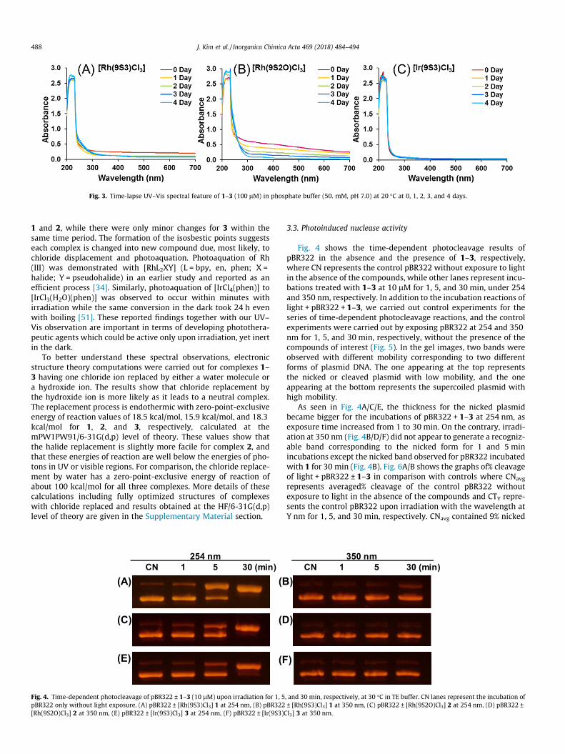
Fig. 3. Time-lapse UV–Vis spectral feature of 1–3 (100 lM) in phosphate buffer (50. mM, pH 7.0) at 20 �C at 0, 1, 2, 3, and 4 days.
488 J. Kim et al. / Inorganica Chimica Acta 469 (2018) 484–494
1 and 2, while there were only minor changes for 3 within thesame time period. The formation of the isosbestic points suggestseach complex is changed into new compound due, most likely, tochloride displacement and photoaquation. Photoaquation of Rh(III) was demonstrated with [RhL2XY] (L = bpy, en, phen; X =halide; Y = pseudohalide) in an earlier study and reported as anefficient process [34]. Similarly, photoaquation of [IrCl4(phen)] to[IrCl3(H2O)(phen)] was observed to occur within minutes withirradiation while the same conversion in the dark took 24 h evenwith boiling [51]. These reported findings together with our UV–Vis observation are important in terms of developing photothera-peutic agents which could be active only upon irradiation, yet inertin the dark.
To better understand these spectral observations, electronicstructure theory computations were carried out for complexes 1–3 having one chloride ion replaced by either a water molecule ora hydroxide ion. The results show that chloride replacement bythe hydroxide ion is more likely as it leads to a neutral complex.The replacement process is endothermic with zero-point-exclusiveenergy of reaction values of 18.5 kcal/mol, 15.9 kcal/mol, and 18.3kcal/mol for 1, 2, and 3, respectively, calculated at themPW1PW91/6-31G(d,p) level of theory. These values show thatthe halide replacement is slightly more facile for complex 2, andthat these energies of reaction are well below the energies of pho-tons in UV or visible regions. For comparison, the chloride replace-ment by water has a zero-point-exclusive energy of reaction ofabout 100 kcal/mol for all three complexes. More details of thesecalculations including fully optimized structures of complexeswith chloride replaced and results obtained at the HF/6-31G(d,p)level of theory are given in the Supplementary Material section.
(A)
(C)
(E)
254 nm .CN 1 5 30 (min)
(B
(D
(F
Fig. 4. Time-dependent photocleavage of pBR322 ± 1–3 (10 lM) upon irradiation for 1, 5pBR322 only without light exposure. (A) pBR322 ± [Rh(9S3)Cl3] 1 at 254 nm, (B) pBR322[Rh(9S2O)Cl3] 2 at 350 nm, (E) pBR322 ± [Ir(9S3)Cl3] 3 at 254 nm, (F) pBR322 ± [Ir(9S3)C
3.3. Photoinduced nuclease activity
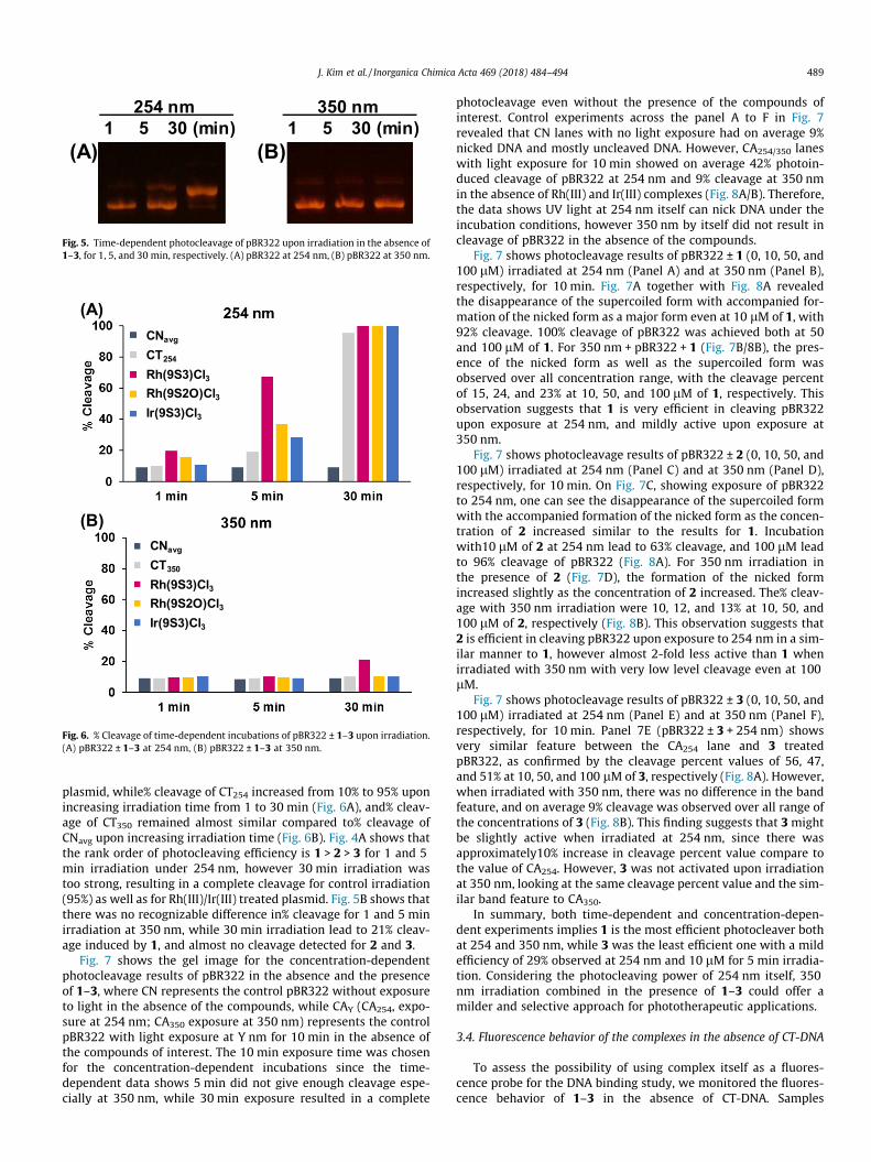
Fig. 4 shows the time-dependent photocleavage results ofpBR322 in the absence and the presence of 1–3, respectively,where CN represents the control pBR322 without exposure to lightin the absence of the compounds, while other lanes represent incu-bations treated with 1–3 at 10 lM for 1, 5, and 30 min, under 254and 350 nm, respectively. In addition to the incubation reactions oflight + pBR322 + 1–3, we carried out control experiments for theseries of time-dependent photocleavage reactions, and the controlexperiments were carried out by exposing pBR322 at 254 and 350nm for 1, 5, and 30 min, respectively, without the presence of thecompounds of interest (Fig. 5). In the gel images, two bands wereobserved with different mobility corresponding to two differentforms of plasmid DNA. The one appearing at the top representsthe nicked or cleaved plasmid with low mobility, and the oneappearing at the bottom represents the supercoiled plasmid withhigh mobility.
As seen in Fig. 4A/C/E, the thickness for the nicked plasmidbecame bigger for the incubations of pBR322 + 1–3 at 254 nm, asexposure time increased from 1 to 30 min. On the contrary, irradi-ation at 350 nm (Fig. 4B/D/F) did not appear to generate a recogniz-able band corresponding to the nicked form for 1 and 5 minincubations except the nicked band observed for pBR322 incubatedwith 1 for 30 min (Fig. 4B). Fig. 6A/B shows the graphs of% cleavageof light + pBR322 ± 1–3 in comparison with controls where CNavg
represents averaged% cleavage of the control pBR322 withoutexposure to light in the absence of the compounds and CTY repre-sents the control pBR322 upon irradiation with the wavelength atY nm for 1, 5, and 30 min, respectively. CNavg contained 9% nicked
350 nm .CN 1 5 30 (min)
)
)
)
, and 30 min, respectively, at 30 �C in TE buffer. CN lanes represent the incubation of± [Rh(9S3)Cl3] 1 at 350 nm, (C) pBR322 ± [Rh(9S2O)Cl3] 2 at 254 nm, (D) pBR322 ±l3] 3 at 350 nm.
(A) (B)
254 nm .1 5 30 (min)
350 nm .1 5 30 (min)
Fig. 5. Time-dependent photocleavage of pBR322 upon irradiation in the absence of1–3, for 1, 5, and 30 min, respectively. (A) pBR322 at 254 nm, (B) pBR322 at 350 nm.
CNavg
CT254
Rh(9S3)Cl3Rh(9S2O)Cl3Ir(9S3)Cl3
CNavg
CT350
Rh(9S3)Cl3Rh(9S2O)Cl3Ir(9S3)Cl3
(A)
(B)
Fig. 6. % Cleavage of time-dependent incubations of pBR322 ± 1–3 upon irradiation.(A) pBR322 ± 1–3 at 254 nm, (B) pBR322 ± 1–3 at 350 nm.
J. Kim et al. / Inorganica Chimica Acta 469 (2018) 484–494 489
plasmid, while% cleavage of CT254 increased from 10% to 95% uponincreasing irradiation time from 1 to 30 min (Fig. 6A), and% cleav-age of CT350 remained almost similar compared to% cleavage ofCNavg upon increasing irradiation time (Fig. 6B). Fig. 4A shows thatthe rank order of photocleaving efficiency is 1 > 2 > 3 for 1 and 5min irradiation under 254 nm, however 30 min irradiation wastoo strong, resulting in a complete cleavage for control irradiation(95%) as well as for Rh(III)/Ir(III) treated plasmid. Fig. 5B shows thatthere was no recognizable difference in% cleavage for 1 and 5 minirradiation at 350 nm, while 30 min irradiation lead to 21% cleav-age induced by 1, and almost no cleavage detected for 2 and 3.
Fig. 7 shows the gel image for the concentration-dependentphotocleavage results of pBR322 in the absence and the presenceof 1–3, where CN represents the control pBR322 without exposureto light in the absence of the compounds, while CAY (CA254, expo-sure at 254 nm; CA350 exposure at 350 nm) represents the controlpBR322 with light exposure at Y nm for 10 min in the absence ofthe compounds of interest. The 10 min exposure time was chosenfor the concentration-dependent incubations since the time-dependent data shows 5 min did not give enough cleavage espe-cially at 350 nm, while 30 min exposure resulted in a complete
photocleavage even without the presence of the compounds ofinterest. Control experiments across the panel A to F in Fig. 7revealed that CN lanes with no light exposure had on average 9%nicked DNA and mostly uncleaved DNA. However, CA254/350 laneswith light exposure for 10 min showed on average 42% photoin-duced cleavage of pBR322 at 254 nm and 9% cleavage at 350 nmin the absence of Rh(III) and Ir(III) complexes (Fig. 8A/B). Therefore,the data shows UV light at 254 nm itself can nick DNA under theincubation conditions, however 350 nm by itself did not result incleavage of pBR322 in the absence of the compounds.
Fig. 7 shows photocleavage results of pBR322 ± 1 (0, 10, 50, and100 lM) irradiated at 254 nm (Panel A) and at 350 nm (Panel B),respectively, for 10 min. Fig. 7A together with Fig. 8A revealedthe disappearance of the supercoiled form with accompanied for-mation of the nicked form as a major form even at 10 lM of 1, with92% cleavage. 100% cleavage of pBR322 was achieved both at 50and 100 lM of 1. For 350 nm + pBR322 + 1 (Fig. 7B/8B), the pres-ence of the nicked form as well as the supercoiled form wasobserved over all concentration range, with the cleavage percentof 15, 24, and 23% at 10, 50, and 100 lM of 1, respectively. Thisobservation suggests that 1 is very efficient in cleaving pBR322upon exposure at 254 nm, and mildly active upon exposure at350 nm.
Fig. 7 shows photocleavage results of pBR322 ± 2 (0, 10, 50, and100 lM) irradiated at 254 nm (Panel C) and at 350 nm (Panel D),respectively, for 10 min. On Fig. 7C, showing exposure of pBR322to 254 nm, one can see the disappearance of the supercoiled formwith the accompanied formation of the nicked form as the concen-tration of 2 increased similar to the results for 1. Incubationwith10 lM of 2 at 254 nm lead to 63% cleavage, and 100 lM leadto 96% cleavage of pBR322 (Fig. 8A). For 350 nm irradiation inthe presence of 2 (Fig. 7D), the formation of the nicked formincreased slightly as the concentration of 2 increased. The% cleav-age with 350 nm irradiation were 10, 12, and 13% at 10, 50, and100 lM of 2, respectively (Fig. 8B). This observation suggests that2 is efficient in cleaving pBR322 upon exposure to 254 nm in a sim-ilar manner to 1, however almost 2-fold less active than 1 whenirradiated with 350 nm with very low level cleavage even at 100lM.
Fig. 7 shows photocleavage results of pBR322 ± 3 (0, 10, 50, and100 lM) irradiated at 254 nm (Panel E) and at 350 nm (Panel F),respectively, for 10 min. Panel 7E (pBR322 ± 3 + 254 nm) showsvery similar feature between the CA254 lane and 3 treatedpBR322, as confirmed by the cleavage percent values of 56, 47,and 51% at 10, 50, and 100 lM of 3, respectively (Fig. 8A). However,when irradiated with 350 nm, there was no difference in the bandfeature, and on average 9% cleavage was observed over all range ofthe concentrations of 3 (Fig. 8B). This finding suggests that 3 mightbe slightly active when irradiated at 254 nm, since there wasapproximately10% increase in cleavage percent value compare tothe value of CA254. However, 3 was not activated upon irradiationat 350 nm, looking at the same cleavage percent value and the sim-ilar band feature to CA350.
In summary, both time-dependent and concentration-depen-dent experiments implies 1 is the most efficient photocleaver bothat 254 and 350 nm, while 3 was the least efficient one with a mildefficiency of 29% observed at 254 nm and 10 lM for 5 min irradia-tion. Considering the photocleaving power of 254 nm itself, 350nm irradiation combined in the presence of 1–3 could offer amilder and selective approach for phototherapeutic applications.
3.4. Fluorescence behavior of the complexes in the absence of CT-DNA
To assess the possibility of using complex itself as a fluores-cence probe for the DNA binding study, we monitored the fluores-cence behavior of 1–3 in the absence of CT-DNA. Samples
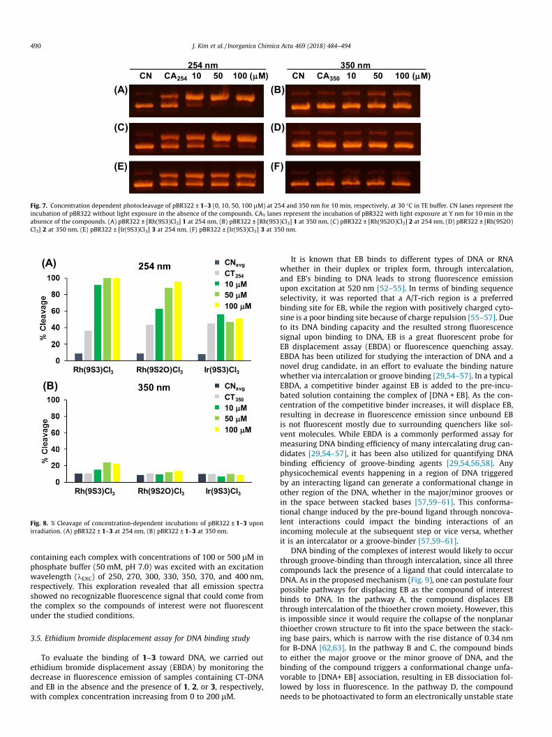
Fig. 7. Concentration dependent photocleavage of pBR322 ± 1–3 (0, 10, 50, 100 lM) at 254 and 350 nm for 10 min, respectively, at 30 �C in TE buffer. CN lanes represent theincubation of pBR322 without light exposure in the absence of the compounds. CAY lanes represent the incubation of pBR322 with light exposure at Y nm for 10 min in theabsence of the compounds. (A) pBR322 ± [Rh(9S3)Cl3] 1 at 254 nm, (B) pBR322 ± [Rh(9S3)Cl3] 1 at 350 nm, (C) pBR322 ± [Rh(9S2O)Cl3] 2 at 254 nm, (D) pBR322 ± [Rh(9S2O)Cl3] 2 at 350 nm, (E) pBR322 ± [Ir(9S3)Cl3] 3 at 254 nm, (F) pBR322 ± [Ir(9S3)Cl3] 3 at 350 nm.
Fig. 8. % Cleavage of concentration-dependent incubations of pBR322 ± 1–3 uponirradiation. (A) pBR322 ± 1–3 at 254 nm, (B) pBR322 ± 1–3 at 350 nm.
490 J. Kim et al. / Inorganica Chimica Acta 469 (2018) 484–494
containing each complex with concentrations of 100 or 500 lM inphosphate buffer (50 mM, pH 7.0) was excited with an excitationwavelength (kEXC) of 250, 270, 300, 330, 350, 370, and 400 nm,respectively. This exploration revealed that all emission spectrashowed no recognizable fluorescence signal that could come fromthe complex so the compounds of interest were not fluorescentunder the studied conditions.
3.5. Ethidium bromide displacement assay for DNA binding study
To evaluate the binding of 1–3 toward DNA, we carried outethidium bromide displacement assay (EBDA) by monitoring thedecrease in fluorescence emission of samples containing CT-DNAand EB in the absence and the presence of 1, 2, or 3, respectively,with complex concentration increasing from 0 to 200 lM.
It is known that EB binds to different types of DNA or RNAwhether in their duplex or triplex form, through intercalation,and EB’s binding to DNA leads to strong fluorescence emissionupon excitation at 520 nm [52–55]. In terms of binding sequenceselectivity, it was reported that a A/T-rich region is a preferredbinding site for EB, while the region with positively charged cyto-sine is a poor binding site because of charge repulsion [55–57]. Dueto its DNA binding capacity and the resulted strong fluorescencesignal upon binding to DNA, EB is a great fluorescent probe forEB displacement assay (EBDA) or fluorescence quenching assay.EBDA has been utilized for studying the interaction of DNA and anovel drug candidate, in an effort to evaluate the binding naturewhether via intercalation or groove binding [29,54–57]. In a typicalEBDA, a competitive binder against EB is added to the pre-incu-bated solution containing the complex of [DNA + EB]. As the con-centration of the competitive binder increases, it will displace EB,resulting in decrease in fluorescence emission since unbound EBis not fluorescent mostly due to surrounding quenchers like sol-vent molecules. While EBDA is a commonly performed assay formeasuring DNA binding efficiency of many intercalating drug can-didates [29,54–57], it has been also utilized for quantifying DNAbinding efficiency of groove-binding agents [29,54,56,58]. Anyphysicochemical events happening in a region of DNA triggeredby an interacting ligand can generate a conformational change inother region of the DNA, whether in the major/minor grooves orin the space between stacked bases [57,59–61]. This conforma-tional change induced by the pre-bound ligand through noncova-lent interactions could impact the binding interactions of anincoming molecule at the subsequent step or vice versa, whetherit is an intercalator or a groove-binder [57,59–61].
DNA binding of the complexes of interest would likely to occurthrough groove-binding than through intercalation, since all threecompounds lack the presence of a ligand that could intercalate toDNA. As in the proposed mechanism (Fig. 9), one can postulate fourpossible pathways for displacing EB as the compound of interestbinds to DNA. In the pathway A, the compound displaces EBthrough intercalation of the thioether crown moiety. However, thisis impossible since it would require the collapse of the nonplanarthioether crown structure to fit into the space between the stack-ing base pairs, which is narrow with the rise distance of 0.34 nmfor B-DNA [62,63]. In the pathway B and C, the compound bindsto either the major groove or the minor groove of DNA, and thebinding of the compound triggers a conformational change unfa-vorable to [DNA+ EB] association, resulting in EB dissociation fol-lowed by loss in fluorescence. In the pathway D, the compoundneeds to be photoactivated to form an electronically unstable state
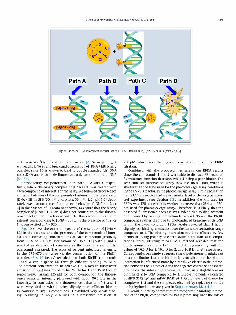
Fig. 9. Proposed EB displacement mechanism of 1–3. M = Rh(III) or Ir(III); X = S or O in [M(9S2X)Cl3].
J. Kim et al. / Inorganica Chimica Acta 469 (2018) 484–494 491
or to generate 1O2 through a redox reaction [2]. Subsequently, itwill lead to DNA strand break and dissociation of [DNA + EB] binarycomplex since EB is known to bind to double stranded (ds) DNAnot ssDNA and is strongly fluorescent only upon binding to DNA[54–56].
Consequently, we performed EBDA with 1, 2, and 3, respec-tively, where the binary complex of [DNA + EB] was treated witheach compound of interest. For the assay, we followed fluorescenceemission behavior of the compounds of interest in the presence of[DNA + EB] in SPB (50 mM phosphate, 50 mM NaCl, pH 7.0). Sepa-rately, we also monitored fluorescence behavior of [DNA + 1, 2, or3] in the absence of EB (data not shown) to ensure that the binarycomplex of [DNA + 1, 2, or 3] does not contribute to the fluores-cence background or interfere with the fluorescence emission ofinterest corresponding to [DNA + EB] with the presence of 1, 2, or3, when excited at k = 520 nm.
Fig. 10 shows the emission spectra of the solution of [DNA +EB] in the absence and the presence of the compounds of inter-est upon increasing concentrations of each compound graduallyfrom 0 lM to 200 lM. Incubations of [DNA + EB] with 1 and 2resulted in decrease of emission as the concentration of thecompound increased. The plots of percent integrated intensityin the 575–675 nm range vs. the concentration of the Rh(III)complex (Fig. 10 insets) revealed that both Rh(III) compounds1 and 2 can displace EB through efficient binding to DNA.The efficient concentration to achieve a 50% loss in fluorescentemission (ECFL50) was found to be 24 lM for 1 and 35 lM for 2,respectively. Passing 125 lM for both compounds, the fluores-cence emission intensity plateaued with about 80% loss in theintensity. In conclusion, the fluorescence behavior of 1 and 2were very similar, with 1 being slightly more efficient binder.In contrast to Rh(III) compounds, 3 exhibited very weak bind-ing, resulting in only 27% loss in fluorescence emission at
200 lM which was the highest concentration used for EBDAtitration.
Combined with the proposed mechanism, our EBDA resultsshow the compounds 1 and 2 were able to displace EB based onfluorescence emission decrease, while 3 being a poor binder. Thescan time for fluorescence assay took less than 1 min, which isshorter than the time used for the photocleavage assay conditionsin the UV–Vis reactor. In the photocleavage assay, 1 min incubationin the UV–Vis reactor had almost similar level of cleavage as a con-trol experiment (see Section 3.3). In addition, the kext used forEBDA was 520 nm which is weaker in energy than 254 and 350nm used for photocleavage assay. Therefore, it is likely that theobserved fluorescence decrease was indeed due to displacementof EB caused by binding interaction between DNA and the Rh(III)compounds rather than due to photoinduced breakage of ds DNAwithin the given condition. EBDA results revealed that 2 has aslightly less binding interaction over the same concentration rangecompared to 1. The binding interaction could be affected by fewfactors including polarity or electrostatic interaction. Our compu-tational study utilizing mPW1PW91 method revealed that thedipole moment values of 1–3 do not differ significantly, with thevalues of 16.6 D for 1, 16.0 D for 2, and 16.9 D for 3, respectively.Consequently, our study suggests that dipole moment might notbe a contributing factor in binding. It is possible that the bindinginteraction is influenced more by a repulsive electrostatic interac-tion between the O atom of 2 and the negative charge of phosphategroups on the interacting groove, resulting in a slightly weakerbinding of 2 to DNA compared to 1. Dipole moments calculatedat HF/6-31G(d,p) and mPW1PW91/6-31G(d,p) levels of theory forcomplexes 1–3 and the complexes obtained by replacing chlorideion by hydroxide ion are given in Supplementary Material.
Overall, our study shows that the recognizable binding interac-tion of the Rh(III) compounds to DNA is promising since the role of
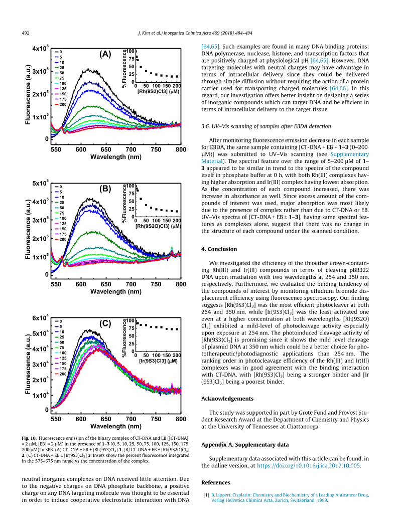
Fig. 10. Fluorescence emission of the binary complex of CT-DNA and EB ([CT-DNA]= 2 lM, [EB] = 2 lM) in the presence of 1–3 (0, 5, 10, 25, 50, 75, 100, 125, 150, 175,200 lM) in SPB. (A) CT-DNA + EB ± [Rh(9S3)Cl3] 1, (B) CT-DNA + EB ± [Rh(9S2O)Cl3]2, (C) CT-DNA + EB ± [Ir(9S3)Cl3] 3. Insets show the percent fluorescence integratedin the 575–675 nm range vs the concentration of the complex.
492 J. Kim et al. / Inorganica Chimica Acta 469 (2018) 484–494
neutral inorganic complexes on DNA received little attention. Dueto the negative charges on DNA phosphate backbone, a positivecharge on any DNA targeting molecule was thought to be essentialin order to induce cooperative electrostatic interaction with DNA
[64,65]. Such examples are found in many DNA binding proteins;DNA polymerase, nuclease, histone, and transcription factors thatare positively charged at physiological pH [64,65]. However, DNAtargeting molecules with neutral charges may have advantage interms of intracellular delivery since they could be deliveredthrough simple diffusion without requiring the action of a proteincarrier used for transporting charged molecules [64,66]. In thisregard, our investigation offers better insight on designing a seriesof inorganic compounds which can target DNA and be efficient interms of intracellular delivery to the target tissue.
3.6. UV–Vis scanning of samples after EBDA detection
After monitoring fluorescence emission decrease in each samplefor EBDA, the same sample containing [CT-DNA + EB + 1–3 (0–200lM)] was submitted to UV–Vis scanning (see SupplementaryMaterial). The spectral feature over the range of 5–200 lM of 1–3 appeared to be similar in trend to the spectra of the compounditself in phosphate buffer at 0 h, with both Rh(III) complexes hav-ing higher absorption and Ir(III) complex having lowest absorption.As the concentration of each compound increased, there wasincrease in absorbance as well. Since excess amount of the com-pounds of interest was used, major absorption was most likelydue to the presence of complex rather than due to CT-DNA or EB.UV–Vis spectra of [CT-DNA + EB ± 1–3], having same spectral fea-tures as complexes alone, suggest that there was no change inthe structure of each compound under the scanned condition.
4. Conclusion
We investigated the efficiency of the thioether crown-contain-ing Rh(III) and Ir(III) compounds in terms of cleaving pBR322DNA upon irradiation with two wavelengths at 254 and 350 nm,respectively. Furthermore, we evaluated the binding tendency ofthe compounds of interest by monitoring ethidium bromide dis-placement efficiency using fluorescence spectroscopy. Our findingsuggests [Rh(9S3)Cl3] was the most efficient photocleaver at both254 and 350 nm, while [Ir(9S3)Cl3] was the least activated oneeven at a higher concentration at both wavelengths. [Rh(9S2O)Cl3] exhibited a mild-level of photocleavage activity especiallyupon exposure at 254 nm. The photoinduced cleavage activity of[Rh(9S3)Cl3] is promising since it shows the mild level cleavageof plasmid DNA at 350 nm which could be a better choice for pho-totherapeutic/photodiagnostic applications than 254 nm. Theranking order in photocleavage efficiency of the Rh(III) and Ir(III)complexes was in good agreement with the binding interactionwith CT-DNA, with [Rh(9S3)Cl3] being a stronger binder and [Ir(9S3)Cl3] being a poorest binder.
Acknowledgements
The study was supported in part by Grote Fund and Provost Stu-dent Research Award at the Department of Chemistry and Physicsat the University of Tennessee at Chattanooga.
Appendix A. Supplementary data
Supplementary data associated with this article can be found, inthe online version, at https://doi.org/10.1016/j.ica.2017.10.005.
References
[1] B. Lippert, Cisplatin: Chemistry and Biochemistry of a Leading Anticancer Drug,Verlag Helvetica Chimica Acta, Zurich, Switzerland, 1999.
J. Kim et al. / Inorganica Chimica Acta 469 (2018) 484–494 493
[2] J.D. Knoll, C. Turro, Control and utilization of ruthenium and rhodium metalcomplex excited states for photoactivated cancer therapy, Coord. Chem. Rev.282–283 (2015) 110–126.
[3] E. Wong, C.M. Giandomenico, Current status of platinum-based antitumordrugs, Chem. Rev. 99 (9) (1999) 2451–2466.
[4] R.A. Alderden, M.D. Hall, T.W. Hambley, The discovery and development ofcisplatin, J. Chem. Educ. 83 (5) (2006) 728–734.
[5] Z.J. Guo, P.J. Sadler, Medicinal inorganic chemistry, Adv. Inorg. Chem. 49 (2000)183–306.
[6] G. Sathyaraj, M. Kiruthika, T. Weyhermuller, B.U. Nair, Oxidative cleavage ofDNA by ruthenium(II) complexes containing a ferrocene/non-ferroceneconjugated imidazole phenol ligand, Organometallics 31 (19) (2012) 6980–6987.
[7] S. Shi, J. Liu, J. Li, K.C. Zheng, C.P. Tan, L.M. Chen, L.N. Ji, Electronic effect ofdifferent positions of the -NO2 group on the DNA-intercalator of chiralcomplexes [Ru(bpy)(2)L](2+) (L = o-npip, m-npip and p-npip), Dalton Trans.(11) (2005) 2038–2046.
[8] A. Chouai, S.E. Wicke, C. Turro, J. Bacsa, K.R. Dunbar, D. Wang, R.P. Thummel,Ruthenium(II) complexes of 1,12-diazaperyiene and their interactions withDNA, Inorg. Chem. 44 (17) (2005) 5996–6003.
[9] G.Y. Li, L.L. Sun, L.N. Ji, H. Chao, Ruthenium(II) complexes with dppz: frommolecular photoswitch to biological applications, Dalton Trans. 45 (34) (2016)13261–13276.
[10] B.C. Poulsen, S. Estalayo-Adrian, S. Blasco, S.A. Bright, J.M. Kelly, D.C. Williams,T. Gunnlaugsson, Luminescent ruthenium polypyridyl complexes withextended ‘dppz’ like ligands as DNA targeting binders and cellular agents,Dalton Trans. 45 (45) (2016) 18208–18220.
[11] Y.J. Sun, L.E. Joyce, N.M. Dickson, C. Turro, DNA photocleavage by an osmium(II) complex in the PDT window, Chem. Commun. 46 (36) (2010) 6759–6761.
[12] Q.X. Zhou, W.H. Lei, J.R. Chen, C. Li, Y.J. Hou, X.S. Wang, B.W. Zhang, A newheteroleptic ruthenium(ii) polypyridyl complex with long-wavelengthabsorption and high singlet-oxygen quantum yield, Chem.-Eur. J. 16 (10)(2010) 3157–3165.
[13] S. Swavey, M. DeBeer, K. Li, Photoinduced interactions of supramolecularruthenium(II) complexes with plasmid DNA: synthesis and spectroscopic,electrochemical, and DNA photocleavage studies, Inorg. Chem. 54 (7) (2015)3139–3147.
[14] H.J. Yu, S.M. Huang, H. Chao, L.N. Ji, Synthesis, crystal structure and anaerobicDNA photocleavage of ruthenium complexes [Ru(tpy)(dpoq)Cl]+ and [Ru(tpy)(dpoq)CH3CN]2+, J. Inorg. Biochem. 149 (2015) 80–87.
[15] M.J. Fernandez, B. Wilson, M. Palacios, M.M. Rodrigo, K.B. Grant, A. Lorente,Copper-activated DNA photocleavage by a pyridine-linked bis-acridineintercalator, Bioconjugate Chem. 18 (1) (2007) 121–129.
[16] P. Ghosh, M. Sinan, D. Lahiri, S. Roy, S. Goswami, Design and synthesis of afunctional derivative of the triazinium cation and its rhodium complex thatshows photoinduced DNA cleavage activity and photocytotoxicity, Eur. J.Inorg. Chem. 29 (2012) 4719–4727.
[17] A. Jacques, Mesmaeker A. Kirsch-De, B. Elias, Selective DNA purine basephotooxidation by bis-terdentate iridium(III) polypyridyl and cyclometalatedcomplexes, Inorg. Chem. 53 (3) (2014) 1507–1512.
[18] Z. Liu, A. Habtemariam, A.M. Pizarro, S.A. Fletcher, A. Kisova, O. Vrana, L.Salassa, P.C. Bruijnincx, G.J. Clarkson, V. Brabec, et al., Organometallic half-sandwich iridium anticancer complexes, J. Med. Chem. 54 (8) (2011) 3011–3026.
[19] B. Maity, B.V.S.K. Chakravarthi, M. Roy, A.A. Karande, A.R. Chakravarty, DNAphotocleavage and cytotoxic properties of ferrocene conjugates, Eur. J. Inorg.Chem. 9 (2011) 1379–1386.
[20] T. Mukherjee, B. Sen, A. Patra, S. Banerjee, G. Hundal, P. Chattopadhyay,Cyclometalated rhodium(III) complexes bearing dithiocarbamate derivative:synthesis, characterization, interaction with DNA and biological study,Polyhedron 69 (2014) 127–134.
[21] M. Roy, S. Saha, A.K. Patra, M. Nethaji, A.R. Chakravarty, Ternary iron(III)complex showing photocleavage of DNA in the photodynamic therapywindow, Inorg. Chem. 46 (11) (2007) 4368–4370.
[22] V. Brabec, J. Pracharova, J. Stepankova, P.J. Sadler, J. Kasparkova, Photo-inducedDNA cleavage and cytotoxicity of a ruthenium(II) arene anticancer complex, J.Inorg. Biochem. 160 (2016) 149–155.
[23] D. Crespy, K. Landfester, U.S. Schubert, A. Schiller, Potential photoactivatedmetallopharmaceuticals: from active molecules to supported drugs, Chem.Commun. 46 (36) (2010) 6651–6662.
[24] G. Gasser, I. Ott, N. Metzler-Nolte, Organometallic anticancer compounds, J.Med. Chem. 54 (1) (2011) 3–25.
[25] A.L. Hurley, D.L. Mohler, Organometallic photonucleases: synthesis andDNA-cleavage studies of cyclopentadienyl metal-substituted dendrimersdesigned to increase double-strand scission, Org. Lett. 2 (18) (2000) 2745–2748.
[26] P. Liu, J. Liu, Y.Q. Zhang, B.Y. Wu, K.Z. Wang, Synthesis, DNA binding andphotocleavage, and cellular uptake of an alkyl chain-linked dinuclearruthenium(II) complex, J. Photochem. Photobiol., B 143 (2015) 89–99.
[27] D.L. Mohler, D.R. Dain, A.D. Kerekes, W.R. Nadler, T.L. Scott, Organometallicphotonucleases: a novel class of DNA-cleaving agents, Bioorg. Med. Chem. Lett.8 (7) (1998) 871–874.
[28] K. Szacilowski, W. Macyk, A. Drzewiecka-Matuszek, M. Brindell, G. Stochel,Bioinorganic photochemistry: frontiers and mechanisms, Chem. Rev. 105 (6)(2005) 2647–2694.
[29] M. Sirajuddin, S. Ali, A. Badshah, Drug-DNA interactions and their study by UV-Visible, fluorescence spectroscopies and cyclic voltametry, J. Photochem.Photobiol. B, Biol. 124 (2013) 1–19.
[30] F.A. Blommaert, H.C.M. Vandijkknijnenburg, F.J. Dijt, L. Denengelse, R.A. Baan,F. Berends, A.M.J. Fichtingerschepman, Formation of DNA-adducts by theanticancer drug carboplatin – different nucleotide-sequence preferences in-vitro and in cells, Biochemistry 34 (26) (1995) 8474–8480.
[31] T.K. Goswami, M. Roy, M. Nethaji, A.R. Chakravarty, Photoinduced DNA andprotein cleavage activity of ferrocene-appended L-methionine reduced schiffbase copper(II) complexes of phenanthroline bases, Organometallics 28 (7)(2009) 1992–1994.
[32] F.A. Cotton, G. Wilkinson, P.L. Gaus, Basic Inorganic Chemistry, third ed., JohnWiley & Sons, Inc., 1995.
[33] D. Loganathan, J.H. Rodriguez, H. Morrison, Photoaquation of methylated cis-dichlorobis(1,10-phenanthroline)rhodium(III)chloride compounds by directpopulation of a photoactive triplet excited state, J. Am. Chem. Soc. 125 (19)(2003) 5640–5641.
[34] M.M. Muir, W.L. Huang, Photoaquation of some complexes of rhodium(III),Inorg. Chem. 12 (8) (1973) 1831–1835.
[35] R. Bieda, I. Ott, M. Dobroschke, A. Prokop, R. Gust, W.S. Sheldrick, Structure-activity relationships and DNA binding properties of apoptosis inducingcytotoxic rhodium(III) polypyridyl complexes containing the cyclic thioether[9]aneS(3), J. Inorg. Biochem. 103 (5) (2009) 698–708.
[36] R. Bieda, I. Ott, R. Gust, W.S. Sheldrick, Cytotoxic rhodium(III) polypyridylcomplexes containing the tris(pyrazolyl)methane coligand: synthesis, DNAbinding properties and structure-activity relationships, Eur. J. Inorg. Chem. 25(2009) 3821–3831.
[37] D.A. Medvetz, K.D. Stakleff, T. Schreiber, P.D. Custer, K. Hindi, M.J. Panzner, D.D.Blanco, M.J. Taschner, C.A. Tessier, W.J. Youngs, Ovarian cancer activity ofcyclic amine and thiaether metal complexes, J. Med. Chem. 50 (7) (2007)1703–1706.
[38] S. Timonen, T.T. Pakkanen, T.A. Pakkanen, Novel single-site catalystscontaining a platinum group metal and a macrocyclic sulfur ligand forethylene polymerization, J. Mol. Catal. A-Chem. 111 (3) (1996) 267–272.
[39] H. Vashi, Advancements in Self-Assembly of Molecular Rhodium(III) andCobalt(III) Cubes and Squares, University of Tennessee at Chattanooga, 2011.
[40] G. Felsenfeld, S.Z. Hirschman, A neighbor-interaction analysis of thehypochromism and spectra of DNA, J. Mol. Biol. 13 (2) (1965) 407–427.
[41] C.J. Cramer, Essentials of Computational Chemistry, John Wiley & Sons, Ltd.,2002.
[42] C. Adamo, V. Barone, Exchange functionals with improved long-range behaviorand adiabatic connection methods without adjustable parameters: The mPWand mPW1PW models, J. Chem. Phys. 108 (2) (1998) 664–675.
[43] P.J. Hay, W.R. Wadt, Abinitio effective core potentials for molecularcalculations – potentials for the transition-metal atoms Sc to Hg, J. Chem.Phys. 82 (1) (1985) 270–283.
[44] R. Krishnan, J.S. Binkley, R. Seeger, J.A. Pople, Self-consistent molecular-orbitalmethods. 20. Basis set for correlated wave-functions, J. Chem. Phys. 72 (1)(1980) 650–654.
[45] M.J. Frisch G.W.T., H.B. Schlegel, G.E. Scuseria, M.A. Robb, J.R. Cheeseman, G.Scalmani, V. Barone, B. Mennucci, G.A. Petersson, H. Nakatsuji, M. Caricato, X.Li, H.P. Hratchian, A.F. Izmaylov, J. Bloino, G. Zheng, J.L. Sonnenberg, M. Hada,M. Ehara, K. Toyota, R. Fukuda, J. Hasegawa, M. Ishida, T. Nakajima, Y. Honda,O. Kitao, H. Nakai, T. Vreven, J. A. Montgomery, Jr., J.E. Peralta, F. Ogliaro, M.Bearpark, J.J. Heyd, E. Brothers, K.N. Kudin, V.N. Staroverov, T. Keith, R.Kobayashi, J. Normand, K. Raghavachari, A. Rendell, J.C. Burant, S.S. Iyengar, J.Tomasi, M. Cossi, N. Rega, J.M. Millam, M. Klene, J.E. Knox, J.B. Cross, V. Bakken,C. Adamo, J. Jaramillo, R. Gomperts, R.E. Stratmann, O. Yazyev, A.J. Austin, R.Cammi, C. Pomelli, J.W. Ochterski, R.L. Martin, K. Morokuma, V.G. Zakrzewski,G.A. Voth, P. Salvador, J.J. Dannenberg, S. Dapprich, A.D. Daniels, O. Farkas, J.B.Foresman, J.V. Ortiz, J. Cioslowski, D.J. Fox, Gaussian 09, Revision C.01. In.Wallingford CT: Gaussian, Inc., 2010.
[46] M.D. Hanwell, D.E. Curtis, D.C. Lonie, T. Vandermeersch, E. Zurek, G.R.Hutchison, Avogadro: an advanced semantic chemical editor, visualization,and analysis platform, J. Chem. Inf. 4 (1) (2012) 17.
[47] S.R. Cooper, S.C. Rawle, R. Yagbasan, D.J. Watkin, Crown thioether chemistry –the rhodium complexes of 1,4,7-trithiacyclononane (9S3) and 1,5,9-trithiacyclododecane (12S3) and the conformational factors that stabilizemonomeric Rh(II) ions, J. Am. Chem. Soc. 113 (5) (1991) 1600–1604.
[48] H.A. Jenkins, S.J. Loeb, Thiacyclophane complexes of rhodium and iridium –synthesis, structure, and reactivity of [M(Cod)(L)][Bf4] (M=Rh, Ir, L= 2,5,8-Trithia[9]-O-Cyclophane (Tt[9]Oc), 5-Oxa-2,8-Dithia[9]-O-Cyclophane (Odt[9]Oc)), Organometallics 13 (5) (1994) 1840–1850.
[49] S.C. Rawle, R. Yagbasan, K. Prout, S.R. Cooper, Crown thioether chemistry –synthesis and structure of [bis(1,4,7-trithiacyclonane)rhodium] tris(triflate) –stabilization of monomeric Rh(Ii), J. Am. Chem. Soc. 109 (20) (1987) 6181–6182.
[50] H.J. Kim, J.H. Lee, I.H. Suh, Y.K. Do, A novel route to dinuclear heterolepticrhodium(III) complexes of 1,4,7-trithiacyclononane, Inorg. Chem. 34 (4) (1995)796–801.
[51] J.A. Broomhead, W. Grumley, Photochemical aquation of iridium (3)phenanthroline and related complexes, Chem. Commun. (20) (1968) 1211.
[52] J. Olmsted 3rd, D.R. Kearns, Mechanism of ethidium bromide fluorescenceenhancement on binding to nucleic acids, Biochemistry 16 (16) (1977) 3647–3654.

494 J. Kim et al. / Inorganica Chimica Acta 469 (2018) 484–494
[53] J. Sambrook, D.W. Russell, Molecular Cloning: A Laboratory Manual, third ed.,Cold Spring Harbor, 2001.
[54] D. Banerjee, S.K. Pal, Simultaneous binding of minor groove binder andintercalator to dodecamer DNA: importance of relative orientation of donorand acceptor in FRET, J. Phys. Chem. B 111 (19) (2007) 5047–5052.
[55] P.V. Scaria, R.H. Shafer, Binding of ethidium-bromide to a DNA triple helix –evidence for intercalation, J. Biol. Chem. 266 (9) (1991) 5417–5423.
[56] J.S. Ren, J.B. Chaires, Sequence and structural selectivity of nucleic acid bindingligands, Biochemistry 38 (49) (1999) 16067–16075.
[57] E. Tuite, U. Sehlstedt, P. Hagmar, B. Norden, M. Takahashi, Effects of minor andmajor groove-binding drugs and intercalators on the DNA association of minorgroove-binding proteins RecA and deoxyribonuclease I detected by flow lineardichroism, Eur. J. Biochem. 243 (1–2) (1997) 482–492.
[58] D. Sardar, P. Datta, S. Das, B. Saha, S. Samanta, D. Bhattacharya, P. Karmakar, C.D. Chen, C.J. Chen, C. Sinha, Synthesis, structure, DNA interaction and nucleaseactivity of rhodium(III)-arylazoimidazole complexes, Inorg. Chim. Acta 394(2013) 98–106.
[59] J.L. Bresloff, D.M. Crothers, Equilibrium studies of ethidium-polynucleotideinteractions, Biochemistry 20 (12) (1981) 3547–3553.
[60] Y.W. Park, K.J. Breslauer, Drug-binding to higher ordered DNA structures –netropsin complexation with a nucleic-acid triple helix, Proc. Natl. Acad. Sci. U.S.A. 89 (14) (1992) 6653–6657.
[61] M.G. Oakley, M. Mrksich, P.B. Dervan, Evidence that a minor groove-bindingpeptide and a major groove-binding protein can simultaneously occupy acommon site on DNA, Biochemistry 31 (45) (1992) 10969–10975.
[62] R. Wing, H. Drew, T. Takano, C. Broka, S. Tanaka, K. Itakura, R.E. Dickerson,Crystal-structure analysis of a complete turn of B-DNA, Nature 287 (5784)(1980) 755–758.
[63] M. Bansal, DNA structure: revisiting the Watson-Crick double helix, Curr. Sci.India 85 (11) (2003) 1556–1563.
[64] J.M. Berg, J.L. Tymoczko, L. Stryer, Biochemistry, sixth ed., W. H. Freeman andCo., New York, 2006.
[65] C.O. Pabo, R.T. Sauer, Protein-DNA recognition, Annu. Rev. Biochem. 53 (1984)293–321.
[66] B. Wang, T. Siahaan, R. Soltero, Drug Delivery: Principles and Applications,John Wiley & Sons, Inc, 2005.
1
Supplementary Material for
Interaction with Calf-thymus DNA and Photoinduced
Cleavage of pBR322 by Rhodium(III) and Iridium(III)
Complexes Containing Crown Thioether Ligands
Jisook Kim,* Ashley D. Cardenal, Hendrik J. Greve, Weinan Chen, Hitesh Vashi, Gregory Grant, and Titus V. Albu
Department of Chemistry and Physics, University of Tennessee at Chattanooga, Chattanooga, TN 37403, USA
In this supplementary material, we present:
– UV-Vis spectra of CT-DNA + EB in the presence of complexes (Figure 1A-1C)
– Figure with HOMO and LUMO for complexes 1-3 determined at the mPW1PW91/6-31G(d,p)
level of theory
– Cartesian coordinates for the optimized geometries of complexes 1-3 at various levels of
theory
– Cartesian coordinates for the optimized geometries of complexes obtained by replacing a
chloride ligand by H2O or OH– at HF/6-31G(d,p) and mPW1PW91/6-31G(d,p) levels of
theory
– Table with calculated energies of reaction for chloride replacement reactions at HF/6-31G(d,p)
and mPW1PW91/6-31G(d,p) levels of theory
– Table with calculated dipole moments at HF/6-31G(d,p) and mPW1PW91/6-31G(d,p) levels
of theory for complexes 1-3 and the complexes obtained by replacing chloride ion by
hydroxide ion
2
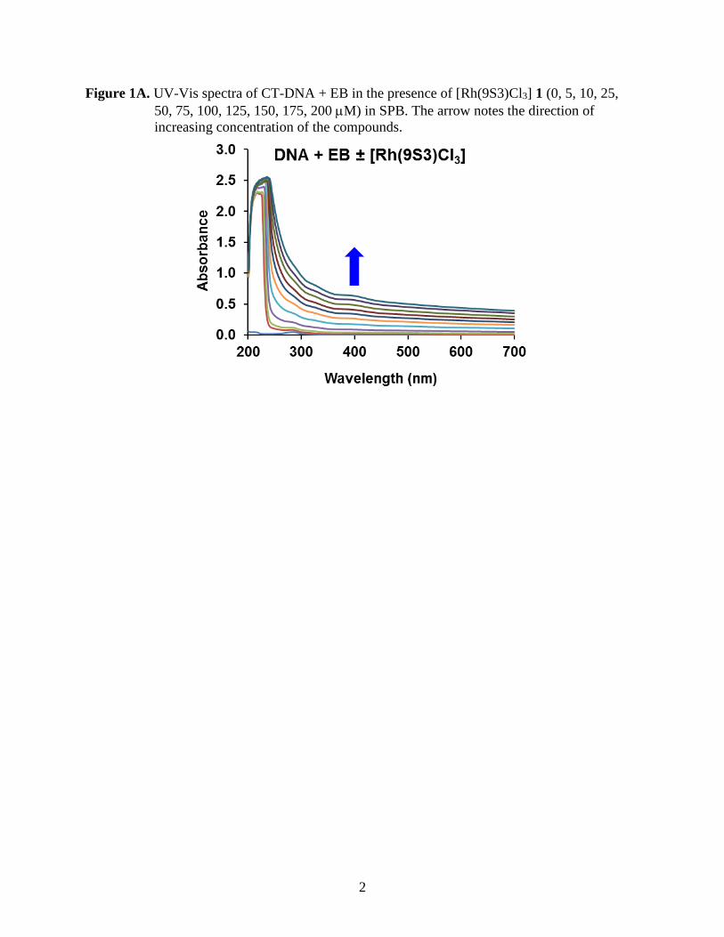
Figure 1A. UV-Vis spectra of CT-DNA + EB in the presence of [Rh(9S3)Cl3] 1 (0, 5, 10, 25,
50, 75, 100, 125, 150, 175, 200 M) in SPB. The arrow notes the direction of
increasing concentration of the compounds.

3
Figure 1B. UV-Vis spectra of CT-DNA + EB in the presence of [Rh(9S2O)Cl3] 2 ( (0, 5, 10, 25,
50, 75, 100, 125, 150, 175, 200 M) in SPB. The arrow notes the direction of
increasing concentration of the compounds.
4
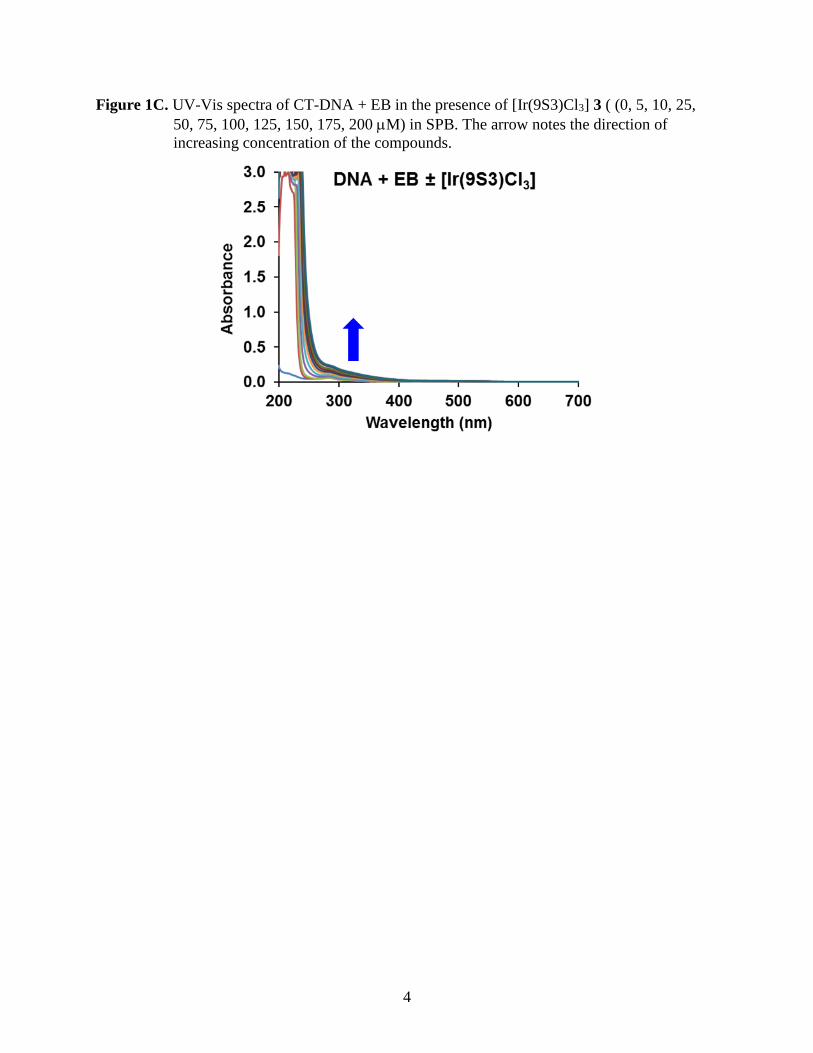
Figure 1C. UV-Vis spectra of CT-DNA + EB in the presence of [Ir(9S3)Cl3] 3 ( (0, 5, 10, 25,
50, 75, 100, 125, 150, 175, 200 M) in SPB. The arrow notes the direction of
increasing concentration of the compounds.

5
Figure 2. HOMO and LUMO for complexes 1-3 calculated at the mPW1PW91/6-31G(d,p)
level of theory
[Rh(9S3)Cl3] - HOMO [Rh(9S3)Cl3] - LUMO
[Rh(9S2O)Cl3] - HOMO [Rh(9S2O)Cl3] - LUMO
[Ir(9S3)Cl3] - HOMO [Ir(9S3)Cl3] - LUMO
6
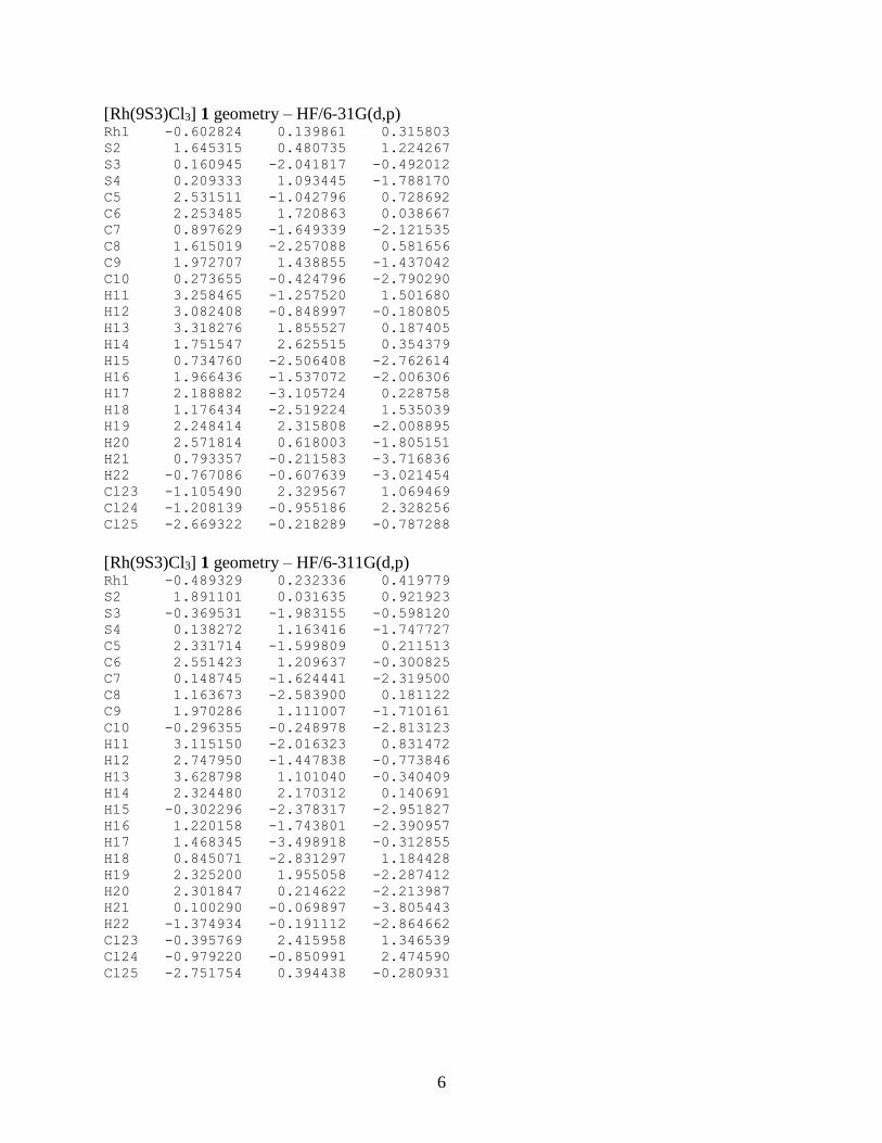
[Rh(9S3)Cl3] 1 geometry – HF/6-31G(d,p) Rh1 -0.602824 0.139861 0.315803
S2 1.645315 0.480735 1.224267
S3 0.160945 -2.041817 -0.492012
S4 0.209333 1.093445 -1.788170
C5 2.531511 -1.042796 0.728692
C6 2.253485 1.720863 0.038667
C7 0.897629 -1.649339 -2.121535
C8 1.615019 -2.257088 0.581656
C9 1.972707 1.438855 -1.437042
C10 0.273655 -0.424796 -2.790290
H11 3.258465 -1.257520 1.501680
H12 3.082408 -0.848997 -0.180805
H13 3.318276 1.855527 0.187405
H14 1.751547 2.625515 0.354379
H15 0.734760 -2.506408 -2.762614
H16 1.966436 -1.537072 -2.006306
H17 2.188882 -3.105724 0.228758
H18 1.176434 -2.519224 1.535039
H19 2.248414 2.315808 -2.008895
H20 2.571814 0.618003 -1.805151
H21 0.793357 -0.211583 -3.716836
H22 -0.767086 -0.607639 -3.021454
Cl23 -1.105490 2.329567 1.069469
Cl24 -1.208139 -0.955186 2.328256
Cl25 -2.669322 -0.218289 -0.787288
[Rh(9S3)Cl3] 1 geometry – HF/6-311G(d,p) Rh1 -0.489329 0.232336 0.419779
S2 1.891101 0.031635 0.921923
S3 -0.369531 -1.983155 -0.598120
S4 0.138272 1.163416 -1.747727
C5 2.331714 -1.599809 0.211513
C6 2.551423 1.209637 -0.300825
C7 0.148745 -1.624441 -2.319500
C8 1.163673 -2.583900 0.181122
C9 1.970286 1.111007 -1.710161
C10 -0.296355 -0.248978 -2.813123
H11 3.115150 -2.016323 0.831472
H12 2.747950 -1.447838 -0.773846
H13 3.628798 1.101040 -0.340409
H14 2.324480 2.170312 0.140691
H15 -0.302296 -2.378317 -2.951827
H16 1.220158 -1.743801 -2.390957
H17 1.468345 -3.498918 -0.312855
H18 0.845071 -2.831297 1.184428
H19 2.325200 1.955058 -2.287412
H20 2.301847 0.214622 -2.213987
H21 0.100290 -0.069897 -3.805443
H22 -1.374934 -0.191112 -2.864662
Cl23 -0.395769 2.415958 1.346539
Cl24 -0.979220 -0.850991 2.474590
Cl25 -2.751754 0.394438 -0.280931
7
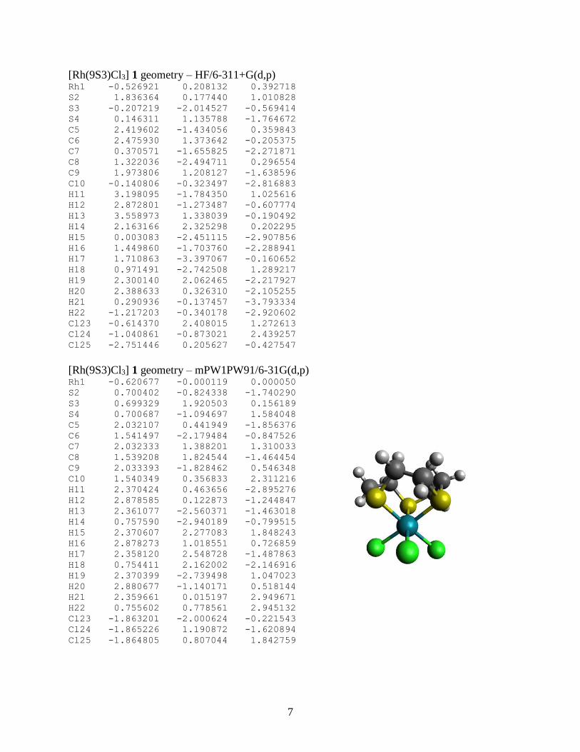
[Rh(9S3)Cl3] 1 geometry – HF/6-311+G(d,p) Rh1 -0.526921 0.208132 0.392718
S2 1.836364 0.177440 1.010828
S3 -0.207219 -2.014527 -0.569414
S4 0.146311 1.135788 -1.764672
C5 2.419602 -1.434056 0.359843
C6 2.475930 1.373642 -0.205375
C7 0.370571 -1.655825 -2.271871
C8 1.322036 -2.494711 0.296554
C9 1.973806 1.208127 -1.638596
C10 -0.140806 -0.323497 -2.816883
H11 3.198095 -1.784350 1.025616
H12 2.872801 -1.273487 -0.607774
H13 3.558973 1.338039 -0.190492
H14 2.163166 2.325298 0.202295
H15 0.003083 -2.451115 -2.907856
H16 1.449860 -1.703760 -2.288941
H17 1.710863 -3.397067 -0.160652
H18 0.971491 -2.742508 1.289217
H19 2.300140 2.062465 -2.217927
H20 2.388633 0.326310 -2.105255
H21 0.290936 -0.137457 -3.793334
H22 -1.217203 -0.340178 -2.920602
Cl23 -0.614370 2.408015 1.272613
Cl24 -1.040861 -0.873021 2.439257
Cl25 -2.751446 0.205627 -0.427547
[Rh(9S3)Cl3] 1 geometry – mPW1PW91/6-31G(d,p) Rh1 -0.620677 -0.000119 0.000050
S2 0.700402 -0.824338 -1.740290
S3 0.699329 1.920503 0.156189
S4 0.700687 -1.094697 1.584048
C5 2.032107 0.441949 -1.856376
C6 1.541497 -2.179484 -0.847526
C7 2.032333 1.388201 1.310033
C8 1.539208 1.824544 -1.464454
C9 2.033393 -1.828462 0.546348
C10 1.540349 0.356833 2.311216
H11 2.370424 0.463656 -2.895276
H12 2.878585 0.122873 -1.244847
H13 2.361077 -2.560371 -1.463018
H14 0.757590 -2.940189 -0.799515
H15 2.370607 2.277083 1.848243
H16 2.878273 1.018551 0.726859
H17 2.358120 2.548728 -1.487863
H18 0.754411 2.162002 -2.146916
H19 2.370399 -2.739498 1.047023
H20 2.880677 -1.140171 0.518144
H21 2.359661 0.015197 2.949671
H22 0.755602 0.778561 2.945132
Cl23 -1.863201 -2.000624 -0.221543
Cl24 -1.865226 1.190872 -1.620894
Cl25 -1.864805 0.807044 1.842759
8
[Rh(9S3)Cl3] 1 geometry – mPW1PW91/6-311G(d,p) Rh1 0.614843 0.000000 0.000185
S2 -0.707047 1.089984 1.584957
S3 -0.706119 0.827762 -1.736792
S4 -0.705899 -1.919011 0.151554
C5 -2.039513 1.825443 0.547896
C6 -1.550942 -0.360330 2.308999
C7 -2.037917 -0.438586 -1.855012
C8 -1.550925 2.179885 -0.843607
C9 -2.039105 -1.387976 1.305848
C10 -1.548695 -1.820962 -1.467101
H11 -2.376166 2.731980 1.053870
H12 -2.881685 1.133491 0.519924
H13 -2.372271 -0.012350 2.939332
H14 -0.774102 -0.780282 2.951037
H15 -2.375551 -0.453398 -2.892771
H16 -2.879351 -0.117180 -1.240706
H17 -2.372152 2.551142 -1.460665
H18 -0.774424 2.946188 -0.800889
H19 -2.377035 -2.279198 1.837515
H20 -2.879890 -1.016987 0.719152
H21 -2.369297 -2.541640 -1.481945
H22 -0.770901 -2.165749 -2.151372
Cl23 1.883208 -0.817793 1.824812
Cl24 1.882163 1.989891 -0.202879
Cl25 1.884188 -1.169782 -1.620665
[Rh(9S3)Cl3] 1 geometry – mPW1PW91/6-311+G(d,p) Rh1 0.617881 -0.000205 0.000087
S2 -0.701763 -0.905805 -1.695618
S3 -0.702587 -1.015477 1.632437
S4 -0.700709 1.922243 0.063442
C5 -2.035389 -1.751413 -0.748220
C6 -1.545973 0.615280 -2.254790
C7 -2.035067 0.228876 1.890433
C8 -1.547412 -2.259741 0.594774
C9 -2.033544 1.525083 -1.143515
C10 -1.545668 1.645727 1.659745
H11 -2.373965 -2.594959 -1.352744
H12 -2.876222 -1.065297 -0.642681
H13 -2.368384 0.338430 -2.918489
H14 -0.769957 1.104144 -2.847353
H15 -2.374331 0.127336 2.923031
H16 -2.875867 -0.021722 1.243003
H17 -2.369606 -2.696206 1.166603
H18 -0.771538 -3.017387 0.466985
H19 -2.370996 2.470495 -1.572459
H20 -2.875178 1.091788 -0.602466
H21 -2.367120 2.359963 1.752337
H22 -0.769369 1.912729 2.379764
Cl23 1.873046 1.014531 -1.730832
Cl24 1.871513 -2.007565 -0.013659
Cl25 1.872919 0.990778 1.744831

9
[Rh(9S2O)Cl3] 2 geometry – HF/6-31G(d,p) Rh1 -0.652324 -0.002493 0.018205
O2 0.794188 0.211099 -1.663879
S3 0.945218 1.591181 0.999201
S4 0.926579 -1.803354 0.586041
C5 1.736105 1.271087 -1.687740
C6 1.266595 -1.060487 -2.085274
C7 2.359136 0.513754 1.439363
C8 1.438270 2.299254 -0.603425
C9 1.949749 -1.802627 -0.934422
C10 1.948860 -0.912885 1.801519
H11 1.691401 1.754985 -2.655832
H12 2.732643 0.862852 -1.563550
H13 1.955920 -0.938376 -2.912935
H14 0.396857 -1.597727 -2.423667
H15 2.844672 0.956428 2.299876
H16 3.075163 0.524639 0.630009
H17 2.295219 2.948033 -0.468253
H18 0.583728 2.901826 -0.874993
H19 2.103165 -2.834689 -1.221317
H20 2.919114 -1.382852 -0.699017
H21 2.833784 -1.501478 2.012621
H22 1.328329 -0.915071 2.688620
Cl23 -1.965748 -1.577635 -1.145424
Cl24 -1.956993 1.844700 -0.646191
Cl25 -1.718433 -0.314652 2.070218
[Rh(9S2O)Cl3] 2 geometry – HF/6-311G(d,p) Rh1 -0.644128 -0.003360 0.023249
O2 0.793840 0.221543 -1.649947
S3 0.949260 1.581704 1.012054
S4 0.927108 -1.807187 0.576274
C5 1.723203 1.290901 -1.683167
C6 1.266970 -1.040554 -2.093524
C7 2.369280 0.500728 1.433218
C8 1.436313 2.309272 -0.586045
C9 1.943565 -1.804510 -0.952548
C10 1.961520 -0.926786 1.791011
H11 1.658043 1.781114 -2.647063
H12 2.725361 0.890819 -1.576397
H13 1.961179 -0.903079 -2.914790
H14 0.401557 -1.572992 -2.449050
H15 2.863343 0.941424 2.289685
H16 3.072385 0.516128 0.613237
H17 2.298266 2.949731 -0.444450
H18 0.584517 2.921210 -0.842322
H19 2.080219 -2.836195 -1.247665
H20 2.917642 -1.398625 -0.713113
H21 2.846600 -1.520291 1.987205
H22 1.348443 -0.936913 2.683061
Cl23 -1.969299 -1.569686 -1.147657
Cl24 -1.961723 1.837952 -0.646338
Cl25 -1.736595 -0.316506 2.061531
10
[Rh(9S2O)Cl3] 2 geometry – HF/6-311+G(d,p) Rh1 -0.648001 -0.003140 0.022083
O2 0.792865 0.215675 -1.661045
S3 0.946322 1.584358 1.005565
S4 0.922817 -1.803322 0.582579
C5 1.730102 1.278180 -1.686094
C6 1.268687 -1.050454 -2.089715
C7 2.363179 0.504844 1.440599
C8 1.441684 2.301814 -0.594691
C9 1.945352 -1.803396 -0.941992
C10 1.952084 -0.921113 1.800324
H11 1.677851 1.767168 -2.651963
H12 2.728772 0.871788 -1.570425
H13 1.963374 -0.920696 -2.912273
H14 0.404465 -1.588694 -2.440517
H15 2.852093 0.948843 2.298539
H16 3.071500 0.517419 0.624967
H17 2.305807 2.939581 -0.453225
H18 0.592859 2.915678 -0.857023
H19 2.088890 -2.835942 -1.231376
H20 2.916452 -1.391368 -0.700893
H21 2.835678 -1.515207 2.002388
H22 1.334502 -0.928030 2.689380
Cl23 -1.965575 -1.573077 -1.146900
Cl24 -1.955219 1.841924 -0.649027
Cl25 -1.730259 -0.311227 2.061582
[Rh(9S2O)Cl3] 2 geometry – mPW1PW91/6-31G(d,p) Rh1 -0.591111 0.000659 0.010557
O2 0.772277 0.168787 -1.726396
S3 0.854660 1.595060 0.948860
S4 0.841713 -1.747738 0.648244
C5 1.784439 1.178098 -1.666085
C6 1.253316 -1.143260 -2.035729
C7 2.282157 0.557941 1.478316
C8 1.413973 2.249552 -0.661679
C9 1.929785 -1.798557 -0.835825
C10 1.860538 -0.848644 1.871299
H11 1.880990 1.632071 -2.657929
H12 2.747943 0.720583 -1.414613
H13 1.943659 -1.091003 -2.884442
H14 0.359583 -1.699029 -2.322457
H15 2.736709 1.049110 2.342182
H16 3.028138 0.548244 0.681252
H17 2.244671 2.944950 -0.517759
H18 0.530803 2.799730 -0.997925
H19 2.125946 -2.848627 -1.062621
H20 2.885213 -1.331795 -0.585390
H21 2.735050 -1.454336 2.124739
H22 1.200158 -0.822501 2.742575
Cl23 -1.910532 -1.592453 -1.097134
Cl24 -1.914231 1.800044 -0.708026
Cl25 -1.721373 -0.240256 2.007103
11
[Rh(9S2O)Cl3] 2 geometry – mPW1PW91/6-311G(d,p) Rh1 -0.585989 0.000090 0.015451
O2 0.771119 0.178054 -1.714355
S3 0.863472 1.587949 0.958777
S4 0.849864 -1.750876 0.639379
C5 1.776745 1.193620 -1.662769
C6 1.253326 -1.127553 -2.043185
C7 2.296163 0.547316 1.467004
C8 1.413260 2.256652 -0.649613
C9 1.929038 -1.796337 -0.852455
C10 1.880142 -0.858826 1.857575
H11 1.856985 1.651303 -2.653011
H12 2.743696 0.739633 -1.423682
H13 1.943268 -1.060490 -2.889595
H14 0.364117 -1.680923 -2.341422
H15 2.759888 1.037264 2.324939
H16 3.027443 0.542655 0.658615
H17 2.246417 2.945761 -0.499571
H18 0.532978 2.814860 -0.973985
H19 2.115621 -2.845317 -1.084898
H20 2.885498 -1.336394 -0.599728
H21 2.755851 -1.467863 2.092985
H22 1.231838 -0.842105 2.736356
Cl23 -1.921265 -1.584293 -1.094089
Cl24 -1.927057 1.789349 -0.705768
Cl25 -1.738059 -0.240897 2.000015
[Rh(9S2O)Cl3] 2 geometry – mPW1PW91/6-311+G(d,p) Rh1 -0.587639 0.000109 0.013645
O2 0.765863 0.168297 -1.721573
S3 0.855777 1.592029 0.948530
S4 0.843346 -1.744466 0.647489
C5 1.775216 1.180680 -1.667753
C6 1.251890 -1.139742 -2.037010
C7 2.287105 0.555800 1.468973
C8 1.413768 2.250120 -0.660964
C9 1.927175 -1.796747 -0.840117
C10 1.870481 -0.848388 1.865065
H11 1.861656 1.633844 -2.660002
H12 2.739278 0.723660 -1.423858
H13 1.943206 -1.078383 -2.883136
H14 0.364926 -1.699360 -2.331396
H15 2.747306 1.051097 2.326025
H16 3.021587 0.547353 0.663461
H17 2.251968 2.933628 -0.511730
H18 0.538529 2.813463 -0.990384
H19 2.117679 -2.846790 -1.065469
H20 2.881623 -1.332316 -0.588041
H21 2.746006 -1.456628 2.104274
H22 1.220338 -0.828177 2.742514
Cl23 -1.913514 -1.593926 -1.090628
Cl24 -1.919184 1.794062 -0.710773
Cl25 -1.723599 -0.234488 2.005569
12
[Ir(9S3)Cl3] 3 geometry – HF/6-31G(d,p) Ir1 0.000075 0.002179 0.561870
S2 0.842672 -1.756929 -0.840431
S3 -1.940216 0.143669 -0.847430
S4 1.097205 1.603403 -0.853497
C5 -0.436184 -1.888658 -2.146217
C6 2.187472 -0.854670 -1.671833
C7 -1.410745 1.309296 -2.158489
C8 -1.828618 -1.476993 -1.668882
C9 1.846065 0.554306 -2.155996
C10 -0.359526 2.312178 -1.683774
H11 -0.473004 -2.925558 -2.454669
H12 -0.123411 -1.310464 -3.004359
H13 2.549811 -1.450068 -2.501358
H14 2.959697 -0.819605 -0.915108
H15 -2.289337 1.857779 -2.472977
H16 -1.063695 0.744254 -3.012202
H17 -2.522794 -1.498021 -2.500494
H18 -2.186752 -2.158797 -0.909226
H19 2.761351 1.039087 -2.470478
H20 1.185898 0.531197 -3.011676
H21 -0.028023 2.918926 -2.517957
H22 -0.773313 2.967722 -0.929297
Cl23 2.035834 -0.199649 1.824230
Cl24 -1.196578 -1.652426 1.830296
Cl25 -0.838524 1.873293 1.816575
[Ir(9S3)Cl3] 3 geometry – HF/6-311G(d,p) Ir1 -0.002765 -0.038459 -0.554171
S2 -0.950459 -1.628732 0.968619
S3 1.946762 0.077198 0.835774
S4 -0.983629 1.727822 0.735845
C5 0.323954 -1.751298 2.282555
C6 -2.231253 -0.584731 1.735576
C7 1.503994 1.369725 2.060073
C8 1.737397 -1.468336 1.777094
C9 -1.795732 0.829653 2.114017
C10 0.518940 2.401950 1.514582
H11 0.289668 -2.763214 2.664640
H12 0.051913 -1.090083 3.092731
H13 -2.621279 -1.096956 2.606983
H14 -3.005069 -0.558160 0.980642
H15 2.420599 1.876705 2.332004
H16 1.123708 0.889144 2.950028
H17 2.433397 -1.466547 2.607411
H18 2.050448 -2.225312 1.071115
H19 -2.673407 1.399169 2.390562
H20 -1.130672 0.826109 2.965651
H21 0.225389 3.085166 2.302547
H22 0.968428 2.975521 0.715584
Cl23 -2.046446 -0.187516 -1.820087
Cl24 1.062370 -1.857356 -1.719714
Cl25 0.956616 1.662952 -1.963490

13
[Ir(9S3)Cl3] 3 geometry – HF/6-311+G(d,p) Ir1 0.004883 -0.052335 -0.556296
S2 -0.674042 -1.737831 1.009937
S3 1.906147 0.414628 0.830090
S4 -1.254354 1.561638 0.694439
C5 0.600152 -1.618271 2.324426
C6 -2.109676 -0.898361 1.753408
C7 1.255431 1.647251 2.022968
C8 1.949591 -1.120467 1.809937
C9 -1.912145 0.577180 2.095830
C10 0.116039 2.490854 1.454098
H11 0.730789 -2.612378 2.732133
H12 0.221289 -0.990491 3.118227
H13 -2.412646 -1.445309 2.638429
H14 -2.876572 -1.017219 1.000063
H15 2.076202 2.303807 2.281443
H16 0.956812 1.132471 2.924962
H17 2.633518 -0.984551 2.639380
H18 2.384302 -1.833936 1.123080
H19 -2.871724 1.002298 2.360450
H20 -1.257011 0.703684 2.945867
H21 -0.286746 3.135807 2.226113
H22 0.468005 3.111201 0.641033
Cl23 -1.982788 -0.568153 -1.805977
Cl24 1.358802 -1.696761 -1.670467
Cl25 0.672068 1.749638 -2.000722
[Ir(9S3)Cl3] 3 geometry – mPW1PW91/6-31G(d,p) Ir1 -0.514279 0.000065 0.000068
S2 0.787596 -0.640399 1.801628
S3 0.787146 -1.240485 -1.455976
S4 0.787806 1.881247 -0.346935
C5 2.126917 -1.628765 1.003896
C6 1.641403 0.942680 2.140105
C7 2.127681 -0.056118 -1.910141
C8 1.639897 -2.326138 -0.254219
C9 2.127823 1.683863 0.907150
C10 1.640537 1.381936 -1.887224
H11 2.449410 -2.378294 1.730245
H12 2.979021 -0.975394 0.804719
H13 2.464026 0.763819 2.837529
H14 0.863447 1.514652 2.653460
H15 2.453538 -0.311108 -2.921172
H16 2.977321 -0.209559 -1.241602
H17 2.462191 -2.841265 -0.757773
H18 0.861427 -3.056066 -0.015309
H19 2.450369 2.687577 1.193355
H20 2.979790 1.185019 0.440307
H21 2.462727 2.075468 -2.082434
H22 0.861994 1.538967 -2.638983
Cl23 -1.809186 1.264520 1.557573
Cl24 -1.809763 -1.980508 0.316612
Cl25 -1.809998 0.716615 -1.873270
14
[Ir(9S3)Cl3] 3 geometry – mPW1PW91/6-311G(d,p) Ir1 0.509969 0.000024 0.000152
S2 -0.792760 -0.018113 -1.909791
S3 -0.792618 -1.645672 0.971123
S4 -0.792962 1.663064 0.939528
C5 -2.132514 -1.211936 -1.476522
C6 -1.650459 1.586585 -1.716146
C7 -2.133928 -0.673536 1.785663
C8 -1.648502 -2.280632 -0.516337
C9 -2.133404 1.884902 -0.310433
C10 -1.650691 0.692265 2.231920
H11 -2.454985 -1.678897 -2.408264
H12 -2.979124 -0.654746 -1.074382
H13 -2.474329 1.633302 -2.431269
H14 -0.879354 2.295986 -2.023476
H15 -2.458351 -1.246986 2.655196
H16 -2.978886 -0.603550 1.100006
H17 -2.471592 -2.924816 -0.200301
H18 -0.876198 -2.899847 -0.977280
H19 -2.454991 2.925583 -0.249290
H20 -2.980515 1.258913 -0.028341
H21 -2.474475 1.288021 2.630397
H22 -0.879730 0.602852 3.000020
Cl23 1.827535 1.701315 -1.044782
Cl24 1.828849 -1.754485 -0.950849
Cl25 1.828637 0.054817 1.995197
[Ir(9S3)Cl3] 3 geometry – mPW1PW91/6-311+G(d,p) Ir1 0.513030 -0.000126 0.000049
S2 -0.790348 -0.116646 1.905465
S3 -0.789183 1.709461 -0.852119
S4 -0.789985 -1.591974 -1.054123
C5 -2.130061 1.103954 1.557356
C6 -1.649330 -1.702692 1.599441
C7 -2.129911 0.798141 -1.733910
C8 -1.646558 2.237995 0.675105
C9 -2.131127 -1.900458 0.175717
C10 -1.647728 -0.533341 -2.275031
H11 -2.454348 1.503775 2.519471
H12 -2.975797 0.575822 1.115847
H13 -2.474795 -1.797929 2.308209
H14 -0.880446 -2.433999 1.857569
H15 -2.454628 1.431448 -2.561057
H16 -2.974948 0.680555 -1.054909
H17 -2.470848 2.901297 0.404227
H18 -0.876114 2.825100 1.179506
H19 -2.455178 -2.933649 0.040923
H20 -2.976627 -1.254125 -0.062059
H21 -2.472869 -1.099141 -2.713002
H22 -0.878081 -0.390786 -3.036598
Cl23 1.817817 -1.772195 0.929068
Cl24 1.819365 1.689274 1.070111
Cl25 1.819400 0.080937 -1.998092
15
[Rh(9S3)Cl2H2O]+ geometry – HF/6-31G(d,p) Rh1 0.693956 -0.001703 -0.237102
S2 -0.243942 -0.046943 1.939089
S3 -0.951483 1.713435 -0.841789
S4 -0.968759 -1.667937 -0.924029
C5 -1.601008 1.183705 1.865273
C6 -1.100096 -1.655090 1.887666
C7 -2.425132 0.743562 -1.333563
C8 -1.352596 2.288932 0.841398
C9 -1.910879 -1.939635 0.625500
C10 -2.073042 -0.618564 -1.925609
H11 -1.668150 1.632672 2.847830
H12 -2.532347 0.667581 1.683064
H13 -1.738634 -1.729153 2.759527
H14 -0.296081 -2.369604 2.002045
H15 -2.951148 1.320558 -2.083226
H16 -3.084040 0.650412 -0.482990
H17 -2.217072 2.938339 0.784690
H18 -0.496969 2.890765 1.115807
H19 -2.208313 -2.979877 0.634670
H20 -2.813101 -1.346886 0.584529
H21 -2.979406 -1.173619 -2.134356
H22 -1.541851 -0.506407 -2.862854
Cl23 2.160991 -1.733188 0.378385
Cl24 2.152894 1.738418 0.374323
O25 1.623502 -0.003977 -2.139139
H26 2.217726 -0.747586 -2.177350
H27 2.160739 0.777988 -2.221046
[Rh(9S3)Cl2H2O]+ geometry – mPW1PW91/6-31G(d,p) Rh1 0.631501 -0.002406 -0.232055
S2 -0.195603 -0.044337 1.895248
S3 -0.898052 1.687688 -0.833620
S4 -0.916894 -1.643026 -0.910749
C5 -1.578939 1.172489 1.849122
C6 -1.068785 -1.651427 1.878179
C7 -2.397042 0.736500 -1.314943
C8 -1.322388 2.278805 0.842901
C9 -1.887671 -1.918086 0.629237
C10 -2.047676 -0.613747 -1.913684
H11 -1.649870 1.601663 2.851689
H12 -2.511698 0.641664 1.651547
H13 -1.691457 -1.714608 2.774843
H14 -0.251998 -2.370600 1.979480
H15 -2.930802 1.334084 -2.058216
H16 -3.049988 0.641127 -0.446006
H17 -2.186046 2.944748 0.770204
H18 -0.450747 2.876925 1.120487
H19 -2.204352 -2.963609 0.621488
H20 -2.787274 -1.301679 0.586346
H21 -2.953714 -1.190950 -2.118175
H22 -1.513792 -0.501122 -2.861097
Cl23 2.127069 -1.712438 0.358886
Cl24 2.119893 1.713374 0.363087
O25 1.637664 0.003346 -2.113254
H26 2.241454 -0.753360 -2.006353
H27 2.206854 0.788880 -2.030213
16
[Rh(9S2O)Cl2H2O]+ geometry – HF/6-31G(d,p) Rh1 0.640730 -0.112661 0.177251
O2 -0.760971 1.211017 1.153787
S3 -0.434726 0.714877 -1.761703
S4 -1.211208 -1.696407 0.488041
C5 -1.452713 2.211484 0.396692
C6 -1.532120 0.517399 2.133659
C7 -2.015042 -0.216555 -1.798622
C8 -0.891122 2.315943 -1.017170
C9 -2.310173 -0.636518 1.502427
C10 -1.904465 -1.614374 -1.196825
H11 -1.336030 3.160914 0.901111
H12 -2.505365 1.961556 0.376497
H13 -2.205335 1.209012 2.623605
H14 -0.824606 0.153279 2.863080
H15 -2.297997 -0.309483 -2.839242
H16 -2.777388 0.371544 -1.308691
H17 -1.600292 2.817401 -1.664016
H18 0.035389 2.871052 -1.029198
H19 -2.729919 -1.257353 2.282936
H20 -3.129261 -0.288584 0.888395
H21 -2.877361 -2.089345 -1.191466
H22 -1.233134 -2.236453 -1.774869
Cl23 2.293905 1.539473 -0.012532
Cl24 1.847720 -1.734450 -0.967416
O25 1.617785 -0.723181 1.953246
H26 2.299302 -0.084930 2.144107
H27 2.055760 -1.556744 1.811930
[Rh(9S2O)Cl2H2O]+ geometry – mPW1PW91/6-31G(d,p) Rh1 0.594147 -0.094010 0.180518
O2 -0.755149 1.303792 1.128389
S3 -0.379829 0.603640 -1.761193
S4 -1.137359 -1.659844 0.550824
C5 -1.519999 2.170031 0.262030
C6 -1.535395 0.596967 2.107501
C7 -1.967162 -0.336026 -1.802422
C8 -0.890584 2.236960 -1.113950
C9 -2.286802 -0.577808 1.493990
C10 -1.845030 -1.692341 -1.133989
H11 -1.532977 3.168417 0.707426
H12 -2.550346 1.806873 0.209138
H13 -2.229685 1.291619 2.589426
H14 -0.813970 0.257176 2.851350
H15 -2.221886 -0.470823 -2.856535
H16 -2.749545 0.275521 -1.350236
H17 -1.567314 2.711234 -1.829377
H18 0.049098 2.793522 -1.094345
H19 -2.728095 -1.184413 2.287963
H20 -3.097707 -0.260696 0.835591
H21 -2.816195 -2.192384 -1.095632
H22 -1.152599 -2.342623 -1.675418
Cl23 2.261932 1.522756 -0.070312
Cl24 1.843062 -1.725678 -0.855546
O25 1.631374 -0.598635 1.970768
H26 2.305506 0.105362 1.991136
H27 2.119257 -1.407341 1.739481
17
[Ir(9S3)Cl2H2O]+ geometry – HF/6-31G(d,p) Ir1 0.576246 -0.001847 -0.177931
S2 -0.427402 -0.039996 1.954516
S3 -1.002086 1.704000 -0.842226
S4 -1.018154 -1.660559 -0.916244
C5 -1.781200 1.190349 1.830164
C6 -1.282728 -1.646812 1.885583
C7 -2.459309 0.742093 -1.398986
C8 -1.486496 2.292202 0.814912
C9 -2.031875 -1.940164 0.588099
C10 -2.090648 -0.626333 -1.966433
H11 -1.885453 1.640807 2.808588
H12 -2.706044 0.676614 1.610939
H13 -1.961885 -1.711885 2.726750
H14 -0.485144 -2.360699 2.043215
H15 -2.944044 1.320322 -2.174822
H16 -3.157357 0.658120 -0.579159
H17 -2.348001 2.939459 0.709999
H18 -0.644756 2.896223 1.126056
H19 -2.319373 -2.983034 0.581589
H20 -2.935642 -1.354153 0.503705
H21 -2.990134 -1.184562 -2.195295
H22 -1.530269 -0.524785 -2.887693
Cl23 2.065294 -1.745293 0.475685
Cl24 2.063141 1.744949 0.473582
O25 1.617121 -0.001964 -2.031301
H26 2.209070 -0.749569 -2.039948
H27 2.163566 0.777792 -2.077139
[Ir(9S3)Cl2H2O]+ geometry – mPW1PW91/6-31G(d,p) Ir1 0.534850 -0.002897 -0.169629
S2 -0.395003 -0.029913 1.910967
S3 -0.946560 1.673233 -0.843900
S4 -0.962882 -1.638662 -0.899546
C5 -1.776539 1.189990 1.801489
C6 -1.273986 -1.635165 1.878057
C7 -2.429251 0.727886 -1.397673
C8 -1.472403 2.286406 0.798674
C9 -2.019427 -1.918020 0.588538
C10 -2.061737 -0.631637 -1.962245
H11 -1.885311 1.625673 2.797311
H12 -2.701599 0.659778 1.568467
H13 -1.947538 -1.677426 2.737994
H14 -0.468111 -2.355879 2.039571
H15 -2.914573 1.324679 -2.173487
H16 -3.126627 0.647313 -0.562241
H17 -2.336012 2.942330 0.663901
H18 -0.622873 2.896205 1.116961
H19 -2.321747 -2.967135 0.564930
H20 -2.920510 -1.309877 0.491389
H21 -2.960211 -1.215129 -2.180761
H22 -1.498179 -0.536615 -2.894080
Cl23 2.056878 -1.708063 0.460852
Cl24 2.053845 1.706248 0.460940
O25 1.654139 0.000797 -1.993402
H26 2.249798 -0.761782 -1.866837
H27 2.225807 0.784093 -1.886317
18
[Rh(9S3)Cl2OH] geometry – HF/6-31G(d,p) Rh1 0.742335 0.003931 -0.279047
S2 -0.285637 -0.052985 2.043839
S3 -0.923634 1.717446 -0.817546
S4 -0.912229 -1.680291 -0.905701
C5 -1.652905 1.155023 1.887981
C6 -1.127576 -1.662157 1.896853
C7 -2.357641 0.726989 -1.372521
C8 -1.385400 2.254474 0.858358
C9 -1.912214 -1.913452 0.606836
C10 -1.946055 -0.621049 -1.963614
H11 -1.783397 1.625789 2.854134
H12 -2.569186 0.624324 1.670283
H13 -1.785365 -1.797462 2.746994
H14 -0.314343 -2.370788 1.986914
H15 -2.865988 1.294985 -2.141409
H16 -3.051871 0.609911 -0.552267
H17 -2.248471 2.906480 0.792813
H18 -0.535948 2.854059 1.158978
H19 -2.252326 -2.941429 0.614268
H20 -2.793216 -1.289891 0.552074
H21 -2.827285 -1.172170 -2.269261
H22 -1.313595 -0.475286 -2.830158
Cl23 2.237028 -1.742066 0.336867
Cl24 2.207494 1.787418 0.305011
O25 1.186042 -0.017700 -2.162698
H26 2.125433 -0.060455 -2.249418
[Rh(9S3)Cl2OH] geometry – mPW1PW91/6-31G(d,p) Rh1 0.646204 0.002702 -0.279935
S2 -0.113068 0.003829 1.981039
S3 -0.900370 1.653843 -0.827186
S4 -0.859652 -1.682955 -0.821559
C5 -1.523765 1.183522 1.899672
C6 -0.965237 -1.613672 1.963353
C7 -2.381466 0.657999 -1.274472
C8 -1.313471 2.258573 0.844922
C9 -1.816572 -1.894408 0.733096
C10 -1.978006 -0.683509 -1.863068
H11 -1.614444 1.660433 2.878611
H12 -2.445036 0.622885 1.727444
H13 -1.562885 -1.721570 2.872578
H14 -0.127448 -2.314616 2.016634
H15 -2.945166 1.223419 -2.020660
H16 -3.022375 0.546108 -0.397339
H17 -2.185236 2.915978 0.784602
H18 -0.437291 2.867077 1.085728
H19 -2.163558 -2.930201 0.768247
H20 -2.702630 -1.256898 0.699927
H21 -2.858006 -1.274535 -2.130088
H22 -1.361859 -0.541402 -2.756291
Cl23 2.209231 -1.680865 0.283504
Cl24 2.125958 1.793400 0.183288
O25 1.060442 -0.076175 -2.220768
H26 2.005539 -0.268472 -2.239356
19
[Rh(9S2O)Cl2OH] geometry – HF/6-31G(d,p) Rh1 -0.707634 -0.138075 -0.217891
O2 0.781784 1.182653 -1.159207
S3 0.485745 0.796942 1.839101
S4 1.169437 -1.722398 -0.422984
C5 1.503634 2.154540 -0.425244
C6 1.500872 0.444882 -2.136725
C7 2.045777 -0.162774 1.817024
C8 0.919414 2.339174 0.970032
C9 2.263673 -0.715334 -1.493208
C10 1.898428 -1.571031 1.238087
H11 1.453828 3.098629 -0.954928
H12 2.545368 1.859762 -0.368310
H13 2.185391 1.101958 -2.661580
H14 0.750694 0.076716 -2.817138
H15 2.383050 -0.256661 2.841541
H16 2.800540 0.402287 1.288665
H17 1.604309 2.923697 1.572301
H18 -0.026388 2.860005 0.915126
H19 2.644200 -1.364029 -2.271345
H20 3.109814 -0.375895 -0.909830
H21 2.863693 -2.063611 1.242080
H22 1.225502 -2.160058 1.849074
Cl23 -2.349384 1.566216 -0.087145
Cl24 -1.951638 -1.747955 0.952612
O25 -1.240671 -0.714988 -1.974658
H26 -2.181703 -0.670619 -2.040228
[Rh(9S2O)Cl2OH] geometry – mPW1PW91/6-31G(d,p) Rh1 0.639955 -0.010178 -0.236540
O2 -0.397433 0.065584 1.781313
S3 -0.925311 1.652377 -0.765364
S4 -0.920231 -1.698445 -0.681394
C5 -1.387826 1.078021 1.949182
C6 -0.834501 -1.263120 2.065844
C7 -2.442821 0.663619 -1.096517
C8 -1.169166 2.204416 0.958126
C9 -1.727886 -1.831227 0.964596
C10 -2.110719 -0.710776 -1.654660
H11 -1.315420 1.478680 2.966446
H12 -2.389158 0.646342 1.836737
H13 -1.360962 -1.285886 3.026911
H14 0.086949 -1.842163 2.143176
H15 -3.038745 1.213783 -1.828881
H16 -3.034550 0.596778 -0.181425
H17 -1.988060 2.926218 1.010762
H18 -0.223347 2.713878 1.163414
H19 -1.905898 -2.890843 1.160447
H20 -2.702275 -1.339149 0.920850
H21 -3.022728 -1.293126 -1.812273
H22 -1.593506 -0.620237 -2.614164
Cl23 2.154473 -1.682118 0.430467
Cl24 2.139180 1.737278 0.250408
O25 1.166277 -0.136878 -2.109772
H26 2.113054 -0.322106 -2.066154

20
[Ir(9S3)Cl2OH] geometry – HF/6-31G(d,p) Ir1 0.592890 0.002709 -0.223332
S2 -0.349740 -0.042512 2.047357
S3 -1.001984 1.696667 -0.795141
S4 -0.989474 -1.671733 -0.863226
C5 -1.722953 1.164968 1.911788
C6 -1.206415 -1.647207 1.936918
C7 -2.445800 0.716854 -1.348945
C8 -1.474733 2.257383 0.870121
C9 -1.991883 -1.909664 0.649326
C10 -2.039419 -0.637326 -1.929602
H11 -1.834352 1.638941 2.878582
H12 -2.641676 0.630933 1.712728
H13 -1.864610 -1.758074 2.790338
H14 -0.398926 -2.360744 2.038291
H15 -2.945336 1.286134 -2.122386
H16 -3.142665 0.609654 -0.529275
H17 -2.348196 2.894175 0.793699
H18 -0.633408 2.872576 1.161670
H19 -2.323895 -2.940098 0.658250
H20 -2.876533 -1.291201 0.592398
H21 -2.921216 -1.196578 -2.218276
H22 -1.411759 -0.500744 -2.801253
Cl23 2.128613 -1.750755 0.394923
Cl24 2.099216 1.794244 0.362185
O25 1.069450 -0.020889 -2.144191
H26 2.007749 -0.054594 -2.237011
[Ir(9S3)Cl2OH] geometry – mPW1PW91/6-31G(d,p) Ir1 0.542574 0.000158 -0.210142
S2 -0.276774 -0.003074 1.987235
S3 -0.951262 1.647017 -0.814150
S4 -0.931113 -1.661301 -0.817526
C5 -1.682510 1.190516 1.884462
C6 -1.157679 -1.608743 1.962481
C7 -2.432960 0.675697 -1.327692
C8 -1.440365 2.264004 0.836612
C9 -1.955763 -1.887648 0.697733
C10 -2.024787 -0.671554 -1.898355
H11 -1.784230 1.662639 2.864212
H12 -2.603871 0.636652 1.691385
H13 -1.797637 -1.687224 2.845430
H14 -0.336662 -2.323835 2.064650
H15 -2.950193 1.253661 -2.096993
H16 -3.112272 0.574359 -0.478416
H17 -2.315338 2.911281 0.732504
H18 -0.580547 2.882328 1.109332
H19 -2.296552 -2.925601 0.707687
H20 -2.841357 -1.252146 0.629114
H21 -2.900661 -1.263660 -2.175725
H22 -1.388822 -0.540020 -2.779257
Cl23 2.118066 -1.691778 0.385808
Cl24 2.070649 1.763640 0.306944
O25 1.075267 -0.058839 -2.148959
H26 2.030386 -0.187489 -2.138503
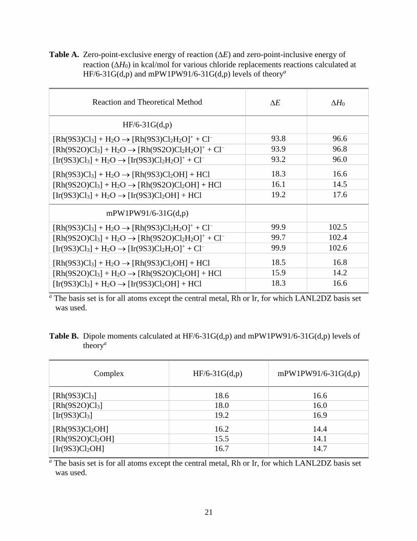
21
Table A. Zero-point-exclusive energy of reaction (E) and zero-point-inclusive energy of
reaction (H0) in kcal/mol for various chloride replacements reactions calculated at
HF/6-31G(d,p) and mPW1PW91/6-31G(d,p) levels of theorya
Reaction and Theoretical Method E H0
HF/6-31G(d,p)
[Rh(9S3)Cl3] + H2O [Rh(9S3)Cl2H2O]+ + Cl– 93.8 96.6
[Rh(9S2O)Cl3] + H2O [Rh(9S2O)Cl2H2O]+ + Cl– 93.9 96.8
[Ir(9S3)Cl3] + H2O [Ir(9S3)Cl2H2O]+ + Cl– 93.2 96.0
[Rh(9S3)Cl3] + H2O [Rh(9S3)Cl2OH] + HCl 18.3 16.6
[Rh(9S2O)Cl3] + H2O [Rh(9S2O)Cl2OH] + HCl 16.1 14.5
[Ir(9S3)Cl3] + H2O [Ir(9S3)Cl2OH] + HCl 19.2 17.6
mPW1PW91/6-31G(d,p)
[Rh(9S3)Cl3] + H2O [Rh(9S3)Cl2H2O]+ + Cl– 99.9 102.5
[Rh(9S2O)Cl3] + H2O [Rh(9S2O)Cl2H2O]+ + Cl– 99.7 102.4
[Ir(9S3)Cl3] + H2O [Ir(9S3)Cl2H2O]+ + Cl– 99.9 102.6
[Rh(9S3)Cl3] + H2O [Rh(9S3)Cl2OH] + HCl 18.5 16.8
[Rh(9S2O)Cl3] + H2O [Rh(9S2O)Cl2OH] + HCl 15.9 14.2
[Ir(9S3)Cl3] + H2O [Ir(9S3)Cl2OH] + HCl 18.3 16.6
a The basis set is for all atoms except the central metal, Rh or Ir, for which LANL2DZ basis set
was used.
Table B. Dipole moments calculated at HF/6-31G(d,p) and mPW1PW91/6-31G(d,p) levels of
theorya
Complex HF/6-31G(d,p) mPW1PW91/6-31G(d,p)
[Rh(9S3)Cl3] 18.6 16.6
[Rh(9S2O)Cl3] 18.0 16.0
[Ir(9S3)Cl3] 19.2 16.9
[Rh(9S3)Cl2OH] 16.2 14.4
[Rh(9S2O)Cl2OH] 15.5 14.1
[Ir(9S3)Cl2OH] 16.7 14.7
a The basis set is for all atoms except the central metal, Rh or Ir, for which LANL2DZ basis set
was used.