information to users - university of hawaii at manoa · 2015-06-08 · information to users this...
TRANSCRIPT

INFORMATION TO USERS
This material was produced from a microfilm copy of the original document. Whilethe most advanced technological means to photograph and reproduce this documenthave been used, the quality is heavily dependent upon the quality of the originalsubmitted.
The following explanation of techniques is provided to help you understandmarkings or patterns which may appear on this reproduction.
1. The sign or "target" for pages apparently lacking from the documentphotographed is "Missing Page(s)". If it was possible to obtain the missingpage(s) or section, they are spliced into the film along with adjacent pages.This may have necessitated cutting thru an image and duplicating adjacentpages to insure you complete continuity.
2. When an image on the film is obliterated with a large round black mark, itis an indication that the photographer suspected that the copy may havemoved during exposure and thus cause a blurred image. You will find agood image of the page in the adjacent frame.
3. When a map, drawing or chart, etc., was part of the material beingphotographed the photographer followed a definite method in"sectioning" the material. It is customary to begin photoing at the upperleft hand corner of a large sheet and to continue photoing from left toright in aqual sections with a small overlap. If necessary, sectioning iscontinued again - beginning below the first row and continuing on untilcomplete.
4. The majority of users indicate that the textual content is of greatest value,~owever, a somewhat higher quality reproduction could be made from"photographs" if essential to the understanding of the dissertation. Silverprints of "photographs" may be ordered at additional charge by writingthe Order Department, giVing the catalog number, title, author andspecific pages you wish reproduced.
5. PLEASE NOTE: Some pages may' have indistinct print. Filmed asreceived.
Xerox University Microfilms300 North Zeeb RoadAnn Arbor, Michigan 48106

75-25,158
CROS1llWAITE, Leola M., 1928.HYPERSENSITIVE 'CELL COLLAPSE., INDUCED IN BELLPEPPER ,(GAP$IWM~ BY'PSEUIXH)NAS
, 'PHASEOLICOIA.AND BY AN ENOOTOXIN ISOIATED FR<M. 'THB BACTBUA. '
University of Hawaii, Ph.D., 1975Agricu1ture, plant pathology
Xerox University Microfilms, Ann Arbor, Michigan 48106
,i

HYPERSENSITIVE CELL COLLAPSE INDUCED IN BELL PEPPER (CAPSICUM ANNUUM)
BY PSEUDOMONAS PHASEOLICOLA AND BY .AN ENDOTOXIN
ISOLATED FROM THE BACTERIA
A DISSERTATION SUBMITTED TO THE GRADUATE DIVISION OF THEUNIVERSITY OF HAWAII IN PARTIAL FULFILLMENT OF
THE REQUIREMENTS FOR THE DEGREE OF
DOCTOR OF PHILOSOPHY
IN
BOTANICAL SCIENCES
MAY 1975
By
Leola M. Crosthwaite
Dissertation Committee:
Suresh S. Patil, ChairmanNoel P. Kefford
Bruce J. CooilCharles H. Lamoureux
Barbara z. Siegel

ABSTRACT
Electron micrographs of pepper tissue infused with 10'/ and 108
cells/ml of Pseudomonas phaseolicola, the causal agent of the halo blight
of bean, show that after 12 hours the bacterial cells aggregate in a
gel-like matrix of electron translucent material on the plant cell walls
facing the intercellular spaces. Within the gel matrix an electron
dense amorphous material is also observed, but its occurrence depends
on the inoculum concentration used to inoculate the plant. In the
plant cells adjacent to the localized bacteria a plasmalemmasome first
develops, followed by deposition of a wall construction, with cell
collapse occurring soon afterwards. Wall constructions and cellular
collapse are also observed in pepper tissue infused with a purified
extract from the bacterial cells. but not from the culture broth.
The purified extract contains a high molecular weight endotoxin which
is capable of causing confluent cell collapse that is not accompanied
by tissue browning or senescence symptoms. It is degraded by pronase,
but not RNAse, and has a molecular weight of approximately 93,000. A
mutant of Pseudomonas phaseolicola which is incapable of producing
halo blight symptoms in its host or producing in culture the inducing
principle also possesses the endotoxin which shows similar properties
to that of the parent pathogenic strain.

ACKNOWLEDGMENTS
Thanks are due to Dr. Paul Dunn and to Dr. Michael Hanson
for their help to the author in mastering the techniques of electron
microscopy and especially to Dr. William S. Sakai for the many
stimulating discussions concerning interactions of Pseudomonas
phaseolicola with pepper tissue.
Special acknowledgment is made to the Electron Microscope
Facility Committee of the St. John Plant Science Laboratory for the
use of the electron microscope.

TABLE OF CONTENTS
Page
ABSTRACT • • • • • •
ACKNOWLEDGMENTS.
LIST OF TABLES • •
LIST OF ILLUSTRATIONS.
iii
iv
vii
viii
CHAPTER I. THE HYPERSENSITIVE REACTION IN PLANT RESISTANCE
Introduction • • • • • • • • • • • • • • 1Literature Review. •• •••• • • • • • • • 7
Plant resistance. • • • • • 7The hypersensitive reaction induced by fungal and
viral pathogens. • • • • • • • • • • • • • 11HR interaction types in fungi. • • • • • • • • • 11Interaction phases in fungal induced HR. 13
Resistance and growth regulators. • • • • • • 16Hypersensitivity induced by bacterial pathogens • • • •• 18Models of the hypersensitive reaction • • • • • 25
Discussion • • • • • 34Literature Cited • • • • • • • • • • • • • • • • 39
CHAPTER II. HYPERSENSITIVE CELL COLLAPSE J:NDUCED IN BELLPEPPER, CAPSICUM ANNUUM BY PSEUDOMONASPHASEOLICOLA
Abstract • • • • • • •Introduction • • • • •Materials and Methods.Results. • • • • • • •Discussion • • • • • •Tables • • • • • • • •Figures •••Literature Cited.
4748505263717491

TABLE OF CONTENTS (continued)
CHAPTER III. ISOLATION AND CHARACTERIZATION OF AN ENDOTOXINFROM PSEUDOMONAS PHASEOLICOLA WHICH INDUCESCELL COLLAPSE IN BELL PEPPER (CAPSICUM ANNUUM)
vi
Page
Abstract · · · · · · · · · · · · · · · · · · · · · · · · · 95Introduction · · · · · · · · · · · · · · · 95Materials and Methods. · · · · · · · ~ · · · · · . . 97Results. · · · · · · · · · 100Discussion · 104Tables . · · · · · · · · · · 108Figures. · · · · · · 112Literature Cited · · · · · · · · · 125
CHAPTER IV. CONCLUSIONS. · · 125
Literature Cited · · · · · · · · · 129
BIBLIOGRAPHY · · · · · · · · · · · · · · · · · · 130
•

Table
1
1
2
LIST OF TABLES
CHAPTER I
Enzyme changes in hypersensitive tissue.
CHAPTER II
Effect of selected bacterial strains onCapsicum annuum. • • • • • • • • •• ••• • • • • •
Effects of inoculum concentration and incubationtime in pepper on po~ulations of Pseudomonasphaseolicola GSO Tox and GSO Tox- at 12 and18 hours • • • • • • • • • • • • • • • • • • • • • • • •
CHAPTER III
Page
24
71
71
1 Effect of ammonium sulfate fractionation ofthe culture broth and bacterial cells of GSO Toxon induction of the hypersensitive reaction inpepper • • • • • • • • • • • • • • • • • • • • • • • •• 108
2
3
Effect of ammonium sulfate fractionation of nonpathogens on the development of the hypersensitivereaction in pepper • • • • • • • • • • • • • • • •
Effect of selected molecules of biological originas potential inducers of cellular necrosis in pepper
109
109
4 Properties of a crude cell-free extract ofPseudomonas phaseolicola isolates. • • • • • • • • . •• 110

Figure
1
LIST OF ILLUSTRATIONS
CHAPTER II
~. phaseolicola GSO Tbx+, 108 cells/rol, fromthe intercellular spaces of C. annuum at 18 h.Note bleb formation in the lower cell (arrow)(x16,000). • • • • • • • •
Page
74
2
3
~. phaseolicola, GSO Tox+, 108the intercellular spaces of £.showing wall separation at thebleb (x45, 000) •
~. phaseolicola, GSO Tbx+, 108the intercellular spaces of C.18 h. showing the distribution(arrows) (x39,600) •••
cells/rol, fromannuum at 12 h.site of the
cells/ml, fromannuum, afterof wall blebs
74
74
4
S
6
7
C. annuum, young tissue, 18 h. after inoculationwith ~. phaseolicola, 108 cells/ml, GSO TOX+,showing an early configuration of the localizationgel. Note the density of plant wall stain issimilar both proximal and distal to the localizedbacteria (x6,400). • • • • • • • • • • • • • • • •
The same section as above shows the plasmalemmapulling away from the plant cell wall at theinfection site . (x16, 740) • •• • • • • • • •
~' phaseolicola, GSO TOX+, 108 cells/rol, at 12 h.show fewer blebs (arrow) per cell than at 18 h.(x7,600) • • • • •• • • • • • • •••
A thin section of P. phaseolicola, GSO Tbx+, in theintercellular space of C. annuum at 18 h. Note theaccumulation of electron opaque material (arrow atlower left). In cells undergoing the localizationreaction both this material and an electron translucent material (arrow at upper right) accumulatein bacterial cells. (x37,000) •••••••••••
76
76
78
78

Figure
8
9
10
11
12
13
LIST OF ILLUSTRATIONS (continued)
c. annuum, 12 h. after inoculation with P.Phaseolicola, GSO TOx+, 108 cells/ml. Notethe dividing bacterium (arrow) inside the electronopaque boundary of the localization gel (LG) \'lhichis broken. A second arrow shows fibrillar materialwhere the bacteria lay in the gel matrix. Adjacentin the plant cell, plasmalemmasomes (PS) develop earlyin the process of localization of the bacteria.(xI1, 800). . . . . . . . . . . . . . . . . . . .
£. annuum, 18 hours after infusing of deionizedwater into the intercellular spaces. (xll,OOO) •
c. annuum, older tissue, 24 h. after inoculationwith ~. phaseolicola, GSO TOx+, 108 cells/ml.Extensive invagination and evagination of theplasmalemma form plasmalemmasomes (PS) whichcontain tubules (arrow) surrounded by electrontranslucent callose deposi ts. Note theosmiophilia of the cell wall. (x8,600) • • • • •
c. annuum, older tissue, 24 h. after inoculationwith ~. phaseolicola, GSO Tox-, 108 cells/ml. Atthe lower right is the edge of the grid and an adjacent artifact. Electron dense amorphous materialis present both inside and surrounding the bacterialocalized inside the localization gel. Near theevagination of the plasmalemma (plasmalemmasomes)in the plant cell dictysomes (D), undilated endoplasmicreticulum (ER) and lipid bodies (L) appear. At thisstage no disruption is observed in either mitochondrialor chloroplast membranes. (xll,170). • • • • • • • • • • •
c. annuum, older tissue, 24 h. after inoculationwith ~. phaseolicola, GSO TOX+, 108 cells/ml.Note callose deposits in evaginations of theplasmalemma. • • • • • • • • • • • • • • •
£. annuum, older tissue, 24 h. after inoculation with~. phaseolicola, GSO Tox-, 108 cells/ml showing theplasmalemmasomes (PS). The tubular structures at thelower left are composed of the evaginated plasmalemma,with cytoplasm, and some endoplasmic reticulum. At theupper right (arrow) note the electron density of thetripartitate tubular membranes. Dense fibrillardeposits are observed at the upper right (arrow).Electron dense amorphous material occur both insideand on the surface of the bacterium (arrow). (x49,SOO)
ix
Page
78
80
80
82
84
84

Figure
14
lS
16
17
18
19
LIST OF ILLUSTRATIONS (continued)
£. annuum, young tissue, 18 h. after inoculation with~. phaseolicola, GSO Tox-, 107 cells/ml showing acollapsed cell. The wall deposit (WD) contains membrane fragments entrapped in both electron opaque andelectron translucent materials. Inside the localization gel only a small amount of electron dense amorphousmaterial surrounds the bacterial cells, as compared tothat found at 108 cells/ml. The bacteria have expandednucleoids with coagulated DNA. (x19,200) ••••••
Wall deposit in C. annuum, young tissue, inoculatedwith the GSO TOX~, 107 cells/ml, where extensivemembrane material (opposing arrows) is surroundedby callose deposi ts. (x2S, 700) • • • • • • • • • •
Wall deposit (WD), in a younger plant of C. annuuminoculated with 107 cells/ml of GSO Tox-. Thisstructure is still attached to the plant cell wallthough the protoplast has collapsed. (xlS,780) ••
A stage in wall deposition where convoluted tubulesoccur in new wall materials of C. annuum afterinoculation of GSO Tox-, 108 cells/ml. Note thedensity of the ribosomes in the adjacent cytoplasmsurrounding the endoplasmic reticulum (ER).(x24,360). • • • •••••••••••••••••••
A localization gel formed in C. annuum with 108
cells/ml of ~. phaseolicola at 18 h. Note thestructural integrity of the bacteria. Adjacentto the localization gel in the plant cell depositsof fibrillar material occur between the plasmalemmaand the wall (arrow). (x26,000) ••••••••
A localization gel surrounding ~. phaseolicolaGSO Tox+, in young tissue of C. annuum at 18 h.,108 cells/ml. The orientation of the bacteriasuggests the presence of electron translucentmaterial adjacent to bacteria which is surroundedby electron dense amorphous material. Notefragmentation of the plant cell wall at the upperleft. (x23,lSO) ••••••••••••••••••
x
Page
86
86
88
88
88
90

Figure
1
2
3
LIST OF ILLUSTRATIONS (continued)
CHAPTER III
Fractionation of Pseudomonas phaseolicola G50 Toxcrude extract on Sephadex G-200. • • • • • • • • •
Tissue collapse induced in pepper by active fractionsof Peak II. The fractions were inoculated seriallyfrom right top to bottom and left top to bottom. •
Elution p~ofile of Peak II endotoxin from SephadexG-200 by gradient elution from DEAE-cellulose•••
xi
Page
112
114
116
4 Molecular weight estimation of Peakwith gel filtration on a calibratedSephadex G-200 (2.5 x 44.5 em) flowml/min • • • • • • .' • • • • • • •
II endotoxincolumn ofrate 0.3
118
5
6
7
Effect of active Peak II endotoxin from Pseudomonasphaseolicola G50 Tox- on l4C02 evolution after 15minutes of glucose-14C incorporation into isolatedbean cells • • • • • • • • • • • • • •
Fluorescil~ material shown in pepper after inoculationof Pseudomonas phaseolicola. • • • • • • • • • •
Fluorescing material shown in pepper after inoculationwith the crude extract from Pseudomonas phaseolicola •
120
122
122

CHAPTER I
THE HYPERSENSITIVE REACTION IN PLANT RESISTANCE
Introduction
Incompatible reactions induced in higher organisms against
potentially injurious agents form the basis for numerous types of
immunological responses which serve as a natural defense of plants
against pathogenic organisms. In the case of higher plants the
presence of specific immune response mechanisms has not yet been
demonstrated, though, in pollen-style incompatible systems, there
are specific protein-protein interactions which have some of the
attributes of antigen-antibody reactions. For example, plant
proteins have been recently isolated from the cell walls which
react specifically with enzymes of phytopathogens which degrade them
(66). However, it is not known if these mechanisms are involved
in the hypersensitive reaction, the most widespread expression of
incompatibility in plants to phytopathogens.
Phytopathogenic bacteria can invade higher plants only passively.
There they multiply in the intercellular spaces or in the conducting
vessels. This milieu has sufficient of the nutrients which are
necessary for proliferation of both compatible and incowpatible
pathogens as well as saprophytes (8, 9, 39). Despite this, the
phytopathogenic bacteria are able to multiply for only a short period
in plants which are not their natural host or in resistant plants
where they may cause rapid necrosis (35, 37, 39, 40). On the other

2
hand, in the susceptible host, phytopathogenic bacteria fail to
trigger the deployment of resistance mechanisms (38, 91). Though plant
metabolism is altered in both host and non-host interactions, these
changes are not specific since the same changes may be elicited by
chemical and physical means (15). Physiologically, the difference in
plant responses between the compatible and incompatible interactions
appear to be one of degree rather than kind (15). Even so, resistance
to the establishment of phytopathogenic bacteria is the norm in nature.
The manifestation of resistance, however, which involves both static
and dynamic aspects, is not absolute. The genetic complement deter
Inines the biochemical potential for resistance, but the actual
expression is often variable (42, 72). This results from the wide
flexibility of regulatory mechanisms which exist between the internal
and external environment (72). The biochemical potential for
resistance is under metabolic controls which are influenced by a
multitude of factors (42). In contrast, from our current understanding
of pathogenesis in plants, susceptibility appears to be a highly
exacting condition and the result of an adaptation, even though for a
limited time, of both host and pathogen (42). The establishment of
infection involves not only overcoming plant defenses, that are
independent of infection, but also overcoming altered plant metabolism
which results from infection (42).
As defined by Wood (96) "resistance of plants to a disease is
the extent to which a plant does not become diseased when growing in
association with phytopathogens." "High and low resistance correspond
to low and high susceptibility, and immunity may be considered the

uppermost level of resistance where no disease symptoms are detectedll
(96) •
3
Generally, resistance is inherited as gen~tically dominant
characters (55). These determinants direct not only the synthesis and
deposition of mechanical barriers and repair mechanisns, but also
regulate production of resistance factors in effective concentrations
(30, 88).
In many plants, resistance factors are preformed and include
such compounds as phenols, alkaloids, steroids, and glycerides.
However, it is remarkable how seldom resistance operates simply because
the plant contains one or more substances toxic to the pathogen (96).
This is demonstrated by experiments which show that phytopathogenic
bacteria begin to multiply in any plant whether susceptible, tolerant,
or resistant (39). Thus preformed resistance factors appear to play
a minor role in plant resistance. Rather, most expressions of
resistance appear to reside in the abil~ty of the pathogen to trigger
these mechanisms in the plant, a property which is apparently lacking
in saprophytes (38) and in compatible pathogens (87).
since Ward's (94) work with Bromus spp. and the pathogen Puccinia
dispersa, phytopathologists have recognized that phytopathogens in
highly resistant or noncongenial plants induce characteristic, rapid,
degenerative effects in the plant-cell protoplast. Concurrently, the
growth of the pathogen may cease abruptly or death may ensue (38, 81).
In 1915, Stakmann (80) used hypersensitivity or the hypersensitive
reaction (HR), a term borrowed from animal immunity, to describe this
type of interaction. The HR may be induced by phytopathogenic bacteria,
fungi, both obligate and facultative, as well as by viruses (38).

The HR is recognized as the most widespread defense mechanism in
higher plants (38). Though HR cannot prevent infection the hyper
sensitive plant cells react quickly to become substrates on which the
pathogen can live for only a short time (96). The reaction is most
commonly characterized by a rapid loss of membrane integrity and
function and the plant cells subsequently collapse and die. This
results in a localized necrosis where the pathogen is confined (30,
34, 37, 38).
Metabolic effects associated with HR can be detected by increased
oxygen consumption, after only two minutes, in apple leaves infiltrated
with Erwinia amylovora (59) and by immediate depolarization of trans
membrane potential in tobacco with Pseudomonas pisi (58). Plant cell
death has been reported to occur in potato cell within 10 minutes after
penetration by incompatible Phytophthora infestans (87). Bacterially
induced HR, in a number of interactions, is characterized by loss of
membrane function as shown by electrolyte loss which occurs within 2-3
hours (36). Membrane disruption and disorganization has been ultra
structurally detected within 6-8 hours (7). Klement and Goodman (38)
claim the reaction was irrevocably induced within 20-25 minutes after
intromission with high concentrations of bacteria (ca. 107 to 108
cells/ml). However, recently, Turner and Novacky (90) found that cell
collapse occurred, though not macroscopically detected, when 104
to
106 cells/ml were introduced into the plant.
The expression of HR is influenced by the nature of plant tissue
(73), humidity and temperature, nutrition, age of the plant, growth
regulation, day length, and stage of development (54). Under standard
4

5
conditions with a given plant and phytopathogen the rapidity with
which confluent necrosis occurs is positively correlated to the
inoculum concentration; however, the time required for the HR to
develop is constant (38).
Cellular collapse which resembles HR may also result from treat
ment with certain salts, sulfhydryl compounds, and hydrolytic enzymes
and HR is also accompanied by physiological responses which are
commonly associated with stress (96). However, before microorganisms
can induce HR they must be pathogens and the plant tissue must be
highly resistant. For the HR to be expressed, genes in the plant
for resistance must interact with genes for avirulence in the
pathogen (18). Hypersensitivi ty and susceptibili ty both have many
physiological and biochemical similarities; yet the distinct require
ments for inducing the two types of responses support a model involving
specificity on the part of both the plant and the pathogen. Many
investigations have already substantiated the observation of Tomiyama
(87), that the HR must be suppressed for the plant to become susceptible
and, where both compatible and incompatible interactions are induced
in the same plant, the incompatible interaction predominates (87).
To the present time, gross metabolic studies have failed to
differentiate between the two responses plants show to invading
pathogens. However, since cellular changes are sensitive indicators
of metabolic activity, ultrastructural studies of these changes in the '
early interaction phases may show whether the pathogen or the host is
affected first. Previous ultrastructural studies conducted on bacterially
induced HR failed to show the nature of plant-pathogen interactions,

6
and have emphasized the degenerative effects of the bacteria on the
plant cells (7,23). Information is needed regarding the specific
nature and progression of the interaction between the plant and
bacterial cells and the mechanism whereby hypersensitive cell
collapse is triggered. One of the most distinctive features of the
inco~patible interaction ~s shown by the restricted population growth.
Most generally, an initial rapid growth phase is followed by a rapid
decline (81), but in some cases the population may remain more or
less static (32). The restricted growth has in some cases been
related to induced substances produced by the plant. In tobacco
infected with an avirulent race of Pseudomonas solanacearum (49) and
~n resistant pepper infected with an incompatible Xanthomonas
vesicatoria, antibacterial substance(s) were detected at the time the
HR appeared (11). In soybean infected with the avirulent P. glycinea
and incompatible ~. lachrymans, the phytoalexin hydroxyphaseolin appears
to be responsible for controlling the bacterial population during the
HR. Nevertheless, thel;>e studies do not explain the relationship between
bacterial growth or death and hypersensitive collapse of plant cells,
or if the events are initiated with the initial physiological inter
action between the plant cell and the pathogen. Thus the question
arises as to whether hypersensitive necrosis, which results in the
resistant or incompatible interaction, is the cause or consequence of
the resistant reaction. At least some of the steps of the interaction
appear to be nonspecific since HR can be blocked with heat killed
bacterial cells (47), by viruses (45), and by infusing subliminal con
centrations of bacterial cells (60). Modification of the plant response

7
may be achieved with cycloheximide (65), cytokinins (57), calcium
salts (12) and by prior introduction of compatible cells followed by a
challenge inoculum of incompatible cells (82).
The objectives of the present investigation are to establi~h the
nature, time and sequence of the first ultrastructural changes in
bacterially induced HR and to determine if the bacterial cells them
selves possess a specific inducing principle(s). This study involves
the halo blight pathogen of bean ~. phaseolicola (Burk) Dowson, race 2,
GSO Tox+ and a UV derived mutant of the wild type GSO tox-, in inter
action with bell pepper, Capsicum annuum. The mutant which is no longer
toxigenic still retains the ability to induce HR in non-host plants.
Such an investigation is warranted since the HR is the most widespread
defense mechanism of plants (38) and little is known of changes induced
in plants by the bacterial plant pathogens. A comparison of cellular
responses of the plant to the pathogen as opposed to that induced by
an HR inducing factor(s) derived from the bacterium would delineate
specific metabolic activity related to HR.
Literature Review
Plant resistance
Higher plants have developed a remarkable array of biochemical
mechanisms for protection against invading phytopathogens. Some of
these pathways involve production of preformed substances, but many
are induced. The induced substances are derived from products of both
established pathways and newly instituted ones in both the host and
the parasite (15, 20, 88). Disease resistance is not an absolute or

8
static condition (42), it depends on the existence of favo~able con
ditions both in the plant and the external environment, as well as the
physiology of the pathogen and the competing microflora (14, 42).
Whether the genetic complement for biochemical resistance is expressed
depends on many factors. ~~ong them are nutrition, growth regula~ion,
temperature, moisture, day length, light intensity, stage of devel()p'~
ment, and nature of the invaded tissue (54, 96). Thus it is not
surprising that susceptibility and resistance under certain conditions
are delicately balanced (42).
The possession of a highly regulated internal environment and
specific defense system dependent on factors of humoral and cellular
origin have not been demonstrated in plants as they have been in
animals (72). The plant host requires different characteristics of the
potential pathogen. For plant pathogens not only infection but also
propagation involves the active penetration of mechanical barriers.
These include cutin, suberin and lignin which are particularly
impervious to penetration or to enzymatic attack. The bacterial
pathogens, which compose the major part of animal pathogens, largely
lack cell-lytic enzymes to actively invade the plant (72).
Klement and Goodman (38) classed plant defense mechanisms with
reference to the bacterial diseases in two general categories:
1. preformed resistance and 2. induced resistance. Preformed factors
are present before the infection and independent of it. Induced
resistance is described in two classes: (a) premunity and (b) hyper-
sensitivity. Premunity is the nonspecific, acquired immunity to a
compatible interaction that may be accomplished by prior inoculation

9
of cell free extracts (3), killed cells (47) and other species of
pathogens (47). Hypersensitivity is an induced defense reaction in
resistant and noncongenial plants to phytopathogens that is
characterized often by a rapid cell collapse due to the loss of membrane
intregrity and function. In cases where membrane damage is initially
minimized, the production of substances contributing to disease
resistance, the phytoalexins, become characteristic of hypersensitive
tissue (32). In reviewing fungal and virally-induced hypersensitive
reaction, Muller (54) defined the HR as "encompassing all morphological
and histological changes produced by an infectious agent that elicit
the premature dying off (necrosis) of the infected tissue as well as
the inactivation and localization of the infectious agent." However,
Tomiyama, et al. (88) recently established with the facultative
pathogen .!:. infes tans in potato tubers that HR per se may not always
limit the pathogen. Secondary responses and the involvement of repair
mechanisms also appear to be associated with the containment.
Experimentally, HR may be delayed, accelerated or reversed by
certain environmental conditions (54) or by influencing the respiration
(54). Masking the visual expression of HR can be achieved with
antibiotics, with growth factors (57) or with narcotics (54), and by
prior infiltration of compatible pathogen before a challenge inoculum
of the incompatible pathogen (82, 87). Presently many investigators
agree that HR is expressed in many different ways and may represent
many different phenomena. As a resistance mechanism, its stability is
demonstrated by the fact that plants may repair HR even after exposure
to supramaximal temperature for a short time (54).

10
The hypersensitive response was first described by Ward (94) when
he observed rust reaction to resistant brome plants. "They turned
brown and died i the destructive action of the infecting tube having
killed the cells too rapidly." GaUmann (54) observed that interaction
led to rapid death of host cells and resulted in local lesions where
the pathogenic species was confined. The term "hypersensitivity,"
borrowed from animal pathology, was first used by Stakmann (80) to
designate the response of plants which are sensitive beyond the norm
to an organism or group of organisms. The cells of the plant in direct
physiological contact with the pathogen showed very early evidence of
the course of biochemical reactions that led to destruction of both
directly affected and adjoining cells (72).
The intensity of expression of the HR was differential throughout
the plant. The response was most acute in leaf mesophyll, lesser in
stem parenchyma and was still less in the cells of the root and the
petals (72). During the development of the plant, sensitivity to HR
increased (54). This indicates that complex, metabolic, biochemical,
and environmental interactions condition the potential for genetic
expression of this defense reaction.
HR appears to be inherent in all higher plant species but depends
on the pathogen's ability to trigger it, given the optimum conditions
(38). Incompatible plants express the reaction more distinctly (24).
In the early phase of the HR an initial normal development of the
pathogen occurs which is independent of the reaction (24). To under
stand HR, the physiological basis for high resistance, which conditions
it, has to be understood. MUller (54) reported that the HR could be
11
converted to the host-pathogen response by respiratory inhibitors,
polyphenol oxidase inhibitors, acids of the Krebs cycle or even by
lowering the temperature in some cases. This may indicate critical
poi.nts where regulatory mechanisms operate.
The hypersensitive reaction induced by fungal and viral pathogens
HR interaction types in fungi
The type of HR interaction shown by fungal pathogens depends on
the evolutionary development of the organism. MUller (54) has reviewed
characteristic reaction types with the different fungal groups. The
rust fungl, which are biotropic fungi and attack cereals, flax, corn,
and beans, show a similar course of HR. In these fungi, the penetration
of the pathogen is independent of the interaction type. Upon infecting
the plant, both compatible and incompatible pathogens grow initially at
the same rate. The range of effects on the plant varies from macro
scopic appearance to extensive necrotic symptoms. Physiological
breakdown is indicated by swelling of membranes, disintegration of
plastids and cell nuclei, and discoloration of cells. Bhattacharya and
Shaw (4) found, in the wheat rust just prior to cell collapse, new
synthesis of both RNA and proteins. In the Synchytridium type HR,
only the meristematic tissue was prone to infection; this resulted in
the formation of tumors which collapsed and died after a few hours.
Much more is known about HR in facultative biotropic fungi of the
Phythophthora type. Tomiyama's group (84, 85, 88, 89) has contributed
extensively to the understanding of HR in both leaves and the potato
root. In the leaves, rapid necrosis of cells preceded the death of the

12
fungus; but in tubers the reaction was slower, mesophyll cells in
resistant tubers undergoing necrosis did not completely localize the
pathogen. An outer rim of cells showing intense biosynthetic activity
and rapid cytoplastic streaming were necessary for repair and the
sealing off of the pathogen (88). The necrotropic fungi, such as the
bean pathogen Colletotrichum lindemithianum, was incapable of a com
patible interaction. The speed of pathogenic destruction and the speed
of counteraction by HR in the plant determined whether the pathogen
could be arrested in time (54).
Hypersensitivity with virus infection is more difficult to
demonstrate. Necrotic tissue which results from viral proliferation
does not exceed a certain size. These are referred to as local lesions.
In many cases virus may be found outside these lesions, but the
necrotic tissue still contains living virus for some time. Since the
size of the local lesions is positively correlated with virus quantity,
HR can be demonstrated by the failure of local lesions to reach optimum
size. In slow growing strains there is a regression in relative virus
quantity in the lesion; this points to an inactivation mechanism (45).
Farkas, Kiraly and Solomosy (19) found evidence for such a mechanism
in the new synthesis of both RNA and protein in tissue surrounding the
lesion (45). Ross and Israel (67) further related ultrastructural
alterations to a newly synthesized defense mechanism.
The metabolic alterations in hypersensitive plants caused by fungi
and viruses are remarkably similar. This is noted in the speed of the
reaction and the nature of enzyme systems activated. However, in both
13
respects they differ from HR induced by bacteria which fail to invade
the plant cell protoplast.
Interaction phases in fungal-induced HR
Many aspects of the incompatible, plant-pathogen interactions
resemble those in host-pathogen combinations. Though the reaction
rates of the responses may vary (54, 88), growth of the pathogen,
increase in plant respiration, and loss of plant electrolytes are
initially similar. However, the mechanisms involved in plant
recognition of the interaction type have not been elucidated. This
is a primary problem in the plant-pathogen encounter which triggers
HR. When Flor (20) discovered that genes for resistance in the plant
required genes for avirulence in the pathogen to induce HR, a concept
of specificity was implied. Albersheim, Jones and English (1)
visualized that an induction process, perhaps involving an interaction
of carbohydrates of the host and of the pathogen, determines the
pathogen's ability to produce degrading enzymes. This reaction would
thus resemble disease inception by pneumococcus polysaccharide
capsules, a-antigenic polysaccharide of ~. coli to resist phagocytoses,
or in recognition of bacterial cell, a-antigenic polysaccharide by
bacterial viruses. with this model, plants having slight differences
in wall polysaccharide would be considered distinct varieties, such as
the a-antigen, distinguished the serotypes of the genus Salmonella (1).
Thus variability in wall polysaccharide would determine differential
resistance to pathogenic infection just as bacteria wit~ slightly
differing a-antigenic polysaccharide often differ in resistance to
phage infection. However, Tomiyarna (85) has expressed doubt that coat

14
components of different serotypes show the necessary specificity needed
for the balance observed for some plant-pathogen combinations.
The recognition of plant pathogens by many plants is followed
by alteration of plant metabolism resulting in the appearance of host
specific, fungi toxic substances called phytoalexins. MUller and Borger
(55) found potato tuber responded to both compatible and incompatible
s trains of Phytoph thora infes tans wi th such subs tances. The phytoalexin
develops more intensely in response to incompatible pathogens, but
there was essentially no difference between varietal, species, or
generic immunity of the plant. Though different fungi induced the
same phytoalexin, they each showed a characteristic ED50 • Recently
Varnes and Kuc (91, 92) found in 11 different cultivars of potato
infected with ~. infestans consistent accumulation of two terpenes
shortly before necrosis occurred. These inhibitory terpenes produced
in both host-pathogen and non-host-pathogen interactions were much
more highly concentrated in the latter. Since different inoculum
concentrations failed to affect the rate of synthesis, the investigators
suggest that rates of production, not specific genetic information, are
involved. HR fails to occur and the rate of accumulation of terpene is
suppressed with an incompatible strain if the potato is inoculated with
compatible strain 12 hours prior to the challenge. If the conditions
are inversed, the disease symptoms are not prevented. This points to
the problem of recognition of the pathogen by the host. These in
vestigators favored the concept that alteration in cell metabolism of
the host during the compatible response suppresses the plants' ability
to respond with HR, but the conveLse was not possible because of the
lack of genetic information to induce disease symptoms. However, it is

15
noteworthy that sonicated homogenates of both virulent and avirulent
races could induce the HR. The view of Varnes and Kuc COLresponds
with that of Metlitskii and Ozerelshovskaya (51) who believe that
compatible pathogens could block HR which was inherent in the plant,
but only after adaptation to a specific host tissue. The unadapted
pathogen would lack genetic information for such interference.
In response to infection with ~. infestans potato tubers show
nonspecific physiological changes that occur very rapidly w~en the
interaction is incompatible. Within 10 minutes, plasma currents and
Browian movement increased with avirulent races whereas the same
responses occurred only after two hours with the virulent race (84).
Increase in numbers of mitochondria and in respiration accompanied all
infection, but for HR the increase was more rapid. Tomiyama (86)
found that in the hypersensitive cells, the early acceleration of
respiration was followed by a slower stage which was resistant to CO2
and HCN. Concurrently, there was increased flow of CO2
to the shikimate
pathway with increased levels of both quinate and shikimate. Higher
levels of these metabolites appeared to be a generalized response from
increased utilization of the HMP shunt (68).
The onset of fungal induced HR in potato was also accompanied by
accelerated oxidative enzyme activity (89). Higher levels of poly
phenoloxidase, peroxidase, cytochrome oxidase, and IAA oxidase
accompanied a rapid synthesis of phenolic compounds in the necrosing
potato tissue. The oxidized products of phenols formed have been shown
to inhibit phosphorylation and dehydrogenase activity of the tissue
(72) and as a consequence, there was a rapid and irreversible oxidation
16
of the redox system (33, 77). 1his effect on the redo)c system was
thought to govern resistance or susceptibility as mediated by phenolic
compounds (19, 33).
As a consequence of oxidation of the redox system, plant cell
respiration decreased (41, 88, 89). Phenols that were oxidized
further condensed to form resins, or reacted with carbohydrates and
amino acids to form brown substances, inactivated enzymes, competed
with enzymes for substrates, and in general contributed to the numerous
reactions associated with necrosis (41,93).
Though some of the physiological and biochemical changes in
cellular metabolism during the HR are known, it has been difficult to
establish a cause-effect relationship between the two. For example,
during the oxidative reactions just discussed, disruption of cell
membranes is prominent. However, the question arises as to whether
the oxidative changes are the cause or the result of the abberration
observed. A causal relationship is supported by data from conductance
measurements of r~lus species infected with venturia inaequalis.
Electrolyte loss induced by this fungus increased twofold 22 hours
prior to symptom develo1?:ment (63).
Resistance and growth regulators
It is not known whether growth regulators playa role in HR (75).
Sequeria (75) reviewed growth regulation with respect to plant disease.
Studies of the diseases induced by phytopathogenic bacteria where
growth regulators are involved have been mainly limited to crown galls.
Some investigators propose that IAA and other growth substances
stimulate phenolic compounds in pathogenic infection, but others believe

17
that accumulations of phenolics only parallel the increased levels of
IAA because they originate from common pathways (41). Thus production
of IAA and phenolics would result from a general stimulation of
aromatic biosynthesis (69). The problem of the actual role that the
growth regulators play in plant diseases is complicated, however, by
the fact some fungi and ba~teria also synthesize the auxin, IAA.
Though it is clear that IAA synthesis proceeds via tryptophan in some
organisms, doubts remain as to involvement of tryptophan in higher
plants (41).
The respiration increase observed in IAA-treated plants results
indirectly from a promotion of energy-requiring processes by increasing
the levels of phosphate acceptors (75). A direct relationship of
IAA to an acceleration of respiration in HR has not been established.
Indirect evidence from several fungal-induced, vascular diseases
indicates that IAA may prevent parasitic invasion. Resistance induced
by IAA is dependent upon calcimn (76). However, the relationship of
IAA to resistance is apparently not a simple one since stahmann (77)
was unable to induce inunun':' ty with synthetic auxin in sweet potato
infected by the fungus Ceratocystis fimbriata.
Ethylene also appears to be involved in infection of plant tissue
by phytopathogens. In sweet potato infected with Ceratocystis
fimbriata. Stahmann (77) implicated ethylene as one of the stimuli
which moved from the area of infection to initiate metabolic changes.
Since ethylene stimulates peroxidase in noninoculated tissue (77) and
the enzyme increases in response to several fungi, ethylene may be
involved in the HR.

18
Hypersensitivity induced by bacterial pathogens
Until recently, little was known about hypersensitivity induced
by the bacterial phyopathogens. The lag in recognition that bacterial
pathogens appeared to induce HR is related to both the nature of the
necrobiotic action and the lack of development in nature of prominent
lesions that could be attributed to a defense reaction of the plant.
Since phytopathogenic bacteria (Pseudomonads, Xanthomonads, and some
Erwinia and Agrobacteria) mainly lack cytolytic enzymes (72), they can
only passively invade the plant where they are initially confined to
the intercellular spaces.
In 1949, Thiers and Lester (83) found bacteria in resistant host
multiply a short time and then stop growing after a short incubation
period. They concluded that since no morphological barriers were
apparent that the effect was due to physiological incompatibility.
When Klement and Lovrekovich (40) developed an injection infil
tration method for intrOducing large populations of bacteria into
plants, studies of experimentally induced HR developed rapidly. Initial
investigations were conducted with bean pods inoculated with both
pathogenic and saprophytic species of Pseudomonas and Xanthomonas (40).
In bean pods phytopathogenic bacteria evoked a marked reaction which
was absent in the presence of nonpathogenic bacteria and saprophytes.
When visible symptoms appeared, the multiplication of the bacteria
was inhibited. This was interpreted as an active response induced by
the growth and metabolic activity of the pathogen. The failure of
animal pathogens to grow in bean pods was interpreted as evidence for
a preformed defense system which did not affect the phytopathogenic

19
bacteria. When two species of phytopathogenic bacteria were
simultaneously injected into bean pods, growth rates were differential
and the defense reaction, induced by the more rapidly growing isolate,
inhibited the growth of the slower growing one (40). From these
studies, Klement's group determined that the time of symptom develop
ment was independent of the amount of inoculum and is mainly regulated
by the rate of growth of the specific bacterium. On the other hand,
the intensity of the reaction of plant tissue depended on the concen
tration of the inoculum (34, 37). Tobacco infected with phytopathogenic
bacteria also demonstrated responses similar to that induced in bean
(34). Prior to the appearance of a rapid necrotization in plant cells
the bacterial growth was i.nhibited. In both compatible and incompatible
interactions, the same response occurred, but with the latter the
growth inhibition and symptom development appeared much earlier.
From the indications of bacterial-induced HR in bean, tobacco
and apple by a number of phytopathogenic bacteria, the response was
not due to nutrient deficiency (2, 38, 61). This fact was indirectly
concluded from the observation that multiplication of compatible
bacteria and symptom development were prevented or delayed if the
tissue was preloaded with incompatible or avirulent phytopathogenic
bacteria. Chand and Walker (9) showed that plant sap from both
susceptible and resistant plants supported equally well the compatible
and incompatible pathogenic bacteria. These investigations support the
hypothesis that infection by some avirulent or incompatible strains of
pathogens rapidly induce the formation of substances that limit their own
20
growth, and, under suitable experimental conditions, also that of
the virulent species (65).
Klement and Goodman (38) described the development of HR as
having 3 phases. 1. Induction--the time that is necessary to trigger
HR and which is independent of the host, 2. the latent period~-the
time following induction, when living bacterial cells are no longer
needed to elicit a plant reaction, to the time just prior to tissue
collapse. The latent period, which occurred independently of living
bacteria, was influenced by the bacterial species and the biological
activity of the plant, 3. rapid collapse of host cells. Evidence
concerning the first phase is as follows: within 20 minutes after
intromission of Pseudomonas syringae into tobacco the induction process
was irrevocably established (64). Some factor in the intact bacteria
appeared to be required for HR to occur since Hoitink (27) was unable
to induce HR in soybean or susceptibility in tobacco with ~. tabaci
gymnoplast. However, induction was also achieved with cell free
extracts of the bacteria and by a soluble protein in the wall (76).
The second phase is sensitive to 37° C temperature (36). Hildebrand
and Riddle (26) found preconditioning the xanthomonads at 16° C
enhanced the HR, however the optimum temperature for HR induced by the
pseudomonads was also the optimum temperature for preconditioning. From
these observations these investigators concluded that the xanthomonads
were restricted in the range of conditions under which HR was induced,
while the pseudomonads were more versatile. Light did not appear to
influence HR induced by the pseudomonads in tobacco (26) or in the
resistance of soybean to~. glycinea (32). However, Lozano and Sequeira

21
(50) found that an incompatible race of P. solanacearum induced no HR
in tobacco in the dark. Light was also a critical factor for the HR
induced by agrobacterium, and this indicated a different system was
involved in induction of HR and may be related to auxin (26).
Though many phytopathologists believe that susceptibility and
hypersensitivity are not distinct phenomena (15, 42, 54), Pinkas and
Novacky (65) recently found evidence to the contrary. HR was delayed
18 hours in tobacco when infused with cycloheximide from 0-3 hours
after inoculation with .!:. pisi or §.. amylovora. In contrast, disease
symptoms in tobacco with .!:. tabaci were unaffected by this treatment.
In the presence of the antibiotic the bacterial population increased
over the control. Since cycloheximide strongly inhibited 14c-leucine
incorporation into tobacco protein, the HR expression appeared to
require protein synthesis. On the other hand, a nonspecific factor
from heat killed bacterial cells appear to affect both interactions in
a similar manner. Recently Lozono and Sequeira (49) found that heat
killed cells of a number of different plant-pathogenic bacteria prevented
the HR of a challenge inoculum, just as symptom development was
prevented with heat-killed bacteria (47). Since a similar effect was
achieved with several species of phytopathogenic bacteria, this
indicated that a nonspecific factor was responsible for the reduction in
the effects of challenge populations of both compatible and incompatible
bacteria.
During the later phases of both host-pathogen and non-host-pathogen
interactions, new synthesis of protein occurred, but was more concentrated
in the latter. This synthesis was observed surrounding TMV lesions in

22
tobacco (57), around invaded cells of resistant potato infected with
~. infestans, and in areas of sweet potato tissue infected with
Ceratocystis fimbriata (95). Resistance appeared to be correlated to
the ability to form such protein following initial infection (95).
Evidence from recent investigations suggests that the lesion
caused by incompatible bacteria that is responsible for HR may be the
plant cell membrane. Novacky (57) found that several cytokins, infused
in tobacco leaves up to 3 hours after inoculation with the incompatible
~. pisi, delayed the HR. The age of the leaf was critical for ex
pression of the kinetin effect and younger leaves were not affected.
Several investigators also observed that kinetin inhibited lesion
development in halo blight of bean (28, 46). Cytokinins are known to
have anti-stress effects initiated by salts, water stress, heat and
cold treatment, detachment and pathogenesis (57). However, as far as
inducing host resistance is concerned, the results are confusing
because of the biological effects which are often opposing when
different concentrations of cytokinins are used (75). The protection
cytokinins confer on plants against stress resembles that for the
calcium salts (57) which has stabilizing effects on cell membranes.
Cytokinin is known to be indirectly responsible for the accumulation
of specific solutes at the site of kinetin application.
At the ultrastructural level, bacterial-induced HR appeared to
cause distinct structural damage to cell membranes. Goodman and Plurad
(23) observed, that in tobacco infected with P. syringae and in apple
infected with !. amoclovora, the chloroplast and tonoplast membranes were
most sensitive and showed disruptive effects due to the presence of the

23
bacteria. Ribosomes and endoplasmic reticulum were more prolific than
in control tissue, but there was a reduction in the number of
mitochondria. The nuclear envelope was resistant to infection, but the
nucleus was somewhat aggregated. Huang, Huang and Goodman (29)
demonstrated an alteration of the properties of structural proteins
of thylakoid membranes of hypersensitive tobacco 3 hours after the
infusion of incompatible bacteria. These investigators were not able
to determine whether the changes of UV absorption, solubility, phosphate
binding properties and changed electrophoretic patterns were due to
altered synthesis of the protein or to denaturation.
Few studies of the biochemical and physiological nature of inter
actions during the bacterially induced HR have been conducted. Recently,
Nemath, Klement and Farkas (56) studied the enzymes that were activated
during the bacterially-induced HR in tobacco with ~. syringae.
Table 1 is a comparison of the enzyme changes in hypersensitive tissue
in response to viral, fungal and bacterial infection. These studies
indicated that the classical peroxidase-polyphenol oxidase complex, so
typical of the fungal (79, 89) and viral (19, 20) induced HR, were
inactive during the bacterially-induced HR in tobacco. The oxidative
metabolism which accompanied the synthesis and oxidation of the phenols
with viral and fungal pathogens (20, 33, 41) appeared to be acti.vated.
A factor probably contributing to this difference is the failure of
bacterial cells to penetrate the plant cells (56). In vitro studies
of pathogenic pseudomonads conducted by Moustafa (Darweish) and
Whittenbury (52) complemented these observations. They reported that
these bacteria not only failed to oxidize phenols to quinones but

24
TABLE 1. Enzyme changes in hypersensitive tissues (56'
Enzyme Parasite
Fungus Virus Bacteria
Ribonuclease + + (+)
Peptidase *0 +,= =
Glucose 6-p dehydrogenase + + +
6-P-gluconate dehydrogenase + + +
polyphenoloxidase + + =
Peroxidase + + =
Cytochrome oxidase + + =
Phenyalanine deaminase + + =
Shikimate dehydrogenase 0 + +
*not tested = equal to control + greater than control
they also reduced low concentrations of quinones by the production of
2-keto-gluconate from glucose. In contrast, Stahmann (77) found that
peroxidase levels were elevated during the host-pathogen interaction
of bean with Pseudomonas phaseolicola. He suggested that the level
of the host peroxidase produced was influenced by the production of a
bacterial catalase. From this relationship, Stahmann proposed that
HR may be related to host peroxidase and bacterial catalase. Thus
incompatible bacteria would produce lower levels of catalase and would
not destroy the peroxides that were necessary oxidants in peroxidase~
catalyzed reactions.
The consistent appearance of ribonuclease in HR induced by
microorganisms was considered to be related to resistance reactions.
Stahmann (77) hypothesized that the hydrolytic breakdown of nucleic
acids may produce nucleotides capable of diffusing into adjoining

25
tissue where they combine with suppressors of the genes for resistance
allowing their expression. Bhattacharya (4) found new synthesis of
DNA, RNA and protein prior to the necrobiotic staga induced by wheat
rust. Such processes also occurred in surrounding lesions caused by
both viruses and fungi (19, 45, 72) and, in addition, the mitochrondria,
endoplasmic reticulum, and membrane bound organelles increased (67).
Wall modification in certain plant-pathogen combinations occurred
adjacent to the lesion (25). This indicated that containment could
only be evaluated in terms of a dynamic response of healthy cells
surrounding the infection sites. It appeared that cellular damage,
if associated with necrobiotic processes, triggered a large variety
of synthetic processes. Enzyme changes with viruses and fungi are
mostly in this category (56). Nemath, et al. (56) suggested that
phenol synthesis and increased polyphenoloxidase and peroxidase activity
may occur during the slower reactions typical of the viruses and fungi.
Presumably, in bacterially-induced HR, the cellular collapse occurred
"too fast" (56) for these reactions to be observed.
Models of the hypersensitive reaction
Various physical and chendcal agents as well as biologically
active compounds are reported to mimic the HR in plants (38). Though
rapid cell collapse may be triggered in several ways, the collapse
induced by microorganisms is unique in three important respects. First,
the competent inducers are pathogens. Second, the biochemical and
physiological environment of the plant cells were highly resistant
or non congenial to the pathogen. Finally, to induce HR, specific
components of both systems are required. 'J.1le latter is supported by

26
the fact that saprophytes and other non pathogens were unable to induce
the response. Susceptible plants were incapable of responding with HR
to compatible pathogens (38), and the potential for the HR was enhanced
by genetic breeding for resistance (54). On the other hand, the ability
of a pathogen to induce HR did not appear to be related to the degree
of its virulence, but to the property determining the potential for
pathogenicity (38, 84). This view is supported by the fact that if
virulence is lost the potential for HR often remains (23, 62).
Explanations for the early localization or death of incompatible
pathogens through the hypersensitive death of the cells, have met many
difficulties. Physiological and biochemical changes, which are distinct
for different plant-pathogen interaction types, are difficult to demon
strate. Most important, many models proposed from these studies fail
to account for the plant's ability to recognize the specific nature
of the invading pathogen.
Since Ward (94) first described the HR, opinions on the basis of
differential plant responses to invading phytopathogens have varied
widely. According to many investigators, nutritional and physiological
factors determine the nature of the plant-pathogen relationships.
If plant substrates were unsuitable for a pathogen, hypersensitive
death of plant cells would presumably result from the death of the
pathogen (54). Fisher and GaUmann (54) proposed that plant proteins
or even carbohydrates of particular structure were a prerequis~te,for
establishment of specific pathogens. However, neither consistent,
qualita'tive or quantitative differences in the plant constituents
could be demonstrated between susceptible and resistant plants. Lewis

27
(44) postulated that the right molecules in the right amounts were most
important. Though patterns were discovered in metabolites derived from
the plant cell environment, no specific mixture or ratio was found to
be necessary for pathogenicity (8, 9). From studies of defective
synthesis occurring in genetic mutants, Garber (21) proposed that
pathogens required specific, small, membrange-permeable molecules such
as amino acids, inorganic acids, or vitamins. The availability of such
required factors in plants would then delimit the range of potential
pathogens. However, Garber's hypothesis was questioned on the basis
that it would be improbable that a mutant could survive competition in
a natural environment. Johnson (54) proposed that genes which control
HR are enzyme producers and phytopathogens could function only when
they possessed the enzymatic system needed for successful parasitism.
This hypothesis failed as a general condition, since dominant genes
are associated with the production of enzymes and recessive genes are
commonly associated with pathogenicity.
Despite these contradictions, the nutrition hypothesis is still
supported by many investigators. Recently, Bogarth (5) postulated
that viral confinement to local lesions may be due to the intense
synthesis of the necrotic area, which may deplete the surrounding tissue
of the essential nutrients necessary for the virus.
Preformed phytotoxins have been consider.ed to be responsible for
HR, but this has been difficult to prove. Many of the presumed in
hibitors to phytopathogens may have resulted from preparative artifacts
(54). Seldom has there been a demonstration of a correlation between
production of the toxin in vivo and the sensitivity of the pathogen.

28
Gassner and Hassebrauk (54) postulated that a quantitative relation
ship existed in the ability of the plant to eliminate toxin produced
by the pathogen. According to their model the resistant plants lack
the ability to eliminate with sufficient speed the toxic material.
However, no such Gpecific agent in plants has been identified (54).
From investigations of potato blight caused by the facultative
fungus Phythophthora infestans, MUller and Borger (55) proposed a more
dynamic interpretation pf the HR. Since this fungus could grow on
nonliving substrate, a nutritional concept for the HR could be
eliminated. In this interaction, plant cells were found to be active
producers of defense factors that could contain the growth of both
compatible and incompatible pathogens, as well as other organisms that
grow on living tissue (55). These factors called phytoalexins were
host specific, but were induced by different fungi (13), fungal
metabolites (13), and antibiotics (74), as well as by stress (96).
The phytoalexins, which are primarily low molecular weight compounds
are believed to be the main line of plant defense involving the non
host-pathogen combination (72), especially involving the fungi.
However, recently Varnes and Kuc (91, 92) found two terpenes were
produced in both compatible and incompatible interactions of P.
infestans with potato. The terpene accumulation was greater in and
correlated with the development of HR. The deployment of resistance
mechanisms appeared to be controlled in tissue infected with the
compatible fungus. Cruickshank (13) suggested that the condition
required for resistance in the incompatible interaction was the
stimulation of phytoalexin to concentrations above the threshold at

29
which the fungus was inhibited. However, Person, Rohringer and
Samborski (64) contradict this view on the basis of the gene for gene
concept. They believe that it is unlikely that genes for virulence
control sensitivity to a phytoalexin. This in effect would counteract
the action of all the genes increasing resistance in the plant, and
thus would be improbable. They proposed instead that virulence may
affect the regulation of phytoalexin production in tne plant. As a
model for HR, the phytoalexin theory has a weakness in two respects.
~~ese inhibitory factors have been demonstrated only in two inter
actions involving bacterially induced HR (11, 13). Some plants fail
to produce detectable levels of phytoalexins.
The phenols, their oxidation products, and the related peroxidase
polyhenoloxidase, enzyme systems were believed by many investigators
to meet many of the requirements for the HR (33, 41, 71). Since quinones
can participate in deaminations, in the formation of 1, 4 addition
products with sulfhydral and amino compounds, and in causing the
oxidation of susceptible functional groups, key microbial enzymes
and metabolites could be inactivated as result of these reactions.
Similar effects could also be expected in host cells (77). Tomiyama's
group (88) further proposed that the phenolic compounds resulting from
HR death of host cells may function to alter the metabolism of healthy
cells surrounding the infection locus thus confining the pathogen.
Stahmann (77) observed that higher levels of peroxidase in resistant
plants were apparently related to ethylene produced by a fungal
pathogen and to the catalase in a bacterial pathogen. Even though
this system may be experimentally feasible, it does not explain the

30
altered metabolism observed in plant cells surrounding the infection
locus, which may be regulated by the pathogen. Furthermore, this
model failed in three important respects. First, this system could
not explain the plant's ability to differentiate between compatible
and incompatible pathogens. Secondly, the system appeared to be of
no consequence in bacterially induced HR (56), and thirdly it is not
certain whether phenol activation precedes or follows the HR (88).
Russian phytopathologists have attempted to establish a relation
ship between the enzymatic equipment of both the plant and the pathogen
and the immunological behavior of plants (71, 72). In this model,
the enzymes that determined the natur.e of the changes occurring in
the plant were adaptive and were considered to be controlled by the
nature of the pathogen and the level of resistance in the plant.
In incompatible combinations, the enzymes would be not only activated
but new isoenzymes would develop. As evidence for such a model, these
investigators cited formation of isoenzymes of peroxidase and oxidative
enzymes possessing resistance to the toxins of the pathogen (78).
Although this model emphasized the dynamic equilibrium which exists
in plant-pathogen interactions, there was no consistent pattern shown
in enzymes activated or the appearance of new isoenzymes. Furthermore,
no specificity was demonstrated that could differentiate the inter
action types.
An inclusive model to explain all aspects of plant-pathogen inter
actions culminating in a hypersensitive response has been difficult
to constxuct. However, organisms must be pathogens to induce disease
symptoms in host plants and avirulent pathogens to induce HR in

31
resistant or non host plants (38). In turn the plant must be either
susceptible or highly resistant to the organism. Thus, the potential
for the HR is related to genetic determinants for pathogenicity (38,
84) as well as to induced plant resistance (38).
Once the reaction is triggered, the HR is autonomous. This is
supported by the ability of the plant to resume expression of HR after
inactivation or after artificial reversal to expression of the symptoms
of susceptibility (54).
Genetic studies of a large number of plant-pathogen interactions
resulting in the HR show that the response is possible only when
products of plant genes for resistance meet those of the pathogen for
avirulence. Such specific requirements from both participants may
indicate that the HR involves a type of immunological response on the
part of both the plant and the pathogen (16). Furthermore, factors
governing specificity could be related to the plant's ability to
distinguish between compatible and incompatible pathogens (38). The
recent discovery that homogenates from both compatible and incompatible
Phytophtora infestans can induce HR in potato substantiates these
observations (91). Specific factors which appear to be inVOlved in
di.sease susceptibility, have previously been shown to be produced
during interactions between host plants and a pathogen of flax which
causes rust, angular leaf spot of cotton, crown gall of sunflower
and tobacco, black rot of sweet potato, and common smut of corn
(10,16). Discovered by cross titrations and precipitin tests, these
factors showed common antigenic properties. Rowley and Jenkins (70)
and Dineen (17), in reviewing the occurrence of common antigens in
32
animal systems, claimed that common antigens could influence the
immunological tolerance of the host to the pathogen.
Common antigens have not been demonstrated for resistant plants
and incompatible pathogens where antigenic disparity would probably be
extensive. DeVay's group (16) did find cell-free antigen preparations
of x. malvacearum which induced the HR. in resistant cotton plants with
little or no response shown for susceptible plants. The active com-
ponent was a polysaccharide-protein complex. Also Sequeira, Aist, and
Ainslie (76) isolated proteins from the incompatible ~. pisis that were
caPable of both inducing and inhibiting HR in tobacco. However, the
role of the antigenic substances in plants has not yet been determined
since antibodies are not known to exist in plants. On the other hand,
plants do possess substances showing antibody-like properties. These
substances, known as the plant lectins, are found in abundance in seeds
of legume plants and in smaller quantities in the leaves and also in
other plant groups (6). Lectins are glycoproteins which can agglutinate
specific blood groups by reacting with polysaccharides on red blood
cells. The role of these antibody-like substances in plants has not,
been determined.
Excellent evidence of specificity in plant-pathogen interactions
is observed in the production of host-specific toxins (43). Also
Jones, Anderson IDld Albersheim (31) discovered that a group of proteins,
isolated from tomato stems, bean hypocotyls and suspension-cultured
sycamore, inhibit specifically the degradation of cell walls by Fusarium
oxysporl~ polygalacturonase. A specific role determining the type
interaction that is established was postulated because purified

33
endopolygalacturonate trans-eleminase, from the potato-rot pathogen
Fusarium roseum was capable of causing necrosis in plant cells (53).
The recent discovery that a cultivar specific factor from an incom
patible interaction protected bean hypocotyl from disease susceptibility
to Colletotrichum lindemuthianum (3) is evidence that these inter
actions are conditioned in a specific manner. This investigation has
shown that specific factors having a genetic basis are capable of
immunizing bean hypocotyls against disease susceptibility. Such
specific reactions may have been already demonstrated with heat killed
bacteria of species capable of inducing either disease susceptibility
or the HR (47,48). Thus, it may be concluded that at least in this
aspect there is a similarity between disease resistance in plants and
immuni ty in animaIs (3) •
The model suggested by the foregoing evidence involves the inter
action of specific factors in both plant and pathogen during the
initial phases of the HR. The factor in plants may be a protein,
since cycloheximide has been shown to inhibit bacterially induced
HR (65). The site where plant~cell breakdown is initiated by such an
active factor is unknown. However, since membrane disruption occurs in
the HR, factors related to membrane synthesis and repair may be
involved. As to the bacterial factor (s) involved in HR, proteins
isolated from bacteria are capable of inducing HR. These proteins
may be the specific factor(s) required by the pathogen for the
induction of HR. Such information should warrant a further search for
inducer proteins in bacterial cells.
34
The potential of a protein to function as a trigger for HR
induction may be visualized in a number of ways. First the well-known
antigenic potential of some proteins suggests a possible function in
recognition of specific sites similar to the classical antigen-antibody
responses in animal systems. Though antibodies have not been demon
strated in plants, the highly exacting requirements of the type shown,
for example, during pollination may also be involved in plant pathogen
interactions. On the other hand, a possibility exists that HR may be
triggered directly by production of bacterial proteins that function
as hydrolytic enzymes. Since the rapid deterioration of membrane
systems is characteristic of the HR, enzymes which either cause
dissolution of the cell wall components necessary for membrane integrity
(38) or direct disruption of membranes, may be involved. Cellulases
or pectinases might function in the former capacity while phosphatidases,
lipases, or proteinases may function in the latter case. The
investigation of hydrolytic potential of a bacterial protein would thus
seem to be of cardinal importance.
Discussion
When products of microorganisms make biological contact with the
protoplasts of higher plants, they may either repress or trigger the
plant defense mechanisms or they may not produce any change. If the
plant-microbe system is incompatible, the defense most commonly
triggered in the plant is the hypersensitive reaction (HR). Except
for reaction rate, the hypersensitivity in plant cells, induced by
many microbial agents, shows biochemical and physiological changes

35
which are similar to susceptibility. This is shown by the initial
rapid growth rate of the pathogen in both types of responses. However,
in contrast to susceptibility, the hypersensitive response shows a more
rapid acceleration of respiration, 02 uptake, utilization of the hexose
monophosphate shunt, activation of oxidative enzymes, synthesis of
phenolics, and loss of membrane integrity with abrupt cessation of
growth in the pathogen.
Genetic studies of a number of plant-pathogen combinations show
evidence that HR, like susceptibility, involves gene products of both
the pathogen and the plant. Such is indicated by the fact that HR is
possible only when a gene product for avirulence in the pathogen
encounters those of a gene for resistance in the plant. The recent
discovery of common antigens in host-pathogen interactions prompted
some investigators to view the HR as involving an immunological
response on the part of both the plant and the pathogen (16). Such
response on the part of the plant finally results in the localized
death of cells which come in contact with the pathogen. A similar
response may be inferred in the case of the pathogen from its ability
to grow for a time on noncongenial substrates.
Such a model holds promise especially in the implied specificity
whereby HR may be differentiated from susceptibility and thus perhaps
from immunological tolerance. Also qualities defining pathogenicity
in microorganisms may be determined. Since interchange of metabolic
products appears to occur soon after contact between interacting pairs,
study of early phases of HR should contribute more to understanding of
this phenomenon than do the later phases as implied by other models,

36
discussed in the body of this paper. Examples are those involving
nutritional aspects, inactivation of toxin, enzymes, and phenolics.
The recognition implied in the above model may resemble that
recently studied by Jones, Anderson and Albersheim (31). These
investigators found a g~oup of proteins in walls of tomato stems,
bean hypocotyls and suspension cultured sycamore cells which inhibited
degradation of cell walls by a fungal polygalacturonase which causea
necrosis of plant cells.
Immunological specificity in many bacteria, pathogenic to animals,
involves polysaccharides. DeVay's group, in one study of plant
pathogens, found a cell free antigen preparation, a polysaccharide
protein complex, from x. malvacearum which induces necrosis in resistant
cotton (16). Recently, sequeira, Aist and Ainslee (76) isolated
proteins from incompatible !:. pisi capable of both inducing and
inhibiting HR in tobacco. Since, in the plants HR has been prevented
both by cycloheximide and cytokinins, the interaction sites may involve
plant-membrane proteins with proteins or conjugated proteins in the
pathogen.
The investigations of HR using the bacterial system offer several
advantages over those with fungi and viruses. First, since these
pathogens do not invade the cell protoplast, fewer products of injury
are contributed to the system. Secondly, since bacteria remain in
the intercellular spaces, they may be removed by centrifugation to
study effects of the interaction on the pathogen. Finally, the
bacteria fail to activate plant enzyme systems typically associated
with stress that occurs during the fungal and virally induced HR.

37
On the other hand, the high concentrations of bacteria (ca. 107
to 108
cells/ml) needed to induce macroscopically visible HR lesions is con
sidered by some investigators to indicate an overwhelming of the plant
defense system. This would appear unlikely since the bacteria are
killed during HR.· Other investigators question its significance
because a high ino~ulum density is required to trigger the response.
However in nature, populations from HR lesions reached 109 to 1010
cells/ml (7) and microscopic evidence of localized cell collapse
occurs with concentrations as low as 104 to 105 cells/ml (90).
Further investigations of the HR in plants is merited not only
because the determination of the basis for recognition systems
involving pathogenic relationships is important but also because such
studies may contribute to the understanding of possible mechanisms
which control development in plants. The antibody-like activity shown
for the lectins in seeds and leaves of legumes may have such a role.
Regarding the process of HR induction, several questions need
to be considered. How does the plant recognize that it is invaded by
a compatible or an incompatible pathogen as opposed to a non pathogen?
When is recognition triggered and what factors in the plant cell
environment influence the inducing fraction in deployment of the
plant defense mechanism?
The purpose of this investigation is to characterize fine
structure changes in a plant-pathogen interaction during the progressive
phases of the HR and to investigate the nature of a biochemical basis
for induction of the HR. The model system used for this study was
Capsicum annuum and Pseudomonas phaseolicola. A non-host plant was

chosen to eliminate triggering metabolic changes related to disease
inception to distinguish those from the ones related to HR. pepper
was chosen because bacterial suspension can be easily infused into the
leaves. The halo blight pathogen of bean was selected because a non-
toxigenic mutant of a toxin-producing strain was available as a marker
to determine if alteration in symptom inducing potential in the host
would also alter those related to HR. The HR induced in pepper by
resistant strains of its pathogen Xanthomonas vesicatoria has already
been studied. Additional information is needed concerning the
mechanisms of HR in plants other than tobacco, the classical test
plant for the HR induction by incompatible bacterial pathogens. Two
+ -isolates of ~. phaseolicola, race 2, GSO Tox and GSO Tox were used.
+GSO Tox is capable of producing a low molecular weight toxin in
culture that is capable of reproducing halo blight symptoms in bean.
The following hypotheses were investigated:
1. Tissue collapse induced in pepper by Pseudomonas phaseolicola
during the hypersensitive reaction is preceded in the early phases of
the interaction by deleterious effects of the plant tissue on the
bacterial cells.
2. Ultrastructural changes which precede tissue breakdown in
pepper are initiated by specific interactions of the bacterial cells
with the plant tissue.
3. A specific inducing principle from the bacterial cells is
responsible for the membrane changes initiated in the plant cell
that ultimately result in tissue collapse.
38

Literature Citea
1. Albersheim, P., T. M. Jones, and P. D. English. 1969. Biochemistry of cell walls in relation to infective processes.Ann. Rev. Phytopathol. 7: 171-194.
39
2. Averre, C. W., and A. Kelman.wil t as influenced by ratioof Pseudomonas solanacearum54: 779-783.
1964. Severity of bacterialof virulent to avirulent cellsin the inoculum. Phytopathology
3. Berard, D. F., J. Kuc, and E. B. Williams. 1972. A cultivarspecific protection factor from incompatible interaction ofgreen bean with Colletotrichium lindemuthianum. Physiol.Plant Pathol. 2: 123-127.
4. Bhattacharya, P. K., and M. Shaw. 1968. The effect of rustinfection on DNA, RNA, and protein in nuclei of Khapliwheat leaves. Can. J. Bot. 46: 96-99.
5. Bogarth, R. F., and A. F. Ross. 1964. Systemic resistanceinduced by localized virus infection. Extent of changesin uninfected parts. Virology 24: 446-455.
6. Boyd, W. C. 1963. The lectins: their present status. Vox,Sanguinis 8: 1-32.
7. Burkowicz, A., and R. N. Goodman. 1970. Ultrastructural changesin apple leaves inoculated with a virulent or an avirulentstrain of Erwinia amylovora. Phytopathol. Z. 68: 258-268.
8. Chamberlain, D. W. 1962. Reactions of resistant and susceptiblesoybean to xanthomonas phaseoli var. soyensis. Plant Dis.Reptr. 46: 707-709.
9. Chand, J. N., and J. C. Walker. 1964. Relation of leaf andvarietal resistance to bacterial multiplication in cucumberinoculated with Pseudomonas lachrymans. Phytopathology54: 49-50.
10. Charudattan, R., and J. E. DeVay. 1971. Common antigens amongvarieties of Gossypium hirsutum and isolates of Fusarium andverticillium species. Phytopathology 62: 230-234.
11. Chen, S. C., R. E. Stall, and A. A. Cook. 1969. Extraction andrange of activity of a bacterial inhibitor from Capsicumannuum. Phytopathology 59: 112.

40
12. Cook, A. A., and R. E. Stall. 1971. Calcium suspression ofelectrolyte loss from pepper leaves inoculated withxanthomonas vesicatoria. Phytopathology 61: 484-487.
13. Crosse, J. E. 1965. Bacterial canker of stone fruits. VI.Inhibition of leaf-scar infection of cherry by a saprophyticbacterium from the leaf surfaces. Ann. Appl. Biol. 56:149-160.
14. Cruickshank, I. A. M. 1964. Phytoalexins. Ann. Rev. Phytopathol.1: 351-374.
15. Day, Peter R. 1972. Genetics of Host Parasite Interactions.w. H. Freeman and Co., San Francisco, 238 p.
16. DeVay, J. E., W. C. Schnathorst, and M. S. Foda. 1967. Commonantigens and host parasite interactions. In Mirocha, C. J.and I. Uritana, eds., The dynamic role of molecular constituents in plant-parasite interaction. American Phytopathological Society, Inc., st. Paul, Minnesota, pp. 313-325.
17. Dineen, J. K. 1963. Innunological aspects of paratism. Nature197: 268-269.
18. Farkas, G. L., Z. Kiraly, and F. Solomosy. 1960. Role ofoxidative metabolism in the localization of plant viruses.Virology 12: 408-421.
19. Farkas, G. L., and M. A. Stahmann. 1966. On the nature ofchanges in perioxidase isoenzymes in bean leaves infectedby southern bean mosiac virus. Phytopathology 56: 669-677.
20. Flor, H. H. 1955. Host-parasite interactions in flax rust.Its genetics and other implications. Phytopathology 45:680-685.
21. Garber, E. D. 1956. A nutrition inhibition hypothesis ofpathogenicity. Am. Nat. 90: 183-197.
22. Goodman, R. N. 1971. Reevaluation of the role of ammonia inthe hypersensitive reaction. Phytopathology 61: 893.
23. Goodman, R. N., and S. R. Plurad. 1971. Ultrastructural changesin tobacco undergoing the hypersensitive reaction caused byplant pathogenic bacteria. Physiol. Plant Pathol. 1: 11-15.
24. Goodman, R. N., Z. Kiraly, and M. Zaitlin. 1967. The biochemistryand physiology of infectious plant disease. Van Nostrand Co.,Princeton, New Jersey, 354 p.

25. Heath, Michele C., and I. B. Heath. 1971. Ultrastructure of animmune and a susceptible reaction of cowpea leaves to rustinfection. Physiol. Plant Pathol. 1: 277-287.
26. Hildebrand, D. C., and Beverley Riddle. 1971. Influence ofenvironmental conditions on reactions induced by infiltrationof bacteria in plant leaves. Hildgardia 41: 33-43.
27. Hoitink, H. A. J., and o. E. Bradfute. 1971. Mechanism ofdisease initiation by Pseudomonas tabaci. In Maas Geesteranus,H. P., ed. Proceedings of the third international conferenceon plant pathogenic bacteria, Wageningen, 14-21 April, 1971.Centre for Agricultural Publishing and Documentation, Wageningen,The Netherlands, pp. 55-58.
28. Hoitink, H. A. J., R. L. Pelletier, and J. G. Coulson. 1966.Toxemia of halo blight of bean. Phytopathology 1062-1065.
29. Huang, Jeng-sheng, and R. N. Goodman. 1.970. The relationshipof phosphatidase activity to the hypersensitive reaction intobacco induced by bacteria. Phytopathology 60: 1020-1021.
30. Israel, H. W., and A. F. Ross. 1967. The fine structure oflocal lesions induced by tobacco mosiac virus in tobacco.Virology 33: 272-276.
31. Jones, T. M., A. J. Anderson, and P. Albersheim. 1972. Hostpathogen interactions. IV. Studies of the polysaccharidedegrading enzyme secreted by Fusarium oxysporurn f. sp.lycopersici. Physiol. Plant. Pathol. 2: 153-166.
32. Keen, N. T., and B. W. Kennedy. 1974. Hydroxyphaseollin andrelated isoflavanoids in the hypersensitive resistancereaction of soy beans to Pseudomonas glycinea. Physiol.Plant Pathol. 4: 173-185.
33. Kiraly, z., and G. L. Farkas. 1962. Relation between phenolmetabolism and stern rust resistance in wheat. Phytopathology52: 657-664.
34. Klement, z. 1963. Rapid detection of the pathogenicity ofphytopathogenic pseudomonads. Nature 199: 299-300.
35. Klement, z. 1963. Method of obtaining fluid from the intercellular spaces of foliage and the fluids merit as substratefor phytobacterial pathogens. Phytopathology 55: 1033-1034.
36. Klement, Z. 1971. The hypersensitive reaction of plants tobacterial infection. Acta Phytopathol. Scient. Hung 6:115-118.

37. Klement, Z., G. L. Farkas, and L. Lovrekovich. 1964. Hypersensitive reaction induced by phytopathogenic bacteria inthe tobacco leaf. Phytopathology 54: 474-477.
38. Klement, Z., and R. N. Goodman. 1967. The hypersensitivereaction to infection by bacterial plant pathogens. Ann.Rev. Phytopathol. 5: 17-44.
39. Klement, Z., and R. N. Goodman. 1967. The role of the livingbacterial cell and the induction time in the hypersensitivereaction of tobacco plants. Phytopathol. 55: 322-333.
40. Klement, z., and L. Lovrekovich. 1961. Defense reactionsinduced by phytopathogenic bacteria in bean pods.Phytopathol. Z. 41: 217-221.
42
41. Kosuge, Tsune.infection.
1969. The role of phenolics in host response toAnn. Rev. of Phytopath6l. 7: 195-222.
42. Kuc, J. 1968. Biochemical control of disease resistance inplants. World Rev. Pest Control 7: 42-55.
43. Kuo, M. S., C. O. Yoder, and R. P. Scheffer. 1970. Comparativespecificity of the toxins of Helminthosporium carbonum andHelminthosporium victorae. Phytopathology 60: 365-368.
44. Lewis, Ralph W.parasitism.
1953. An outline of the balance hypothesis ofAm. Nat. 87: 273-281.
45. Lobenstein, G. 1972. Localization and induced resistance invirus infected plants. Ann. Rev. Phytopathol. 10: 177-206.
46. Lovrekovich, L., and G. L. Farkas. 1963. Kinetin as anantagonist of toxic effect of Pseudomonas tabaci. Nature 196:710.
47. Lovrekovich, L., G. L. Farkas. 1965. Induced protection againstwildfire disease in tobacco leaves treated with heat killedbacteria. Nature 205. 823-824.
48. Lovrekovich, L., H. Lovrekovich, and R. N. Goodman. 1970.Ammonia as a necrotoxin in hypersensitive reaction causedby bacteria in tobacco leaves. Can. J. Bot. 48: 167-171.
49. Lozano, J. C., and Luis Sequeira. 1970. Prevention of thehypersensitive reaction in tobacco leaves by heat killedbacterial cells. Phytopathology 60: 675-679.
50. Lozano, J. C., and L. Sequeira. 1970. Differentiation of racesof Pseudomonas solanacearum by a leaf infiltration technique.Phytopathology 60: 833-838.

51. Metlikskii, L. V., and R. Ozerelskovskaya. 1968. Plantimmunity. Biochemical aspects of plant resistance toparasitic fungi. Plenum Press, New York, New York, 114 p.
52. Moustafa (Darweish), F. A., and R. Whittenbury. 1971.Properties which appear to allow phytopathogenicpseudomonads to counteract plant defense mechanisms.Phytopathol. Z. 67: 214-224.
53. Mullen, J. M., and D. F. Bateman. 1971. Production of anendopolygalacturonate trans-eliminase by a potato dry rotpathogen, Fusarium roseum avenaceum in culture and diseasetissue. Physiol. Plant Pathol. 1: 363-373.
54. Muller, K. O. 1959. Hypersensitivity. In Horsfall, J. G.,and A. E. Dimond, eds., Plant pathology 1. Academic Press,New York, pp. 470-519.
55. Muller, K. 0., and H. Borger. 1940. Experimentalle Untersuchungenuber die Phythophthora-Resistenz der Kartoffel; zuglich einBeitrag zmn Problem der "erworbenen Resistenz" in Pflanzenreich.Arb. bio1. Rei chsens talt. Land-u. Fortwirtach. Berlin-Dahlem23: 189-231.
56. Nemath, J., Z. Klement, and G. L. Farkas. 1968. An enzymologicalstudy of the hypersensitive reaction induced by Pseudomonassyringae in tobacco leaf tissue. Phytopathol. Z. 65: 267-278.
57. Novacky, A. 1972. Suppression of the bacterially inducedhypersensitive reaction to cytokinins. Physio1. PlantPathol. 2: 101-104.
58. Novacky, A. 1974. Transmembrane potential in bacterialhypersensitivity. Sixty-sixth annual meeting. AmericanPhytopathological Society, August 11-15, 1974, Vancouver,Canada.
59. Novacky, A., G. Acedo, and R. N. Goodman. 1973. Prevention ofbacterially induced hypersensitive reaction by living bacteria.Physiol. Plant Pathol. 3: 133-136.
60. Novacky, A., and R. N. Goodman. 1973. Prevention of bacteriallyinduced hypersensitive reaction by living incompatible bacteria,Pseudomonas pisi. Phytopathol. 62: 780-781.
61. Orner, M. E., and R. K. S. Wood. 1969. Growth of Pseudomonasphaseolicola in susceptible and resistant bean plants. Ann.Appl. Biol. 63: 103-116.
62. Patil, S. S., A. C. Hayward, and R. Emmons. 1974. An ultraviolet-induced nontoxigenic mutant of Pseudomonas phaseolicolaof altered pathogenicity. Phytopathology 64: 590-595.

63. Pellizzari, E. D., J. Kuc, and E. B. Williams. 1970. Thehypersensitive reaction in Malus species: Changes inleakage of lectrolytes from apple leaves after innoculationwith Venturia inaequalis. Phytopathology 60: 373-376.
64. Person, C., D. J. Samborski, and R. Rohringer. 1962. The genefor gene concept. Nature 194: 561-562.
65. Pinkas, Y., and A. Novacky. 1971. The differentiation betweenbacteria hypersensitive reaction and pathogenesis by use ofcycloheximide. Phytopathology 61: 906.
66. Ries, Stephen H., and Peter Albersheim. 1973. Purification ofa protease secreted by Colletotrichium lindemuthianum.Phytopathology 63: 625-629.
67. Rohringer, R., and D. J. Samborski. 1967. Aromatic compoundsin the host-parasite interactions. Ann. Rev. Phytopathol. 7:77-86.
68. Rohringer, R., D. J. Samborski, and C. o. Person.Ribonuclease activity in rusted wheat leaves.Bot. 39: 77-84.
1961.Can. J.
69. Ross, A. Frank, and H. W. Israel. 1970. Use of heat treatmentin the study of acquired resistance to tobacco mosiac virusin hypersensitive tobacco. Phytopathology 60: 755-770.
70. Rowley, D., and C. R. Jenkins. 1962. Antigenic cross reactionbetween host and parasite as a possible cause of pathogenicity.Nature 193: 151-154.
71. Rubin, B. A., and V. A. Arkenova. 1957. The participation ofthe polyphenolase system in the defense reaction of potatoagainst Phytophthora infestans. Biokhimiya 22: 202-208.
72. Rubin, B. A., and Yeo V. Artsikhovskaya. 1963. Biochemistryand physiology of plant immunity. The Macmillan Company,New York, 358 p.
73. Rubin, B. A., and E. V. Arcichovakaia. 1967. The biochemicaland physiological background of plant immunity. Phytopathol.z. 58: 101-102.
74. Schwochau, M. E., and L. A. Hadwiger. 1968. stimulation ofpisatin production in Pisum sativurr. by Actinomycin D andother compounds. Arch. Biochem. Biophys. 126: 731-733.
75. Sequeira, L. 1963. Growth regulators in plant disease.Ann. Rev. Phytopathol. 1: 5-30.

76. Sequeira, L., S. Aist, and V. Ainslie. 1972. Prevention ofthe hypersensitive reaction in tobacco by proteinaceousconstituents of Pseudomonas solanacearum. Phytopathology62: 536-541.
49
77. stahmann, M. A. 1967. Influence of host-parasite interactionson proteins, enzymes and resistance. In Mirocha, C. J., andI Uritani, eds., The dynamic role of molecular constituentsin plant-parasite interaction. American PhytopathologicalSociety. St. Paul, Minnesota, pp. 357-364.
78. St~~mann, M. A., B. G. Clare, and W. Woodbury. 1966. Increaseddisease resistance and enzyme activity induced by ethyleneand ethylene production by black rot infected sweet potatotissue. Plant Physiol. 41: 1505-1512.
79. Stahmann, M. A., I. Uritani, and K. Tomiyama. 1960.Changes in potato proteins induced by fungus infection.Phytopathology 50: 655.
80. Stakmann, E. C. 1914. A study in cereal rust, physiologicalraces. U. Minn. Agri. Exper. st. Bull. 138.
81. Stall, R. E., and A. A. Cook. 1966. Multiplication ofXanthomonas vesicatoria and lesion development in resistantand susceptible pepper leaves. Phytopathology 58: 1584-1587.
82. Stall, R. E., J. A. Bartz, and A. A. Cook. 1974.hypersensitivity to Xanthomonads in pepper afterwith virulent cells of Xanthomonas vesicatoria.64: 731-735.
DecreasedinoculationPhytopathology
83. Thiers, H. D., and M. B. Lester. 1949. Histological studiesof bacterial blight infection of the cotton plant.Phytopathology 39: 499-500.
84. Tomiyarna, K. 1956. Cell physiological studies on the resistanceof potato plants to Phytophthora infestans. IV. On themovement of cytoplasm of the host cell induced by invasion ofPhytophthora infestans. Ann. Path. Soc. Japan 21: 54-62.
85. Tomiyama, K. 1960. Some factors affecting the death ofhypersensitive cells infected by Phythophthora infestans.Phytopathol. Z. 39: 134-138.
86. Tomiyama, K. 1963. Physiology and biochemistry of diseaseresistance in plants. Ann. Rev. Phytopathol. 1: 295-324.
87. Tomiyama, K. 1966. Double infection by a compatible raceof Phythophthora infestans of a potato cell which had beenpreviously infected by a compatible race. Ann. Phytopathol.Soc. Japan 32: 181-185.

88. Tomiyama, K., R. Sakai, T. Sakuma, and N. Ishizaka. 1967.The role of polyphenols in the defense reaction in plantsinduced by infection. In Mirocha, C. J., and I. Uritani,eds., The dynamic role of molecular constituents in plantparasite interaction. American Pathological Society, st.Paul, Minnesota, pp. 165-179.
89. Tomiyama, K., and M. A. Stahmann. 1964. Alteration ofoxidative enzymes in potato tuber tissue by infection withPhythophthora infestans. Plant Physiol. 39: 483-490.
90. Turner, John G., and Anton Novacky. 1974. The quantitativerelation between plant and bac'terial cells involved in thehypersensitive reaction. Phytopathology 64: 885-890.
91. Uritani, I., T. Asahi, T. Minamikawa, H. Hyodo, K. Oshima, andM. Kojima. 1967. The relation of metabolic changes ininfected plants to changes in enzyme activity. In Mirocha, C.,and I. Uritani, eds, The dynamic role of molecular constituentsin plant-parasite interaction. American Pathological Society,st. Paul, Minnesota, pp. 342-355.
92. Uritani, I., and M. A. Stahmann. 1961. Changes in nitrogenmetabolism in sweet potato with black rot. Plant Physiol.36: 770-782.
93. Varns, J. L., and J. Kuc. 1971.phytotuberin accumulation andpotato by compatible races ofPhytopathology 61: 178-181.
Suppression of risitin andhypersensitive response inPhythophthora infestans.
94. Walker, J. C., and M. A. Stahmann. 1955. Chemical nature ofdisease resistance in plants. Ann. Rev. of Plant Physiol.6: 351-366.
95. Ward, A. M. 1902. On the relations between host and parasitein the Bromes and their brown rust, Puccinia dispersa (Erikas)Ann. Bot. London 16: 233-315.
96. Wood, R. K. S. 1967. Physiological plant pathology. BlackwellScientific Publications, Oxford, England, 570 p.

CHAPTER II
HYPERSENSITIVE CELL COLLAPSE INDUCED IN BELL PEPPER,
CAPSICUM ANNUUM BY PSEUDOMONAS PHASEOLICOLA
Abstract
In leaves of Capsicum annuum the hypersensitive reaction is
induced within 12-18 hours after infiltration with 107 _108
cells/ml
of (race 2), of a toxin producing bacterial pathogen of bean,
Pseudomonas phaseolicola, GSO TOX+, or a non toxin producing mutant,
GSO Tox. Ultrastructure studies of plant tissue prior to cell
collapse show that the plant cells, having the bacteria aggregated
in an electron translucent gel matrix on the cell wall, are the first
to show deviation from normal appearance. This is first exemplified
by invagination of the plasmalemma with formation of a plasmalemmasome.
Subsequent cellular changes suggest intensified synthesis in the plant
cell. This is associated with increased density of ribosomes,
endoplasmic reticulum and dictyosomes which appear at the infection
site. Cellular disruption is first observed as disorganization of the
chloroplast and later as the loss of structural integrity of other cell
organelles. At infection sites of tissue infueed with 107
cells/ml,
a wall deposit forms between the cell wall and the ":'::"!v~~l:nated
plasmalemma.
In the intercellular spaces of pepper tissue, the two isolates are
differentially affected by the hypersensitive reaction (HR). Eighteen
hours after infiltration into leaf tissue, the free bacteria show wall

lesions which suggest loss of structural integrity. These lesions
appear twice as frequently in GSO Tax+ as in GSO TOX-, when the
inoculum density is 108
cells/mI. If the inoculum concentration is
710 cells/ml the inverse occurs. However, those bacteria, enveloped
in the gel matrix in which an electron dense amorphous material is
observed, appear to be unaffected by the plant. If the inoculum
concentration used is 108
cells/ml, cell division for each isolate
remains constant at both 12 and 18 hours, but is 3-fold greater in
favor of GSO Tox-. with the lower inoculum concentration the
division rate is equal for both isolates.
Four developments associated with bacterially induced HR are
reported for the first time: a localization reaction in leaves
involving the bacteria, wall lesions in the free bacterial population,
and, in the plant cell, the formation of plasmalemmasomes and wall
deposits at the infection sites.
Introduction
Numerous hypotheses have been formulated to explain the rapid
hypersensitive collapse of plant cells which occurs during inter-
actions involving incompatible plant-pathogen systems. Those inves-
tigated most extensively were nutritional imbalance (11, 25), toxic
products or hydrolytic enzymes secreted by the pathogen (13, 17, 23),
production of phytoalexins or activation of the polyphenoloxidase and
peroxidase enzyme systems in the plant (22, 27, 32, 35) or the
appearance of new isoenzymes (33). However, Flor's (9) discovery that
gene products for resistance in the plant must encounter gene products
48

49
for avirulence in the pathogen to induce hypersensitive reaction (HR)
imply a concept of specificity in host-parasite interactions. Though
this concept was extended by some investigators (1) to involve
specific plant substrates required by the pathogen, others visualized
the involvement of a specific immunological response on the part of
both the plant and the pathogen (3, 7). Information is needed to
reconstruct the specific nature and progression of the interaction
of the pathogen with the plant cell in incompatible plant-pathogen
systems •
Few ultrastructural studies have been conducted on bacterially
induced HR (2, 13). They have emphasized the degenerative effects of
bacteria on the plant cells. Nothing has been reported concerning the
effect of the incompatible plant environment on the bacteria themselves
to relate these changes to potential effects on the ultrastructure
of the plant cell. To determine which partner is affected first may
give clues as to the nature of HR.
The object of this investigation was to establish the nature and
time sequence of the early ultrastructural changes of the plant
bacterial interaction between Capsicum annuum, L. and the bean pathogen
Pseudomonas phaseolicola (Burk.) Dowson, race 2, GSO ToX+ and a UV
derived mutant, GSO TOX-, derived from the wild type. The mutant
shows altered pathogenicity to its natural host and fai.ls to produce
its characteristic toxin in culture (30). It was included in this
study as a potential genetic marker to determine if specific cellular
changes induced in pepper during the HR may be related to its altered
pathogenicity. For such observations, the mutant may be valuable since

50
its growth potential and nutritional requirements appear to be
unaffected (30). Quantiative determinations of the effect of the
plant environment on the bacteria were made by removing the
bacteria from the leaf intercellular spaces at various time intervals
an~ observing them with electron microscopy using negative staining.
In complementary studies, the effects of bacteria on the plant cells
at various time intervals after infection were followed in ultra-thin
sections of the plant tissue. Four models are proposed to explain the
nature of the interaction between the plant tissue and the bacterial
cells.
Materials and Methods
Pseudomonas phaseolicola G50 TOX+ and the mutant G50 Tox were
cultured on YDC slants. Liquid shake cultures containing Watanabe's
medium (39) were inoculated with the two isolates and grown at 19° C
at 150 rpm (Gyratory shaker) for 12-14 hours. The suspension was
centrifuged at 3000 g, washed twice, and resuspended in 0.85% saline
solution or deionized water. The final suspension was adjusted to 107
8and 10 cells/mI. Inoculum suspension of the non-pathogens listed
in Table 1 were similarly prepared from new slants of Nutrient agar.
Leaf infiltration was performed using a hypodermic syringe with a fine
needle. Approximately 2 centimeter areas of leaf tissue were inoculated
in a Latin Square plot design to randomize the effects of leaf age and
inoculum concentration. The host plants Capsicum annuum (Yolo wonder)
were grown in a controlled, growth chamber set to a 12 hour day/night
cycle at a relative humidity of 70-75% with a 28° C day/24° C night

51
ten\perature. Most of the investigations were performed with 3-5
month old plants, but plants 6-7 months old were used for complementary
studies.
Samples prepared for evaluating the effect of the plant on the
bacteria (approximately 75 and 35 1 cm discs for 107 and 108
cells/ml
respectively) were harvested at 3, 6, 9, 12 and 18 hrs and put in cold
0.1M phosphate buffer at pH 7.2. The tissue discs were washed and
infiltrated in vacuo with sterile 0.85% saline. After blotting the
excess fluid with filter paper, the discs were centrifuged (4° C) at
1100 g to remove the intercellular fluid, and which was recentrifuged
to sediment the bacteria. These bacteria, negatively stained according
to Fuerst and Hayward (10), were observed by electron microscopy and
observed for dividing or non dividing cells on the basis of the presence
or absence of wall constrictions, and as cells showing irregular
structure or deformations. Bacterial ultrastructure was studied with
thin sections of cells from the intercellular fluid, prepared by the
method of Kellengberger, Ryter and Sechaud (20).
For ultrastructure studies of infected tissue, leaf tissue
samples, 1 mm square, were excised under cold phosphate buffer and
transferred immediately to Karnovsky's glutaraldehyde fixative (18)
buffered with 0.5M phosphate buffer at pH 7.2 for 2 hours. The sections
were then washed in 4 changes of the buffer during 2 hours and were
post fixed in 2% osmium for 2 hours. Dehydration was performed with
a graded series of acetone. After three changes of propylene oxide,
the tissue was embedded in Epon 812 and sectioned with a diamond knife.
The thin sections were stained on coated grids with 2% aqueous uranyl

52
acetate for 15 minutes at 25 0 C, followed by lead citrate for 3
minutes (31). All electron microscopy was performed on an Hitache
HS-85 electron microscope. Electron micrographs of the two isolates
interacting with pepper at different bacterial concentrations, times,
and tissue maturity were observed. Twenty-five different subjective
criteria were used to determine if qualitative or quantitative
variability between the two isolates could be detected. The criteria
not only included observations of ultrastructural alterations related
to synthesis and/or degeneration, but also the frequency that the
changes occurred.
The significance of host effects on the bacteria was determined
by testing the null hypothesis of no difference with the "t" test
using the standard error of difference between percentages.
Results
Effect of selected bacterial isolates on pepper tissue. On
infiltration of 108 cells/ml of Pseudomonas phaseolicola G50 TOX+
and G50 Tox- into fully expanded leaves of young pepper plants
(prior to blooming) the first macroscopic indication of the inter
action is a silvery-like sheen observed on the underside of the leaf
8-9 hours after inoculation. Within 12-18 hours, extensive areas of
cellular collapse occur where the leaf tissue becomes tissue-thin and
water congested. Afterwards the total area of collapsed tissue
increases until it becomes confluent. Within 24 hours tissue browning
occurs and the infused area dries out. Surrounding the infused zone,
the leaf tissue becomes chlorotic and leaf abscission occurs soon

afterwards.
53
6When 10 cells/ml are introduced, no macroscopic cellular
collapse is evident even after 48 hours. The expression of the
hypersensitive reaction (HR) is reduced or completely blocked if prior
to infiltration of bacterial cells the leaf shows evidence of chlorosis,
senescence or pathogenic attack. It is delayed 6-8 hours in older
plants (after fruit set).
To determine if hypersensitive cell collapse caused by pathogenic
bacteria is a unique property of these organisms, selected bacterial
strains were tested in pepper at a concentration of 108 cells/mI.
Among those tested were gram negative bacteria, both capsulated and
non capsulated forms, gram positive species, as well as a saprophyte
(Table 1). Control leaves were infused with sterile deionized water.
Even with repeated experiments using 109 cells/ml as inoculum
density, the nonpathogens failed to induce tissue necrosis or HR in
pepper leaves. On the other hand, the bacterial, plant pathogens
induce HR in tobacco, and the non toxigenic mutant induces the same
response in chinese yam, an alternate host of ~. phaseolicola (Hayward,
unpublished) •
Effects of pepper-leaf tissue on plant-pathogenic bacteria and
on a saprophyte. The bacterial cells were removed from inoculated
pepper discs 12 and 18 hours after infusing the tissue with 107
or
108
cells/ml with either P. phaseolicola GSO Tox+ or GSO Tox
or P. florescens, a saprophyte. The cells were observed with electron
microscopy using negative staining and counted for cells showing
morphological alterations, normal appearing cells, or dividing cells.
Lysed cells were not counted.

54
since the saprophyte causes no apparent tissue lesions, this
bacterium was used to determine if saprophytes survive the interaction
with the plant. After 3 hours, the cells of P. florescens show dividing
cells; however, it is difficult to determine if these represented
cells carried over in division stages from the culture medium. After
6 hours of incubation all the cells recovered were completely lysed.
In contrast to this even after 18 hours, flagellated~. phaseolicola
were recovered by low speed centrifugation from the intercellular
spaces (Table 2). All of the bacteria had normal appearance until
9 hours. After 12 hours, malformations appeared as blebs in the wall
of the bacterial cells and apparently resulted from a structural
weakening of wall components (Fig. 1). When the incubation was
increased to 18 hours, the frequency of cells with wall blebs increased
and the number of these deformations per cell also increased. When
either isolate was infused at a density of 107
cells/ml, the division
rates for both isolates were similar at 18 hours (Table 2). Similar
division rates were also observed for in vitro and in vivo culture
(30). The rate of bleb formation at the same inoculum density in the
mutant was 2-fold greater than the wild type, G50 Tox+ (Table 2).
However, when the inoculum concentration was increased to 108
cells/rol,
the interaction at 18 hours differed. In this case the division rate
was 5-fold in favor of the mutant, while the frequency of bleb
formation was one-half that of the toxigenic strain (Table 1). Even at
12 hours the multiplication rate of the mutant exceeded that of the
toxigenic strain, at which time the frequency of wall blebs in the two
isolates was the same. Cell division and bleb formation were not mutually
exclusive events, since dividing cells often possessed wall blebs.

55
Ultrastructure of the interactions of Pseudomonas phaseolicola
with Capsicu.m annuum. Thin sections of young pepper leaves sampled
at 12, 18, and 24 hours show that the bacterial cells are progressively
aggregated within an electron translucent matrix having an electron
opaque boundary on the free surface of the plant cell wall (Figs. 4-5).
This matrix is referred to as the localization gel. Plant cells
harboring the localized bacteria are the first ones to show evidence
of disorganization in response to the interaction (Figs. 4-5). Until
9 hours no cellular changes are apparent either in the bacteria or
plant cells and the bacteria remain free and flagellated within the
intercellular spaces. Even though flagellated bacteria were removed
from the leaf intercellular spaces at 18 hours, the localized
bacteria lack flagella.
The inoculum concentration apparently influences the nature of
the balance established between the localized bacteria and the plant
cells. At 18 hours, if a 107 inoculum concentration is used, the
progressive changes related to both the bacteria and the plant cells
at the site of the localization reaction are relatively homogenous
in the infused tissue. In contrast, when the inoculum density was
108
cells/ml, a wide variability was noted in the progression of the
interactions between the plant cell and bacteria. For both GSa
+Tox and GSa Tox the changes occurring at either inoculum concentration
appear to be very similar.
Negatively stained cells removed from the intercellular spaces
of pepper at 12 and 18 hours are shown in Figures 1, 2, 3. The two
types of cells observed at an 18 hour sampling appear to be normal

56
cells and cells possessing wall blebs, e.g., cells having bulges in
the cell walls into which the cellular contents often protruded
(Fig. 1). These blebs developed randomly and were observed both in
dividing and non dividing cells (Figs. 2, 3).
In the initial phases of the localization reaction the bacteria,
minus flagella, appear to be suspended within a fluid-like, electron
translucent substance having greater staining density on the boundary
facing the intercellular spaces (not shown). At this stage, the
electron opaque boundary was tightly appressed to the aggregated,
bacterial cells and to the surface of the cell wall. A primary
response of the plant cells to this phase of the localization was the
development of a plasmalemmasome between the cell wall and the
invagination of the plasmalemma (see later electron micrographs). The
structural integrity of the plant cell wall appears to be intact, but
staining density is often variable (Figs. 4-5). Though the primary
components of the plant cell wall are normally electron translucent in
electron microscopy, in these preparations the walls sometimes were
dense staining. A glancing section through the plant cell in Figure 4
indicates that the wall distal and proximal to the localized bacteria
has similar staining density.
The configuration of the electron-opaque layer surrounding the
bacteria in Figure 5 suggests that it bounds a substance with a gel
like consistancy, that is apparent both by the separated orientation
of the bacterial cells and by the tension of this material on the
curved surface of the plant cell. Though the composition of the
gel is unknown its electron translucent property in these preparations

57
may suggest that it is a polysaccharide. In further reference to
this material it will be called the localization gel. The phase of
development of the localization gel shown in Figure 5 is the one
most frequently observed in the 12 hour samples. The bacterial
cells, though lacking flagella, at this time have normal appearance
with centrally dispersed fibrillar DNA, peripheral distribution of
ribosomes, often dispersed with polyribosomes, and have an undulating
trilaminar wall (Figs. 4-5).
Bacterial cells removed from leaf tissue after 12 hours of
incubation show only a few widely dispersed wall blebs (Fig. 6).
Formation of these blebs may be responsible for the loss of structural
integrity of lysing bacteria observed in the thin sections of the
electron translucent localization gel (see later micrographs).
However, these structures were poorly defined after preparative
procedures for thin sectioning were performed.
Preceding lysis, characteristic changes occur (Figure 7).
In the bacterium on the left, masses of electron-dense amorphous
material occur among the polyribosomes located within dense peripheral
ribosomes. The cellon the right shows a greater distribution of
this electron-dense amorphous mate~ial. In the latter cell, an
unbounded electron-translucent zone occurs which resembles the
lysogenous space which forms in response to chemical stress (16).
Prior to lysis these spaces accumulate in large numbers in R.
phaseolicola. However, the bacteria in the localization gel which
are surrounded by dense deposits of the electron-dense amporphous
material show normal morphology.

58
A section taken at the 12 hour sampling demonstrates more
distinctly the gel-like properties of the localization gel (Fig. 8).
In this case where the electron opaque outer portion is ruptured in
preparation for electron microscopy, opaque fibrillar material and
electron dense amorphous material occur. The adjacent plant cell
wall where the bacterial cells are aggregated, appears to be intact
and lacks evidence of solubilization. In the plant cell, within
the space between the plant cell wall and the invaginated plasmalemma,
the plasmalemmasome is present and in the protoplast intense synthetic
activity is apparent by the dense ribosomes and the development of
the endoplasmic reticulum. The chloroplast, shown in Figure 8,
which is one of the first plant organelles to be affected due to
the HR, shows no abnormalities as compared to the control (Fig. 9).
The first indications (not shown) of disorganization occur in the
chloroplast lamellae~ the starch grains show abnormal osmiophilia,
and the electron opaque lipid globules become less frequent.
The rapid cellular changes which occur in young pepper tissue
undergoing the HR make it difficult to follow the intermediate
phases between initiation of the response and subsequent cellular
collapse. However, in preliminary investigations it was observed
that in older tissue, at fruit set, the HR was delayed 6-8 hours.
Thin sections of this tissue showed that not only a delay in cellular
changes occurs, but also that there is a corresponding delay in the
localization process. This was evident from the large accumulation
of bacterial cells before the localization reaction occurs (Fig. 10).

59
In this case some of the bacterial cells appear to be lysing and
possess a large number of lysogenous spaces. The damage to the
adjacent plant cell chloroplast was variable, showing lamellar dis
organization as well as formation of crystals (not shown). However,
in this tissue the development of the plasmalemmasome could be
followed (Fig. 10). It appears first (Fig. 10) as a deep invagination
of the plasmalemma into the cytoplasm with the formation of tubular
structures containing some cytoplasm and endoplasmic reticulum. An
electron translucent amorphous material surrounds these tubules, and
the spacial relationship suggests this material is deposited by the
tubules. Electron opaque lipid deposits (5) are prominent in the
older tissue and are found surrounded by rough and smooth endoplasmic
reticulum (Fig. 11). These structures were not observed in young
tissue. Prior to ultrastructural disorganization in the chloroplast,
cytoplasmic organization becomes very complex at the infection site
(Fig. 11). Mitochrondria with dilated cristae, dictysomes, lipid
bodies surrounded with rough endoplasmic reticulum, and deep tubules
into the cytoplasm are sites of synthetic activity at this time
(Fig. 11). Within the localization gel, some electron dense amorphous
material accumulates, along with fragments that appear to be a lysed
bacterium (Fig. 11). As progressive cellular deterioration and
vesiculation of the cytoplasm occurs, the electron translucent amorphous
material surrounding the tubules accumulate near the plant cell wall
(Fig. 12). From its staining properties, it appears to be the same
material that is involved in the formation of wall deposits of young
pepper tissue (see later). A high magnification of a section of the

60
older tissue also shows deposits of electron opaque fibrillar material
in the space between the wall and the invaginated plasmalemma (Fig.
13). The dictyosomes which were often 'observed at the infection
site after the localization gel is formed may be responsible for the
synthesis of this material (Fig. 11). In this section the electron
dense amorphous material located on the bacterial cell surface appears
to peel off into the surrounding localization gel and is visible only
within the thin, dense-staining boundary layer of the localization
gel.
The localization gel of the 12 hour samplings contains only
small deposits of the electron del~e amorphous material (not shown).
The quantity of this material did not increase in the 18 hour
samples if an inoculum density of 107 cells/ml was used (Fig. 14).
In contrast, if concentration of 108
cells/ml was infused, most of
the reaction sites showed that this substance filled the entire gel
matrix, though a few sites having the 12-hour configuration remained.
It is uncertain if the localization gel itself is responsible for the
bacterial lysis observed, or if it results from a plant secreted
substance(s). However, bacterial cells which have normal appearance
are always found when the entire gel matrix is filled with the electron
dense amorphous material.
In contrast to the interaction of younger and older tissue
infused with 108 cells/ml inoculum density, young tissue infused
with 107 cells/ml demonstrates a response to the localized bacteria
which involves a massive deposition of the newly synthesized
translucent amorphous material (Figs. 14-17). This material, previously

61
described, accumulates between the plant cell wall and the invaginated
plasmalemma (Fig. 14). The localization gel in this tissue contains
only 2-3 bacteria, but a single bacterium was commonly found (Fig. 16).
All of the bacteria at 107 cells/ml inoculum concentration appear
to be moribund since they are flattened or angular and have
coagulated DNA within the expanded mucleoid surrounded with
condensed peripheral ribosomes. Synthesis of the electron-dense,
amorphous material appears to be aborted since only a small amount
of this material is present. The wall deposits in the plant
cell (Fig. 15) were composed of small quantity of fibrillar material
and large quantities of the electron translucent amorphous material in
which was entrapped membranous material (Fig. 15). The primary
component of these deposits appears to be callose, a 1,3 glucan, since
the infection sites fluoresce bright yellow with fluorescence
microscopy after staining with Aniline blue (10) (see Chapter III).
After the plasmalemma pulls away from the cell wall the structural
integrity of these wall-deposits remains intact (Fig. 16).
The development of the interaction phases in the tissue infused
with 107 cells/ml is more homogenous than in tissue using the higher
inoculum density. The affected plant cells at this concentration
are either collapsed or very necrotic at the 18 hour sampling (Fig. 17).
In the 18 hour samples of tissue infused with 108 cells/ml a
differential in the development of the localization gel was observed
and the affected cell showed a corresponding degree of damage. The
development phase most commonly observed shows that the gel matrix
is completely filled with electron dense amorphous material that

62
appears to be of bacterial origin (Fi.g. 18). In close proximity
to the phase of interaction shown in Figure 18 were other phases
where the localization gel had little or none of tllis material
(Fig. 4), and the plant cell showed little or no disruption.
The plant cell adjoining the localization gel such as illustrated in
Figure 18 often, but not always, was distended into the space of the
invaginated plasmalenuna. Loose fibrillar material which appears in
the space of the invaginated plasmalemma (Fig. 19) and where dictysomes
were observed (Fig. 13) may contribute structural elements to the
distended wall. If the wall distention results from solubilization
of the wall, it should be noted that disruption appears to be greater
in the plant cell wall distal than proximal to the localization gel
(Fig. 19). The bacteria within the mature localization gel remain
remarkably intact and appear to be surrounded by a non staining zone.
It is uncertain whether its appearance is due to unutilized nutrients
or due to shrinkage during preparation for electron microscopy.
After evaluating the interaction phases of the two isolates of
P. phaseolicola at di.fferent sampling times using 25 different criteria
for synthesis and/or degradation, no qualitative differences between
the two isolates were found. The plasmalemma is initially affected
by the presence of the bacteria followed subsequently by chloroplast
disorganization. Changes in other cellular organelles were
discerned only in the later phases, just prior to cell collapse. In
most cases the mitochrondria appear to be resistant to the bacterial
infection, but degeneration was first evident with loss of stroma
material. Only in a few very necrotic cells did dense staining masses

63
appear in the vacuole. They resemble phenols reported in other plant
pathogen interactions. Though 12 hour samples show that the plant
cell walls have variable staining-density, they appear to be virtually
intact at the time the localization reaction is first observed. Dense
staining materials are present in the wall at the interaction sites
at 18 hours and in addition some encrusting material is also evident;
this could be the electron dense amorphous material produced by the
free bacterial cells. Solubilization and/or synthesis of plant cell
·walls often, but not always, occurs in cells adjacent to the local
ization reaction when 108
cells/ml are introduced, and disruption
and erosion of outer wall layers is observed in the 24 hour samples.
Discussion
Hildebrand and Riddle (16) experimentally established that most
bacterial plant pathogens of the Pseudomonaceae, many of the
Xanthomonaceae, and Agrobacteriaceae were capable of inducing the HR
in tobacco. The different genera·produced characteristic lesions,
although specific environmental conditions for optimal expression in
every case were not the same.
In the interaction between pepper and ~. phaseolicola, conditions
which maintain the plant in an optimal vegetative condition appear
to facilitate optimum expression of HR. When the reproductive cycle
is initiated, the HR expression is both delayed and the localization
reaction inhibited as compared to the young vegetative plant. Under
optimal conditions that favor expression of the HR with ~. phaseolicola,
P. florescens and other members of the Enterobacteriaceae fail to

64
induce necrosis in pepper tissue even when the inoculum density is
910 cells/ml. However, high molecular weight substances (endotoxins)
from~. coli, ~. florescens as well as ~. phaseolicola induce cellular
collapse in pepper (see Chapter III), but the specific activity of the
endotoxin from ~. phaseolicola was more than 2000-fold greater than
those derived from nonpathogens. This may explain why no necrosis
occurs in pepper tissue even though complete lysis of ~. florescens
occurs within 6 hours.
The basis for plant recognition of the incompatible pathogen and
the relationship of this recognition to subsequent hypersensitive cell
collapse has not been established. Information that is available does
not indicate that HR results from a single universal reaction. Even
in pepper, HR may result by different mechanisms. This is indicated
by the difference in the pattern of electrolyte loss which results
when pepper is infected by either of two incompatible strains of
Xanthomonas vesicatoria and is exposed to the same environmental
and chemical parameters (6). Also stall, Bartz and Cook (37) have
shown that when isolates of compatible and incompatible Xanthomonas
vesicatoria are mixed and infused into pepper, HR is the determining
reaction even when the population is 9:1 in favor of the compatible
pathogen. If a primary inoculum of the compatible isolate is
followed by a challenge inoculum of the incompatible isolate, HR is
attenuated. The same is true if the challenge inoculum is another
species of the xanthomonads. However, if the challenge inoculum was
a pseudomonad, full expression of the HR occurs and the process of
recognition was unaltered. These findings may suggest that recognition
65
of different incompatible pathogens is also related to different
mechanisms. In soybean infected with incompatible ~. glycinea,
phytoalexin production rather than. membrane disruption occurs during
HR (19).
Alterations in the normal ultrastructural appearance of plant
and bacterial cells occur in the pepper~~. phaseolicola system shortly
before the first macroscopic evidence of cell collapse is visible.
Twelve hours after the introduction of the bacteria disorganization of
plant cells and wall blebs in bacterial cells occur. The significance
to HR of the observed alterations and of the sequence of cellular
changes which follow can be determined by comparing the changes
with those of other host-pat-hogen interactions. Though other ultra
structural studies of pepper with bacterial pathogens have not been
done, a study has been done on the halo blight lesions of bean
caused by ~. phaseolicola. The bacteria found in the water soaked
zones of these lesions, from whence antibacterial substances have
been isolated (29), develop wall malformations similar to the wall
blebs found during the HR in pepper (24). Materials similar to the
electron dense amorphous material described in pepper were also reported
to occur on the cell walls of bean. However, no localization reaction
or wall deposits were reported in the host of this pathogen. Though
extensive leakage of metabolites, accumulation of organic compounds
and partial destruction of cell walls and cell organelles occur, bean
cells do not collapse until after several days (34). There are three
lesions induced in pepper by ~. phaseolicola that do not occur in bean.
These are cells prone to collapse, a localization reaction, and wall
66
deposits. Pepper tissue, in response to this organism, shows both
HR and synthesis of massive wall deposits, while in immune cowpea
infected with rust (15) these events are mutually exclusive and are
interpreted to reflect mechanisms which block the advancing fungus.
Since bacterial pathogens remain in the intercellular spaces, the
occurrence of wall deposits may instead reflect effects of injury to
the plant cell (5).
A hypothesis relating the changes in ultrastructure of the
bacterial cells to subsequent, hypersensitive cell-collapse in pepper
is difficult to propose since variability of interactions occurs with
the two inoculum densities. Three observations favor using the
results obtained with the lower density as a basis for formulating a
hypothesis. First the growth rate in pepper for the two bacterial
isolates is similar, which corresponds with results obtained in bean
leaves and in culture (30); second, at the lower concentration tile
tissue response to the infused bacteria is more homogeneous; and
third, Turner and Novacky (38, found a linear relationship between
the number of collapsed cells and the introduced bacteria in tobacco
when the population was 103 - 105 cells/mI. A departure from linearity
7occurred with inoculum concentrations above 10 cells/mI. Often
phytopathologists have observed that plant resistance can be overcome
by high inoculum densities (33). In the present investigation,
increasing the inoculum density from 107 to 108 cells/ml resulted in
an increase in wall lesions in the free popUlation of bacterial cells.
However, the localized population show a decrease in disruptive effects
of the plant with increase of the inoculum concentration. Specific

67
ultrastructural changes observed in plant cells appear to correspond to
the development of a localization reaction. This suggests that tl1e
two events are related, and may be most important for relating
changes in the bacterial population to hypersensitive cell collapse.
There are at least 4 models which may explain how the ultra
structural changes in the bacterial cells are related to the hyper
sensitive response in plant tissue. First, inhibition of growth of
the bacterial cells or damage to them may cause production or release
of toxic substances responsible for plant cell collapse. This model
was also suggested by Kiraly, Barna and Er.sek (21) to explain exper
imental findings using 3 nonrelated, fungal, host-parasite systems.
These investigators found that even compatible pathogens, when killed
or damaged by appropriate antibiotic treatment of infected tissue,
produced a hypersensitive necrosis rather than typical, symptomatic
lesions. From these observations cell collapse was considered to result
when the damaged pathogen released an endotoxin. By this interpretation,
the HR was an unknown resistance mechanism responsible for the damage
to the invading fungus.
Damage and/or inhibition of growth of bacterial cells in pepper
does not appear until after 12 hours. Though bacterial cells which
appeared to be moribund are localized on plant cells undergoing HR,
plant cell degeneration is also consistently observed in cells
adjacent to localized bacteria showing normal morphology. The latter
observations were made when an inoculum density of 108 cells/ml were
used and where the gel matrix is filled with the electron dense
amorphous material. At this concentration, dividing cells were

68
observed in the electron-translucent gel matrix, but it may be possible
that inhibition of further growth would be a sufficient stimulus to
cause release of toxic substance(s).
I present two observations against this model: First, an
endotoxin capable of causing cell collapse in pepper has been
isolated from log phase cells of P. phaseolicola (see Chapter III).
This toxin requires the same time to induce the cell collapse as do
the bacterial cells, and release of a hypothetical toxin due to
stress on the bacteria would not account for a lag of 12 hours.
Second, incompatible~. phaseoli in pepper causes cell collapse
whether or not the bacterial cells are killed (36).
The three remaining models consider the same requirement
necessary for the isolated endotoxin from ~. phaseolicola (see
Chapter III) to cause cellular collapse after release from the
bacterial cells. The second and third models would propose that soon
after the bacteria encounter the plant cell the endotoxin is released
from the bacteria possibly by the enzyme action of the plant.
The released endotoxin may interfere with membrane synthesis or
membrane repair mechanisms or may even stimulate the production of
plant substances causing membrane degeneration. This mechanism would
presume that the ultrastructural changes in plant cells associated with
the localized bacteria constitute changes induced in cells already
destined for collapse. The homogeneity of response shown when 107
cells/ml inoculum concentration is used supports this view. In
this case homogeneity of the response would suggest the stimulus was
simultaneous. This aspect of models two and three would agree
with (a) observations with other bacteria in pepper, (b) the

69
initiation of cellular collapse being dependent on the initial,
introduced population and not the final moribund population, (c)
cellular collapse occurring in plant cells having either moribund
or normal appearing b~cteria in the gel matrix, and (d) in many
plants the damaged pathogen is unable to cause hypersensitive cell
collapse. Models two and three would differ only in the explanation
of the basis of the dispar.ity observed between the interaction phases
in tissues infused with 108
or 107 cells/ml. The second model
proposes that plant tissues, on initial infusion of a high inoculum
density of bacteria, may be affected physiologically so that the
cells are unable to recognize the aggregated bacteria. The differ
ences in interaction phases at the high inoculum concentration would
thus result because of the differential ability of the cells themselves
to recover the facili ty of recognition. According to the third model,
the aggregated bacteria themselves would directly affect the plant
cell production of the localization gel. When 107 cells/ml inoculum
concentrations are used, the plant tissue is capable of consistently
responding with a localization reaction even to a single bacterium.
Though similar effects would finally result if either model is opera
tive, they would differ as to the nature of the population of the
bacteria inducing the change. In the former case, the infused
population would determine the response whereas in the latter one
the population already affected by the plant environment would determine
the secondary alterations in plant cell ultrastructure.
A fourth model is proposed whereby the HR results directly from
the release of endotoxin from bacterial cells during the onset of the

70
localization reaction. At this phase bacterial cells were observed
in division stages in the fluid-like gel matrix, when 108 cells/ml
are introduced. Since threshold levels of the HR-inducing principle
would be present in a single bacterium, additional triggering potential
would be redundant. The fourth model is favored by involving fewer
steps. However, there is a 12 hour lag between the time required
for the isolated endotoxin to induce the HR and the time of the
localization reaction. The fourth model may not provide sufficient
time for the cell collapse to be precipitated. This may not be
important if the endotoxin encounters problems in reaching the active
site.

TABLE 1. Effect of selected bacterial strains on Capsicum annuum
Klebsiella pneumoniae
Enterobacter aerogenes
Escherichia coli
Pseudomonas aeruginosa
Pseudomonas florescens
Pseudomonas phaseolicola GSO (TOX+) ~
Pseudomonas phaseolicola GSO (Tox-) +
Staphylococcus aureus
Bacillus subtilis
a . 1 .no macroscop~c es~ons
b .. 11macroscop~c t~ssue co apse
71
TABLE 2. Effects of inoculum concentration and incubation time inpepper on populations of Pseudomonas phaseolicolaGSO Tox+ and GSO Tox- at 12 and 18 hours
Inoculum Percentage Percentage percentage Total numberIsolate density dividing non divid- cells with of bacteria
cells ing cells wall blebs counted
GSO Tox+ (12 hr) 108 8 84 8 100GSO Tox- (12 hr) 108 18 72 10 109
GSO Tox+ (18 hr) 107 15 68 12 194GSO Tox+ (18 hr) 108 3 52 45 175
GSO Tox- (18 hr) 107 15 60 25 140GSO Tox- (18 hr) 108 17 60 23 197

List of Abbreviations Used in Figures
B--bacteria
Ch-chloroplast
c--callose
D--dictyosome
ER-endoplasmic reticulum
IS-intercellular space
L--lipid body
LG-localization gel
PL-plasmalemma
PS-plasmalemmasome
M--mitochondria
V--vacuole
WD-wall deposit
W--plant cell wall
72

1'3
Fig. 1.
Fig. 2.
Fig. 3.
P. phaseolicola GSO TOX+, 108 cells/rol, from theintercellular spaces of C. annuum at 18 h. Notebleb formation in the lower cell (arrow)(x16,000).
+ 8~. phaseolicola, GSO Tox , 10 cells/ml, from theintercellular spaces of ~. annuum at 12 h. showingwall separation at the site of the bleb(x4S,000).
+ 8~. phaseolicola, GSO Tox , 10 cells/ml, from theintercellular spaces of ~. annuum, after 18 h.showing the distribution of wall blebs (arrows)(x39,600) •

3
74

75
Fig. 4.
Fig. 5.
c. annuum, young tissue, 18 h. after inoculation with~. phaseolicola, 108 cells/ml, G50 Tox+, showing anearly configuration of the localization gel. Notethe density of plant wall stain is similar bothproximal and distal to the localized bacteria.(x6,400).
The same section as above shows the plasmalemmapulling away from the cell wall at the infectionsite. (x16,740).

76
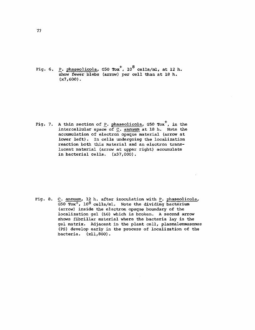
17
Fig. 6.
Fig. 7.
Fig. 8.
+ 8P. phaseolicola, GSO Tox , 10 cells/ml, at 12 h.show fewer blebs (arrow) per cell than at 18 h.(x7,600).
A thin section of ~. phaseolicola, GSO TOX+, in theintercellular space of c. annuum at 18 h. Note theaccumulation of electron opaque material (arrow atlower left). In cells undergoing the localizationreaction both this material and an electron translucent material (arrow at upper right) accumulatein bacterial cells. (x37,000).
£. annuum, 12 h. after inoculation with ~. phaseolicola,GSO TOX+, 108 cells/mJ.. Note the dividing bacterium(arrow) inside the electron opaque boundary of thelocalization gel (LG) which is broken. A second arrowshows fibrillar material where the bacteria lay in thegel matrix. Adjacent in the plant cell, plasmalemmasomes(PS) develop early in the process of localization of thebacteria. (xll,800).

78
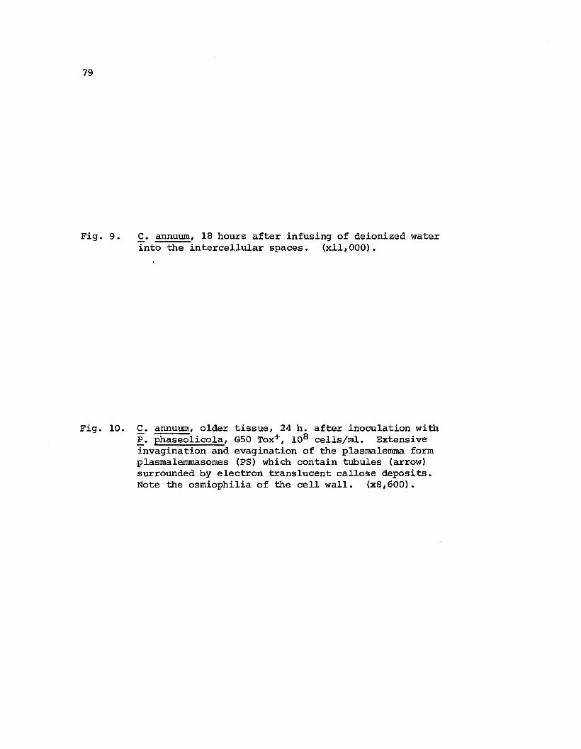
79
Fig. 9.
Fig. 10.
£. annuum, 18 hours after infusing of deionized waterinto the intercellular spaces. (xll,OOO).
£. annuum, older tissue, 24 h. after inoculation with~. phaseolicola, GSO Tox+, 108 cells/mI. Extensiveinvagination and evagination of the plasmalemma formplasmalemmasomes (PS) which contain tubules (arrow)surrounded by electron translucent callose deposits.Note the osmiophilia of the cell wall. (x8,600).

80

81
Fig. 11. C. annuum, older tissue, 24 h. after inoculation with~. phaseolicola, GSO Tox-, 108 cells/ml. At the lowerright is the edge of the grid and an adjacent artifact.Electron dense amorphous material is present bothinside and surrounding the bacteria localized insidethe localization gel. Near the evagination of theplasmalemma (plasmalemmasomes) in the plant celldictysomes (D), undilated endoplasmic reticulum(ER) and lipid bodies (L) appear. At this stageno disruption is observed in either mitochondrialor chloroplast membranes. (xll,170).

82

83
Fig. 12.
Fig. 13.
c. annuum, older tissue, 24 h. after inoculation with~. phaseolicola, GSO Tox+, 108 cells/ml. Note callosedeposits in evaginations of the plasmalemma.
~. annuum, older tissue, 24 h. after inoculation with~. phaseolicola, GSO Tox-, 108 cells/ml showing theplasmalemmasomes (PS). The tubular structures at thelower left are composed of the evaginated plasmalemma,with cytoplasm, and some endoplasmic reticulum. Atthe upper right (arrow) note the electron density of thetripartitate tubular membranes. Dense fibrillar depositsare observed at the upper right (arrow). Electron denseamorphous material occur both inside and on the surfaceof the bacterium (arrow). (x49,SOO).

t
84
'.
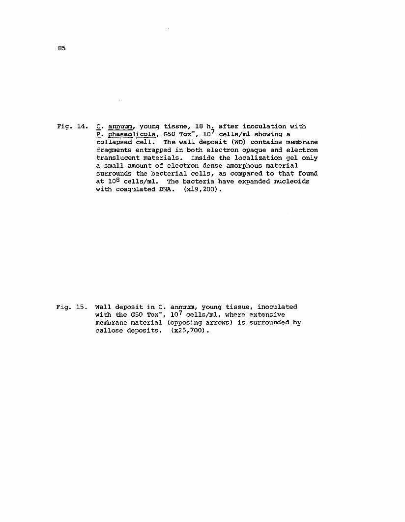
85
Fig. 14.
Fig. 15.
.£. annuum, young tissue, 18 h. after inoculation with~. phaseolicola, GsO Tox-, 107 cells/ml showing acollapsed cell. The wall deposit (WO) contains membranefragments entrapped in both electron opaque and electrontranslucent materials. Inside the localization gel onlya small amount of electron dense amorphous materialsurrounds the bacterial cells, as compared to that foundat 108 cells/ml. The bacteria have expanded nucleoidswith coagulated DNA. (x19,200).
Wall deposit i.n C. annuum, young tissue, inoculatedwith the GsO Tox-, 107 cells/rol, where extensivemembrane material (opposing arrows) is surrounded bycallose deposits. (x2s,700).

86

87
Fig. 16. Wall deposit (WD), in a younger plant of C. annuuminoculated with 107 cells/ml of GSO Tox-.- Thisstructure is still attached to the plant cell wallthough the protoplast has colla.psed. (xlS, 780),
Fig. 17. A stage in wall deposition where convoluted tubules occurin new wall materials of C. annuum after inoculation ofGSO Tox-, 108 cells/ml. Note the density of theribosomes in the adjacent cytoplasm surrounding theendoplasmic reticulum (ER). (x24,360).
Fig. 18. A localization gel formed in C. annuum with 108
cells/ml of ~. phaseolicola at 18 h. Note thestructural integrity of the bacteria. Adjacentto the localization gel in the plant cell depositsof fibrillar material occur between the plasmalemmaand the wall (arrow). (x26,000).

88

89
Fig. 19. A localization gel surrounding ~. phaseolicolaGSa TOX+, in young tissue of £. annuum at 18 h.,108 cells/mI. The orientation of the bacteriasuggests the presence of electron translucentmaterial adjacent to bacteria which is surroundedby electron dense amorphous material. Notefragmentation of the plant cell wall at theupper left. (x23,lSO).

'19
..
.'.,,
JS
"'.
1
'.• .#
·..·t·~.i.· ~" .. '''\'.... ' -a~'..... ,. ,
' •., ..,··4 '/'
t.
'~
, ,
, .
90

91
Literature Cited
1. Albersheim, P., T. M. Jones, and P. D. English. 1969.Biochemistry of cell walls in relation to infectiveprocesses. Ann. Rev. Phytopathol. 7: 171-194.
2. Burkowicz, A., and R. N. Goodman. 1970. Ultrastructural changesin apple leaves inoculated with avirulent or an avirulentstrain of Erwinia amylovora. Phytopathol. Z. 68: 258-268.
3. Charudattan, R., and J. E. DeVay. 1971. Common antigens amongvarieties of Gossypium hirsutum and isolates of Fusarium andVerticillium species. Phytopathology 62: 23 -234.
4. Chen, S. C., R. E. Stall, and A. A. Cook. 1969. Extraction andrange of ac~ivity of a bacterial inhibitor from Capsicumannuum. Phytopathology 59: 112.
5. Clowes, F. A. L., and B. E. Juniper. 1968. Plant cells.Blackwell Scientific Publications, Ltd., Oxford and Edinburgh,546 p.
6. Cook, A. A. 1973. Characterization of the hypersensitivereaction in Capsicum annuum induced by tomato strain ofXanthomonas vesicatoria. Phytopathology 63: 915-918.
7. DeVay, J. E., W. C. Schnathorst, and M. S. Foda. 1967. Commonantigens and host parasite interactions. In Mirocha, C. J.and I. Uritana, eds., The dynamic role of molecular con-s tituents in plant-parasi te interaction. American Phytopathological Society, Inc., st. Paul, Minnesota, pp. 313-325.
8. Faulkner, G., W. C. Kimmins, and R. G. Brown. 1972. The use offlurochromes for the identification of a (1,3) glucans. Can. J.Bot. 51: 1503-1504.
9. Flor, H. H. 1955. Host-parasite interactions in flax rust.Its genetics and other implications. Phytopathology 45:680-685.
10. Fuerst, J. A., and A. C. Hayward. 1969. Surface appendagessimilar to firnbrae (pili) on Pseudomonas species. J. Gen.Microbiol. 58: 227-237.
11. Garber, E. D. 1956. A nutrition inhibition hypothesis ofpathogenicity. Am. Nat. 90: 183-187.
12. Goodman, R. N. 1971. Reevaluation of the role of ammonia inthe hypersensitive reaction. Phytopathology 61: 893.

13. Goodman, R. N., and S. R. Plurad. 1971. Ultrastructuralchanges in tobacco undergoing the hypersensitive reactioncaused by plant pathogenic bacteria. Physiol. PlantPathol. 1: 11-15.
14. Heath, Michele C., and I. B. Heath. 1971. Ultrastructureof an immune and a susceptible reaction of cowpea leavesto rust infection. Physiol. Plant Pathol. 1: 277-287.
15. Hildebrand, D. C., and Beverley Riddle. 1971. Influenceof environmental conditions on reactions induced byinfiltration of bacteria in plant leaves. Hildgardia41: 33-43.
16. Horino, Osamu. 1973. Ultrastructure of Xanthomonas oryza~
and its morphologi.cal changes by chemical treatment.Ann. Phytopathol. Soc. Japan 39: 14-26.
17. Huang, Jeng-sheng, and R. N. Goodman. 1970. The relationship of phosphatidase activity to the hypersensitivereaction in tobacco induced by bacteria. Phytopathology60: 1020-1021.
18. Karnovsky, M. J. 1965. A formaldehyde-glutaraldehyde fixativeof high osmolality for use in electron microscopy. J. CellBiol. 27: l37A.
19. Keen, N. T., and B. W. Kennedy. 1974. Hydroxyphaseollin andrelated isoflavnoids in the hypersensitive resistancereaction of soy beans to Pseudomonas glycinea. Physiol.Plant Pathol. 4: 173-185.
20. Kellenberger, E., A. Ryter, and J. Sechaud. 1958. Electronmicroscope study of DNA containing plasma. II. Vegetativeand mature phage DNA as compared with normal bacterialnucleoids in different physiological states. J. Biophys.Biochem. Cytol. 4: 671-676.
21. Kiraly, Z., B. Barna, and T. Ersek. 1972. Hypersensitivity asconsequence, not the cause of plant resistance to infection.Nature 239: 456-457.
92
22. Kiraly, Z., and G. L. Farkas. 1962. Relation between phenolmetabolism and stem rust resistance in wheat. Phytopathology52: 657-664.
23. Klement, Z., and R. N. Goodman. 1967. The hypersensitivereaction to infection by bacterial plant pathogens. Ann.Rev. Phytopathol. 5: 17-44.

93
24. Le Normand, M., A. Coleno, J. P. Gourret, and H. Fernandez-Arias.1971. Etude de la graisse du haricot et de agent Pseudomonasphaseolicola (Burk.) Dowson. Ann. Phytopathol. 3: 390-418.
25. Lewis, Ralph W.parasitism.
1953. An outline of the balance hypothesis ofAm. Nat. 87: 273-281.
26. Lovrekovich, L., and H. Lovrekovich. 1970. Tissue necrosis intobacco caused by a saprophytic bacterium. Phytopatholoqy60: 1279-1280.
27. Mullen, J. M., and D. F. Bateman. 1971. Production of anendopolygalacturonate trans-eliminase by a potato dry rotpathogen, Fusari~ roseum avenaceum in culture and diseasetissue. Physiol. Plant Pathol. 1: 363-373.
28. Novacky, A., G. Acedo, and R. N. Goodman. 1973. Preventionof bacterially induced hypersensitive reaction by livingbacteria. Physiol. Plant Pathol. 3: 133-136.
29. O'Brien, Francis, and R. K. S. Wood. 1973. Antibacterialsubstances in hypersensitive response induced by bacteria.Nature 242: 532-533.
30. Patil, S. s., A. C. Hayward, and R. Emmons. 1974. An ultraviolet-induced nontoxigenic mutant of Pseudomonas phaseolicolaof altered pathogenicity. Phytopathology 64: 590-595.
31. Reynolds, E. S. 1963. The use of lead citrate at high pH asan electron-opaque stain in electron microscopy. J. CellBiol. 17: 208-212.
32. Rubin, B. A., and V. A. Arkenova. 1957. The participation ofthe polyphenolase system in the defense reaction of potatoagainst Phytophthora infestans. Biokhimiya 22: 202-208.
33. Rubin, B. A., and Yeo V. Artsikhovskaya. 1963. Biochemistryand physiology of plant immunity. The Macmillan Company,New York, 358 p.
34. Rudolph, K. 1971. Reactions of the host plant to specificpathogens. In Maas Geesteranus, H. P., ed., Proceedingsof the third-rnternational conference on plant pathogenicbacteria, April 14-21, 1971, Wageningen, The Netherlands,pp. 179-190.
35. Rudolph, K., and M. A. stahmann. 1964. Interaction ofperoxidase and catalases between Phaseolus vulgaris andPseudomonas phaseolicola (halo blight of bean) Nature204: 474-475.

36. Sasser, Myron, A. K. Andrews, and Z. U. Doganay. 1974.Inhibition of photosynthesis diminishes antibacterialaction of pepper plants. Phytopathology 64: 770-772.
37. Stall, R. E., J. A. Bartz, and A. A. Cook. 1974. Decreasedhypersensitivity to Xanthomonads in pepper after inoculationwith virulent cells of Xanthomonas vesicatoria.Phytopathology 64: 731-735.
38. Turner, John G., and Anton Novacky. 1974. The quantitativerelation between plant and bacterial cells involved in thehypersensitive reaction. Phytopathology 64: 885-890.
39. Varns, J. L., J. Kuc, and E. B. Williams. 1971. Terpenoidaccumulation as a biochemical response of the potato tuberto Phythophthora infestans. Phytopathology 61: 174-177.
94

CHAPTER III
ISOLATION AND CHARACTERIZATION OF AN ENDOTOXIN FROM
PSEUDOMONAS PHASEOLICOLA WHICH INDUCES CELL COLLAPSE
IN BELL PEPPER (CAPSICUM ANNUUM)
Abstract
A high molecular weight endotoxin capable of causing cell
collapse in bell pepper (Capsicum annuum) was purified from bacterial
+cells of two isolates of Pseudomonas phaseolicola, GSO Tox and GSO
Tox respectively, the toxin and non toxin producing isolates of the
causal agent of halo blight of bean (Phaseolus vulgaris L.). The
endotoxin from either isolate has similar chemical and biological
properties. A molecular weight determination of the endotoxin from
GSO Tox indicates a molecular weight of approximately 93,000. At
concentrations as low as 2 ~g/ml, confluent necrosis is induced in
pepper without browning, senescence symptoms or leaf abscission. The
endotoxin is thermostable, and is degraded by pronase, but not RNAse.
The lesion induced in pepper by the endotoxin requires the same time
to develop as the lesions induced by the bacterial cells.
Introduction
The hypersensitive reaction (HR), classically associated with
plant resistance, and exemplified by rapid cell collapse when an
incompatible pathogen is introduced into non host or resistant plants,
is mimicked by crude extracts obtained from the pathogen (14, 16).
So far, however, only in a single case has a HR-inducing principle

96
been purified (5). Apart from inducing HR, this purified material
exhibits pectin transe1iminase activity. From the information avai1-
able concerning induction mechanisms involved in HR, the reaction
does not appear to utilize a universal mechanism. In pepper, Cook (1)
found the HR induced by two different, incompatible strains of
Xanthomonas vesicatoria was affected differentially when subjected to
the same physical and chemical treatment. Since it appears that the
HR is suppressed during the host-pathogen interactions, it is important
to understand how the reaction is triggered in this, the most widespread
mechanism of plant resistance.
The purpose of this investigation was to determine if discrete,
inducing principles which induce HR in pepper can be isolated from
+ -cultures of Pseudomonas phaseo1ico1a, GSO Tox and GSO Tox , the causal
agent of halo blight of bean. This organism produces a low molecular
weight exotoxin in culture which has halo blight inducing potential
in bean. The UV-derived, nontoxigenic mutant was included in this
study to determine if alterations in genetic characters affecting halo
blight symptoms would also affect the properties of the HR inducing
princip1e(s). Information is available concerning HR induced by
Xanthomonas vesicatoria in pepper. Studies using this plant would
extend progress in understanding HR in systems other than those using
tobacco, the classical indicator for testing the HR inducing potential
of pathogenic bacteria.

97
Materials and Methods
Assay of fractions from cultures of P. phaseolicola for HR
inducing potential was routinely performed by infusing test suspensions
in 0.025 M phosphate buffer into pepper leaves with a hypodermic
syringe. Biological activity was arbitrarily graded by the degree of
tissue collapse which occurred in the infused zone.
+Log phase cells of ~. phaseolicola, G50 Tox and G50 Tox were
collected from Watanabe's medium (18) by centrifugation at 6000 g for
20 minutes. Bacterial cells were subjected to either sonication or
osmotic shock (11).
Cells (30-50 gms) subjected to sonication were washed three times
in 0.025 M phosphate buffer, pH 6.8, and resuspended in 5 gm/20 ml of
a medium composed of the same buffer, 0.5% sodium ascorbate and 0.5M
sucrose. Sonication was performed at high frequency with a Bronwill
Biosonik III probe at 80% power source, for 6 minutes in 15 second
bursts while cooling the medium in an acetone and ice bath. Cellular
fragments were removed first by two successive centrifugations at
27,000 g for 40 minutes and then filtering the supernatant through 0.45
~m Millipore filter. Release of wall derived fractions by osmotic shock
was performed by the procedure of Neu and Chou (11). The cells were
washed with 0.01 M Tris buffer, pH 7.3, at 4 0 C and suspended in 20%
sucrose-0.03 M Tris buffer, pH 7.3, with 0.1 roM EDTA for 10 minutes.
The cells removed by centrifugation were resuspended in 0.05 mM MgC12
at 4 0 C for 5-10 minutes. After centrifugation the supernatant was
concentrated with Aquacide II.

98
A precipitate was obtained from the supernatant prepared both by
sonication and osmotic shock by adding (NH4)2S04 to a final concentration
of 50%. This precipitate was collected by centrifugation, dissolved
in 0.025 M phosphate buffer, pH 6.8, and dialyzed against several
changes of the same buffer for 24 hours. After heat precipitation over
a boiling water bath for 3-7 minutes, the supernatant was again
precipitated with protamine sulfate. After centrifugation the
resulting supernatant was dialyzed in the above buffer for 24 hours
with several changes and then subjected to gel filtration on
Sephadex G-25 (2.5 x 35 em) previously equilibrated with the same
buffer. The activity which emerged in the void volume is referred to
as the crude extract. The crude extract was applied to Sephadex G-200
(2.5 x 44.5 em) equilibrated with 0.025 M, pH 6.8, phosphate buffer.
This yielded several protein peaks as determined by Lowry's method
(10), but only two of them caused tissue collapse in pepper. One was
a fast-moving Peak I activity and the other a slower-moving Peak II
fraction. Since only the Peak II fraction caused pepper tissue
collapse corresponding to protein concentration, it was chosen for
further purification. However, the yield is very low and its biological
activity is rapidly lost. Approximately 13 mg of Peak II protein was
recovered from 100 gm of bacterial cells. After dialysis of the Peak II
fraction against 0.01 M, pH 7.5, phosphate buffer it was loaded on a
DEAE-cellulose column (1.0 x 17 em) equilibrated with the same buffer.
The column was washed with deionized water and chromatographed with a
linear gradient of 0.01-0.025 M, pH 7.5, phosphate buffer.

99
Protein determinations were made using the method of Lowry ~ ale
(10) with bovine serum albumin as the standard.
Pronase digestion was performed at 37° C in phosphate buffer
(pH 7.5). The ratio of pronase concentration used to endotoxin con-
centration was 2:1 (i.e., 50 ~g/ml pronase/25 ~g/ml endotoxin).
Estimation of molecular weight was performed on a calibrated
sephadex G-200 column (2.5 x 44.5 em) in pH 6.8 buffer using a flow
rate of 0.3 ml/min. (3). Molecular weight of standard proteins used
were peroxidase 3.98 x 10⁴ , alkaline phosphatase 1.00 x 10⁵, S amylase
5 51.52 x 10 and urease 2.43 x 10 •
Isolated cells from various plant tissues were prepared using the
procedures of Jensen, Francki and zaitlin (6) a.nd were labelled either
14 14 14with methyl-D- C-glucose, C-glucose or NaH C03 • Leakage of
14. d' d d f . th 11 b . 11'C-~nterme ~ates were etecte a ter remov~ng e ce s y ~ ~pore
filter and counting the supernatant in Bray's solution with a Packard
Model 2420 scintillation counter. The pepper discs were exposed to
l4co or 14 C-glucose and infiltrated in vacuo with the active2
Peak II fractions. After appropriate incubation intervals the discs
were infiltrated with water and centrifuged for 30 minutes at 1000 g.
The fluid from the intercellular spaces was counted in Bray's solution.
In experiments testing incorporation of the active fraction Peak II
into isolated bean cells after labelling with l4c-glucose, l4co2 was
collected in Hyamine (Rohm and Haas) and counted in Bray's solution.

100
Results
Isolation of an inducing principle which causes cellular collapse
in pepper from two isolates of Pseudomonas phaseolicola. As shown in
Table 1 the culture medium failed to produce any evidence of inducing
principle(s) capable of causing cell collapse. However, two active
components were precipitated by 50% satuation with (NH4)2S04 of the
soluble sonicates of the bacterial cells. One of these components which
appeared in the osmotic shock fluid was selected for further purifica
tion. According to Neu and Chou (11), the latter procedure presumably
does not kill the bacteria. However, since the quantity of active
material recovered by this method is roughly 10% of that recovered
by osmotic shock, sonication was routinely used. The tissue lesions
caused by the rnaterial recovered after 50% satuation of soluble
sonicates with (NH4)2S04 were identical in appearance to those
produced by the bacteria. '£he time required for the lesion to develop
was also the same. However, the intensity of the cell collapse induced
in pepper increased after precipitation of the soluble fraction over a
boiling water bath (3-10 minutes). It appeared that some substance(s)
which interfered with factor(s) causing hypersensitive cell collapse
was inactivated by this treatment, since after gel filtration of both
heat treated and non-treated samples the activity was recovered at the
same elution volume. Subsequently, 3-5 minutes of heat precipitation
was used in the purification procedures.
Ammonium sulfate fractionations of soluble sonicates of two non
pathogens ~. coli and ~. florescens were also performed. Both non
pathogens yielded biologically active fractions causing cell collapse

101
in pepper in the same saturation range as did ~. phaseolicola. How
ever, in contrast to the active fractions obtained from ~. phaseolicola
isolates, these substances were heat labile and required more than a
2000-fold greater protein concentrations to cause tissue collapse •.
The high molecular weight substances from E. coli did not yield
active fractions after gel filtration.
Since high molecular weight components of bacterial origin were
capable of causing cell collapse in pepper, naturally occurring com
pounds of high molecular weight were tested for potential toxicity to
pepper tissue (Table 3). The substances tested included nucleic acids,
proteins, polysaccharides, bacterial endotoxins, peptides and pectinase,
enzymes. None of these at the designated concentrations caused visible
necrosis. Macerozyme (Kanematsu-Goshu, Ltd., Tokyo, Japan), the
pectinase enzymes derived from Aspergillus oryzae, also failed to
induce any lesions at concentrations to 10 mg/ml.
Properties of the crude extract of ~. phaseolicola GSO ToX+
and GSO Tox shown in Table 4 represent the stage of purification just
prior to gel filtration with Sephadex G-200. The active components
appear to be high molecular weight substances having unusual stability
to heat and pH change. When this crude extract was subjected to
Sephadex G-lOO, the active components emerged near the void volume.
When the same extract was subjected to Sephadex G-200, the activity
eluted as a fast-moving component, Peak I (Fig. 1) which showed no
correspondence in activity to protein concentration, and a slower
moving component, Peak II, which showed a correspondence with a protein
peak. The biological activity of Peak II was lost within 30 minutes

102
with pronase hydrolysis (Table 4), and this fraction was chosen for
further purification. Though heat stable, Peak II failed to retain
biological activity beyond 2 days in storage at 0 0 C. Both lyophili
zation and concentration greatly reduced the activity so that active
material was never recovered when Peak II was recycled Sephadex G-200.
When the crude extract was chromatographed on DEAE-cellulose (1.0 x
17 em) equilibrated with 0.025 M potassium phosphate buffer, pH 7.5,
loaded in the same buffer and eluted stepwise with 0.025, 0.1, 0.2,
0.3, and 0.4 M of the above buffer, all the activity was recovered
in the 0.025 M buffer eluate. When this fraction was concentrated
and eluted from Sephadex G-200, cell collapse was detected only in
the elution volume of Peak II activity. However, the protein concen
tration was too low to obtain an elution profile by Lowry's method.
The central peak fraction of Peak II activity from several runs on
Sephadex G-200 was concentrated, and subjected to gradient elution on
DEAE-cellulose using 0.01-0.025 M phosphate buffer, pH 7.5. This
concentrate showed 4 closely-eluting protein peaks (Fig. 3). However,
since the peak had already lost biological activity, it was impossible
to determine which of the eluted peaks caused cell collapse in pepper.
Biologically active products were never recovered either with preparative
or quantitative procedures using disc gel electrophoresis. These
procedures were carried out according to the method of Davis (2) using
6% and 7% polyacrilamide gel concentrations and a discontinuous buffer
system of Tris-glycine (pH 8.8).
The biological life of Peak II is not extended significantly by
freezing, by several buffer systems, using BSA for osmotic stabilization
103
and a buffering effect, reducing agents, dilute alcohol or by recom-
bining with post-fraction, slow-moving peaks. Both lyophilization and
concentration using the Amicon centriflo cone and Aquacide II greatly
reduce the activity compared with nontreated fractions having the same
protein concentration.
The ratio of the elution volume of the Peak II active fraction to
the elution of blue dextran (Ve!Vo) was 2.309. On a column calibrated
with proteins of known molecular weight, the Peak II fraction eluted
near alkaline phosphatase and has a molecular weight of approximately
93,000 (Fig. 5).
Since pepper tissue was shown to lose electrolytes within 3 hours
after infusion of Xanthomonas vesicatoria (17), Peak II fractions from
~. phaseolicola were tested for the ability to cause leakage of
l4C-intermediates from isolated cells of tobacco, bean and passion
14 14 14fruit labelled with methyl-D- C-glucose, C-glucose or CO2 or from
pepper discs labelled with l4C02 or l4C-glucose. Though the active,
Peak II fractions caused confluent collapse of the leaf tissue of
intact pepper plants, neither the labelled cells nor the pepper discs
showed any significant loss of l4C-intermediates above the control for
a period of 6 hours. Evidence showing the Peak II fraction is active
in this system, at least initially, is suggested by the rapid increase
followed by a decline in l4co evolved from bean cells, after incor-2
14poration of C-glucose. This indicates that the faction was trans-
ported across the cell membranes (Fig. 5).
The best assay for Peak II activity proved to be the intact
plant. The fractions as they eluted from Sephadex G-200 caused tissue

104
collapse corresponding to protein concentration of the fractions in ~~e
protein peak (Fig. 2). Diluting or concentrating these fractions cause~
inconsistent responses which made end-point dilution studies meaning
less. Pepper seedlings, fresh water and marine algae, blood cells,
or beet disc were tested for potential assay material, but all proved
to be ineffective.
The appearance of the lesion induced in pepper progressively
changes through purification steps. The fraction precipitated with
(NH4)2S04 produces symptoms identical to the intact bacterial cells.
With heat precipitation chlorosis surrounding the lesions was reduced
and leaf abscission was delayed. At this step of purification the
crude extract, like the bacterial cells, induces callose formation in
pepper tissue as indicated by the bright yellow fluorescence observed
with UV fluorescence microscopy after staining with Aniline Blue (4)
(Figs. 6, 7). The principle responsible for causing the browning
response and senescence is removed with Peak I on Sephadex G-200.
Since neither cellular browning nor early membrane leakage (to 6 hours)
is observed with Peak II activity, these responses which occur during
HR may result from complex interaction of plant and bacterial cells.
Discussion
Sonicated cells of the two isolates of Pseudomonas phaseolicola,
race 2, GSO Tox+ and GSO Tox-, each yield 2 active components which
elute at the same place from Sephadex G-200 that are capable of causing
cell collapse in pepper. Though by definition the active fractions
are endotoxins, toxic substances not released into the culture medium,

105
Peak II could be associated with the cell membrane, cell wall or the
periplasmic space.
Two observations suggest that Peak II endotoxin may be a
proteinaceous compound. These are: first, the loss of biological
activity with pronase digestion (Table 4) and second, the correspondence
of biological activity to the protein peak eluted from Sephadex G-200
(Fig. 2). Though the molecular weight of 93,000 is near the size of
many enzymes, its thermostability suggests the presence of a non-
proteinaceous component.
Sequeira, Aist and Ainslie (15), and Sleesman, Perley and Hoitink
(16) have reported that proteinaceous substances isolated from P.
solanacearum and~. tabaci and ~. pisi were capable of causing cell
collapse in tobacco, but no further information has been reported
concerning their purification. Recently, Gardner and Kado (6) induced
cell collapse in tobacco with a protein isolated from Erwinia
rubrifaciens which also showed pectin transeliminase activity (5).
However, Macerozyme which contains the pectic enzymes derived from
Aspergillus oryzae required 10 mg/ml before tissue collapse in pepper
was induced. In contrast, only 2 ~g/ml of t~e endotoxin was required
to cause confluent collapse. Keen and Williams (7) found a
lipopolysaccharide from~. lachrymans, the causal agent of angular
wilt of cucumber, had noncatalytic protease activity, but the
endotoxin from P. phaseolicola failed to cleave Azacol (12) indicating
that it has no proteolytic activity under the conditions used here.
The endotoxin from P. phaseolicola failed to cause tissue necrosis
in tobacco, tomato, passion fruit and a number of dicotyledonous plants

106
growing in their natural environment. Though this endotoxin appears
to be specific, it has not been determined if environmental parameters
or problems of transport may be involved in the failure of lesion
formation in other plants. However in tobacco, lesions were not formed
when the leaves were infused with the endotoxin under conditions that
are conducive to lesion production when the leaves are infiltrated with
intact bacteria. BSA did not inhibit lesion formation in pepper when
infused both prior to or after introduction of the endotoxin. BSA and
other foreign substances prevent the expression of the HR in tobacco
(9) •
Recently Kiraly, Barna and Ersek (8) have claimed that both
hypersensitive, tissue necrosis and phytoalexin production occur in an
infected plant as the result of an already-damaged or dying pathogen
and are not causally related to plant resistance. This conclusion was
based on the finding that killing the pathogen in infected tissue with
antibiotics in 3 different host-pathogen systems caused tissue necrosis
in each instance. Phytoalexin production was also found in the tested
compatible system. Although heat killed cells of P. phaseolicola are
capable of causing inconsistent flecks in pepper tissue, it is not
known if this is the same activity exhibited by the purified endotoxin.
Ultrastructural studies of pepper leaves infected with ~. phaseolicola
show that the first visible disruptive effects on the bacteria occur
12 hours after infusion and tissue collapse occurs within 18 hours
(see Chapter II). The time required for tissue collapse induced by
the endotoxin is also 18 hours. If some factor does not delay the

107
endotoxin in reaching the active site, it appears that death of
bacterial cells is not required to induce cellular collapse in the
pepper ~. phaseolicola interaction. This is consistent with the
observation of Sasser, Andrews and Doganay (13) who showed that in an
incompatible system involving pepper and Xanthomonas phaseoli cell
collapse occurred regardless of whether or not the bacteria were dead.
The basis for this conclusion was the observation that inhibition of
antibacterial substances either in the dark or by infusing
3-(p-chlorophenyl)-1, I-dimethyl urea to prevent photosynthesis
allowed population growth comparable to that observed in the com
patible pepper E~ vesicatoria system. In either case whether the
production of antibacterial substance was blocked or not pepper tissue
collapse occurred within the same time period. Thus, tissue collapse
was not causally related to the production of antibacterial substances.
In terms of a resistance mechanism the cell collapse in the P.
phaseolicola-pepper system may be related to the physical delimitation
of the pathogen by the death of plant tissue.

TABLE 1. Effects of ammonium sulfate fractionation of theculture broth and bacterial cells of G50 Tox+ andG50 Tox- on induction of the hypersensitive reaction in pepper
108
Source
Culture filtrate
Cells subjected to osmoticshock treatment
Sonicated cells
203040506080
90-100
2030405060
80-100
20304050
60-7080-100
HR response
++++
++++

109
TABLE 2. Effect of ammonium sulfate fractionation of solublesonicates derived from nonpathogens on the development of the hypersensitive reaction in pepper
Bacteria
Escherichia coli
Pseudomonas florescens
204060
80-100
204060
80-100
HRresponse
++
++
TABLE 3. Effect of selected naturally occurring high molecular weightsubstance as potential inducers of cellular necrosis inpepPer
Molecules of biological origin
DNA (sperm)
§.. coli LPS W (endotoxin)
Salmonella typhimurium (endotoxin)
Bovine serum albumin (Fraction IV)
Gelatin hydrolysate
Casein hydrolysate
Dextrans 40,000 MW
Dextrans 4,000 MW
Pectinase Macerozyme
Concentration
10 mg/ml
3 mg/ml
3 mg/ml
25 mg/ml
10 mg/ml
10 mg/ml
25 mg/ml
25 mg/ml
10 mg/ml
Tissue necrosis
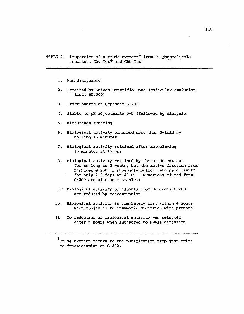
TABLE 4.
1.
2.
3.
4.
5.
6.
7.
8.
1Properties of a crude e~tract from P. phaseolicolaisolates, G50 Tox+ and G50 Tox- -
Non dialyzable
Retained by Amicon Centriflo Cone (Molecular exclusionlimit 50,000)
Fractionated on Sephadex G-200
Stable to pH adjustments 5-9 (followed by dialysis)
Withstands freezing
Biological activity enhanced more than 2-fold byboiling 15 minutes
Biological activity retained after autoclaving15 minutes at 15 psi
Biological activity retained by the crude extractfor as long as 3 weeks, but the active fraction fromSephadex G-200 in phosphate buffer retains activityfor only 2-3 days at 4° C. (Fractions eluted fromG-200 are also heat stable.)
110
9.·' Biological activity of eluants from Sephadex G-200are reduced by concentration
10. Biological activity is completely lost within 4 hourswhen subjected to enZYmatic digestion with pronase
11. No reduction of biological activity was detectedafter 5 hours when subjected to RNAse digestion
1Crude extract refers to the purification step just priorto fractionation on G-200.

111
Fig. 1. Fractionation of Pseudomonas phaseo1ico1a GSO Toxcrude extract on Sephadex G-200
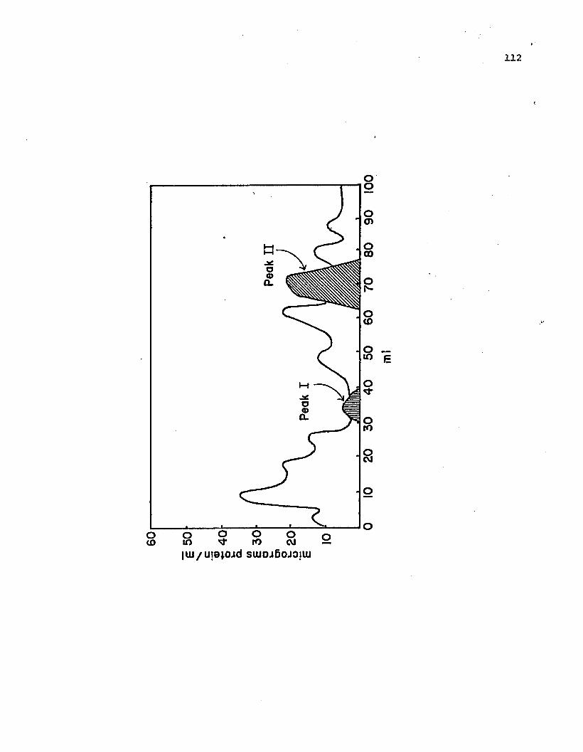
oto
112
0'0-0en
;\ 0en
Q)0a.~
0to .'
010 E
0~
0rt)
~
a-._-.__~_~__...L-_...::::a._~O
0 00 00U') V rt) t\I
IWI U!9~OJd SWDJf)OJ:l!W

113
Fig. 2. Tissue collapse induced in pepper by activefractions of Peak II. The fractions wereinoculated serially from right top to bottomand left top to bottom

114

115
;./
".( "\
" .'.,,'
Fig. 3. Elution profile of Peak II endotoxin fromSephadex G-200 by gradient elution fromDEAE-cellulose

micrograms protein I mII\l CIoI ~
000 0q-------------.....
(.l,Io
~o
-3 01-0-
eno
ena
CDa
0'------------.....a
116

117
Fig. 4. Molecular weight estimation of Peak II endotoxinwith gel filtration on a calibrated column ofSephadex G-200 (2.5 x 44.5 cm) flow rate 0.3 ml/min

118
Vel Vo
alkaline phosphatase( 100.000)
b ~ g01..--------..-------,-----:;::..--------'7'
r0
«0
<53:=E N- bx<5
I(JJ-
<:> f amylase(152.000)
urease(243.000)

119
Fig. 5. Effect of active Peak II endotoxin fromPseudomonas phaseolicola G50 Tox- on14C02 evolution after 15 minutes of glucosel4C incorporation into isolated bean cells

1000
EQ.(,)
_ 400Q)
c
Light contrai (buffer) --Dark control (buffer>--Active fraction --
33 ug chiorophyillreactionvessel
120
300
200
100
5 10
5 ug/ml
~
0.05ug/ml) //
/
15 20 25 30 35 40minutes

121
Fig. 6. Fluorescing material shown in pepper after inoculationof Pseudomonas phaseolicola
Fig. 7. Fluorescing material shown in pepper after inoculationwith the crude extract from Pseudomonas phaseolicola

122
Literature Cited
1. Cook, A. A., and R. E. Stall. 1971. Calcium suspression ofelectrolyte loss from pepper leaves inoculated withXanthomonas vesicatoria. Phytopathology 61: 484-487.
2. Davis, B. J. 1964. Disc electrophoresis. II. Method andapplication to human serum protein. Ann. N.Y. Acad. Sci.121: 404-427.
3. Deterrnann, Helmut. 1969. Gel chromotography, Second edition.Springer-verlag, New York, Inc., New York, 202 p.
4. Faulkner, G., W. C. Kimmins, and R. G. Brown. 1972. The useof flurochromes for the identification of a (1,3) glucans.Can. J. Bot. 51: 1503-1504.
5. Gardner, J. M., and C. I. Kado. 1972. Induction of the hypersensitive reaction in tobacco with specific high-molecularweight substances derived from the osmotic shock fluid ofErwinia rubrifaciens. Phytopathology 62: 759.
123
6. Jensen, R. G., R. I. B. Francki, and M.Metabolism of separated leaf cells.photosynthetically active cells fromPhysiol. 48: 9-13.
zaitlin. 1971.I. Preparation oftobacco. Plant
7. Keen, N. T., P. H. Williams, and J. C. Walker. 1967.Characterization of a protease produced by Pseudomonaslachrymans. Phytopathology 57: 257-262.
8. Kiraly, Z., B. Barna, and T. Ersek. 1972. Hypersensitivity asconsequence, not the cause of plant resistance to infection,Nature 239: 456-457.
9. Klement, Z. 1971. The hypersensitive reaction of plants tobacterial infection. Acta Phytopathol. Scient. Hung. 6:115-118.
10. Lowry, O. H., N. J. Rosebrough, A. L. Rosebrough, A. L. Farr,and R. J. Randall. 1951. Protein measurement with Folinphenol reagent. J. Biol. Chern. 193: 265-275.
11. Neu, Harold C., and James Chou. 1967. Release of surfaceenzymes in Enterobacteriaceae by osmotic shock. J.Bacteriol. 94: 1934-1945.
12. Ries, Stephen H., and Peter Albersheim. 1973. Purificationof a protease secreted by Colletotrichium lindemuthianurn.Phytopathology 63: 625-629.

124
13. Sasser, Myron, A. K. Andrews, and Z. U. Doganay. 1974. Inhibitionof photosynthesis diminishes antibacterial action of pepperplants. Phytopathology 64: 770-772.
14. Sequeira, L. 1963. Growth regulators in plant disease.Ann. Rev. Phytopatho1. 1: 5-30.
15. Sequeira, L., S. Aist, and V. Ainslie. 1972. Prevention ofthe hypersensitive reaction in tobacco by proteinaceousconstituents of Pseudomonas solanacearum. Phytopathology62: 536-541.
16. Sleesman, H. C., J. E. Perley, and H. A. J. Hoitink. 1970.Susceptible and hypersensitive reactions in tobacco andtheir prevention by cell free extractions of Pseudomonastabaci and Pseudomonas glycinea. Phytopathology 60: 1314.
17. Stall, R. E., and A. A. Cook. 1966. Multiplication ofXanthomonas vesicatoria and lesion development in resistantand susceptible pepper leaves. Phytopathology 58: 15841587.
18. Watanabe, Minoru. 1963.of Xanthomonas oryzae.
Studies on the nutritional physiologyAnn. Path. Soc. Japan 28: 201-208.

CHAPTER IV
CONCLUSIONS
Both bacterial cells and a crude endotoxin, isolated from the
two isolates of Pseudomonas phaseolicola, GSO Tox+ and GSO Tox-,
induced in pepper callose-containing, wall deposits and hypersensitive
cell collapse followed by tissue browning, senescence symptoms, and
leaf abscission. The time required for cellular collapse to occur
was the same for the crude extract as for the bacterial cells.
Purification of the endotoxin removed the factor(s) responsible for
tissue browning, senescence and abscission. Thus the leaf tissue
remained green while the time required for cell collapse to occur was
unchanged. Though further research will be needed to determine if the
purified endotoxin has a role in hypersensitive collapse in vivo,
such a role may be indicated by its unusual properties, high specific
activity, high molecular weight, and the fact that BSA, when inoculated
into pepper tissue prior to or after the endotoxin is injected, does
not inhibit HR.
The HR induced in tobacco by Pseudomonas phaseolicola is inhibited
by BSA and other foreign proteins (4). Since the endotoxin, under the
conditions tested, failed to cause cell collapse in tobacco, it is not
known if plant pathogenic bacteria possess more than one factor that
may cause HR in different plant species. There is evidence that, in
a single, incompatible, plant species, different mechanisms are
involved in HR when triggered by different pathotypes of the same
bacterial species. Cook (1) found that HR triggered in pepper by either

126
a tomato or a pepper strain of Xanthomonas vesicato~was differentially
affected when subjected to different environnlental and chemical
treatments. At least in pepper, a single universal mechanism is not
involved in induction of HR. Correspondingly, it is conceivable that
different, HR-inducing factors may be detected in a single, plant
pathogenic bacteria depending on the plant used. However, a discrete
number of genetic determinants would be expected depending on the
latitude of tolerance involved for both plant and pathogen.
In some cases, factors which induce HR appear to be specific.
This was shown in resistant apple to the fungus Venturia inaequalis,
the causal agent of apple blight. Raa and Sijpesteijn (6) found strong
evidence that the fungus secreted a compound which was selectively
toxic to resistant apples and caused HR, whereas the susceptible
varieties were not affected by the toxic principle. In non host plants,
such specificity would not be expected. However, an explanation is
needed for the inabili ty of BSA to inhibi t the HR in pepper induced by
the endotoxin.
If the endotoxin is responsible for induction of HR by ~.
phaseolicola, in vivo, it must be inferred that the factor is released
at some time during the interaction with pepper tissue. The similarity
of time-lapse required for lesion development by both the endotoxin and
by the bacterial cells suggests this may occur soon after the bacteria
encounter the plant cells. However, if the endotoxin is delayed in
reaching the active site in the plant cell, this may occur when the
localization reaction is initiated. That bacterial-cell death is not
required for release of such a factor is indicated by bacterial cells
127
showing normal appearance during ER induced in pepper both by
Xanthomonas phaseoli (8) and Pseudomonas phaseolicola (2). The time
lag required for the endotoxin to cause cell collapse in pepper may
indicate that plant cell synthesis is affected. It is possible that
the endotoxin interferes with plant, cell-membrane repair or
synthesis or may activate a subst~~~e in the plant which casues membrane
breakdown.
The present investigation has not established if the endotoxins
of the two isolates of ~. phaseolicola are the same, but several of
their properties show that they are qualitatively similar. Further
studies will be needed to determine if the endotoxins are also
affected when genetic determinants responsible for disease symptoms
are altered.
The HR observed in pepper suggests a dynamic process. If the
reaction is triggered soon after the bacterial cells contact the
plant cells, then the model inferring involvement of toxic products,
degradative enzymes, and of changes in oxidative metabolism associated
with later phases of the interaction, may be precluded. These
phenomena may be secondary and thus not causally related to HR. Also,
since the death of the bacteria does not appear to have cause-effect
relationships to HR in pepper, phytoalexins may be excluded from
consideration. Further study is needed to establish if the endotoxin
can cause cell collapse in other plant species. The failure to observe
cell collapse in other plants tested may be due to problems in transport.
Thus, at the present state of the study, it cannot be determined if the
endotoxin may act as a specific bacterial fact0~ that reacts with a
specific plant factor.

128
This investigation has demonstrated that similar plant cell
modifications occur during the bacterially induced HR as was reported
for HR induced by a virus (5). The modifications include formation
of plasmalemmasome and wall deposits (see Chapter II). Recently, a
localization reaction involving bacterial pathogens has been demon
strated in the vascular tissue (3, 4). Further study of the isolated
endotoxin may provide an insight into the metabolic pathways involved
in cell collapse caused by HR.

129
Literature Cited
1. Cook, A. A. 1973. Characterization of the hypersensitivereaction in Capsicum annuum induced by tomato strain ofXanthomonas vesicatoria. Phytopathology 63: 915-918.
2. Goodman, R. N. 1974. Induced resistance and hypersensitivereaction to bacterial infection. In Induction mechanismsof biochemical and cytological responses in diseased plants.Proceedings of-post congress Osaka Symposium on phytopathology9-11 September 1974 Suita, Osaka, Japan.
3. Horino, Osamu. 1974. Induction of bacterial leaf blightresistance by incompatible strains of Xanthomonas oryzae.~ Induction mechanisms of biochemical and cytologicalresponses in diseased plants. Proceedings of post congressOsaka Symposium on phytopathology 9-11 September 1974Suita, Osaka, Japan.
4. Klement, Z. 1971. The hypersensitive reaction of plants tobacterial infection. Acta Phytopathol. Scient. Hung 6:115-118.
5. Lobenstein, G. 1972. Localization and induced resistance invirus infected plants. Ann. Rev. Phytopathol. 10: 177-206.
6. Raa, J., and A. Kaars Sijpesteijn. 1968. A biochemical mechanismof natural resistance of apples to Venturia inaequalis.Neth. J. Plant Pathol. 74: 229-231.
7. Sasser, Myron, A. K. Andrews, and Z. U. Doganay. 1974.Inhibition of photosynthesis diminishes antibacterialaction of pepper plants. Phytopathology 64: 770-772.

BIBLIOGRAPHY
Albersheim, P., T. M. Jones and P. D. English. 1969.of cell walls in relation to infective processes.Phytopathol. 7: 171-194.
BiochemistryAnn. Rev.
Averre, C. W., and A. Kelman. 1964. Severity of bacterial wilt asinfluenced by ratio of virulent to avirulent cells of Pseudomonassolancearurn in the inoculum. Phytopathology 54: 779-783.
Berard, D. F., J. Kuc, and E. B. Williams. 1972. A cultivar specificprotection factor from incompatible interaction of green bean withColletotrichiurn lindemuthianum. Physiol. Plant Pathol. 2: 123-127.
Bhattacharya, P. K., and M. Shaw. 1968. The effect of rust infectionon DNA, RNA, and protein in nuclei of Khapli wheat leaves. Can. J.Bot. 46: 96-99.
Bogarth, R. F., and A. F. Ross. 1964. Systemic resistance induced bylocalized virus infection. Extent of changes in uninfected parts.Virology 24: 446-455.
Boyd, W. C. 1963. The lectins: their present status. Vox,Sanguinis 8: 1-32.
Burkowicz, A., and R. N. Goodman. 1970. Ultrastructural changes inapple leaves inoculated with a virulent or an avirulent strain ofErwinia amylovora. Phytopathol. Z. 68: 258-268.
Chamberlain, D. W. 1962. Reaction of resistant and susceptible soybean to Xanthomonas phaseoli var. soyensis. Plant Dis. Reptr.46: 707-709.
Chand, J. N., and J. C. Walker. 1964. Relation of leaf and varietalresistance to bacterial multiplication in cucumber inoculated withPseudomonas lachryrnans. Phytopathology 54: 49-50.
Charudattan, R., and J. E. DeVay. 1971. Common antigens amongvarieties of Gossypiurn hirsuturn and isolates of Fusarium andVerticilliurn species. Phytopathology 62: 230-234.
Chen, S. C., R. E. Stall, and A. A. Cook. 1969. Extraction and rangeof activity of a bacterial inhibitor from Capsicum annuum.Phytopathology 59: 112.
Clowes, F. A. L., and B. E. Juniper. 1968. Plant cells. BlackwellScientific Publications, Ltd., Oxford and Edinburgh, 546 p.

131
Cook, A. A. 1973. Characterization of the hypersensitive reactionin Capsicum annuum induced by tomato strain of Xanthomonasvesicatoria. Phytopathology 63: 915-918.
Cook, A. A., and R. E. Stall. 1971. Calcium suspression ofelectrolyte loss from pepper leaves inoculated with Xanthomonasvesicatoria. Phytopathology 61: 484-487.
Corden, M. E., and A. E. Dimond. 1959. The effect of growth regulatingsubstances on disease resistance and plant growth. Phytopathology49: 68-72.
Crosse, J. E. 1965. Bacterial canker of stone fruits. VI. Inhibitionof leaf-scar infection of cherry by a saprophytic bacterium fromthe leaf surfaces. Ann. Appl. Biol. 56: 149-160.
Cruickshank, I. A. M. 1964. Phytoalexins. Ann. Rev. Phytopathol.1: 351-374.
Daley, J. M., L. R. Krupka, and A. A. Bell. 1962. Influence ofhormones on respiratory metabolism of healthy and rust affectedtissues. Plant Physiol. 37: 130-134.
Davis, B. J. 1964. Disc electrophoresis. II. Method and applicationto human serum protein. Ann. N.Y. Acad. Sci. 121: 404-427.
Day, Peter R. 1972. Genetics of Host Parasite Interactions.w. H. Freeman and Co., San Francisco, 238 p.
Determann, Helmut. 1969. Gel chromotography, Second edition.Springer-Verlag, New York, Inc., New York, 202.p.
DeVay, J. E., W. C. Schnathorst, and M. S. Foda. 1967. Commonantigens and host parasite interactions. In Mirocha, C. J. andI. Uritana, eds., The dynamic role of molecular constituents inplant-parasite interaction. American Phytopathological Society,Inc., st. Paul, Minnesota, pp. 313-325.
Dineen, J. K. 1963. Immunological aspects of paratism. Nature197: 268-269.
Doubly, J. A., H. H. Flor, and C. o. Clagett. 1960. Relation ofantigens of Melapsora lini and Linum usitatissium to resistanceand susceptibility. Science 13: 229.
Ercolani, G. L. 1970. Bacterial wilt of tomato. IV. The interac~ion betwp.en virulent and avirulent strains of Corynebacteriummichiganese (E. F. Sm.) Jens. in vivo. Phytopathol. medit. 9:151-159.

132
Farkas, G. L., z. Kiraly, and F. Solomosy. 1960. Role of oxidativemetabolism in the localization of plant viruses. Virology 12:408-421.
Farkas, G. L., and M. A. Stahmann. 1966. On the nature of changesin perioxidase isoenzymes in bean leaves infected by southern beanmosiac virus. Phytopathology 56: 669-677.
Faulkner, G., W. C. Kimmins, and R. G. Brown. 1972. The use offlurochromes for the identification of 6 (1,3) glucans. Can. J.Bot. 51: 1503-1504.
Flor, H. H. 1955. Host-parasite interactions in flax rust. Itsgenetics and other implications. Phytopathology 45: 680-685.
Fuerst, J. A., and A. C. Hayward. 1969. Surface appendages similarto fimbrae (pili) on Pseudomonas species. J. Gen. Microbiol.58: 227-237.
Galston, A. W., and W. K. Purves. 1960. The mechanism of action ofauxin. Ann. Rev. of Plant Physiol. 11: 239-276.
Garber, E. D. 1956. A nutrition inhibition hypothesis of pathogenicity.Am. Nat. 90: 183-197.
Gardner, J. M., and C. I. Kado. 1972. Induction of the hypersensitivereaction in tobacco with specific high-molecular weight substancesderived from the osmotic shock fluid of Erwinia rubrifaciens.Phytopathology 62: 759.
Goodman, R. N. 1968. ~he hypersensitive reaction in tobacco as areflection of changes in host cell permeability. Phytopathology58: 872-873.
Goodman, R. N. 1971. Reevaluation of the role of ammonia in thehypersensitive reaction. Phytopathology 61: 893.
Goodman, R. N. 1974. Induced resistance and hypersensitive reactionto bacterial infection. In Induction mechanisms of biochemical andcytological responses in diseased plants. Proceedings of postcongress Osaka Symposium on phytopathology 9-11 September 1974Suita, Osaka, Japan.
Goodman, R. N., and S. R. Plurad. 1971. Ultrastructural changes intobacco undergoing the hypersensitive reaction caused by plantpathogenic bacteria. Physiol. Plant Pathol. 1: 11-15.
Goodman, R. N., Z. Kiraly, and M. Zaitlin. 1967. The biochemistryand physiology of infectious plant disease. Van Nostrand Co.,Princeton, New Jersey, 354 p.

133
Heath, Michele C., and I. B. Heath. 1971. Ultrastructure of animmune and a susceptible reaction of cowpea leaves to rustinfection. Physiol. Plant Pathol. 1: 277-287.
Hildebrand, D. C., and Beverley Riddle. 1971. Influence of environmental conditions on reactions induced by infiltration of bacteriain plant leaves. Hildgardia 41: 33-43.
Hoitink, H. A. J., and o. E. Bradfute. 1971. Mechanism of diseaseinitiation by Pseudomonas tabaci. In Maas Geesteranus, H. P., ed.,Proceedings of the third international conference on plantpathogenic bacteria, Wageningen, 14-21 April, 1971. Centre forAgricultural Publishing and Docjmentation, Wageningen, The Netherlands, pp. 55-58.
Hoitink, H. A. J., R. L. Pelletier, and J. G. Coulson. 1966. Toxemiaof halo blight of bean. Phytopathology 1062-1065.
Horino, Osamu. 1973. Ultrastructure of Xanthomonas oryzae and itsmorphological changes by chemical treatment. Ann. Phytopathol.Soc. Japan 39: 14-26.
Horino, Osamu. 1974. Induction of bacterial leaf blight resistanceby incompatible strains of Xanthomonas oryzae. In Inductionmechanisms of biochemical and cytological responses in diseasedplants. Proceedings of post congress Osaka symposium on phytopathology 9-11 September 1974 Suita, Osaka, Japan.
Huang, Jeng-sheng, and R. N. Goodman. 1970. The relationship ofphosphatidase activity to the hypersensitive reaction in tobaccoinduced by bacteria. Phytopathology 60: 1020-1021.
Huang, Jeng-sheng, Pi-yu Huang, and Robert N. Goodman. 1973. Reconstitution of membrane-like structures with structural proteins andlipids isolated from tobacco thylakoid membranes. Am. J. Bot.60: 80-85.
Israel, H. W., and A. F. Ross. 1967. The fine structure of locallesions induced by tobacco mosiac virus in tobacco. Virology 33:272-276.
Jensen, R. G., R. I. B. Francki, and M. Zaitlin. 1971. Metabolismof separated leaf cells. I. Prepar~tion of photosyntheticallyactive cells from tobacco. Plant PhyGiol. 48: 9-13.
Jones, T. M., A. J. Anderson, and P. Albersheim. 1972. Host-pathogeninteractions. IV. Studies of the polysaccharide degrading enzymesecreted by Fusarium oxysporum f. sp. lycopersici. Physiol. PlantPathol. 2: 153-166.
Karnovsky, M. J. 1965. A formaldehyde-glutaraldehyde fixative of highosmolality for use in electron microscopy. J. Cell BioI. 27: 137A.

134
Keen, N. T., and B. W. Kennedy. 1974. Hydroxyphaseollin and relatedisoflavanoids in the hypersensitive resistance reaction of soy beansto Pseudomonas glycinea. Physiol. Plant Pathol. 4: 173-185.
Keen, N. T., P. H. Williams, and J. C. Walker. 1967.of a protease produced by pseudomonas'lachryrnans.57: 257-262.
CharacterizationPhytopathology
Kellenberger, E., A. Ryter, and J. Sechaud. 1958. Electron microscopestudy of DNA containing plasma. II. Vegetative and mature phageDNA as compared with normal bacterial nucleoids in differentphysiological states. J. Biophys. Biochem. Cytol. 4: 671-676.
Kelman, A., and L. Sequeira. 1972. Resistance in plants to bacteria.Proc. Roy. Soc. Lond. B. 181: 247-266.
Kiraly, Z. 1974. Tissue necrosis and plant disease resistance.Sixty-sixth annual meeting, American Phytopathological Society,August 11-15, 1974, Vancouver, Canada.
Kiraly, Z., B. Barna, and T. Ersek. 1972. Hypersensitivity as consequence, not the cause of plant resistance to infection. Nature239: 456-457.
Kiraly, Z., and G. L. Farkas. 1962.and stem rust resistance in wheat.
Relation between phenol metabolismPhytopathology 52: 657-664.
Klement, Z. 1963. Rapid detection of the pathogenicity of phytopathogenic pseudomonads. Nature 199: 299-300.
Klement, Z. 1963. Method of obtaining fluid from the intercellularspaces of foliage and the fluids merit as substrate for phytobacterialpathogens. Phytopathology 55: 1033-1034.
Klement, Z. 1971. The hypersensitive reaction of plants to bacterialinfection. Acta Phytopathol. Scient. Hung. 6: 115-118.
Klement, z., G. L. Farkas, and L. Lovrekovich. 1964. Hypersensitivereaction induced by phytopathogenic bacteria in the tobacco leaf.Phytopathology 54: 474-477.
Klement, Z., and R. N. Goodman. 1967. The hypersensitive reaction toinfection by bacterial plant pathogens. Ann. Rev. Phytopathol.5: 17-44.
Klement, Z., and R. N. Goodman. 1967. The role of the livingbacterial cell and the induction time in the hypersensi.tive reactionof tobacco plants. Phytopathol. 55: 322-333.
Klement, z., and L. Lovrekovich. 1961. Defense reactions induced byphytopathogenic bacteria in bean pods. Phytopathol. Z. 41: 217-221.

Kosuge, Tsune.infection.
1969. The role of phenolics in host response toAnn. Rev. of Phytopathol. 7: 195-222.
135
Kuc, J. 1968. Biochemical control of disease resistance in plants.World Rev. Pest Control 7: 42-55.
Kuo, M. S., C. o. Yod~r, and R. P. Scheffer. 1970. Comparativespecificity of the toxins of Helminthosporium carbonum andHelminthosporium victorae. Phytopathology 60: 365-368.
Le Normand, M., A. Coleno, J. P. Gourret, and H. Fernandez-Arias.1971. Etude de la graisse du haricot et de agent Pseudomonasphaseolicola (Burk.) Dowson. Ann. phytopathol. 3: 390-418.
Lewis, Ralph W.parasitism.
1953. An outline of the balance hypothesis ofAm. Nat. 87: 273-281.
Tissue necrosis inPhytopathology 60:
Lobenstein, G. 1972. Localization and induced resistance in virusinfected plants. Ann. Rev. Phytopathol. 10: 177-206.
Lovrekovi.ch, L., and G. L. Farkas. 1963. Kinetin as an antagonist oftoxic effect of Pseudomonas tabaci. Nature 196: 710.
Lovrekovich, L., G. L. Farkas. 1965. Induced protection againstwildfire disease in tobacco leaves treated with heat killed bacteria.Nature 205: 823-824.
Lovrekovich, L., and H. Lovrekovich. 1970.tobacco caused by a saprophytic bacterium.1279-1280.
Lovrekovich, L., H. Lovrekovich, and R. N. Goodman. 1970. Ammoniaas a necrotoxin in hypersensitive reaction caused by bacteria intobacco leaves. Can. J. Bot. 48: 167-171.
Lowry, O. H., N. J. Rosebrough, A. L. Farr, and R. J. Randall. 1951.Protein measurement with Folin phenol reagent. J. BioI. Chern.193: 265-275.
Lozano, J. C., and Luis Sequeira. 1970. Prevention of the hypersensitive reaction in tobacco leaves by heat killed bacterialcells. Phytopathology 60: 675-679.
Lozano, J. C., and. L. Sequeira. 1970. Differentiation of races ofPseudomonas solanacearum by a leaf infiltration technique.Phytopathology 60: 833-838.
Metlikskii, L. V., and R. Ozerelskovskaya. 1968. Plant immunity.Biochemical aspects of plant resistance to parasitic fungi. PlenumPress, New York., New York, 114 p.

136
Misaghi, Iraj, and R. G. Grogan. 1969. Nutritional and biochemicalcomparisons of plant pathogenic and saprophytic pseudomonads.Phytopathology 59: 1436-1450.
Moustafa (Darweish), F. A., and R. Whittenbury. 1971. Propertieswhich appear to allow phytopathogenic pseudomonads to counteractplant defense mechanisms. Phytopathol. Z. 67: 214-224.
Mullen, J. M., and D. F. Bateman. 1971. Production of anendopolygalacturonate trans-eliminase by a potato dry rotFusarium roseum avenaceum in culture and disease tissue.Plant Pathol. 1: 363-373.
pathogen,Physio1.
Muller, K. o. 1959. Hypersensitivity. In Horsfall, J. G., andA. E. Dimond, eds., Plant pathology 1. Academic Press, NewYork, pp. 470-519.
Muller, K. 0., and H. Borger. 1940. Experimentalle untersuchungenuber die Phythophthora-Resistenz der Kartoffel; zuglich einBeitrag zum Problem der Il erworbenen Resistenz ll in Pflanzenreich.Arb. biol. Reichsenstalt. Land-u. Fortwirtach. Berlin-Dahlem23: 189-231.
Nemath, J., Z. Klement, and G. L. Farkas. 1968. An enzymologicalstudy of the hypersensitive reaction induced by Pseudomonassyringae in tobacco leaf tissue. Phytopathol. Z. 65: 267-278.
Neu, Harold C., and James Chou. 1967. Release of surface enzymesin Enterobacteriaceae by osmotic shock. J. Bacteriol. 94: 19341945.
Novacky, A. 1972. Suppression of the bacterially induced hypersensitive reaction to cytokinins. Physiol. Plant Pathol. 2:101-104.
INovacky, A. 1974. Transmembrane potential in bacterial hypersensitivity.
Sixty-sixth annual meeting. American Phytopathological Society,August 11-15, 1974, Vancouver, Canada.
Novacky, A., G. Acedo, and R. N. Goodman. 1973. Prevention ofbacterially induced hypersensitive reaction by living bacteria.Physiol. Plant Pathol. 3: 133-136.
Novacky, A., and R. N. Goodman. 1973. Prevention of bacteriallyinduced hypersensitive reaction by living incompatible bacteria,Pseudomonas pisi. Phytopathol. 62: 780-781.
O'Brien, Francis, and R. K. S. Wood. 1973. Antibacterial substancesin hypersensitive response induced by bacteria. Nature 242: 532533.

137
Orner, M. E., and R. K. S. Wood. 1969. Growth of Pseudomonasphaseolicola in susceptible and resistant bean plants. Ann.Appl. Biol. 63: 103-116.
Patel, P. N., and J. C. Walker. 1963. Changes in free amino acidand amide content of resistant and susceptible beans afterinfection with the halo blight organism. Phytopathology 53:522-528.
Patil, Suresh S. 1974. Toxins produced by phytopathogenic bacteria.Ann. Rev. Phytopathol. 12: 259-279.
Patil, S. S., A. C. Hayward, and R. Emmons. 1974. An ultravioletinduced nontoxigenic mutant of Pseudomonas phaseolicola of alteredpathogenicity. Phytopathology 64: 590-595.
Pellizzari, E. D., J. Kuc, and E. B. Williams. 1970. The hypersensitive reaction in Malus species: Changes in leakage oflectrolytes from apple leaves after innoculation with venturiainaegualis. Phytopathology 60: 373-376.
Person, C., D. J. Samborski, and R. Rohringer. 1962. The gene forgene concept. Nature 194: 561-562.
Pinkas, Y., and A. Novacky. 1971. The differentiation betweenbacteria hypersensitive reaction and pathogenesis by use ofcycloheximide. Phytopathology 61: 906.
Raa, J., and A. Kaars Sijpesteijn. 1968. A biochemical mechanismof natural resistance of apples to Venturia inaequalis.Neth. J. Plant Pathol. 74: 229-231.
Reynolds, E. S. 1963. The use of lead citrate at high pH as anelectron-opaque stain in electron microscopy. J. Cell Biol. 17:208-212.
Ries, Stephen H., and Peter Albersheim. 1973. Purification of aprotease secreted by Colletotrichium lindemuthianum. Phytopathology63: 625-629.
Rohringer, R., and D. J. Samborski.the host-parasite interactions.
1967. Aromatic compounds inAnn. Rev. Phytopathol. 7: 77-86.
Rohringer, R., D. J. Samborski, and C. o. Person. 1961. Ribonucleaseactivity in rusted wheat leaves. Can. J. Bot. 39: 77-84.
Ross, A. Frank, and H. W. Israel. 1970. Use of heat treatment in thestudy of acquired resistance to tobacco mosiac virus in hypersensitivetobacco. Phytopathology 60: 755-770.

138
Rowley, D., and C. R. Jenkins. 1962. Antigenic cross ~eaction betweenhost and parasite as a possible cause of pathogenicity. Nature193: 151-154.
Rubin, B. A., andV. A. Arkenova. 1957. The participation of thepolyphenolase system in the defense reaction of potato againstPhytophthora infestans. Biokhimiya 22: 202-208.
Rubin, B. A., an~ Yeo V. Artsikhovskaya. 1963. Biochemistry andphysiology of plant innnunity. The Macmillan Company, New York,358 p.
Rubin, B. A., and E. V. Arcichovakaia. 1967.physiological background of plant immunity.58: 101-102.
The biochemical andPhytopathol. Z.
Rudolph, K. 1964. Analysis of proteins and enzymes of the interaction between Phaseolus vulgaris and Pseudomonas phaseolicola(halo blight of bean) Phytopathology 54: 904.
Rudolph, K. 1971. Reactions of the host plant to specific pathogens.In Maas Geesteranus, H. P., ed., Proceedings of the third international conference on plant pathogenic bacteria, April 14-21,1971, Wageningen, The Netherlands, pp. 179-190.
Rudolph, K., and M. A. Stahrnann. 1964. Interaction of peroxidaseand catalases between Phaseolus vulgaris and Pseudomonasphaseolicola (halo blight of bean) Nature 204: 474-475.
Sakuma, T., and K. Tomiyama. 1967. The role of phenolic compoundsin the resistance of potato tuber tissue to infection by Phytophthorainfestans. Ann. Path. Soc. Japan 33: 48-58.
Sands, D. C., M. N. Shroth, and D. C. Hildebrand. 1970. Taxonomy ofphytopathogenic pseudomonads. J. Bacteriol. 101: 9-23.
Sands, D. C., and M. L. Zucker. 1971. Impaired amino acid biosynthesisin phytopathogenic pseudomonads. Phytopathology 61: 90.
Sasser, Myron, A. K. Andrews, and Z. U. Doganay. 1974. Inhibition ofphotosynthesis diminishes antibacterial action of pepper plants.phytopathology 64: 770-772.
Schnathorst, W. C., and J. E. DeVay. 1963. Common antigens inXanthomonas malvacearurn and Gossypiurn hirsutum and their possiblerelationship to host specificity and disease resistance.phytopathology 53: 1142.
Schwochau, M. E., and L. A. Hadwiger. 1968. Stimulation of pisatinproduction in Pisurn sativurn by Actinomycin D and other compounds.Arch. Biochem. Biophys. 126: 731-733.

139
Sequeira, L. 1963. Growth regulators in plant disease. Ann. Rev.Phytopathol. 1: 5-30.
Sequeira, L., S. Aist, and V. Ainslie. 1972. Prevention of the hypersensitive reaction in tobacco by proteinaceous constituents ofPseudomonas solanacearum. Phytopathology 62: 536-541.
Shepard, James F. 1970. The potential significance of potatohemagglutinins (lectins) in serodiagnosis. Phytopathology 60:1623-1625.
Sleesman, H. C., J. E. Perley, and H. A. J. Hoitink. 1970.Susceptible and hypersensitive reactions in tobacco and theirprevention by cell free extractions of Pseudomonas tabaci andPseudomonas glycinea. Phytopathology 60: 1314 •
.Solomosy, F., and G. L. Farkas. 1962. Simultaneous activation ofpentose phosphate shunt enzymes in a virus-infected local lesionhost plant. Nature 195: 835.
Stahmann, M. A. 1967. Influence of host-parasite interactions onproteins, enzymes and resistance. In Mirocha, C. J., and I.Uritani, eds. The dynamic role of molecular constituents inplant-parasite interaction. American Phytopathological Society.st. Paul, Minnesota, pp. 357-369.
Stahmann, M. A., B. G. Clare, and W. Woodbury. 1966. Increaseddisease resistance and enzyme activity induced by ethylene andethylene production by black rot infected sv.eet potato tissue.Plant Physiol. 41: 1505-1512.
Stahmann, M. A., I. Uritani, and K. Tomiyama. 1960. Changes inpotato proteins induced by fungus infection. Phytopathology 50: 655.
Stakmann, E. C. 1914. A study in cereal rust, physiological races.U. Minn. Agri. Exper. st. Bull. 138.
Stall, R. E., and A. A. Cook. 1966. Multiplication of Xanthomonasvesicatoria and lesion development in resistant and susceptiblepepper leaves. Phytopathology 58: 1584-1587.
Stall, R. E., J. A. Bartz, and A. A. Cook. 1974. Decreased hypersensitivity to Xanthomonads in pepper after inoculation withvirulent cells of Xanthomonas vesicatoria. Phyto~athology 64:731-735.
Tani, T., H. Yamamoto, T. Onae, and N. Naito. 1974. Primaryrecognition and following expression of resistance in oat leaveshypersensitively responding to the crown rust fungus. !~
Proceedings of post congress Osaka symposium on phytopathology.9-11, September, Suita, Osaka, Japan

140
Thiers, H. D., and H. B. Lester. 1949. Histological studies ofbacterial blight infection of the cotton plant. Phytoflathology39: 499-500.
Tomiyama, K. 1956.potato plants tocytoplasm of theinfes tans • Ann.
Cell physiological studies on the resistance ofPhytophthora infestans. IV. On the movement ofhost cell induced by invasion of PhytophthoraPath. Soc. Japan 21: 54-62.
Tomiyama, K. 1960. Some factors affecting the death of hypersensitivecells infected by Phythophthora infestans. Phytopatho1. Z. 39:134-138.
Tomiyama, K.in plants.
1963. Physiology and biochemistry of disease resistanceAnn. Rev. Phytopathol. 1: 295-324.
Tomiyama, K. 1966 n Double i.nfection by a compatible race of Phythophthorainfestans of a potato cell which had been previously infected by acompatible race. Ann. Phytopatho1. Soc. Japan 32: 181-185.
Tomiyarna, K., R. Sakai, T. Sakuma, and N. Ishizaka. 1967. The roleof polyphenols in the defense reaction in plants induced byinfection. In I~rocha, C. J., and I. Uritani, eds., The dynamicrole of molecular constituents in plant-parasite interaction.American Pathological Society, St. Paul, Minnesota, pp. 165-179.
Tomiyama, K., and M. A. Stahmann. 1964. Alteration of oxidativeenzymes in potato tuber tissue by infection with Phythophthorainfestans. Plant Physiol. 39: 483-490.
Turner, John G., and Anton Novacky. 1974. The quantitative relationbetween plant and bacterial cells involved in the hypersensitivereaction. Phytopathology 64: 885-890.
Uritani, I., T. Asahi, T. Minamikawa, H. Hyodo, K. Oshima, andM. Kojima. 1967. The relation of metabolic changes in infectedplants to changes in enzyme activity. In Mirocha, C., and I.Uritani, eds., The dynamic role of molecular constituents in plantparasite interaction. American Pathological Society, st. Paul,Minn~sota, pp. 342-355.
Uritani, I., and M. A. Stahrnann. 1961. Changes in nitrogen metabolismin sweet potato with black rot. Plant Physiol.. 36: 770-782.
Varns, J. L., and J. Kuc. 1971. Suppression of risitin andphytotuberin accumulation and hypersensitive response in potatoby compatible races of Phythophthora infestans. Phytopathology61: 178-181.
Varns, J. L., J. Kuc, and E. B. Williams. 1971. Terpenoid accumulationas a biochemical response of the potato tuber to Phythophthorainfestans. Phytopathology 61: 174-177.

141
Walker, J. C., and M. A. Stahmann. 1955. Chemical nature of diseaseresistance in plants. Ann. Rev. of Plant Physiol. 6: 351-366.
Ward, A. M. 1902. On the relations between host and parasite in theBromes and their brown rust, Puccinia dispersa (Erikas) Ann. Bot.London 16: 233-315.
Watanabe, Minoru. 1963. Studies on the nutritional physiology ofXanthomonas oryzae. Ann. Path. Soc. Japan 28: 201-208.
Wimalajeewa, D. L. S., and J. E. DeVay. 1971. The occurrence andcharacterization of a common antigen relationship between Ustilagomaydis and Zea mays. Physiol. Plant Pathol. 1: 523-535.
Wood, R. K. S. 1967. Physiological plant pathology. BlackwellScientific Publications, Oxford, England, 570 p.