information for users
TRANSCRIPT

Pathology Molecular Pathology
RWF-CP-MOL-LI174 Revision 1.3
Document title: Molecular Pathology Guidelines for Users Page 1 of 19 WARNING: This document is only controlled if viewed electronically from its original location if the hard copy is validated Printed Copy No. Approved by: Service Manager & Clinical Lead Validated by: (signature) Date of issue: Feb 2021 Master copy registered on Pathology Qpulse database
Molecular Pathology Department
Maidstone and Tunbridge Wells NHS Trust
Information for Users
Molecular Pathology
Hermitage Lane
Maidstone
Kent
ME16 9QQ
Tel: 01622 225643

Pathology Molecular Pathology
RWF-CP-MOL-LI174 Revision 1.3
Document title: Molecular Pathology Guidelines for Users Page 2 of 19 WARNING: This document is only controlled if viewed electronically from its original location if the hard copy is validated Printed Copy No. Approved by: Service Manager & Clinical Lead Validated by: (signature) Date of issue: Feb 2021 Master copy registered on Pathology Qpulse database
Table of Contents
Introduction .................................................................................................................................. 3 Molecular Pathology Team .......................................................................................................... 4
Laboratory Opening hours ........................................................................................................ 5 Reports, results and turnaround times...................................................................................... 5
Clinical Advice .............................................................................................................................. 6
Costs ........................................................................................................................................ 6
Consent, storage and authorisation .......................................................................................... 7
Transport of Samples ............................................................................................................... 7
Problems and Queries .............................................................................................................. 7 Requests ...................................................................................................................................... 7
Specimen Requirements .......................................................................................................... 8
Quality Assurance and National Guidelines for Testing............................................................ 8 Factors known to affect Molecular Pathology testing ............................................................... 9
1. Molecular Testing for Targeted Treatments in Lung Cancer ........................................... 10
a. EGFR mutation Analysis .............................................................................................. 10
b. ALK Testing ................................................................................................................. 10 c. ROS1 Testing .............................................................................................................. 10
d. PDL1 Testing ............................................................................................................... 11
2. Molecular Testing for Targeted Treatments in Colorectal Cancer ................................... 13 a. RAS mutation analysis ................................................................................................. 13
b. BRAF mutation analysis .............................................................................................. 14
c. Mismatch Repair Testing ............................................................................................. 14
3. Molecular Testing for Targeted Treatments in Malignant Melanoma............................... 16 a. BRAF mutation analysis .............................................................................................. 16
4. Molecular Testing for Targeted Treatments in Breast and Gastric Cancers .................... 17
a. Her-2 immunohistochemistry analysis ......................................................................... 17 b. Fluorescent in-situ hybridisation (FISH) analysis ......................................................... 17
5. Appendix A. Request Form ............................................................................................ 19

Pathology Molecular Pathology
RWF-CP-MOL-LI174 Revision 1.3
Document title: Molecular Pathology Guidelines for Users Page 3 of 19 WARNING: This document is only controlled if viewed electronically from its original location if the hard copy is validated Printed Copy No. Approved by: Service Manager & Clinical Lead Validated by: (signature) Date of issue: Feb 2021 Master copy registered on Pathology Qpulse database
Introduction
Maidstone and Tunbridge Wells NHS Trust is a large acute hospital Trust in the South East of
England. The Trust prides itself in putting the patient first with respect to innovation, service
delivery and excellence.
Molecular tests offer novel and state-of-the-art laboratory methods primarily used to personalise patients treatment based on the molecular phenotype of a tumour (somatic mutation profile, gene and protein expression). The Molecular Pathology Department occupies a purpose built suite of laboratories (opened in 2011) which is situated within the Cellular & Molecular Pathology department within the Division of Diagnostic and Clinical Support services at Maidstone Hospital. The aim of the service is to produce a report in timely fashion, incorporating appropriate interpretation of results primarily to predict response to therapies, but also to aid prognosis and diagnosis where appropriate. The service is led by a Clinical Scientist and the team comprises of a number of dedicated specialist Biomedical Scientists together with support staff. Consultant Pathologists provide clinical support. The Lead Consultant Pathologist chairs the Molecular Pathology Governance group which provides clinical direction and leadership together with supporting the ongoing development of the department. The repertoire of the service is under constant review and a full development programme is ongoing to allow for repatriation of clinically appropriate outsourced tests. The department has undertaken full validation of all testing procedures prior to implementation.
We participate in National External Quality Schemes for all tests in our repertoire. We are
currently accredited to ISO15189:2012 by UKAS and our schedule of accreditation can be
found on the UKAS website .

Pathology Molecular Pathology
RWF-CP-MOL-LI174 Revision 1.3
Document title: Molecular Pathology Guidelines for Users Page 4 of 19 WARNING: This document is only controlled if viewed electronically from its original location if the hard copy is validated Printed Copy No. Approved by: Service Manager & Clinical Lead Validated by: (signature) Date of issue: Feb 2021 Master copy registered on Pathology Qpulse database
Molecular Pathology Team
Clinical Lead for Cellular Pathology
Dr Dominic Chambers MBBS BSc (Hons) FRCPath. Consultant Histopathologist
Tel: 01622 224055
Email: [email protected]
Clinical Scientist (Lead for Molecular Pathology)
Mrs Gillian Donald MSc CSci FIBMS
Tel: 01622 224060
Email: [email protected]
Laboratory Manager
Mr Peter Deal Bsc(Hons) CSci FIBMS
Tel: 01622 228445
Email: [email protected]
Lead Consultant Pathologists:
Breast HER-2 testing: Dr Sonia Saw
Gastric HER-2 testing: Dr Gary Rushton
Lung Cancer Testing Dr Dominic Chambers
Lower GI Testing: Dr Dominic Chambers
Dermatology Testing: Dr Ann Fleming
Scientific Team Clerical Team
Mr Ajay Ruparel Mrs Natalie Taylor
Mr Kwaku Ayensu Mrs Mandy Bolton
Mrs Ros Brewer
Mr Cimarun Gill Laboratory Support
Mrs Harminder Chagger
Miss Natasha Spiteri
Ms Stephanie Holmes
e-mail: [email protected] Tel: 01622 225643
Pathology Molecular Pathology
RWF-CP-MOL-LI174 Revision 1.3
Document title: Molecular Pathology Guidelines for Users Page 5 of 19 WARNING: This document is only controlled if viewed electronically from its original location if the hard copy is validated Printed Copy No. Approved by: Service Manager & Clinical Lead Validated by: (signature) Date of issue: Feb 2021 Master copy registered on Pathology Qpulse database
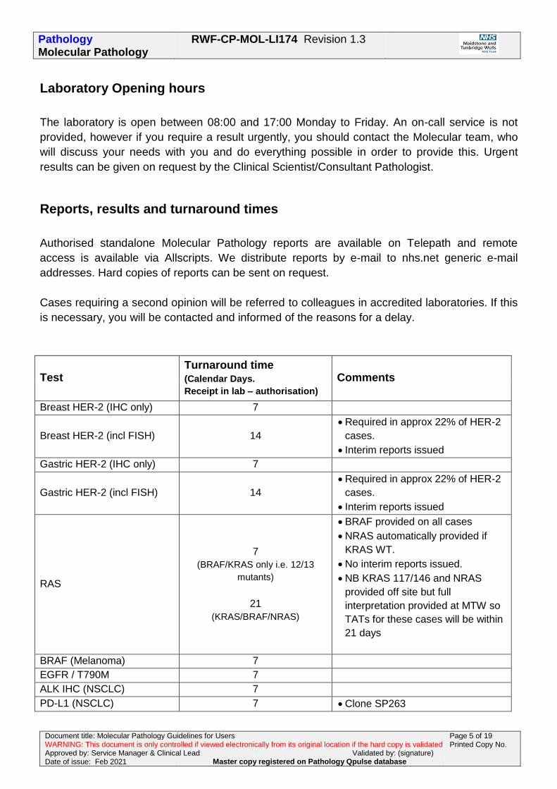
Laboratory Opening hours
The laboratory is open between 08:00 and 17:00 Monday to Friday. An on-call service is not
provided, however if you require a result urgently, you should contact the Molecular team, who
will discuss your needs with you and do everything possible in order to provide this. Urgent
results can be given on request by the Clinical Scientist/Consultant Pathologist.
Reports, results and turnaround times
Authorised standalone Molecular Pathology reports are available on Telepath and remote
access is available via Allscripts. We distribute reports by e-mail to nhs.net generic e-mail
addresses. Hard copies of reports can be sent on request.
Cases requiring a second opinion will be referred to colleagues in accredited laboratories. If this
is necessary, you will be contacted and informed of the reasons for a delay.
Test Turnaround time (Calendar Days.
Receipt in lab – authorisation) Comments
Breast HER-2 (IHC only) 7
Breast HER-2 (incl FISH) 14
Required in approx 22% of HER-2
cases.
Interim reports issued
Gastric HER-2 (IHC only) 7
Gastric HER-2 (incl FISH) 14
Required in approx 22% of HER-2
cases.
Interim reports issued
RAS
7
(BRAF/KRAS only i.e. 12/13
mutants)
21
(KRAS/BRAF/NRAS)
BRAF provided on all cases
NRAS automatically provided if
KRAS WT.
No interim reports issued.
NB KRAS 117/146 and NRAS
provided off site but full
interpretation provided at MTW so
TATs for these cases will be within
21 days
BRAF (Melanoma) 7
EGFR / T790M 7
ALK IHC (NSCLC) 7
PD-L1 (NSCLC) 7 Clone SP263

Pathology Molecular Pathology
RWF-CP-MOL-LI174 Revision 1.3
Document title: Molecular Pathology Guidelines for Users Page 6 of 19 WARNING: This document is only controlled if viewed electronically from its original location if the hard copy is validated Printed Copy No. Approved by: Service Manager & Clinical Lead Validated by: (signature) Date of issue: Feb 2021 Master copy registered on Pathology Qpulse database
PD-L1 (Triple negative
breast Cancer) 7
Clone SP142
Currently outsourced.
PD-L1 (H&N)
(pembrolizumab) 7-10
Clone 22C3
Currently outsourced
PD-L1 (Urology) 7-10
Clone 22C3 (Pembrolizumab)
Clone SP142 (Atezolizumab)
Currently outsourced
PD-L1 (melanoma)
(nivolumab) 7-10
Clone 28/8
Currently outsourced
ROS1 (NSCLC) 7
ROS1 FISH (NSCLC) 7-10 Currently outsourced
MMR 7
BRAF / methylation testing if loss
observed (7 additional days if
required)
IHC Results on CPATH as part of
histology report.
BRAF results on HAEM and e-
mailed to requestor
Methylation testing outsourced and
results e-mailed to requestors
Gene Expression Profiling
Oncotype DX
Endopredict.
Prosigna
Outsourced
Clinical Advice
Clinical advice can be provided from the Clinical Scientist or relevant pathologists involved in
the service. Please contact a member of the Molecular team who will put you in contact with the
relevant individual.
Costs
Price of testing is available from the Laboratory Manager on request.
Some tests are currently within tariff and you will be charged for each test. These include
HER-2 (all indications and tests performed), MMR assessment (including reflex tests) and
EGFR testing.
Pathology Molecular Pathology
RWF-CP-MOL-LI174 Revision 1.3
Document title: Molecular Pathology Guidelines for Users Page 7 of 19 WARNING: This document is only controlled if viewed electronically from its original location if the hard copy is validated Printed Copy No. Approved by: Service Manager & Clinical Lead Validated by: (signature) Date of issue: Feb 2021 Master copy registered on Pathology Qpulse database
NHSE currently fund ALK IHC, ALK FISH, RAS, BRAF, PD-L1, ROS1 and Breast gene
expression profiling. Please discuss invoicing arrangements with the Molecular Management
team.
Invoices are prepared on a monthly basis to those departments not eligible for free testing and
private providers.
Prices are reviewed in line with inflation prior to the start of the new financial year and users are
informed of impending increase during March by email.
Consent, storage and authorisation
Please note, in accordance with the requirements of the Human Tissue Act, it is the
responsibility of the referring clinician to ensure that appropriate informed consent has been
obtained before any testing is undertaken. The laboratory must be informed of any restrictions
to this consent. Unless stated, the laboratory will process all samples with the understanding all
appropriate consent has been obtained from the patient for the tests requested, and for storage
of the derived DNA for future use.
Samples from patients who do not consent to storage and future use of their DNA must be
accompanied by a “closed consent” form indicating the limits of the consent granted. If in doubt,
contact a member of the Molecular Pathology team to discuss.
Transport of Samples
It is the responsibility of those taking and dispatching specimens to the laboratory to ensure that
these samples are sent in accordance with any national guidelines and/or local policies for the
packaging, labelling and transport of biological material.
Problems and Queries
For any further information, issues, complaints please contact a member of the Molecular
Pathology team who will refer you appropriately.
Requests
Requests can be made using the request form (see p18) and e-mailing to mtw-

Pathology Molecular Pathology
RWF-CP-MOL-LI174 Revision 1.3
Document title: Molecular Pathology Guidelines for Users Page 8 of 19 WARNING: This document is only controlled if viewed electronically from its original location if the hard copy is validated Printed Copy No. Approved by: Service Manager & Clinical Lead Validated by: (signature) Date of issue: Feb 2021 Master copy registered on Pathology Qpulse database
All requests require a completed request form, a copy of the histology report and FFPE block of
representative tissue
Specimen Requirements
The request form and FFPE should be sent to the laboratory following local protocols for
transport of pathology samples. The following information must be legible on the request form:
Patient hospital number
NHS number
Surname
Forename
Date of Birth
Gender
Details of GP
NHS/PP status
If the sample is from a private patient, we will invoice you as the requestor.
FFPE blocks must be labelled with the unique identifier. Accompanying slides must be labelled
with the unique identifier and patient Surname and Forename (or initial). Samples without
minimum data, or when data on the sample and accompanying documents do not match, will
not be accepted and will be returned to the sender. Failure to comply with this policy will result
in the sample being rejected or the result delayed.
Requesters should ensure invasive carcinoma is in the block sent for testing and that the
sample represents the tumour.
Please note, we are unable to accept unstained slides
Quality Assurance and National Guidelines for Testing
All assays are subject to rigorous Internal Quality Control (IQC) measures and the laboratory
participates in External Quality Assurance (EQA) schemes for which results are available on
request:

Pathology Molecular Pathology
RWF-CP-MOL-LI174 Revision 1.3
Document title: Molecular Pathology Guidelines for Users Page 9 of 19 WARNING: This document is only controlled if viewed electronically from its original location if the hard copy is validated Printed Copy No. Approved by: Service Manager & Clinical Lead Validated by: (signature) Date of issue: Feb 2021 Master copy registered on Pathology Qpulse database
UK NEQAS Molecular Genetics forColorectal cancer, lung cancer and Melanoma testing
UK NEQAS ICC & ISH for Breast and Gastric Cancer, PD-L1, MMR, ROS1 and ALK testing
Service users can be assured with the large number of samples being tested, extensive
experience with all methodology, complete training of all staff a high quality and accurate
service is provided. Centralised testing also gives value for money.
Uncertainty in assessment and reporting has been considered for all tests provided and multiple
measures are in place to mitigate these risks where they exist. Details are available from the
laboratory on request.
Factors known to affect Molecular Pathology testing
Testing can be performed on metastatic deposits, core biopsies, cell blocks of cytology
specimens, EBUS samples and samples of the resected tumour. There is some evidence that
elements of processing the FFPE block may interfere with molecular analysis. Specialised
molecular Pathology analyses can be problematic in obtaining optimal results, particularly with
factors outlined below. Invalid tests will be repeated once (at no additional cost). The service
user will be contacted and informed of the reasons for a delay.
Decalcification in an acid containing solution, fast acid decalcification (including surface
decalcification) or fixation in Bouin’s solution is known to degrade the DNA within a sample
making subsequent molecular analysis difficult. Please provide decalcification details if
performed (chemical used and duration).
Alcohol fixation is a contraindication with many immunohistochemical assays. If the sample
has been exposed to an alcoholic fixative (e.g. clots / EBUS etc.), this should be made clear
on the request form. Failure to do so may result in false negative results.
Tumour tissues, particularly small biopsies, may yield too little DNA for molecular analysis. If
this is the case the sample will be reported as insufficient.
DNA from non-neoplastic cells within the sample may dilute the analyte of interest beyond
the level of detection for the assay. Tumour burden and macrodissection if considered
necessary, is undertaken in order to minimise this occurrence and will be present on the
report.
The quality of tissue fixation- including ischaemic time, length of fixation, the size of the
sample, penetration of formalin and processing protocols can affect the quality of DNA
isolated from FFPE tissues; and therefore the results of molecular testing. Large, poorly
fixed samples often produce inconsistent and technically difficult results.
Marking ink and Eosin can interfere with fluorescent signals during FISH interpretation.

Pathology Molecular Pathology
RWF-CP-MOL-LI174 Revision 1.3
Document title: Molecular Pathology Guidelines for Users Page 10 of 19 WARNING: This document is only controlled if viewed electronically from its original location if the hard copy is validated Printed Copy No. Approved by: Service Manager & Clinical Lead Validated by: (signature) Date of issue: Feb 2021 Master copy registered on Pathology Qpulse database
1. Molecular Testing for Targeted Treatments in Lung Cancer
a. EGFR mutation Analysis
Epidermal growth factor receptor (EGFR) tyrosine kinase inhibitors (TKIs) are currently used in
patients with advanced metastatic non-small cell lung cancer (NSCLC). Somatic mutations in
the EGFR gene are used to predict response to EGFR TKIs including Erlotinib (Tarceva®),
Afatinib (Gilotrif®), and Gefitinib (Iressa®).
At present, mutation analysis of exons 18-21 of the EGFR gene are carried out as reflex tests on all biopsied or resected non-squamous NSCLCs (requested by Pathologists). The test used is the Roche COBAS EGFR v2 kit. This method screens for 42 mutations in exons 18-21 (29 deletions in exon 19, T790M, L858R, G719X, S768I, L861Q, 5 Insertions in exon 20).
Reports are standalone reports available on the remote LIMS viewer or e-mailed to external
users. The turnaround time for this test is 7 days.
b. ALK Testing
The Anaplastic Lymphoma Kinase (ALK) gene encodes a tyrosine kinase normally expressed in
neuronal cells. The ALK gene can, by deletion or translocation, form a fusion protein, which is
abnormally expressed in approximately 4-7% of NSCLC patients. The presence of this ALK
gene fusion protein predicts response to ALK inhibitors.
Tests are carried out as reflex tests on all non-squamous NSCLCs (requested by Pathologists). ALK testing is carried out using the Ventana D5F3 Rabbit monoclonal antibody. Reports are available on Telepath as a standalone report.
c. ROS1 Testing
The ROS1 gene (6q22) encodes a transmembrane receptor protein with one of the largest
extracellular domains amongst all human receptor tyrosine kinases. Genomic rearrangements
involving ROS1 occur in 1–2 % of NSCLCs and these tumours can now be treated with
crizotinib (Xalkori®). See https://www.nice.org.uk/guidance/ta529.
Tests are requested as reflex tests on all non-squamous NSCLCs (requested by Pathologists). The test is performed using the Roche SP384 ROS1 monoclonal antibody (CE-IVD).

Pathology Molecular Pathology
RWF-CP-MOL-LI174 Revision 1.3
Document title: Molecular Pathology Guidelines for Users Page 11 of 19 WARNING: This document is only controlled if viewed electronically from its original location if the hard copy is validated Printed Copy No. Approved by: Service Manager & Clinical Lead Validated by: (signature) Date of issue: Feb 2021 Master copy registered on Pathology Qpulse database
Reports are available on Telepath as a standalone report. The turnaround time for this test is 7
days.
Cases showing any level of staining are referred to Queen Elizabeth Hospital in Birmingham for
FISH testing to make a definitive assessment of translocation status. A report will be available
as a final report addendum to the ROS1 immunohistochemistry result within 2 weeks.
d. PD-L1 Testing
PD-L1 is a protein that binds to the PD-1 checkpoint on immune cells that can determine response to PD-1 checkpoint inhibitors such as Nivolumab (Opdivo®) and Pembrolizumab (Keytruda®). Several other PD-1/PD-L1 checkpoint inhibitors are also in late-stage clinical testing.
Currently PD-L1 tests are provided as a reflex test on all squamous and non-squamous NSCLCs. The test provided is the Roche SP263 clone for which the interpretation must only be used for the following indications:
Indication for use Therapy PD-L1 Expression- Therapeutic Line
NSCLC KEYTRUDA® ≥ 50% TC – First Line
≥ 1% TC – Second Line
OPDIVO® ≥ 1%, ≥ 5% and ≥ 10% TC – Second Line
Testing for melanoma, urothelial carcinoma, triple negative breast cancer, head & neck carcinomas and other indications will be outsourced. Please discuss with the management team and mark requests clearly, including the treatment being considered for the patient. Sample Requirements:
Formalin fixed, paraffin embedded tumour blocks should accompany a copy of the
Histopathology report along with a completed request form. Samples can be sent according to
local protocols. Please note, we cannot accept unstained slides
FFPE blocks must be labelled with your unique identifier. Accompanying slides (if present) must
be labelled with the unique identifier as well as patient family name and given name (or initial).

Pathology Molecular Pathology
RWF-CP-MOL-LI174 Revision 1.3
Document title: Molecular Pathology Guidelines for Users Page 12 of 19 WARNING: This document is only controlled if viewed electronically from its original location if the hard copy is validated Printed Copy No. Approved by: Service Manager & Clinical Lead Validated by: (signature) Date of issue: Feb 2021 Master copy registered on Pathology Qpulse database
Samples without minimum data, or when data on the sample and accompanying documents do
not match, will not be accepted and will be returned to the sender.
Requesters should ensure appropriate carcinoma is in the block sent for testing and that the
sample represents the tumour. If more than one tumour lesion is present representative blocks
of each tumour should be sent.
If requesting multiple tests on small samples, it is helpful to indicate which tests should be given
priority. If no indication is given, PD-L1 testing will take precedence.

Pathology Molecular Pathology
RWF-CP-MOL-LI174 Revision 1.3
Document title: Molecular Pathology Guidelines for Users Page 13 of 19 WARNING: This document is only controlled if viewed electronically from its original location if the hard copy is validated Printed Copy No. Approved by: Service Manager & Clinical Lead Validated by: (signature) Date of issue: Feb 2021 Master copy registered on Pathology Qpulse database
2. Molecular Testing for Targeted Treatments in Colorectal Cancer
Anti-EGFR monoclonal antibody therapies such as Cetuximab and panitumumab target EGFR
and are approved for use in patients with metastatic colorectal cancer. These are currently used
in combination with chemotherapy in metastatic colorectal tumours that carry no mutations in
the RAS family (KRAS and NRAS genes). Mutation testing is required to indicate suitability to
receive anti-EGFR antibody therapy. The presence of a RAS mutation predicts the lack of
response to these therapies.
a. RAS mutation analysis
All samples submitted for RAS analysis are assessed for the need to enrich the tumour by
macro-dissection. This is an important prerequisite for molecular genetic testing in pathology as
failure to enrich tumour DNA, which may harbour somatic mutations, can result in false negative
results.
An H&E slide is requested with every submission which should be marked up with tumour
circled together with an estimate of tumour burden within the marked area and identified by
initials / date of the individual making the assessment (see example below). Please note we will
retain this slide. A minimum of 10% tumour burden is required for the successful analysis of
KRAS/NRAS genes
Our testing strategy is to test all patients with the Roche COBAS KRAS real time mutation
assay for 19 different KRAS mutations covering the majority of clinically relevant ‘hot-spot’
mutations in codons 12, 13 and 61. Detection of a mutation will result in a final report being
issued. Please note the mutation present will not be characterised. Those samples where no

Pathology Molecular Pathology
RWF-CP-MOL-LI174 Revision 1.3
Document title: Molecular Pathology Guidelines for Users Page 14 of 19 WARNING: This document is only controlled if viewed electronically from its original location if the hard copy is validated Printed Copy No. Approved by: Service Manager & Clinical Lead Validated by: (signature) Date of issue: Feb 2021 Master copy registered on Pathology Qpulse database
mutation is detected will be outsourced for mutation detection in KRAS codons 117 & 146 as
well as NRAS (codons 12/13, 59-61).
b. BRAF mutation analysis
BRAF is a proto-oncogene with a central role in signalling pathways that control cellular
proliferation, differentiation and cell death/ apoptosis. In normal cells, BRAF is part of a highly
regulated signalling pathway that mediates the effects of growth factor receptors. Oncogenic
mutations in BRAF, V600E result in a gain of kinase function, activating signalling pathways in
the absence of the typical growth factors.
Molecular Pathology performs the ROCHE cobas BRAF real-time PCR assay intended for the
identification of mutations in codons V600E of the BRAF mutations in DNA derived from human
colorectal tissues in all cases submitted for RAS analysis. A minimum of 50% tumour burden is
required for the successful analysis of the BRAF gene.
RAS Reports are available following all tests performed as standalone reports on Telepath.
c. Mismatch Repair Testing
Microsatellite instability (MSI), a hypermutable phenotype caused by the loss of DNA, mismatch
of repair activity is a mechanism of colorectal carcinogenesis. MSI is detected in about 15% of
all colorectal cancers; 12% caused by sporadic, acquired hypermethylation and 3% associated
with hereditary non-polyposis colorectal cancer (HNPCC), Lynch syndrome. Patients with
HNPCC, Lynch syndrome, have an 80% percent chance of developing colorectal cancer.
Tumours with MSI have slightly better prognosis. Discovery of MSI has increased awareness of
the implications for specialised management of patients.
At Maidstone & Tunbridge Wells NHS Trust we perform MMR immunohistochemistry in order to
assess the presence or loss of mis-match repair proteins MLH1, MSH2, PMS2 and MSH6 as a
surrogate for MSI testing. No losses indicate preserved expression of the genes and sporadic
nature of the disease.
If loss of expression is detected in MLH1 +/- PMS2, then a BRAF test is undertaken to elucidate
whether or not a BRAF V600E mutation is present (which indicates a probable sporadic
pathway). Those cases where no mutation is detected are followed up with MLH1 promotor
methylation studies to further stratify those patient that need to be referred for specialist genetic
testing and advice with regards to Lynch Syndrome. MLH1 promotor methylation testing is
outsourced.
Pathology Molecular Pathology
RWF-CP-MOL-LI174 Revision 1.3
Document title: Molecular Pathology Guidelines for Users Page 15 of 19 WARNING: This document is only controlled if viewed electronically from its original location if the hard copy is validated Printed Copy No. Approved by: Service Manager & Clinical Lead Validated by: (signature) Date of issue: Feb 2021 Master copy registered on Pathology Qpulse database
Currently some tests are provided as a reflex based upon the Bethesda criteria and others by
clinical request on our request form or via e-mail to [email protected] .
Results of MMR immunohistochemistry can be found on the Histopathology report. If applicable,
standalone reports will be made available for BRAF and MLH1 promotor methylation for
interpretation and final report production by the Consultant Histopathologist.
Pathology Molecular Pathology
RWF-CP-MOL-LI174 Revision 1.3
Document title: Molecular Pathology Guidelines for Users Page 16 of 19 WARNING: This document is only controlled if viewed electronically from its original location if the hard copy is validated Printed Copy No. Approved by: Service Manager & Clinical Lead Validated by: (signature) Date of issue: Feb 2021 Master copy registered on Pathology Qpulse database
3. Molecular Testing for Targeted Treatments in Malignant Melanoma
a. BRAF mutation analysis
Vemurafenib (Zelboraf®) is a BRAF enzyme inhibitor developed by Plexxikon and Genentech
for the treatment of late-stage melanoma. Response to therapy is achieved in tumours that
harbour BRAF codon 600 mutations.
Molecular Pathology performs the ROCHE cobas BRAF real-time PCR assay intended for the
identification of mutations in codons V600E of the BRAF gene in DNA derived from human
melanoma tissue. A minimum of 50% tumour burden is required for the successful analysis of
the BRAF gene.
Some requests are reflexed based on diagnostic criteria and requests from clinicians are
accepted via e-mail to [email protected] . BRAF Reports are available as
standalone reports on Telepath.

Pathology Molecular Pathology
RWF-CP-MOL-LI174 Revision 1.3
Document title: Molecular Pathology Guidelines for Users Page 17 of 19 WARNING: This document is only controlled if viewed electronically from its original location if the hard copy is validated Printed Copy No. Approved by: Service Manager & Clinical Lead Validated by: (signature) Date of issue: Feb 2021 Master copy registered on Pathology Qpulse database
4. Molecular Testing for Targeted Treatments in Breast and Gastric
Cancers
The human epidermal growth factor HER-2/neu/c-erbB-2 (HER-2) is amplified in approximately
15% of invasive breast cancers. HER-2 status is of interest due to a number of targeted
therapies in breast cancer. HER-2 positivity is a pre-requisite for the humanised monoclonal
antibody Trastuzumab (Herceptin ™) and other HER-2 dependent therapies to be prescribed to
patients. All invasive breast carcinomas are reflexed for a HER-2 at diagnosis.
Advanced gastric/GOJ cancers benefit from the administration of Herceptin in patients shown to
over-express the protein due to gene amplification. Trastuzumab, in combination with cisplatin
and capecitabine or 5-fluorouracil, is recommended as an option for the treatment of people
with HER-2 positive metastatic adenocarcinoma of the stomach or gastro-oesophageal junction
not previously having received treatment for their metastatic disease or have tumours
expressing high levels of HER-2. HER-2 in gastric carcinomas are performed on clinical
request only
a. Her-2 immunohistochemistry analysis
The antibody clone 4B5 from Roche is used on all cases and is scored using a semi-
quantitative system. The majority will be reported as negative or positive on the
immunohistochemistry results. Those falling into the equivocal category of 2+ are reflexed to be
further assessed using Fluorescent in-situ hybridisation. An interim report is issued in these
circumstances.
b. Fluorescent in-situ hybridisation (FISH) analysis
Fluorescent in-situ hybridisation (FISH) is carried out on equivocal 4B5 cases as a reflex test on
both breast and gastric cases. The HER-2 gene is examined using fluorescent labels to
ascertain evidence of gene amplification. Scoring is carried out according to UK guidelines
(Rakha EA et al, J Clin Pathol 2014;0:1-7. Wong NACS et al, J Clin Pathol 2018;71:388-394)
Cases showing HER-2 / CEP17 ratio of <1.79 and average copy numbers of <4.0 per cell are
considered NEGATIVE (NOT AMPLIFIED)
Cases showing HER-2:CEP17 ratio of 1.8 – 1.99 or <2.0 with average copy numbers of 4.0 –
5.99 per cell are considered EQUIVOCAL/ BORDERLINE NON-AMPLIFIED. A repeat test is

Pathology Molecular Pathology
RWF-CP-MOL-LI174 Revision 1.3
Document title: Molecular Pathology Guidelines for Users Page 18 of 19 WARNING: This document is only controlled if viewed electronically from its original location if the hard copy is validated Printed Copy No. Approved by: Service Manager & Clinical Lead Validated by: (signature) Date of issue: Feb 2021 Master copy registered on Pathology Qpulse database
requested, either on the excision when available, another block of the same sample, or another
sample. Following the repeat test, readings in these ranges are reported as Not amplified or
Amplified.
Cases showing HER-2:CEP17 ratio >2.0 or with average copy numbers of >6.0 are reported as
POSITIVE (AMPLIFIED)
Breast FISH cases showing HER2 CEP17 ratio >=2.0, HER2 copy number <4 are reported
following current guidance issued by UK National Coordinating Committee for Breast Pathology
Group
Occasionally, the clinical picture may dictate an alternative outcome to that described above
and all such cases will be fully explained within the comments of the report. Further clinical
advice and guidance can be sought from the Clinical Scientist

Pathology Molecular Pathology
RWF-CP-MOL-LI174 Revision 1.3
Document title: Molecular Pathology Guidelines for Users Page 19 of 19 WARNING: This document is only controlled if viewed electronically from its original location if the hard copy is validated Printed Copy No. Approved by: Service Manager & Clinical Lead Validated by: (signature) Date of issue: Feb 2021 Master copy registered on Pathology Qpulse database
Appendix A. Request Form