influence of microgroove dimension on cell behavior of
TRANSCRIPT
Influence of Microgroove Dimension on Cell Behavior of Human Gingival Fibroblasts Cultured on Titanium
Substrata
Suk Won Lee
The Graduate School Yonsei University
Department of Dental Science

Influence of Microgroove Dimension on Cell Behavior of Human Gingival Fibroblasts Cultured on Titanium
Substrata
A Dissertation Submitted to the Department of Dental Science and the Graduate School of Yonsei University
in partial fulfillment of the requirements for the degree of
Doctor of Philosophy
Suk Won Lee
June 2007

This certifies that the dissertation of
Suk-Won Lee is approved.
___________________________
Thesis Supervisor: Keun-Woo Lee
___________________________
Moon-Kyu Chung
___________________________
Dong-Hoo Han
___________________________
In-Chul Rhyu
___________________________
Won-Yoon Chung
The Graduate School
Yonsei University
June 2007

감사의감사의감사의감사의 글글글글
먼 길을 달려오는 동안 마지막까지 어려운 고비마다 깊이
격려해주시고 올바른 길로 이끌어 주신 이근우 지도교수님께 진심으로
감사드립니다. 탁월한 통찰력으로 논문의 틀을 바로잡아주신 정문규
교수님과 날카로운 지적으로 논문의 완성도를 높여주신 한동후 교수님께
깊이 감사드립니다. 본 논문에서 사용된 마이크로그루브에 대한 해박한
지식으로 그 임상적 의미를 일깨워주신 서울치대 치주과학교실의 류인철
교수님과 부족한 제가 세포 실험이 어떠한 것이라는 것을 어렴풋이나마
알게 해주신 구강생물학교실 정원윤 교수님께도 깊은 감사의 마음을
전합니다. 치과의사는 어떻게 살아야 하는지를 몸소 보여주신 이호용
교수님과 본 논문의 아이디어를 처음 접하게 해주신 한종현 교수님께
감사의 말씀을 올립니다. 학창시절 담임 선생님이셨으며 전공 선택을
이끌어주신 강우진 교수님, 언제나 깊은 관심으로 챙겨주시는 문홍석,
심준성 교수님과 지금 이 시각에도 연구에 몰두하고 계실 김선재, 이재훈,
배은경 교수님, 그리고 본 논문에 특히 깊은 관심을 가져주셨던 황선홍
교수님께 자랑스럽게 이 논문을 드리고 싶습니다. 든든한 선배로서, 또한
선배 연구자로서 언제나 질문에 답을 주셨던 인하의대 치과학교실의
오남식 교수님, 시편을 아름답고 신속하게 제작해주신 멤스웨어 조수제
사장님과 윤혜진씨께 감사드립니다. 주말도 반납하며 헌신적으로 실험을
도와주신 성빈센트병원 임상의학연구소 김수연 선생님, 밤이 깊은 줄
모르고 연구에 대한 토론에 응해주셨던 배진완 교수님과 성빈센트병원
동료 교수님들, 그리고 김종수 기공실장님께 역시 감사드립니다.
언제나 깊은 격려로 힘을 주시는 장인어른, 장모님, 올바른 가치관을
심어주시고 삶의 길잡이이신 아버지, 그리고 이 세상에서 나를 가장
사랑해주시는 어머니와 더불어 하나님이 주신 아름다운 선물 혜준이
예준이와 나의 믿음이며 안식처인 사랑하는 아내에게 이 논문을 바칩니다.
2007년 6월
저 자 씀

i
TABLE OF CONTENTS
LIST OF FIGURES......................................................................................... ii
LIST OF TABLES.......................................................................................... iv
ABSTRACT(ENGLISH).....................................................................v
I . INTRODUCTION............................................................................. 1
II . MATERIALS AND METHODS....................................................5
A . Cell culture .................................................................................. 5
B . Fabrication of Ti substrata........................................................... 6
C . Adhesion assay ............................................................................ 8
D . Scanning electron microscopy .................................................... 8
E . XTT assay.................................................................................... 9
F . RT-PCR........................................................................................ 9
G . Statistical analysis...................................................................... 10
III . RESULTS........................................................................................ 12
A . Adhesion analysis...................................................................... 12
B . Morphological analysis ............................................................. 14
C . Assessment of viability and proliferation.................................. 18
D . Gene expression analysis .......................................................... 21
IV . DISCUSSION................................................................................ 23
V . CONCLUSION............................................................................... 43
VI . REFERENCES.............................................................................. 45
ABSTRACT(KOREAN).................................................................... 54

ii
LIST OF FIGURES
Fig. 1. Fabrication of Ti substrata using photolithography........................... 7
Fig. 2. Fabricated surfaces of the Ti substrata with microgrooves ............... 7
Fig. 3. Effect of structural dimensions of surface microgrooves on human
gingival fibroblast adhesion to Ti substrata..................................... 13
Fig. 4. Scanning electron microscopic (SEM) observations on human
gingival fibroblast morphology and orientation at 16 h of culture.. 15
Fig. 5. Scanning electron microscopic (SEM) observations on human
gingival fibroblast morphology and orientation at 48 h of culture.. 15
Fig. 6. Scanning electron microscopic (SEM) observations on human
gingival fibroblast morphology at 16 h of culture in
higher magnification........................................................................ 16
Fig. 7. Scanning electron microscopic (SEM) observations on human
gingival fibroblast morphology at 16 and 48 h of culture ............... 16
Fig. 8. Scanning electron microscopic (SEM) observations on human
gingival fibroblast lying on the edge at 16 h of culture................... 17
Fig. 9. Scanning electron microscopic (SEM) observations on human
gingival fibroblast with abundant filopodia projections
at 16 h of culture.............................................................................. 17
Fig. 10. Effect of structural dimensions of surface microgrooves on human
gingival fibroblast viability and proliferation on Ti substrata ......... 20
Fig. 11. Analysis on expression of genes involved in
cell-matrix adhesion and G1/S cell cycle progression in RT-PCR .. 22
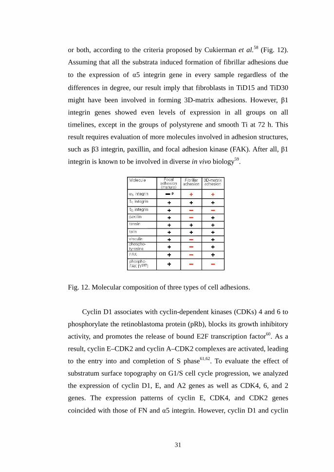
Fig. 12. Molecular composition of three types of cell adhesions.................. 31
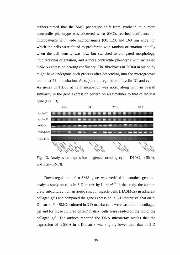
Fig. 13. Analysis on expression of genes encoding
cyclin D1/A2, α-SMA, and TGF-βR-I/II......................................... 36
Fig. 14. Analysis on expression of genes encoding
cyclin D1/A2 and p21cip1 ................................................................. 40
Fig. 15. Analysis on expression of genes encoding cyclin E and p21cip1 ...... 42

iii
Fig. 16. Analysis on expression of genes encoding
Skp2, NF-κB1, and TGF-βR-I........................................................... 42

iv
LIST OF TABLES
Table 1. Gene-specific primers used in RT-PCR........................................... 11
Table 2. Comparison of fibroblast adhesion to Ti substrata
at 1 and 2 h incubation by structural dimensions of
surface microgrooves...................................................................... 12
Table 3. Comparison of fibroblast viability and proliferation on Ti substrata
after 24, 48, 72, and 96 h of culture
by structural dimensions of surface microgrooves.......................... 19

v
ABSTRACT
Influence of Microgroove Dimension on Cell Behavior of
Human Gingival Fibroblasts Cultured on Titanium
Substrata
Suk-Won Lee, D.D.S., M.S.D.
Department of Dental Science, Graduate School, Yonsei University
(Directed by Prof. Keun-Woo Lee, D.D.S., M.S.D., Ph.D.)
To enhance interactions between titanium (Ti) oral implants and the
surrounding gingival soft tissues, the effects of Ti-surface microgrooves on
cell behavior have extensively been investigated. Narrow microgrooves on
Ti substrata were verified to induce changes in morphology, cell-substratum
adhesion, and gene expression of cultured connective tissue cells, such as
fibroblasts. However, their effect on enhancing cell proliferation in vitro or
on the ability to effectively reduce epithelial down-growth in vivo, is not yet
clear.
In this study, we hypothesized that surface microgrooves of appropriate
depth and extensive width on Ti substrata that enable the cells to readily
descend into themselves, would alter various cell behaviors including
viability and proliferation of cultured human gingival fibroblasts. For the
evaluation, cell adhesion, morphology, viability and proliferation, and gene
expression of human gingival fibroblasts cultured on Ti substrata with
various dimensions of surface microgrooves were analyzed. The purpose of
this study was to determine the dimension of surface microgrooves on Ti
substrata that shows the greatest positive influence on characterizing specific
cell behavior of cultured human gingival fibroblasts.
Commercially pure Ti discs with surface microgrooves of monotonous

vi
3.5 µm in depth and respective 15, 30, and 60 µm in width were fabricated
using photolithography and used as the culture substrata in the three
experimental groups in this study (TiD15, TiD30, and TiD60 groups),
whereas the smooth Ti disc was used as the control substrata (smooth Ti
group). Human gingival fibroblasts were cultured on the four groups of
titanium substrata on successive timelines. Cell behaviors, such as adhesion,
morphology, viability and proliferation, and gene expression were analyzed
and compared between all groups using crystal violet stain, scanning
electron microscopy (SEM), XTT assay, and reverse transcriptase-
polymerase chain reaction (RT-PCR), respectively. One-way analysis of
variance was used in the statistical analyses on the adhesion as well as the
viability and proliferation data. From the results of the analyses on various
biological activities of human gingival fibroblasts, the following results were
obtained.
1. There was no difference between the numbers of human gingival
fibroblasts adhered to smooth Ti substrata and those adhered to Ti
substrata with surface microgrooves at 1 and 2 h incubation.
Fibroblasts merely formed initial cell-substratum contact by the
times.
2. In SEM, contact guidance of human gingival fibroblasts parallel to
the direction of microgrooves was observed. Cells were able to
readily descend into the microgrooves of 30 µm in width and 3.5 µm
in depth at the early phase of culture, whereas at the later phase,
cells in all groups were found both in the grooves and on the ridges.
Cells on the ridge edges or in groove corners were spindle shaped
with abundant filopodia formation towards the acid-etched surface
inside the microgrooves, thus mimicked the shape of the fibroblasts
cultured in three-dimensional (3D) nanoenvironment.
3. On successive timelines, human gingival fibroblasts cultured on Ti
substrata with various dimensions of surface microgrooves showed
vii
differences in the rate of reaching their confluence. Human gingival
fibroblasts cultured on Ti substrata with microgrooves of 15 µm in
width and 3.5 µm in depth significantly increased their viability and
proliferation compared with those cultured on smooth Ti substrata
after 72 h of culture and decreased after 96 h, whereas the cells on
the microgrooves of 30 µm in width and 3.5 µm in depth continued
increasing their viability and proliferation up to after 96 h of culture.
4. A joint up-regulation of matrix-assembly genes, such as fibronectin
and �5 integrin genes, was noted in human gingival fibroblasts
cultured on titanium substrata with microgrooves of 15 and 30 µm in
width and an equal 3.5 µm in depth.
5. Gene expression pattern specific to the cells in 3D-matrix culture,
such as down-regulation of �-smooth muscle actin gene along with
up-regulation of fibronectin and p21 genes, was pronounced in
human gingival fibroblasts cultured on Ti substrata with
microgrooves of 30 µm in width and 3.5 µm in depth.
In reference to the results above, two conclusions were made.: 1)
Surface microgrooves of 15 µm in width and 3.5 µm in depth on titanium
substrata increase the viability and proliferation, as well as the expression of
genes involved in the matrix assembly of cultured human gingival
fibroblasts. 2) Surface microgrooves of 30 µm in width and 3.5 µm in depth
on titanium substrata provide human gingival fibroblasts with a three-
dimensional context of culture, thus show corresponding gene expression.
Key words: Titanium, Microgroove, Fibroblast, Proliferation,
Gene expression
1
Influence of Microgroove Dimension on Cell
Behavior of Human Gingival Fibroblasts
Cultured on Titanium Substrata
Department of Dental Science,
Graduate School, Yonsei University
(Directed by Prof. Keun-Woo Lee, D.D.S., M.S.D., Ph.D.)
Suk-Won Lee
I . INTRODUCTION
Since the establishment of osseointegration as a unique concept in oral
implantology, researchers now seek for methods to enhance the interactions
between titanium implants and the surrounding soft tissues, known as peri-
implant soft tissue reactions. The major class of cells found in peri-implant
soft tissues includes gingival fibroblasts whose changes in cell behaviors
such as cell-matrix adhesions on microtopographic features have long been
the topic of studies done by Brunette1, Jansen2, and their coworkers. They
suggested that microfabricated grooved surfaces would produce orientation
and directed locomotion of epithelial cells in vitro, thus inhibit epithelial
down-growth on titanium oral implants in vivo. Moreover, since surface
topography was considered important in establishing connective-tissue
organization adjacent to titanium oral implants, fibroblast shape and
orientation on such grooved surfaces had extensively been evaluated and the
effects of the surface topography of microgrooved titanium substrata on cell
behavior in vitro and in vivo were suggested to depend on the groove
dimensions. Among the structures varying in shape and dimension,

2
microgrooves used in these studies were mainly fabricated from the
micromachining technique. In most studies, V-shaped micromachined
grooves and their morphologic derivatives were fabricated so as to allow the
cell to confront its substratum with defined edges. The degree of cell
spreading on such geometry was maximized in two directions. The one, an
elongation or a polarization in shape parallel to the long axis of
grooves/ridges was considered one of the results from a phenomenon called
‘contact guidance’, whereas the other, from forming bridges between ridges
leading to cell spreading3. On sensing such geometry, focal contacts were
considered to stiffen4 and the cellular traction forces exerted through focal
contacts increased5, leading to increased amount of focal adhesions6. The
adhesion strength even surpassed the amount of externally applied local
mechanical force7,8. Using focal adhesions as anchors, contractile traction
forces exerted by a cell, also called cytoskeletal tension per se or cytoskeletal
prestress, together with extracellular matrix (ECM) and cytoskeletal
structure were considered to play decisive roles in the control of various
biological activities including the gene expression and growth9. It has been
hypothesized that surface topography could induce direct cell
mechanotransduction, thus leads to changes in the probability of gene
expression10. In these biomechanical models, environments were provided so
as to induce changes in cell shape and cell spreading on micropatterned or
nanostructured surfaces. Another example would be the strengthened cell
adhesions and the resultant increase in traction forces exerted by a cell on
sharp edges of microstructures in specific directions11.
Several studies have reported the induction of the changes in cell shape
by microgrooves and these changes were found to be tightly coupled to DNA
synthesis and growth in adherent cells12. Indeed, human fibroblasts grown on
the microgrooved substrata, compared to those on the smooth ones, were
significantly elongated and orientated along the grooves leading to an
increase in the amount of fibronectin mRNA/cell13, or displayed alterations
in the expression of numerous genes responsible for various biological

3
activities14. However, the induction of the changes in cell shape by
microgrooves have mainly been restricted to the use of grooves with several
micrometers-wide spacing, which was considered narrower than the
diameter of a single human fibroblast. So far, only a few studies compared
the proliferating activity of fibroblasts on various dimensions of
microgrooves. Majority of these in vitro studies used substrata with narrow
grooves of 1-10 µm in width, and in contrast to the effective increase in the
rate of cell orientation, either the presence of surface microgrooves or
groove dimensions were verified to increase the proliferating activity of
adhered fibroblasts15,16,17. In an in vivo attempt to inhibit or reduce the extent
of epithelial down-growth using microgrooved implants, contradictory
results were reported from the Brunette18 vs. the Jansen19 groups. Two
differences in the experiment designs were noted between the studies, which
were the flexibility of the implanted material and the structural dimension of
the parallel microgrooves provided.
Taken together, microgrooves narrower than the diameter of an adhered
single fibroblast were considered to induce changes in cell shape leading to
increased formation of focal adhesion assembly and alteration in gene
expression, but their in vitro effect on enhancing cell proliferation or the
ability to effectively reduce epithelial down-growth in vivo is not yet clear.
In this study, we hypothesized that surface microgrooves of appropriate
depth and extensive width on titanium substrata that enable the cells to
readily descend into themselves, would alter various cell behaviors including
proliferation of cultured human gingival fibroblasts. The timeline analysis on
the viability and proliferation of fibroblasts cultured on microgrooved Ti
substrata has been reported in several studies. However, to our knowledge,
gene-expression analysis on the cells in such microenvironment in relation to
successive timelines has not been reported so far.
The purpose of this study was to determine an optimal dimension of
microgrooves for enhancing cell behavior by analyzing adhesion,
morphology, proliferation, and gene expression of human gingival

4
fibroblasts cultured on titanium substrata with various dimensions of surface
microgrooves.
5
II . MATERIALS AND METHODS
A . Cell culture
Healthy gingival tissues were obtained from patients who underwent
oral surgery for removing impacted wisdom teeth at St. Vincent’s Hospital
Department of Dentistry. In all cases, tissues were obtained from subjects
following informed consent as prescribed in an approved St. Vincent’s
Hospital Institutional Review Board (IRB) protocol. Tissues were incubated
for 16-22 h in Hank's balanced salt solution (HBSS, Gibco BRL, Grand
Island, NY, USA) at 4°C for the purpose of separating connective tissue
from epithelium. Obtained connective tissues were cut into small pieces and
placed in Petri dishes (direct explant method) in Dulbecco’s modified
Eagle’s medium (DMEM, Gibco BRL, Grand Island, NY, USA)
supplemented with penicillin G sodium (50 IU/ml), streptomycin sulfate
(50(g/ml), and amphotericin B and were kept overnight at 4°C. Cells or
explants were washed 3 times in phosphate-buffered salines (PBS, Gibco
BRL, Grand Island, NY, USA) and suspended in DMEM supplemented with
10 % fetal bovine serum (FBS, Sigma-Aldrich Co., St. Louis, MO, USA)
and antibiotics. The composition and concentration of the solution were
maintained to be used as the culture medium in every experiment in this
study (DMEM supplemented with 10% FBS and antibiotics). Suspended
fibroblasts were seeded into a T-75 flask (enzymatic dissociation) and
incubated in a humidified incubator at 37°C with 5% CO2 in 95% air. When
cells reached 80% confluence (about once per week), they were removed
and suspended using a trypsin-EDTA solution (0.25% trypsin and 0.1%
glucose dissolved in 1 mM of EDTA-saline, Sigma-Aldrich Co., St. Louis,
MO, USA), washed, centrifuged and reseeded. The culture medium was
changed every second day after seeding. Human gingival fibroblasts with
3rd-4th passage were used in all experiments in this study.
6
B . Fabrication of titanium substrata
A 0.2 mm-thick sheet of commercially pure titanium (Ti) was cut in
circles of 10 and 25 mm in diameter to be slightly greater in area compared
to the floors of the 96- and 24-well tissue culture plates, respectively. The
prepared Ti discs were washed and dried in acetone, mechanically polished
to obtain a finish surface with Ra ≤ 0.15 µm, and used as the culture
substrata in the control groups, smooth Ti, in this study. The finish surface
was created so as to mimic the surfaces of the trans-gingival areas of
commercially available Ti dental implant abutments. The Ti substrata used in
other experimental groups were fabricated with photolithography
(MEMSware Inc., Gyoenggi, Kwangju, Korea). In brief, the mechanically
polished smooth Ti discs were coated with a UV-sensitive polymer (photo-
resist, DTFR, Dongjin, Seoul, Korea). The coated surfaces were exposed to
ultraviolet (UV) light through a patterned photo-mask to develop the initial
micropatterns. The photo-mask presented the geometric feature of
continuously repeated micropatterns of various dimensions. The mask was
made of a quartz plate selectively coated with a thin layer of non-transparent
chromium to create light-accessible windows. The design of the mask was
previously created with computer-aided design (CAD) software. Future
grooves were designed to have an equal depth of 3.5 µm and widths of 15,
30, and 60 µm, respectively. The widths of the automatically created ridges
were identical to those of the grooves. A monotonous depth of 3.5 µm was
chosen because with photolithography, it was the greatest depth available
coupled with 15 µm-wide grooves. The micropatterns were developed by
immersing the coated Ti discs in a solvent (2.38% tetra-methyl-ammonium-
hydroxide, TMAH, Dongjin, Seoul, Korea) that only dissolves the exposed
areas. Grooves were created by the micropatterns etched into the
mechanically polished smooth Ti surfaces with 1% hydrofluoric acid (HF)
and the residual photo-resist coatings were removed in a solvent (acetone
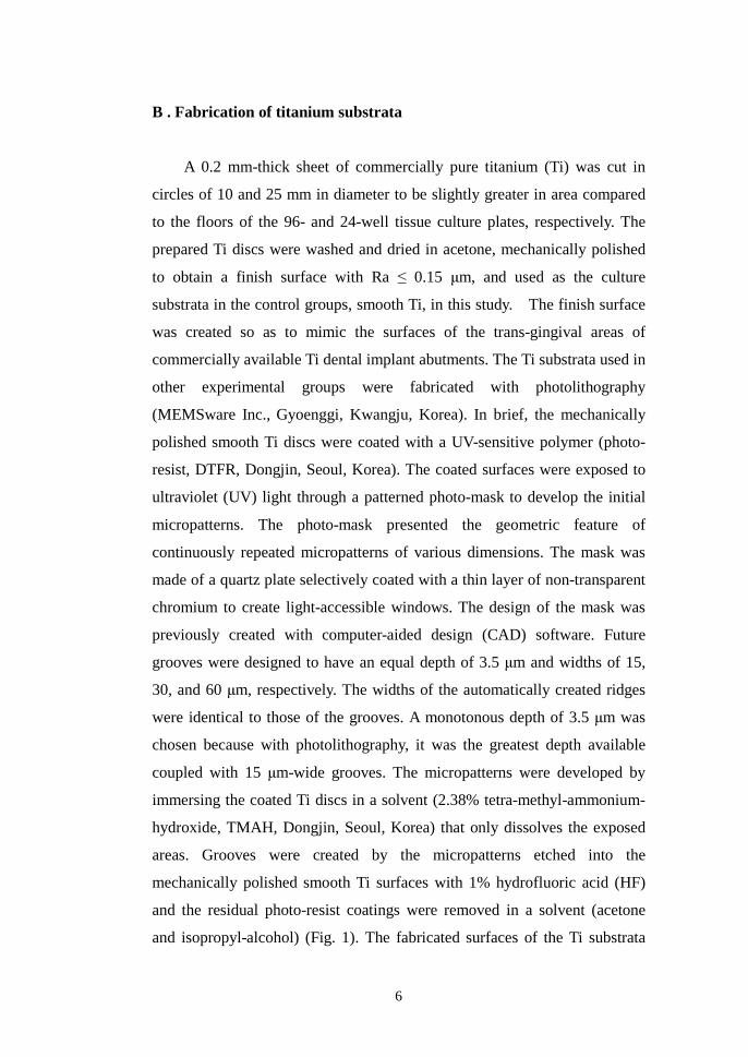
and isopropyl-alcohol) (Fig. 1). The fabricated surfaces of the Ti substrata
7
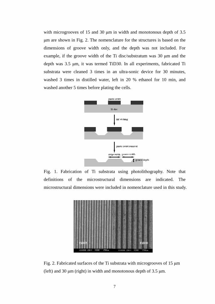
with microgrooves of 15 and 30 µm in width and monotonous depth of 3.5
µm are shown in Fig. 2. The nomenclature for the structures is based on the
dimensions of groove width only, and the depth was not included. For
example, if the groove width of the Ti disc/substratum was 30 µm and the
depth was 3.5 µm, it was termed TiD30. In all experiments, fabricated Ti
substrata were cleaned 3 times in an ultra-sonic device for 30 minutes,
washed 3 times in distilled water, left in 20 % ethanol for 10 min, and
washed another 5 times before plating the cells.
Fig. 1. Fabrication of Ti substrata using photolithography. Note that
definitions of the microstructural dimensions are indicated. The
microstructural dimensions were included in nomenclature used in this study.
Fig. 2. Fabricated surfaces of the Ti substrata with microgrooves of 15 µm
(left) and 30 µm (right) in width and monotonous depth of 3.5 µm.

8
C . Adhesion assay
The floors of 96-well plates were removed and the remaining plastic
cylinders were attached to the fabricated surfaces of the 10 mm-diameter Ti
discs using a silicone bonding agent. As a result, a total of twenty four 96-
well Ti substrata were prepared and divided into the four groups of smooth
Ti, TiD15, TiD30, and TiD60. Cultured human gingival fibroblasts (3rd-4th
passage) were detached from subculture with trypsin-EDTA, resuspended,
and plated simultaneously on the 96-well Ti substrata at a cell population
density of 3 10ⅹ3 cells/ml in DMEM supplemented with 10% FBS and
antibiotics. From three samples out of six in each group, the media were
removed at respective 1 and 2 h incubation and the unattached cells were
washed away twice with warmed PBS. Attached cells were stained with a
solution of 0.2 % crystal violet in 10 % ethanol for 5 min at room
temperature. Stained cells were washed with PBS and the bound dye was
solubilized with 100 µl of solubilization buffer (50/50 mixture of 0.1 M
NaH2PO4, Ph 4.5 and 50 % ethanol) to be transferred to 96-well plates.
Absorbance (optical density, OD) was measured using ELISA analyzer
(Spectra MAX 250, Molecular Devices Co., Sunnyvale, CA, USA) at 570
nm.
D . Scanning electron microscopy
Adhesion and morphology of fibroblasts on the surfaces of fabricated Ti
substrata of smooth Ti, TiD15, TiD30, and TiD60 was analyzed under
scanning electron microscopic (SEM) observations. At 16 and 48 h plating
and incubation, samples were fixed in 4% paraformaldehyde for 2 h and
rinsed twice in 0.1M PBS for 10 min. Samples were again fixed for another
2 h in 1% osmium tetroxide. Samples were then dehydrated by immersing
for 10 min in each of 20, 50, 60, 70, 80, 90, and 100% ethanol dilutions
followed by drying with a critical point dryer (Jumb, Bio-Rad Laboratories
9
Inc., Hercules, CA, USA). Samples were sputter coated with a 10-nm gold
film (SEM coating system, E 5150, Bio-Rad Laboratories Inc., Hercules, CA,
USA) and imaged with S-800 FE-SEM® (HITACHI, Tokyo, Japan).
E . XTT assay
A total of forty eight 24-well Ti substrata were prepared and divided
into the four groups of smooth Ti, TiD15, TiD30, and TiD60. Cultured
human gingival fibroblasts were trypsinized and simultaneously plated on
the 24-well Ti substrata at a cell population density of 1ⅹ104 cells/ml in
DMEM supplemented with 10% FBS and antibiotics. Cells were incubated
in a humidified incubator at 37°C with 5% CO2 in 95% air for 24, 48, 72,
and 96 h. In all groups, the viability and proliferation of fibroblasts was
determined by XTT assay (Cell Proliferation Kit II, Roche Applied Science,
Mannheim, Germany) as described by Roehm et al.20 (1991). In brief, XTT
labeling reagent (soium 3'-[1-[(phenylamino)-carbonyl]-3, 4-tetrazolium]-
bis(4-methoxy-6- nitro)benzene-sulfonic acid hydrate) and electron coupling
reagent (N-methyl dibenzopyrazine methyl sulfate, PMS in PBS) were
thawed. Each vial was thoroughly mixed and a clear solution was obtained.
XTT labeling mixture was prepared by mixing 50 µl of XTT labeling reagent
and 1 µl of electron coupling reagent. 50 µl of XTT labeling mixture was
added per well and incubated for 4 h in a humidified incubator at 37°C with
5% CO2 in 95% air. In all groups, formazan products were transferred to 96-
well plates and the absorbance was measured using ELISA analyzer (Spectra
MAX 250, Molecular Devices Co., Sunnyvale, CA, USA) at 470 nm with a
reference wavelength at 650 nm.
F . RT-PCR
Fibroblasts were trypsinized at 24, 48, 72, and 96 h incubation. Gene
expression of human gingival fibroblasts cultured on the untreated floors of
10
24-well polystyrene microplates served as the positive control. Total RNA in
each sample was extracted using Trizol (Trizol: U.S. Patent No. 5, 346,994,
Gibco BRL, Grand Island, NY, USA) according to the manufacturer’s
instructions. The concentration of extracted total RNA in each sample was
estimated by the absorption at 260nm. 1 mg of total RNA samples was
converted to cDNA with reverse transcriptase (Promega Co., Madison,
Wisconsin, USA). The polymerase chain reaction was carried out using Taq
polymerase (Roche Diagnostics, Mannheim, Germany), 10 x buffer, 25mM
MgCl2 and 25mM dNTPs (dGTP, dCTP, dATP and dTTP). Expression of
various genes involved in cell-matrix adhesion and cell-cycle progression
were analyzed in reverse transcriptase-polymerase chain reaction (RT-PCR)
(Table 1). The PCR primer of β-actin was used as the housekeeping gene.
The amplification was done in a PCR thermal cycler (Bio-Rad Laboratories
Inc., Hercules, CA, USA) under the following conditions: 35 cycles of 94 ℃
for 30 sec, 58 for 45 sec, and 72 for 30 sec. The amplification products ℃ ℃
were electrophoresed on 2% agarose gels, and visualized with ethidium
bromide. The density of each mRNA was compared using quantitative
analysis software (Bio-Rad Laboratories Inc., Hercules, CA, USA) between
the groups on successive timelines.
G . Statistical analysis
Experiments were repeated simultaneously and independently in
triplicate. The mean values and standard deviations of the data from the
adhesion and XTT assay were calculated. One-way analysis of variance
(ANOVA) in SPSS 13.0 software program was used to compare the mean
values of the data between the groups of smooth Ti, TiD15, TiD30, and
TiD60 (p<0.05).

11
Table 1. Gene-Specific Primers used in RT-PCR
Target Sense Antisense Bp
FN21 5'-CGAAATCACAGCCAGTAG-3' 5'-ATCACATCCACACGGTAG-3' 639
α5 integrin22 5'-ACCAAGGCCCCAGCTCCATTAG-3' 5'-GCCTCACACTGCAGGCTAAATG-3' 376
β1 integrin23 5'-GAGCAGCAAGGACTTTGGG-3' 5'-GAGCAGCAAGGACTTTGGG-3' 537
cyclin D124 5'-ATTAGTTTACCTGGACCCAG-3' 5'-GATGGAGCCGTCGGTGTAGATGCA-3' 399
cyclin E25 5'-CAGCCTTGGGACAATAATGC-3' 5'-TGCAGAAGAGGGTGTTGCTC-3' 254
cyclin A224 5'-ATTAGTTTACCTGGACCCAG-3' 5'-CACAAACTCTGCTACTTCTG-3' 443
CDK426 5'-CCAAAGTCAGCCAGCTTGACTGTT-3' 5'-CATGTAGACCAGGACCTAAGGACA-3' 193
CDK626 5'-TGATGTGTGCACAGTGTCACGAAC-3' 5'-CTGTATTCAGCTCCGAGGTGTTCT-3' 737
CDK226 5'-ACGTACGGAGTTGTGTACAAAGCC-3' 5'-GCTAGTCCAAAGTCTGCTAGCTTG-3' 405
p2126 5'-AGTGGACAGCGAGCAGCTGA-3' 5'-TAGAAATCTGTCATGCTGGTCTG-3' 380
p2726 5'-AAACGTGCGAGTGTCTAACGGGA-3' 5'-CGCTTCCTTATTCCTGCGCATTG-3' 454
Skp227 5'-CAACTACCTCCAACACCTATC-3' 5'-TCCTGCCTATTTTCCCTGTTCT-3' 326
α-SMA28 5'-GTCCACCGCAAATGCTTCTAA-3' 5'-AAAACACATTAACGAGTCAG-3' 141
TGF-βR-I29 5'-ATTGCTGGACCAGTGTGCTTCGTC-3' 5'-TAAGTCTGCAATACAGCAAGTTCCATTCTT-3' 668
TGF-βR-II29 5'-CGCTTTGCTGAGGTCTATAAGGCC-3' 5'-GATATTGGAGCTCTTGAGGTCCCT-3' 395
NF-κB130 5'-CCTGGATGACTCTTGGGAAA-3' 5'-CTAGCCAGCTGTTTCATGTC-3' 174
β-actin 5'-ATCGTGGGCCGCCCTAGGCA-3' 5'-TGGCCTTAGGGTTCAGAGGGG-3' 345
FN: fibronectin, CDK: cyclin-dependent kinase, p21: cyclin-dependent kinase inhibitor 1A,
p27: cyclin-dependent kinase inhibitor 1B, Skp2: S-phase kinase-associated protein-2,
α-SMA: α-smooth muscle actin, TGF-βR-I: type I transforming growth factor (TGF-)β
receptor, TGF-βR-II: type II transforming growth factor (TGF-)β receptor, NF-κB1: nuclear
factor of kappa light polypeptide gene enhancer in B-cells 1
12
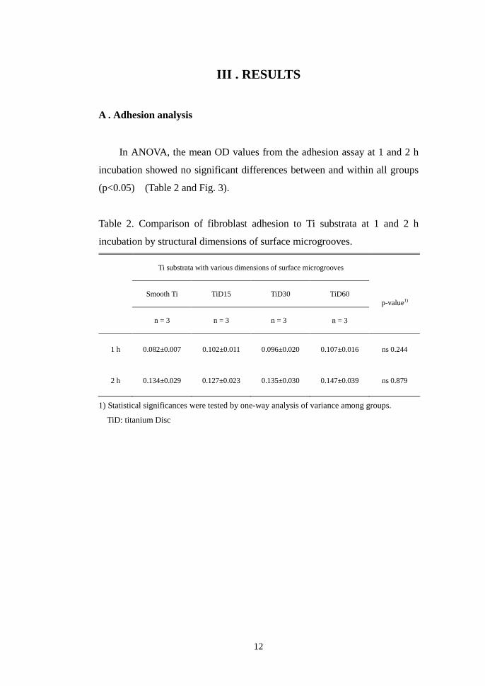
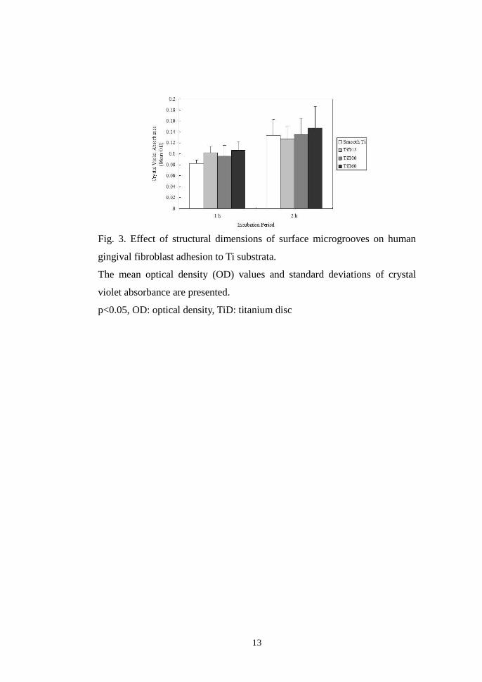
III . RESULTS
A . Adhesion analysis
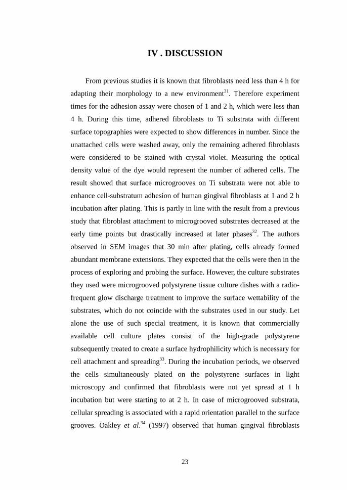
In ANOVA, the mean OD values from the adhesion assay at 1 and 2 h
incubation showed no significant differences between and within all groups
(p<0.05) (Table 2 and Fig. 3).
Table 2. Comparison of fibroblast adhesion to Ti substrata at 1 and 2 h
incubation by structural dimensions of surface microgrooves.
Ti substrata with various dimensions of surface microgrooves
Smooth Ti TiD15 TiD30 TiD60
p-value1)
n = 3 n = 3 n = 3
n = 3
1 h 0.082±0.007 0.102±0.011 0.096±0.020 0.107±0.016 ns 0.244
2 h 0.134±0.029 0.127±0.023 0.135±0.030 0.147±0.039
ns 0.879
1) Statistical significances were tested by one-way analysis of variance among groups.
TiD: titanium Disc
13
Fig. 3. Effect of structural dimensions of surface microgrooves on human
gingival fibroblast adhesion to Ti substrata.
The mean optical density (OD) values and standard deviations of crystal
violet absorbance are presented.
p<0.05, OD: optical density, TiD: titanium disc

14
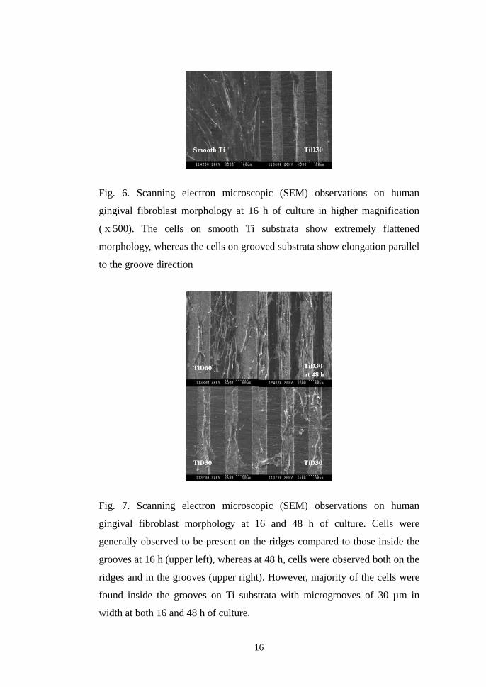
B . Morphological analysis
In SEM, orientation of fibroblasts along the microgrooves on TiD15,
TiD30, and TiD60 were observed at 16 h incubation, whereas the cells on the
smooth Ti were observed to be oriented in random directions (Fig. 4). At 48
h, more extensive orientations on TiD15, TiD30, and TiD60 were observed
(Fig. 5). On both timelines, the degree of fibroblast alignment according to
the orientation angle in relation to the direction of the microgrooves was not
significantly different between TiD15, TiD30, and TiD60. Fibroblasts on the
smooth Ti substrata were extensively spread and showed a flat morphology.
In contrast, fibroblasts on microgrooved Ti substrata were elongated and less
flat compared to those on smooth Ti substrata. (Fig. 6) General morphology
of the cells did not vary with the groove dimensions. On all microgrooved Ti
substrata, cells were present inside the microgrooves as well as outside on
the ridges with a difference in the tendency between 16 and 48 h. Cells were
generally observed to be present on the ridges rather than inside the grooves
at 16 h, whereas at 48 h, cells were observed both on the ridges and in the
grooves (Fig. 7). Notably, majority of the cells were found inside the grooves
on TiD30 both at 16 and 48 h (Fig. 7). In all groups, the entire surfaces of the
substrata were filled up with fibroblasts at 48 h, suggesting that fibroblasts
almost reached their confluence, except in TiD15 where relatively fewer
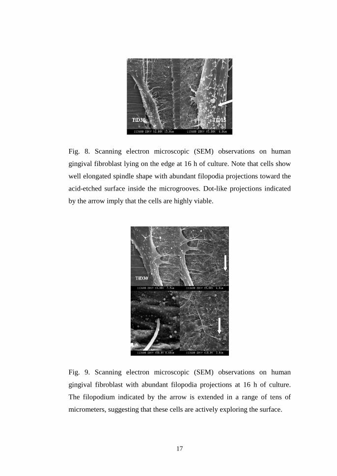
cells were found inside the grooves compared with those in the ridges. Cells
descending into the grooves were generally found on the edges or in the
corners of the microstructures. These cells showed well elongated spindle
shape and increased formation of filopodia (Fig. 8). Numerous filopodia
from the cells lying across the borders between ridges and grooves projected
towards the acid-etched surfaces inside the grooves, with some extending in
a range of tens of micrometers (arrows in Fig. 9), suggesting that these cells
were actively exploring the surfaces (Fig. 9). The diameter of a spindle-
shaped single fibroblast appeared to be identical to that of the microgrooves
in TiD15.

15
Fig. 4. Scanning electron microscopic (SEM) observations on human
gingival fibroblast morphology and orientation at 16 h of culture. Note that
the cells are aligned in random directions on smooth Ti substrata. Contact
guidance is observed in cells cultured on microgrooved Ti substrata.
Fig. 5. Scanning electron microscopic (SEM) observations on human
gingival fibroblast morphology and orientation at 48 h of culture. Note that
the cells are more oriented parallel to the microgrooves than the cells at 16 h
of culture.
16
Fig. 6. Scanning electron microscopic (SEM) observations on human
gingival fibroblast morphology at 16 h of culture in higher magnification
(ⅹ500). The cells on smooth Ti substrata show extremely flattened
morphology, whereas the cells on grooved substrata show elongation parallel
to the groove direction
Fig. 7. Scanning electron microscopic (SEM) observations on human
gingival fibroblast morphology at 16 and 48 h of culture. Cells were
generally observed to be present on the ridges compared to those inside the
grooves at 16 h (upper left), whereas at 48 h, cells were observed both on the
ridges and in the grooves (upper right). However, majority of the cells were
found inside the grooves on Ti substrata with microgrooves of 30 µm in
width at both 16 and 48 h of culture.
17
Fig. 8. Scanning electron microscopic (SEM) observations on human
gingival fibroblast lying on the edge at 16 h of culture. Note that cells show
well elongated spindle shape with abundant filopodia projections toward the
acid-etched surface inside the microgrooves. Dot-like projections indicated
by the arrow imply that the cells are highly viable.
Fig. 9. Scanning electron microscopic (SEM) observations on human
gingival fibroblast with abundant filopodia projections at 16 h of culture.
The filopodium indicated by the arrow is extended in a range of tens of
micrometers, suggesting that these cells are actively exploring the surface.

18
C . Assessment of viability and proliferation
In ANOVA, the mean OD values of the formazan absorbance at 72 h
incubation were significantly different between and within all groups
(p<0.05). According to the data using the Ti discs with various dimensions of
surface microgrooves as culture substrata, the results from the XTT assay
were significantly related between those obtained at 72 h incubation.
Multiple comparison of the fibroblast viability and proliferation data from
the XTT assay at 72 h incubation showed the mean OD value of TiD15 was
significantly greater compared to that of the control group, smooth Ti
(p<0.05) (Table 3 and Fig. 10). All other comparisons between groups were
not statistically significant. As was noted in the timeline analysis, the mean
OD value in TiD15 continuously increased up to 72 h, with a burst of
increase between the time points of 48 and 72 h, but markedly decreased at
96 h. However, the mean OD values in the groups of smooth Ti and TiD30
continuously increased up to 96 h, with the latter showing slightly higher
rate of increase.

19
Table 3. Comparison of fibroblast viability and proliferation on Ti substrata
after 24, 48, 72, and 96 h of culture by structural dimensions of surface
microgrooves.
Ti substrata with various dimensions of surface microgrooves
Smooth Ti TiD15 TiD30
TiD60
p-value1)
n = 3 n = 3 n = 3
n = 3
24 h 0.310±0.010 0.294±0.047 0.343±0.030 0.327±0.043
ns 0.417
48 h 0.439±0.050 0.471±0.058 0.492±0.005 0.460±0.022
ns 0.472
72 h 0.513±0.063 0.795±0.143 0.636±0.080 0.561±0.107
< 0.05
T2) a b a,b a,b
96 h 0.586±0.103 0.586±0.163 0.718±0.082 0.566±0.024
ns 0.328
1) Statistical significances were tested by one-way analysis of variance among groups.
2) The same letters indicate non-significant difference between groups based on Tukey’s
multiple comparison tests.
TiD: Titanium Disc

20
Fig. 10. Effect of structural dimensions of surface microgrooves on human
gingival fibroblast viability and proliferation on Ti substrata.
The mean optical density (OD) values and standard deviations of formazan
absorbance are presented.
*: Significantly greater compared with the smooth Ti (control) group
p<0.05, OD: optical density, TiD: titanium disc

21
D . Gene expression analysis
Increased levels of expression of transcripts in TiD15 and TiD30 were
noted with the genes encoding FN, α5 integrin, β1 integrin, cyclin E, CDK2,
and CDK4 at 24, 48, and 72 h incubation compared with those in the groups
of polystyrene and smooth Ti. On the other hand, decrease in cyclin D1,
cyclin A2, and �-SMA gene expression levels were noted in TiD15 and
TiD30 on all timelines compared with those in the groups of polystyrene and
smooth Ti. In comparison between groups on successive timelines of culture,
CDK6, p21, p27, Skp2, TGF-βR-I, TGF-βR-II, and NF-κB1 genes showed
controversial results in the expression levels (Fig. 11).

22
Fig. 11. Analysis on expression of genes involved in cell-matrix adhesion
and G1/S cell cycle progression in RT-PCR.
TiD: titanium disc, RT-PCR: reverse transcriptase-polymerase chain reaction
FN: fibronectin, CDK: cyclin-dependent kinase, p21: cyclin-dependent
kinase inhibitor 1A, p27: cyclin-dependent kinase inhibitor 1B, Skp2: S-
phase kinase-associated protein-2, α-SMA: α-smooth muscle actin, TGF-βR-
I: type I transforming growth factor (TGF-)β receptor, TGF-βR-II: type II
transforming growth factor (TGF-)β receptor, NF-κB1: nuclear factor of
kappa light polypeptide gene enhancer in B-cells 1
23
IV . DISCUSSION
From previous studies it is known that fibroblasts need less than 4 h for
adapting their morphology to a new environment31. Therefore experiment
times for the adhesion assay were chosen of 1 and 2 h, which were less than
4 h. During this time, adhered fibroblasts to Ti substrata with different
surface topographies were expected to show differences in number. Since the
unattached cells were washed away, only the remaining adhered fibroblasts
were considered to be stained with crystal violet. Measuring the optical
density value of the dye would represent the number of adhered cells. The
result showed that surface microgrooves on Ti substrata were not able to
enhance cell-substratum adhesion of human gingival fibroblasts at 1 and 2 h
incubation after plating. This is partly in line with the result from a previous
study that fibroblast attachment to microgrooved substrates decreased at the
early time points but drastically increased at later phases32. The authors
observed in SEM images that 30 min after plating, cells already formed
abundant membrane extensions. They expected that the cells were then in the
process of exploring and probing the surface. However, the culture substrates
they used were microgrooved polystyrene tissue culture dishes with a radio-
frequent glow discharge treatment to improve the surface wettability of the
substrates, which do not coincide with the substrates used in our study. Let
alone the use of such special treatment, it is known that commercially
available cell culture plates consist of the high-grade polystyrene
subsequently treated to create a surface hydrophilicity which is necessary for
cell attachment and spreading33. During the incubation periods, we observed
the cells simultaneously plated on the polystyrene surfaces in light
microscopy and confirmed that fibroblasts were not yet spread at 1 h
incubation but were starting to at 2 h. In case of microgrooved substrata,
cellular spreading is associated with a rapid orientation parallel to the surface
grooves. Oakley et al.34 (1997) observed that human gingival fibroblasts
24
generally spread radially across the ridges and grooves and gradually
elongate in a direction parallel to the grooves within 6 h of seeding. However,
the difference in the degree of alignment between 15- and 3-µm to 5-µm
grooves diminished at 24 h. Results from the separate studies, including the
one from ours, suggest that elongated and aligned fibroblasts on
microgrooved substrata would start to show increased adhesion, compared to
those on smooth substrata, at a specific time point from 4 to 6 h. Our result
from the adhesion assay at 1 and 2 h incubation strongly implies that primary
human gingival fibroblasts were not capable of forming cell-substratum
adhesion, but merely formed the initial cell-substratum contact to the
surfaces of Ti substrata on the experimental timelines.
In natural tissues, gingival fibroblasts are arranged in a three-
dimensional (3D) organization that provides appropriate functional and
spatial conditions. To mimic this environmental characteristic of natural
tissues in vitro, we prepared the Ti substrata with surface microgrooves of
reasonable width and depth. The dimensions of the microstructures used in
this study were designed to be ensured that human gingival fibroblasts would,
at the same, lie inside the grooves and display contact guidance35. To verify
this, we plated fibroblasts at a cell population density of 1ⅹ103 cells/ml and
observed at 16 and 48 h incubation. The plated cell number was selected so
as to inhibit intercellular collision. 16 h was selected according to the studies
demonstrating that, on both materials of Ti and polyacrylamide, fibroblasts
deposit sufficient amount of fibronectin (FN) onto the substrates for cell-
matrix adhesion by the time point13,36. Deposition of FN by the cells was
considered to enable them to form cell-matrix adhesions leading to
subsequent spreading or elongation. In SEM, majority of the typical spindle-
shaped fibroblasts were found on ridge edges or in groove corners. Filopodia
formation was abundant in these cells as has previously been reported37.
Also abundant were the small dot-like protrusions on cell dorsa which were
considered to be metabolite-secreting vesicles or filopodia budding (arrow in
Fig. 8). Fibroblasts featuring abovementioned characteristics in SEM images

25
were considered to be highly viable and very similar in overall shape to
those of fibroblasts cultured in 3-D nanoenvironment38,39. The observed
filopodia mainly projected towards the acid-etched surfaces of the grooves,
implying that these cells were actively exploring the surfaces before
migrating (arrows in Fig. 9). This result supports other results from two very
recent studies demonstrating the importance of filopodia in cell behavior. A
study by Partridge and Marcantonio40 (2006) verified that filopodia
containing both integrin and actin formed the initial cell-matrix contacts and
further generated mature focal adhesions. Another recent study by Galbraith
et al.41 (2007) suggested that actin and β1 integrin in cellular projections
including filopodia at the leading edge of a migrating cell, probe ligand and
create sticky fingers. Since fibroblasts are the most abundant cells found in
gingival connective tissues, active projection of filopodia onto the acid-
etched surfaces inside the microgrooves observed in this study encourages
the use of such surface in enhancing peri-implant connective tissue
attachment42.
One of the most frequently used colorimetric assays, XTT, used in this
study is a method well established in investigating the influence of a specific
substance/material and/or process on cell survival. The results of the XTT
assay for cell viability and proliferation are influenced not only by the
number but also by the metabolic activity of cells. Cleavage of XTT by
dehydrogenase enzymes of metabolically active cells yields a highly colored
formazan product which is water soluble. This feature enables the product to
be diluted in medium, eliminates the need for formazan crystal solubilization
prior to absorbance measurements as is required in MTT assay, protects
cellular membranes from being damaged, and leads the cells to survive
throughout the test process20. On planning for this experiment, we expected
the fibroblasts to show increased proliferation on the microgrooved Ti
substrata compared with the smooth ones. The results from the timeline
proliferation analysis using XTT assay gave us no reason to confirm such
expectation except the result, at 72 h incubation, that fibroblasts in TiD15
26
showed a burst of increase in proliferation, but suddenly decreased on the
successive timeline at 96 h. This, in part, corresponds with the results in the
study by Dalby et al.14 (2003) that on the quartz substrata with surface
microgrooves of 12.5 µm in width, fibroblasts showed up-regulation of
genes involved in cell signaling, DNA transcription, RNA-protein translation,
and extracellular matrix formation and remodeling after 24 h of culture,
while all the genes were down-regulated by day 5. They suggested two
reasons for this unique result. First, cell types of mesenchymal origin are
highly proliferative initially (Days 0–3), and then, as confluence is
approached, this proliferation slows and the cells start to differentiate and
produce matrix, ultimately making new tissue. This is also true with the
time–line of cultured cells’ proliferation in microenvironments43. Fibroblasts
on both TiD15 were considered to have reached confluence at a certain time
point around 72 h of culture. We suggest that human gingival fibroblasts
cultured on Ti substrata with surface microgrooves of 15 µm in width and
3.5 µm in depth reach their confluence earlier compared with those cultured
on the smooth Ti or Ti substrata with greater dimensions of surface
microgrooves. The second reason suggested by Dalby14 (2003) and his
coworkers was that as the cells align in response to the grooves, cell
activities are initially increased and thus take on far less random
morphologies than are produced on the flat surfaces. The nuclei shapes
would be restricted leading to the restriction of chromosomal arrangement
and result in the down-regulation at later phases. This suggestion does not
coincide with our results from the morphologic analysis that, in SEM at 48 h,
the majority of fibroblasts were still observed to be incapable of moving into
the grooves of 15 µm in width. It seems unlikely that any of the fibroblasts
cultured in our experiment would have experienced such nucleic
deformation. In TiD30 however, a gradual increase in proliferating activities
was noted on successive timelines up to 96 h.
Since proliferating data of the cells cultured on substrata with relatively
greater microgrooves in dimension are so scarce, we found a somewhat

27
corresponding result from a very recent study analyzing on microgroove-
related cell migration and alignment44. They used 3T3 fibroblastic cells
plated on structured Ti-alloy surfaces with groove depth/ridge height of 5-22
µm, and spacing of 5-30 µm. Cells migrating on surface structures with
groove or ridge width up to 20-30 µm exhibited the highest mean migration
velocity. Similar to our findings in SEM, the authors found no significant
difference in the frequencies of a certain type of cell shape among the
surface structures with different dimensions. Also, they strongly implied that
the width of 30 µm roughly represents the main diameter of the cells. This
also corresponds with our SEM findings that at 16 h incubation, compared
with the results at 48 h, only the fibroblasts on TiD30 substrata were
generally found inside the grooves compared with other grooves, and that, a
single cell in one groove. The importance of the presence of cell contact on
the bottom of the grooves in relation to contact guidance in mechanical point
of view has been suggested by Walboomers et al.17 (1999). The authors also
concluded that at confluence, microgrooves with relatively wider grooves of
20 µm in width compared to the narrower ones were able to support greater
numbers of cells. In the groups of smooth Ti and TiD60, fibroblasts showed
no more increase in viability and proliferation at 96 h compared to that at 72
h, implying that they were or, at least, were about to reach their confluence at
around 72 h. However, a continuous increase was seen in TiD30 even at 96 h,
suggesting that the fibroblasts were still proliferating at the time. Indeed, in a
study using polymethylmethacrylate (PMMA) surfaces with precise 3D
microgrooved structures, wider and deeper grooves of 30 µm in width and
20 µm in depth mostly stimulated fibroblast proliferation45. Taken together,
we suggest that microgrooves of 30 µm in width enable human gingival
fibroblasts to readily descend into the grooves in early phases of culture, thus
lead the cells to proliferate for longer period of time, but this suggestion
leaves much to be investigated using appropriate methods to analyze the
differences in the rate of cell proliferation. Once the cells lie in a groove, the
probability of protrusion attachment outside the groove becomes small due

28
to the convex character of the groove edge46. Since microgrooves have been
verified to influence the deposition patterns of ECM proteins47 and the ECM
guides the orientation of the cell division axis48, proliferation of the cells
lying in a groove would be influenced by the dimension of the structure.
In this study, the purpose of using both adhesion and XTT assay was
twofold: 1) to assay the cell-substratum adhesion and proliferation of
fibroblasts plated on titanium substrata with different dimensions of
microgrooves and 2) to determine, after plating, the optimal time period of
incubation at which fibroblasts on the microgrooved Ti substrata would
show significantly greater extent of viability and proliferation compared with
those on smooth Ti substrata. It was under the verifications of the
abovementioned conditions that expression of genes related to cell-
substratum adhesion and adhesion-dependent cell cycle progression of plated
fibroblasts were analyzed in RT-PCR. However, we statistically failed to
verify the specific timeline as well as dimension of microgrooves on which
human gingival fibroblasts would show enhanced both cell-substratum
adhesion and viability/proliferation compared to those in other experimental
conditions. For most types of cells, the opportunities for anchorage and
attachment depend on the surrounding matrix, which is usually made by the
cell itself. Thus, a cell can create an environment that then acts back on the
cell to reinforce its differentiated state. Furthermore, the ECM that a cell
secretes forms part of the environment for its neighbors as well as for the
cell itself, and thus tends to make neighboring cells differentiate in the same
way, which in another term, contact guidance49. Fibroblasts also need
adhesions to the ECM for their proliferation and growth. Fibroblasts undergo
cell spreading on adhesion to the ECM such as FN fibrils. In the event, the
generated forces by the cells align and stiffen ECM and partially unfold
ECM proteins50. Up-regulation of FN gene in TiD15 and TiD30 at 24 and 72
h incubation compared with the expression in the groups of polystyrene and
smooth Ti supports the hypothesis that, after such geometry is sensed and
considered to be favorable, cells create their own environment by secreting
29
the ECM proteins, and that, in a continuous feedback fashion51 (Fig. 11). Our
result in FN gene analysis corresponds, in part, with the results of two
previous studies by Chou et al.13,52 (1995 and 1996) from the Brunette group.
In the first previous study, human gingival fibroblasts cultured on Ti reduced
the FN mRNA level by 58% at 16 h, but increased it by 2.6-fold at 90 h
compared to those on culture plastics, although the cell numbers on the two
surfaces were essentially the same52. In our study, down-regulation of FN
genes in the smooth Ti group compared to those in the control group at 24 h
supports the above result. However, FN gene expression levels in the smooth
Ti group were noted to be essentially the same with those in the polystyrene
group at 48 and 72 h, or even reduced at 96 h. The reduction at 96 h seems to
be due to the marked expression of FN gene in the polystyrene group
compared with any other group on the same timeline. This result is in
accordance with the other results of our study analyzing genes such as �5
integrin, cyclin E, CDK2, and CDK4. One possible explanation is that the
fibroblasts in the polystyrene group were slower in reaching their confluence
than the ones in the experimental groups. However, it still does not
correspond with our viability and proliferation data that the fibroblasts in all
groups, except those in TiD30, seems to have reached their confluence at 48
or 72 h. In the second previous study by Chou et al.13 (1995), the authors
compared the FN mRNA levels of fibroblasts cultured on smooth Ti and Ti
with microgrooves of 3 µm in depth and 6-10 µm in width. In the study, the
grooved surface increased the amounts of FN mRNA/cell approximately 3.5-
fold at 16 hours, 1.9-fold at 40 hours and 2.2-fold at 90 hours. Despite the
differences in microgroove dimensions, our result from the analysis on FN
gene on successive timelines supports this previous result. It is confirmed
that surface microgrooves of 15 and 30 µm in width and of equal 3.5 µm in
depth on Ti substrata acted to up-regulate human gingival fibroblasts’ FN
gene on successive timelines from 24 to 72 h. However, due to the
possibility of confluence at around 72 h of culture, the results at 96 h cannot
be confirmed. After all, our data from the comparative analysis on FN gene

30
expression between the groups of various microgroove dimensions do not
correspond with the result from the study by Chou et al.13 (1995). Gene
expression analysis at 96 h as well as at later phases needs further
investigation.
As was mentioned earlier, cells align and stiffen ECM and partially
unfold ECM proteins and, acting back, the ECM proteins reorganize the
cytoskeleton of the cells. Integrins act to couple the two structures and,
together with diverse intracellular signaling proteins recruited to the site,
integrate signals in both inside-out and outside-in directions. Integrins
recognize the outside-in signals such as positional cues encoded by the
extracellular matrix and convert them into biochemical signals that control
the cell’s response to soluble growth factors and cytokines53. To analyze the
cell-substratum adhesion of human gingival fibroblasts to the substrata used
in this study, �5β1 integrin, a fibronectin receptor54, has been analyzed in
this study. Marked down-regulation of �5 integrin gene was noted in the
groups of polystyrene and smooth Ti at 48 and 72 h of culture. Also, the gene
was up-regulated in TiD15 and TiD30 at 24, 48, and 72 h (Fig. 11). The
expression profile of �5 integrin gene in 20 samples on successive timelines
coincided with that of FN gene, suggesting that human gingival fibroblasts
in TiD15 and TiD30 were more active in FN assembly, namely the FN
fibrillogenesis55,56, than those in the groups of polystyrene and smooth Ti.
Integrins can be localized in different adhesion structures called focal
complexes, focal adhesions, fibrillar adhesions, and 3D-matrix adhesions.
These structures reflect different stages of interaction between cells and
ECM57. 3D-matrix adhesion was verified in a comprehensive study by
Cukierman et al.58 (2001). The joint up-regulation of FN and �5 integrin
genes in TiD15 and TiD30 compared to the groups of polystyrene and
smooth Ti suggests that human gingival fibroblasts cultured on Ti substrata
with reasonably deep and wider microgrooves than the diameter of a single
fibroblast actively formed either fibrillar adhesions or 3D-matrix adhesions,
31
or both, according to the criteria proposed by Cukierman et al.58 (Fig. 12).
Assuming that all the substrata induced formation of fibrillar adhesions due
to the expression of �5 integrin gene in every sample regardless of the
differences in degree, our result imply that fibroblasts in TiD15 and TiD30
might have been involved in forming 3D-matrix adhesions. However, β1
integrin genes showed even levels of expression in all groups on all
timelines, except in the groups of polystyrene and smooth Ti at 72 h. This
result requires evaluation of more molecules involved in adhesion structures,
such as β3 integrin, paxillin, and focal adhesion kinase (FAK). After all, β1
integrin is known to be involved in diverse in vivo biology59.
Fig. 12. Molecular composition of three types of cell adhesions.
Cyclin D1 associates with cyclin-dependent kinases (CDKs) 4 and 6 to
phosphorylate the retinoblastoma protein (pRb), blocks its growth inhibitory
activity, and promotes the release of bound E2F transcription factor60. As a
result, cyclin E–CDK2 and cyclin A–CDK2 complexes are activated, leading
to the entry into and completion of S phase61,62. To evaluate the effect of
substratum surface topography on G1/S cell cycle progression, we analyzed
the expression of cyclin D1, E, and A2 genes as well as CDK4, 6, and 2
genes. The expression patterns of cyclin E, CDK4, and CDK2 genes
coincided with those of FN and �5 integrin. However, cyclin D1 and cyclin

32
A2 genes showed exactly inverse expression profiles with those of cyclin E,
CDK4, and CDK2 genes. The tendency was even true with the comparison
between FN and �5 integrin genes (Fig. 11). It has previously been proposed
that cyclin D1 in actively proliferating cells, whose elevated level is
maintained through G1 phase but reduced in S phase and then elevated again
in G2 phase for proliferation to continue, serves as a cell cycle regulatory
switch63. In accordance to a view that cyclin D1 is inhibitory to DNA
synthesis64, the decline of cyclin D1 expression in S phase is not considered
to be dependent on the environment of the cell, but directly regulated by cell
cycle progression. DNA duplication occurs during S phase, which requires
10-12 hours and occupies about half of the cell-cycle time in most
mammalian cells. A flattened morphology has been verified to be a
prominent feature of S-phase cells and, in turn, the spread cells that are
generally adhered to substratum are found during the S-phase65,66. Our result
from the analyses on various cyclin- and CDK-genes is reminiscent of the
fact that, in non-transformed cells, progression through cell cycle is tightly
regulated on both translation and transcription levels. We suggest prudently
that, referring to the viability and proliferation data in our study, human
gingival fibroblasts on microgrooved and smooth Ti substrata were
progressing through different stages of cell cycle at specific experiment time
points, leading to the difference in the rate of reaching the confluence. We
would like to point out that, so far, very little is known about the control of
cell cycle progression after the cells have already started cycling. Since most
of the cell cycle studies use serum-starved cultures, analysis on the steps
involved in cell cycle progression from serum-deprived cultures is so rare63.
Therefore, the results from our gene expression analyses on the molecules
involved in cell cycle regulation cannot be directly applied to actively
cycling cells due to the fact that all our experiments were basically done in
serum-derived cultures. However, the fact that the complex nature of
mammalian cell cycle regulation has not yet been fully uncovered still leaves
a possibility that our results from gene analysis would yield a novel proposal,

33
assuming that more molecules be analyzed. For example, the gene
expression data of cyclin D1 and cyclin A2 genes showing inverse
expression profiles with those of FN gene, cyclin E gene, and so on, are the
data that need research related to 3-D cell behavior. A study of gene
expression profiling on vascular smooth muscle cells cultured in 3-D
collagen matrices reported that a CDK inhibitor p21cip1, known to have
opposing regulatory functions to those of cyclin D1 in G1 phase cell cycle
control, showed much higher gene expression level compared to the cells
cultured on 2-D surfaces67.
Regulation of �-SMA gene expression is a complex process and shows
substantial tissue specificity68. Expression of �-SMA is one of the most
reliable markers for myofibroblast differentiation and the prominent features
of myofibroblasts are the development of a strong actin network reinforced
by �-SMA and the presence of enhanced cell adhesion mechanisms69. TGF-
β1 promotes the induction of the myofibroblastic phenotype through the
activation of the RhoA–ROCK and JNK signaling pathways in human
gingival fibroblasts. These phenotypical changes include the ability of cells
to adhere and spread over fibronectin, the reinforcement of focal adhesion
complexes, the organization of actin stress fibers, and the further induction
of the myofibroblast marker �-SMA70. In our study, up-regulation of �-SMA
gene in the polystyrene and smooth Ti group suggests that not only the tissue
culture plastics but also the commercially pure titanium are capable of
inducing myofibroblastic phenotype in human gingival fibroblasts, which
was considered to be promoted by endogenous TGF-β1 or exogenous TGF-
β1 in FBS. Down-regulation of �-SMA gene in TiD15 and TiD30 on all
timelines in our study is in contrast to the result from Thampatty and Wang71
(2007). In their study, human tendon fibroblasts cultured on the
microgrooved surfaces increased �-SMA protein expression after treatment
with TGF-β1. The authors suggested that TGF-β1 treated fibroblasts in
microgrooves became differentiated into myofibroblasts. There are two

34
notable points to be discussed from their study. First, they did not compare
the extent of �-SMA protein expression between the cells plated on smooth
and microgrooved substrates. All the cells were plated only on microgrooved
membranes and the group lacking in the TGF-β1 treatment was used as a
control. Second, they used fibronectin-coated silicone membranes with
surface microgrooves of 10 µm in groove and ridge width, and 3 µm in
groove depth as culture substrata. The tendon fibroblasts showing extreme
elongation appeared to be trapped inside the microgrooves, which might
have affected the result. Since the microgrooves used in our study were
greater in dimension compared to those used in the study by Thampatty and
Wang, it may be inappropriate to compare the results of the two studies. The
only similarity in the experimental designs lies in the fact that both studies
used culture substrata with surface microgrooves. In addition, fibroblasts in
both studies were treated with TGF-β1, since the growth factor was
considered to be abundant in DMEM supplemented with 10% FBS used in
all experiments in our study, whereas Thampatty and Wang incubated the
cells in DMEM containing both 1% FBS and 5.0 ng/ml of TGF-β1 for 24 h.
It has been postulated that growth response of human gingival fibroblasts to
TGF-β1 or PDGF-BB stimulation in serum-free medium was equivalent to
growth obtained in medium containing 10% FBS without added growth
factors72. As verified in SEM, human gingival fibroblasts in our study were
not observed to be trapped inside, but rather spontaneously crawled into the
microgrooves. Therefore, we examined both TGF-βR-I and TGF-βR-II
whose genes and proteins were verified in a previous study to show
alterations in expression in relation to the change in fibroblasts’ sensitivity to
TGF-β1-induced �-SMA expression in 3-D environment compared to those
in 2-D73. Although the authors used several types fibroblasts and a number of
experimental designs, only their result from normal skin fibroblasts routinely
cultured in supplemented DMEM containing 10% fetal calf serum without
TGF-β1 treatment were compared with the result of our study. In their study,
the result from Western blot analysis revealed that the fibroblasts in 3-D

35
culture expressed lower levels of �-SMA and TGF-βR-II but higher levels of
TGF-βR-I compared to those cultured in 2-D monolayer culture. However,
they used a unique 3-D culture system called ‘spheroid culture’, which lead
the fibroblasts to differ in shape to that of the cells in TiD15 or TiD30 in our
study. Presuming that the polystyrene/smooth Ti substrata and the
microgrooved Ti substrata used in our study correspond to the 2-D and the 3-
D environment used in the study by Kunz-Schughart et al.73, respectively,
the results from the two studies related to the �-SMA expression of the
cultured cells correspond with each other. Likewise, the overall expression
pattern of TGF-βR-II genes on all timelines in our study and the TGF-βR-II
expression in the study by Kunz-Schughart et al.73, both in relation to the �-
SMA expression, correspond with each other except that the TGF-βR-II gene
in TiD30 showed an inverse expression profile with that of the �-SMA gene
(Fig. 13). This inverse gene expression profile was totally unexpected and
strongly requires further investigation. It has been verified more than a
decade ago that, unlike TGF-β1 with the ability to directly bind to TGF-βR-
II, TGF-β2 binding to TGF-βR-II requires coexpression of TGF-βR-I74,
leading to an efficient formation of TGF-βR-I/R-II heteromer followed by
activation of TGF-β1 signaling pathway. In our study, joint up-regulation of
TGF-βR-I and TGF-βR-II was noted in TiD30 after 24 and 96 h of culture,
while the �-SMA gene was down-regulated. Taken together, it is implied that
titanium substrata with surface microgrooves of 30 µm in width and 3.5 µm
in depth may act to trigger TGF-β1 signaling pathway. An interesting result
from our study is an unexpected up-regulation of �-SMA gene in TiD60 after
72 h of culture. A recent study evaluating SMC behavior on 3-D
microchannels in biodegradable polymeric films by Shen et al.75 (2006)
reported that the cells grown on flat film showed even expression of �-SMA
after 3 days’ and 7 days’ culture, whereas the �-SMA expression of SMCs
grown on 160 µm-wide and 25 µm-deep micropatterns was significantly
enhanced after 7 days’ culture compared to that after 3 days’ culture. The
36
authors stated that the SMC phenotype shift from synthetic to a more
contractile phenotype was observed when SMCs reached confluence on
micropatterns with wide microchannels (80, 120, and 160 µm wide), in
which the cells were found to proliferate with random orientation initially
when the cell density was low, but switched to elongated morphology,
unidirectional orientation, and a more contractile phenotype with increased
�-SMA expression nearing confluence. The fibroblasts in TiD60 in our study
might have undergone such process after descending into the microgrooves
around at 72 h incubation. Also, joint up-regulation of cyclin D1 and cyclin
A2 genes in TiD60 at 72 h incubation was noted along with an overall
similarity in the gene expression pattern on all timelines to that of �-SMA
gene (Fig. 13).
Fig. 13. Analysis on expression of genes encoding cyclin D1/A2, α-SMA,
and TGF-βR-I/II.
Down-regulation of �-SMA gene was verified in another genomic
analysis study on cells in 3-D matrix by Li et al.67 In the study, the authors
grew subcultured human aortic smooth muscle cells (HASMCs) in adherent
collagen gels and compared the gene expression in 3-D matrix vs. that on 2-
D matrix. For SMCs cultured in 3-D matrix; cells were cast into the collagen
gel and for those cultured on 2-D matrix; cells were seeded on the top of the
collagen gel. The authors reported the DNA microarray results that the
expression of α-SMA in 3-D matrix was slightly lower than that in 2-D
37
matrix. They confirmed the result in RT-PCR as well as in Western blot
analysis and concluded that both α-SMA gene and protein are down-
regulated in 3-D collagen matrix compared to the 2-D matrix. Comparison of
our RT-PCR results from the analyses on genes other than α-SMA with the
results of the study by Li et al.67 offers us a number of correspondences as
well as several points to be discussed. The first gene to be compared is the
p21 gene, also known as WAF1, p21CIP1, CDK-interaction protein 1, DNA
synthesis inhibitor, and cyclin-dependent kinase inhibitor 1A. This gene
encodes a potent cyclin-dependent kinase inhibitor. The encoded protein
binds to and inhibits the activity of cyclin-CDK2 or -CDK4 complexes, and
thus functions as a regulator of cell cycle progression at G1. The expression
of this gene is tightly controlled by the tumor suppressor protein p53,
through which this protein mediates the p53-dependent cell cycle G1 phase
arrest in response to a variety of stress stimuli. This protein can interact with
proliferating cell nuclear antigen (PCNA), a DNA polymerase accessory
factor, and plays a regulatory role in S phase DNA replication and DNA
damage repair. Li et al.67 reported the significantly higher expression level of
p21 gene in 3-D matrix by more than 2-fold increase compared with that in
2-D matrix and suggested that p21 may be responsible for the lower
proliferation rate in 3-D matrix. In our study, relatively higher expression of
p21 gene in primary human gingival fibroblasts was obvious in TiD30, and
in a degree, in TiD60, compared with that in the groups of polystyrene,
smooth Ti, and TiD15 after 24, 48, 72 h of culture. However at 96 h
incubation, an inverse expression profile was noted with that on other
timelines resulting in up-regulation of the gene in TiD15. This seems to be
strongly related to the sudden decrease in the viability and proliferating
activity of the fibroblasts in TiD15 at 96 h compared with those at 72 h, as
has been confirmed by the XTT assay in this study. We suggested earlier that
the result provides a strong implication of reaching the cells’ confluence at
around 72 h incubation. Also in mouse embryonic stem cells, p21 gene has
been reported to be significantly up-regulated in 3-D porous tantalum

38
scaffold with the pore size up to 150 µm76. With our results from the analysis
on α-SMA and p21 gene expression and with the results of the several
studies discussed, we suggest that the fibroblasts on TiD30 in our study
express specific genes that are characterized in 3-D matrix culture. Further,
we confirmed this in SEM analysis on cellular morphology and conclude
that, titanium substrata with surface microgrooves of 30 µm in width and 3.5
µm in depth provided human gingival fibroblasts with three-dimensional
microenvironment. Indeed, Li et al.67 characterized cell morphology by
staining SMCs in 3-D or on 2-D collagen matrix on actin and vinculin, and
found that the cells on 2-D matrix had prominent stress fibers and
lamellipodia, whereas the cells in 3-D matrix had less actin stress fibers and
usually had multiple filopodia that might follow the collagen fibrils due to
contact guidance. We were able to verify this feature of cells in 3-D collagen
matrix in TiD30 under SEM observations in our study. The second gene to
be compared is cellular fibronectin (FN). Li et al.67 postulated that higher
expression levels of p21 and collagen I genes are the most prominent
features in 3-D matrix culture compared with 2-D, which was again
confirmed by Liu et al.76 However, Li et al.67 used collagen matrix for both
2-D and 3-D environment. Had it been the fibronectin matrix that they used
for culture environment, their result would possibly have been a distinct one.
In fact, in searching for clues for overcoming the failures for most
biomaterials, Vogel and Baneyx46 insisted upon using fibronectin matrix, not
collagen. One of the reasons they proposed was that only fibronectin binds to
�5β1 integrins and plays a central role in soliciting cellular behavior and
integrin signaling that compares well to those found for cells in naturally
occurring matrices. Following the view from Vogel and Baneyx46, we
analyzed expression of FN, �5 integrin, and β1 integrin genes and verified
that FN genes were up-regulated and the expression was most pronounced in
TiD30 at 24 and 48 h. In order to analyze the stimulatory effects of a 3-D
microenvironment on cell-mediated fibronectin fibrillogenesis, Mao and
Schwarzbauer77 (2005) cultured fibroblasts on a pre-assembled 3D

39
fibronectin matrix and found significant stimulation of fibronectin fibril
assembly compared to the cells in 2D culture. Again, the pronounced
expression of FN and �5 integrin genes in TiD30 in our study suggests that
titanium substrata with surface microgrooves of 30 µm in width and 3.5 µm
in depth provided human gingival fibroblasts with three-dimensional context
of culture and implies that the cells might have formed 3D-matrix adhesion.
The third and the last gene to be compared is the cyclin A2 gene. Li et al.67
assessed the SMCs’ proliferation rate in 3-D or on 2-D matrices by
subjecting the cells to flow cytometry analysis for DNA content after 24 h of
culture and reported that SMCs in 3-D matrix had less cells in S phase than
SMCs on 2-D matrix, but had more cells in G0/G1 phase, suggesting that
SMCs in 3-D matrix had a lower proliferation rate. Indeed, they confirmed
in microarray analysis that cyclin A1 gene was down-regulated in 3-D matrix,
although expression of the gene is known to be specific to germ cells. Lower
expression of cyclin A2 gene in TiD15, TiD30, and TiD60 at 24 h incubation
compared with that in the groups of polystyrene and smooth Ti was noted in
our study. Because Li et al.67 did not continue to assess SMC proliferation at
phases later than 24 h of culture, our result from the proliferation data using
XTT assay on successive timelines were not compared with their result.
Instead, we discuss here, further analysis on cyclin A2 gene at later phases of
culture. The overall expression pattern of cyclin A2 gene corresponded with
that of cyclin D1 gene on all timelines in our study. However, comparing the
expression profiles of the two genes with that of p21 gene revealed that
cyclin D1/A2 gene vs. p21 gene are inversely regulated, except in TiD15 at
48 h incubation and in all groups at 72 h incubation (Fig. 14). Assuming that
the cells in different groups displayed varying proliferation rate, as has been
confirmed in the timeline analysis with XTT assay, it is suggested that
primary human gingival fibroblasts in our study underwent alterations in cell
cycle progression and proliferation rate in a time period between 48 and 72 h
of culture.

40
Fig. 14. Analysis on expression of genes encoding cyclin D1/A2 and p21cip1.
To further confirm the complex regulation of the cell cycle genes, we
analyzed gene expression of p27 and Skp2, the result of which merely
aggravated the complexity. However, we found some of the interesting
comparisons to be discussed. Comparison of the overall expression pattern
of p27 gene with that of cyclin E gene revealed that the two genes showed a
nearly exact inverse expression profile after 48, 72, and 96 of culture, except
in TiD15 at 48 h, suggesting that p27 played a specific role in regulating the
expression of cyclin E in human gingival fibroblasts cultured on titanium
substrata with various dimensions of surface microgrooves (Fig. 15).
Moreover, some of the results of marked p27 gene expressions even
coincided with the expression profiles of cyclin D1/A2 genes. Among them,
exclusion of the results of the marked p27 gene expression in the groups of
polystyrene and smooth Ti leaves us the gene expression profiles under the
two critical experimental conditions in our study, the TiD15 at 48 h and the
TiD60 at 72 h. Pronounced expression of several genes under such
conditions in this study strongly suggests that further analysis on additional
genes be the key to unveiling the signal transduction pathways exclusive to
sensing microgrooves. In addition to the reputed role in preventing the
activation of cyclin E-CDK2 or cyclin D-CDK4 complexes, and thus
controlling the cell cycle progression at G1, p27 has been reported to be
involved in numerous other biological activities. Among them, a role has
been suggested for cytoplasmic p27kip1 in the regulation of cell migration
independent of cyclin–CDK inhibition78. Also, p27 has even been suggested

41
to play a role in assembling and maintaining the stability of cyclin D1-CDK4
complexes79. It would be valuable to further establish the role of p27kip1 in
sensing the microenvironment, such as the microgrooves used in our study.
Predominantly in S phase, activated p27kip1 is specifically recognized by an
F-box protein, S-phase kinase-associated protein-2 (Skp2 or p45). This
protein interacts with S-phase kinase-associated protein 1 (SKP1 or p19) and,
together with other F-box proteins constitutes one of the four subunits of
ubiquitin protein ligase complexes called SCFs (SKP1-cullin-F-box).
SCFSkp2 is responsible for phosphorylation-dependent ubiquitination, that is
the degradation, of p27kip1. Recently, this Skp2 dependent-degradation of
p27kip1 has been verified to be regulated by focal adhesion kinase (FAK),
thereby rendering the activity of Skp2 adhesion-dependent, while levels of
p21cip1 were regulated independent of Skp280. We analyzed Skp2 gene and,
again, the result merely aggravated the complexity in relation to the
comparison with the p27 gene expression. However, on comparison between
Skp2, NF-κB1, and TGF-βR-I genes, we found that the expression profiles
of these genes almost exactly coincide with each other (Fig. 16). The genes
were up-regulated in the groups of microgrooved Ti substrata at 24 and 96 h
of culture, whereas nearly none of the three genes were readily expressed at
48 and 72 h of culture. The Rho GTPase, Rac1, has been shown to be an
important downstream of Ras in cyclin D1 transcriptional regulation, and 3-
D nanofibrillar surfaces have been suggested to induce sustained activation
of Rac in NIH3T3 fibroblasts and normal rat kidney cells81. Also in mouse
embryonic stem cells, enhanced proliferation and self-renewal of the cells on
3-D nanofibrillar surfaces have been shown to be correlated with the
activation of Rac and phosphoinositide 3-kinase (PI3K) pathway. Since Rac1
induction of cyclin D1 transcription has been proposed to involve activation
of a transcription regulator, NF-κB82, we analyzed the expression of NF-κB1
in order to determine whether Rac-dependent signaling was involved in the
induction of cyclin D1 transcription in fibroblasts cultured on microgrooved
substrata. Comparing the expression profile of NF-κB1 gene with that of
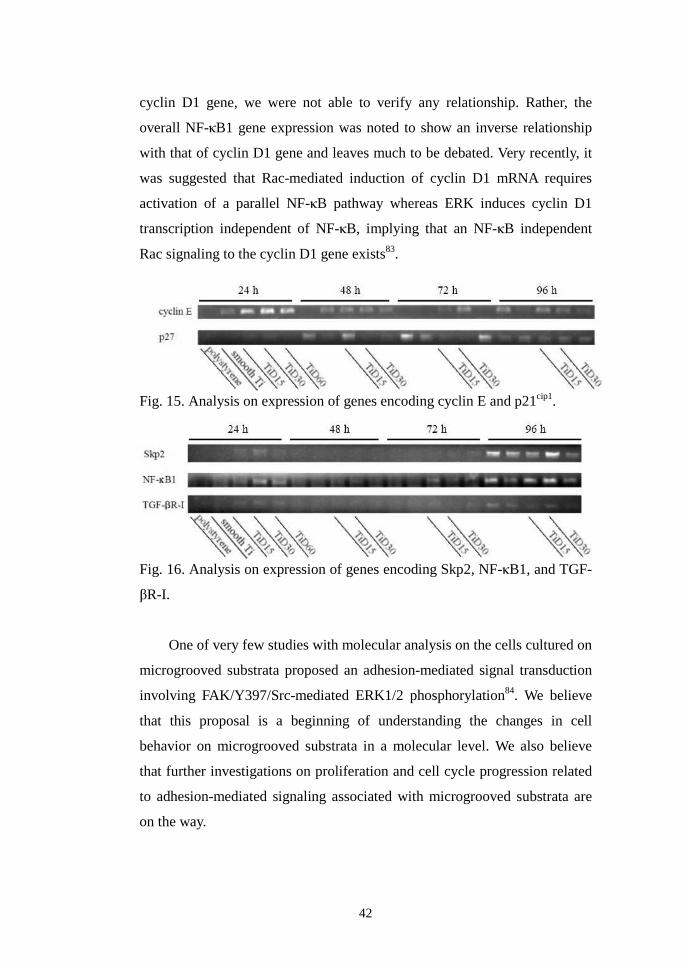
42
cyclin D1 gene, we were not able to verify any relationship. Rather, the
overall NF-κB1 gene expression was noted to show an inverse relationship
with that of cyclin D1 gene and leaves much to be debated. Very recently, it
was suggested that Rac-mediated induction of cyclin D1 mRNA requires
activation of a parallel NF-κB pathway whereas ERK induces cyclin D1
transcription independent of NF-κB, implying that an NF-κB independent
Rac signaling to the cyclin D1 gene exists83.
Fig. 15. Analysis on expression of genes encoding cyclin E and p21cip1.
Fig. 16. Analysis on expression of genes encoding Skp2, NF-κB1, and TGF-
βR-I.
One of very few studies with molecular analysis on the cells cultured on
microgrooved substrata proposed an adhesion-mediated signal transduction
involving FAK/Y397/Src-mediated ERK1/2 phosphorylation84. We believe
that this proposal is a beginning of understanding the changes in cell
behavior on microgrooved substrata in a molecular level. We also believe
that further investigations on proliferation and cell cycle progression related
to adhesion-mediated signaling associated with microgrooved substrata are
on the way.

43
V . CONCLUSION
In this study, we hypothesized that surface microgrooves of appropriate
depth and extensive width on Ti substrata that enable the cells to readily
descend into themselves, would alter various cell behaviors including
viability and proliferation of cultured human gingival fibroblasts. The
purpose of this study was to determine the dimension of surface
microgrooves on Ti substrata that shows the greatest positive influence on
characterizing specific cell behavior of cultured human gingival fibroblasts.
For this study, we prepared commercially pure titanium discs divided
into four groups of smooth titanium substrata and the titanium substrata with
surface microgrooves of monotonous 3.5 µm in depth and 15, 30, and 60 µm
in width using photolithography. Adhesion, morphology, viability and
proliferation, and gene expression of human gingival fibroblasts cultured on
these substrata on successive timelines was analyzed and compared between
groups. From the results, the following conclusions were made.
1. There was no difference between the numbers of human gingival
fibroblasts adhered to smooth Ti substrata and those adhered to Ti
substrata with surface microgrooves at 1 and 2 h incubation.
Fibroblasts merely formed initial cell-substratum contact by the
times.
2. In SEM, contact guidance of human gingival fibroblasts parallel to
the direction of microgrooves was observed. Cells were able to
readily descend into the microgrooves of 30 µm in width and 3.5 µm
in depth at the early phase of culture, whereas at the later phase,
cells in all groups were found both in the grooves and on the ridges.
Cells on the ridge edges or in groove corners were spindle shaped
with abundant filopodia formation towards the acid-etched surface

44
inside the microgrooves, thus mimicked the shape of the fibroblasts
cultured in three-dimensional (3D) nanoenvironment.
3. On successive timelines, human gingival fibroblasts cultured on Ti
substrata with various dimensions of surface microgrooves showed
differences in the rate of reaching their confluence. Human gingival
fibroblasts cultured on Ti substrata with microgrooves of 15 µm in
width and 3.5 µm in depth significantly increased their viability and
proliferation compared with those cultured on smooth Ti substrata
after 72 h of culture and decreased after 96 h, whereas the cells on
the microgrooves of 30 µm in width and 3.5 µm in depth continued
increasing their viability and proliferation up to after 96 h of culture.
4. A joint up-regulation of matrix-assembly genes, such as fibronectin
and �5 integrin genes, was noted in human gingival fibroblasts
cultured on titanium substrata with microgrooves of 15 and 30 µm in
width and an equal 3.5 µm in depth.
5. Gene expression pattern specific to the cells in 3D-matrix culture,
such as down-regulation of �-smooth muscle actin gene along with
up-regulation of fibronectin and p21 genes, was pronounced in
human gingival fibroblasts cultured on Ti substrata with
microgrooves of 30 µm in width and 3.5 µm in depth.
In reference to the results above, two conclusions were made.: 1)
Surface microgrooves of 15 µm in width and 3.5 µm in depth on titanium
substrata increase the viability and proliferation, as well as the expression of
genes involved in the matrix assembly of cultured human gingival
fibroblasts. 2) Surface microgrooves of 30 µm in width and 3.5 µm in depth
on titanium substrata provide human gingival fibroblasts with a three-
dimensional context of culture, thus show corresponding gene expression.

45
VI . REFERENCES
1. Brunette, D. M. and Chehroudi, B.: The effects of the surface
topography of micromachined titanium substrata on cell behavior in
vitro and in vivo. J. Biomech. Eng. 121: 49-57, 1999.
2. Jansen, J. A., den Braber, E. T., Walboomers, X. F., and de Ruijter, J. E.:
Soft tissue and epithelial models. Adv. Dent. Res. 13: 57-66, 1999.
3. Weiss, P.: Experiments on cell and axon orientation in vitro: the role of
colloidal exudates in tissue organization. J. Exp. Zool. 100: 353–86,
1945.
4. den Braber, E. T., de Ruijter, J. E., Ginsel, L. A., von Recum, A. F., and
Jansen, J. A.: Quantitative analysis of fibroblast morphology on
microgrooved surfaces with various groove and ridge dimensions.
Biomater. 17: 2037-44, 1996.
5. Wang, N., Ostuni, E., Whitesides, G. M., and Ingber, D. E.:
Micropatterning tractional forces in living cells. Cell Motil. Cytoskeleton
52: 97-106, 2002.
6. Chen, C. S., Alonso, J. L., Ostuni, E., Whitesides, G. M., and Ingber, D.
E.: Cell shape provides global control of focal adhesion assembly.
Biochem. Biophys. Res. Commun. 307: 355-61, 2003.
7. Loesberg, W. A., Walboomers, X. F., van Loon, J. J., and Jansen, J. A.:
The effect of combined cyclic mechanical stretching and microgrooved
surface topography on the behavior of fibroblasts. J. Biomed. Mater. Res.
A 75: 723-32, 2005.
8. Loesberg, W. A., Walboomers, X. F., van Loon, J. J., and Jansen, J. A.:
The effect of combined hypergravity and micro-grooved surface
topography on the behaviour of fibroblasts. Cell Motil. Cytoskeleton 63:
384-94, 2006.
9. Ingber, D. E.: Tensegrity II. How structural networks influence cellular
information processing networks. J. Cell Sci. 116: 1397-1408, 2003.

46
10. Dalby, M. J.: Topographically induced direct cell mechanotransduction.
Med. Eng. Phys. 27: 730-41, 2005.
11. Thery, M., Pepin, A., Dressaire, E., Chen, Y., and Bornens, M.: Cell
distribution of stress fibres in response to the geometry of the adhesive
environment. Cell Motil. Cytoskeleton 63: 341-55, 2006.
12. Folkman, J. and Moscona, A.: Role of cell shape in growth control.
Nature 273: 345-49, 1978.
13. Chou, L., Firth, J. D., Uitto, V. J., and Brunette, D. M.: Substratum
surface topography alters cell shape and regulates fibronectin mRNA
level, mRNA stability, secretion and assembly in human fibroblasts. J.
Cell Sci. 108: 1563-73, 1995.
14. Dalby, M. J., Riehle, M. O., Yarwood, S. J., Wilkinson, C. D., and Curtis,
A. S.: Nucleus alignment and cell signaling in fibroblasts: response to a
micro-grooved topography. Exp. Cell Res. 284: 274-82, 2003.
15. den Braber, E. T., de Ruijter, J. E., Smits, H. J., Ginsel, L. A., von
Recum, A. F., and Jansen, J. A.: Quantitative analysis of cell
proliferation and orientation on substrata with uniform parallel surface
micro-grooves. Biomater. 17: 1093-99, 1996.
16. Walboomers, X. F., Croes, H. J., Ginsel, L. A., and Jansen, J. A.: Contact
guidance of rat fibroblasts on various implant materials. J. Biomed.
Mater. Res. 47: 204-12, 1999.
17. Walboomers, X. F., Monaghan, W., Curtis, A. S., and Jansen, J. A.:
Attachment of fibroblasts on smooth and microgrooved polystyrene. J.
Biomed. Mater. Res. 46: 212-20, 1999.
18. Chehroudi, B. and Brunette, D. M.: Subcutaneous microfabricated
surfaces inhibit epithelial recession and promote long-term survival of
percutaneous implants. Biomater. 23: 229-37, 2002.
19. Walboomers, X. F. and Jansen, J. A.: Effect of microtextured surfaces on
the performance of percutaneous devices. J. Biomed. Mater. Res. A 74:
381-7, 2005.
20. Roehm, N. W., Rodgers, G. H., Hatfield, S. M., and Glasebrook, A. L.:

47
An improved colorimetric assay for cell proliferation and viability
utilizing the tetrazolium salt XTT. J. Immunol. Methods 142: 257-65,
1991.
21. Zhang, C., Meng, X. F., Zhu, Z. H., Yang, X., and Deng, A. G.: Role of
connective tissue growth factor in plasminogen activator inhibitor-1 and
fibronectin expression induced by transforming growth factor β1 in renal
tubular cells. Chinese Med. J. 117: 990-6, 2004.
22. Yao, M., Zhou, X. D., Zha, X. L., Shi, D. R., Jian Fu, J., He, Lu, H. F.,
and Tang, Z. Y.: Expression of the integrin �5 subunit and its mediated
cell adhesion in hepatocellular carcinoma. J. Cancer Res. Clin. Oncol.
123: 435-40, 1997.
23. Bonson, S., Jeansonne, B. G., and Lallier, T. E.: Root-end Filling
Materials Alter Fibroblast Differentiation. J. Dent. Res. 83: 408-13, 2004.
24. Shen, G., Xu, C. J., Chen, C., Hebbar, V., and Kong, A. N.: p53-
independent G1 cell cycle arrest of human colon carcinoma cells HT-29
by sulforaphane is associated with induction of p21CIP1 and inhibition of
expression of cyclin D1. Cancer Chemother. Pharmacol. 57: 317–27,
2006.
25. Sawasaki, T., Shigemasa, K., Shiroyama, Y., Kusuda, T., Fujii, T.,
Parmley, T. H., O’Brien, T. J., and Ohama, K.: Cyclin E mRNA
overexpression in epithelial ovarian cancers: inverse correlation with
p53 protein accumulation. J. Soc. Gynecol. Investig. 8: 179-85, 2001.
26. Wong, H. and Riabowol, K.: Differential CDK-inhibitor gene expression
in aging human diploid fibroblasts. Exp. Gerontol. 3: 311-25, 1996.
27. Mammoto, A., Huang, S., Moore, K., Oh, P., and Ingber D. E.: Role of
RhoA, mDia, and ROCK in cell shape-dependent control of the Skp2-
p27kip1 pathway and the G1/S transition. J. Biol. Chem. 279: 26323-30,
2004.
28. Baouz, S., Giron-Michel, J., Azzarone, B., Giuliani, M., Cagnoni, F.,
Olsson, S., Testi, R., Gabbiani, G., and Canonica, G. W.: Lung
myofibroblasts as targets of salmeterol and fluticasone propionate:
48
inhibition of α-SMA and NF-κB. Int. Immunol. 17: 1473-81, 2005.
29. Boumédiene, K., Takigawa, M., and Pujol, J. P.: Cell density-dependent
proliferative effects of transforming growth factor (TGF)-beta 1, beta 2,
and beta 3 in human chondrosarcoma cells HCS-2/8 are associated with
changes in the expression of TGF-beta receptor type I. Cancer Invest.
19: 475-86, 2001.
30. Okada, Y., Kato, M., Minakami, H., Inoue, Y., Morikawa, A., Otsuki, K.,
and Kimura, H.: Reduced expression of flice-inhibitory protein (FLIP)
and NFkappaB is associated with death receptor-induced cell death in
human aortic endothelial cells (HAECs). Cytokine 15: 66-74, 2001.
31. Grinnell, F., Ho, C. H., Tamariz, E., Lee, D. J., and Skuta, G.: Dendritic
fibroblasts in three-dimensional collagen matrices. Mol. Biol. Cell 14:
384-95, 2003.
32. Walboomers, X. F., Ginsel, L. A., and Jansen, J. A.: Early spreading
events of fibroblasts on microgrooved substrates. J. Biomed. Mater. Res.
51: 529–34, 2000.
33. Shen, M. and Horbett, T. A.: The effects of surface chemistry and
absorbed proteins on monocyte/macrophage adhesion to chemically
modified polystyrene surfaces. J. Biomed. Mater. Res. 57: 336-45, 2001.
34. Oakley, C., Jaeger, N. F., and Brunette, D. M.: Sensitivity of fibroblasts
and their cytoskeletons to substratum topographies: Topographic
guidance and topographic compensation by micromachined grooves of
different dimensions. Exp. Cell Res. 234: 413-24, 1997.
35. Brunette, D. M. Fibroblasts on micromachined substrate orient
hierarchically to grooves of different dimensions. Exp. Cell Res. 164: 11-
26, 1986.
36. Jiang, G., Huang, A. H., Cai, Y., Tanase, M., and Sheetz, M. P.: Rigidity
sensing at the leading edge through αvβ3 integrins and RPTPα. Biophys.
J. 90: 1804-9, 2006.
37. Walboomers, X. F., Croes, H. J., Ginsel, L. A., and Jansen, J. A.: Growth
behavior of fibroblasts on microgrooved polystyrene. Biomater. 19:
49
1861-8, 1998.
38. Tillman, J., Ullm, A., and Madihally, S. V.: Three-dimensional cell
colonization in a sulfate rich environment. Biomater. 27: 5618–26, 2006.
39. Noh, H. K., Lee, S. W., Kim, J. M., Oh, J. E., Kim, K. H., Chung, C. P.,
Choi, S. C., Park, W. H., and Min, B. M.: Electrospinning of chitin
nanofibers: degradation behavior and cellular response to normal human
keratinocytes and fibroblasts. Biomater. 27: 3934-44, 2006.
40. Partridge, M. A. and Marcantonio, E. E.: Initiation of attachment and
generation of mature focal adhesions by integrin-containing filopodia in
cell spreading. Mol. Biol. Cell 17: 4237-48, 2006.
41. Galbraith, C. G., Yamada, K. M., and Galbraith, J. A.: Polymerizing actin
fibers position integrins primed to probe for adhesion sites. Science 315:
992-5, 2007.
42. Kim, H., Murakami, H., Chehroudi, B., Textor, M., and Brunette, D. M.:
Effects of surface topography on the connective tissue attachment to
subcutaneous implants. Int. J. Oral Maxillofac. Implants 21: 354-65,
2006.
43. Bottaro, D. P., Liebmann-Vinson, A., and Heidaran, M. A.: Molecular
signaling in bioengineered tissue microenvironments. Ann. N. Y. Acad.
Sci. 961: 143-53, 2002.
44. Kaiser, J. P., Reinmann, A., and Bruinink, A.: The effect of topographic
characteristics on cell migration velocity. Biomater. 27: 5230-41, 2006.
45. Sun, F., Casse, D., van Kan, J. A., Ge, R., and Watt, F.: Geometric
control of fibroblast growth on proton beam-micromachined scaffolds.
Tissue Eng. 10: 267-72, 2004.
46. Alaerts, J. A., De Cupere, V. M., Moser, S., van den Bosh de Aguilar, P.,
and Rouxhet, P. G.: Surface characterization of poly(methyl
methacrylate) microgrooved for contact guidance of mammalian cells.
Biomater. 22: 1635-42, 2001.
47. den Braber, E. T., de Ruijter, J. E., Ginsel, L. A., von Recum, A. F., and
Jansen, J. A.: Orientation of ECM protein deposition, fibroblast
50
cytoskeleton, and attachment complex components on silicone
microgrooved surfaces. J. Biomed. Mater. Res. 40: 291–300, 1998.
48. Thery, M., Racine, V., Pepin, A., Piel, M., Chen, Y., Sibarita, J. B., and
Bornens, M.: The extracellular matrix guides the orientation of the cell
division axis. Nat. Cell Biol. 7: 947-53, 2005.
49. Chen, C. S., Mrksich, M., Huang, S., Whitesides, G. M., and Ingber, D.
E.: Geometric control of cell life and death. Science 276:1425-28, 1997.
50. Vogel, V. and Baneyx, G.: The tissue engineering puzzle: A molecular
perspective. Ann. Rev. Biomed. Eng. 5: 441-63, 2003.
51. Vogel, V.: Mechanotransduction involving multimodular proteins:
converting force into biochemical signals. Annu. Rev. Biophys. Biomol.
Struct. 35: 459-88, 2006.
52. Chou, L., Firth, J. D., Nathanson, D., Uitto, V. J., and Brunette, D. M.:
Effects of titanium on transcriptional and post-transcriptional regulation
of fibronectin in human fibroblasts. J. Biomed. Mater. Res. 31: 209-17,
1996.
53. Giancotti, F. G. and Tarone, G.: Positional control of cell fate through
joint integrin/receptor protein kinase signaling. Annu. Rev. Cell. Dev.
Biol. 19: 173-206, 2003.
54. Mould, A. P., Askari, J. A., Aota, S., Yamada, K. M., Irie, A., Takada, Y.,
Mardon, H. J., and Humphries, M. J.: Defining the topology of integrin
α5β1-fibronectin interactions using inhibitory anti-α5 and anti-β1
monoclonal antibodies. J. Biol. Chem. 272: 17283-92, 1997.
55. Pankov, R., Cukierman, E., Katz, B. Z., Matsumoto, K., Lin, D. C., Lin,
S., Hahn, C., and Yamada, K. M.: Integrin dynamics and matrix
assembly: tensin-dependent translocation of α5β1 integrins promotes
early fibronectin fibrillogenesis. J. Cell Biol. 148: 1075-90, 2000.
56. Clark, K., Pankov, R., Travis, M. A., Askari, J. A., Mould, A. P., Craig, S.
E., Newham, P., Yamada, K. M., Humphries, and M. J.: A specific α5β1
integrin conformation promotes directional integrin translocation and
fibronectin matrix formation. J. Cell Sci. 118: 291-300, 2005.

51
57. Zaidel-Bar, R., Cohen, M., Addadi, L., and Geiger, B.: Hierarchical
assembly of cell–matrix adhesion complexes. Biochem. Soc. Transact.
32: 416-20, 2004.
58. Cukierman, E., Pankov, R., Stevens, D. R., and Yamada, K. M.: Taking
cell-matrix adhesions to the third dimension. Science 294: 1708–12,
2001.
59. Brakebusch, C. and Fassler, R.: β1 integrin function in vivo: adhesion,
migration and more. Cancer Metastasis Rev. 24: 403-11, 2005.
60. Sherr, C. J. and Roberts, J. M.: CDK inhibitors: positive and negative
regulators of G1-phase progression. Genes Dev. 13: 1501-12, 1999.
61. Girard, F., Strausfeld, U., Fernandez, A., and Lam, N. C.: Cyclin A is
required for the onset of DNA replication in mammalian fibroblasts. Cell
67: 1169-79, 1991.
62. Weinberg, R. A.: The retinoblastoma protein and cell cycle control. Cell
81: 323– 30, 1995.
63. Stacey, D. W.: Cyclin D1 serves as a cell cycle regulatory switch in
actively proliferating cells. Curr. Opin. Cell Biol. 15: 158-63, 2003.
64. Pagano, M., Theodoras, A. M., Tam, S. W., Draetta, G. F., and Chen, J.:
Cyclin D1-mediated inhibition of repair and replicative DNA synthesis
in human fibroblasts. Genes Dev. 8: 1627-39, 1994.
65. Ingber, D. E., Prusty, D., Sun, Z., Betensky, H., and Wang, N.: Cell
shape, cytoskeletal mechanics, and cell cycle control in angiogenesis. J.
Biomech. 28: 1471–84, 1995.
66. Meredith, D. O., Owen, G. R., ap Gwynn, I., and Richards, R. G.:
Variation in cell-substratum adhesion in relation to cell cycle phases. Exp.
Cell Res. 293: 58-67, 2004.
67. Li, S., Lao, J., Chen, B. P., Li, Y. S., Zhao, Y., Chu, J., Chen, K. D., Tsou,
T. C., Peck, K., and Chien, S.: Genomic analysis of smooth muscle cells
in 3-dimensional collagen matrix. FASEB J. 17: 1376, 2003.
68. Mack, C. P., Somlyo, A. V., Hautmann, M., Somlyo, A. P., and Owens, G.
K.: Smooth muscle actin differentiation marker gene expression is
52
regulated by RhoA-mediated actin polymerization. J. Biol. Chem. 276:
341–347, 2001.
69. Tomasek, J. J., Gabbiani, G., Hinz, B., Chaponnier, C., and Brown, R.
A.: Myofibroblasts and mechano-regulation of connective tissue
remodelling. Nat. Rev. Mol. Cell Biol. 3: 349–363, 2002.
70. Smith, P. C., Cáceres, M., and Martinez, J.: Induction of the
myofibroblastic phenotype in human gingival fibroblasts by
transforming growth factor-beta1: role of RhoA-ROCK and c-Jun N-
terminal kinase signaling pathways. J. Periodontal Res. 41: 418-25,
2006.
71. Thampatty, B. P. and Wang, J. H.: A new approach to study fibroblast
migration. Cell Motil. Cytoskeleton 64: 1-5, 2007.
72. Anderson, T. J., Lapp, C. A., Billman, M. A., and Schuster, G. S.: Effects
of transforming growth factor-beta and platelet-derived growth factor on
human gingival fibroblasts grown in serum-containing and serum-free
medium. J. Clin. Periodontol. 25: 48-55, 1998.
73. Kunz-Schughart, L. A., Wenninger, S., Neumeier, T., Seidl, P., and
Knuechel, R.: Three-dimensional tissue structure affects sensitivity of
fibroblasts to TGF-beta 1. Am. J. Physiol. Cell Physiol. 284: C209-19,
2003.
74. Rodriguez, C., Chen, F., Weinberg, R. A., and Lodish, H. F.: Cooperative
binding of transforming growth factor (TGF)-β2 to the types I and II
TGF-β receptors. J. Biol. Chem. 270: 15919-22, 1995.
75. Shen, J. Y., Chan-Park, M. B., He, B., Zhu, A. P., Zhu, X., Beuerman, R.
W., Yang, E. B., Chen, W., and Chan, V.: Three-dimensional
microchannels in biodegradable polymeric films for control orientation
and phenotype of vascular smooth muscle cells. Tissue Eng. 12: 2229-40,
2006.
76. Liu, H., Lin, J., and Roy, K.: Effect of 3D scaffold and dynamic culture
condition on the global gene expression profile of mouse embryonic
stem cells. Biomater. 27: 5978-89, 2006.
53
77. Mao, Y. and Schwarzbauer, J. E.: Stimulatory effects of a three-
dimensional microenvironment on cell-mediated fibronectin
fibrillogenesis. J. Cell Sci. 118: 4427-36, 2005.
78. Assoian, R. K.: Stopping and going with p27kip1. Dev. Cell 6: 458-9,
2004.
79. Bagui, T. K., Mohapatra, S., Haura, E., and Pledger, W. J.: P27kip1 and
p21Cip1 are not required for the formation of active D cyclin-cdk4
complexes. Mol. Cell Biol. 23: 7285–90, 2003.
80. Bryant, P., Zheng, Q., and Pumiglia, K.: Focal adhesion kinase controls
cellular levels of p27/Kip1 and p21/Cip1 through Skp2-dependent and -
independent mechanisms. Mol. Cell Biol. 26: 4201-13, 2006.
81. Nur-E-Kamal, A., Ahmed, I., Kamal, J., Schindler, M., and Meiners, S.
Three dimensional nanofibrillar surfaces induce activation of Rac.
Biochem. Biophys. Res. Commun. 331: 428-34, 2005.
82. Boyer, L., Travaglione, S., Falzano, L., Gauthier, N. C., Popoff, M. R.,
Lemichez, E., Fiorentini, C., and Fabbri A.: Rac GTPase instructs NF-
κB activation by conveying the SCF complex and IκB-α to the ruffling
membranes. Mol. Biol. Cell 15: 1124-33, 2004.
83. Klein, E. A., Yang, C., Kazanietz, M. G., and Assoian, R. K.: NFkappaB-
independent signaling to the cyclin D1 gene by Rac. Cell Cycle 6: 1115-
21, 2007.
84. Hamilton, D. W. and Brunette, D. M.: The effect of substratum
topography on osteoblast adhesion mediated signal transduction and
phosphorylation. Biomater. 28: 1806-19, 2007.

54
국문 요약
표면 마이크로그루브의 크기가 티타늄 기판 상에서
배양된 인간치은섬유아세포의 세포행동에 미치는 영향
연세대학교 대학원 치의학과 (지도교수 이 근 우)
이 석 원
치과용 티타늄 임플란트와 주위 연조직간의 반응을 증진시킬 것으로
기대되는 티타늄 표면 마이크로그루브가 실제 세포행동에 미치는 영향을
규명하려는 노력이 전개되고 있다. 이러한 연구들에서 널리 사용된
마이크로그루브들은 대부분 배양된 세포의 직경보다 작은 1-10μm의
폭을 가지며 섬유아세포의 형태 변화를 야기하여 focal adhesion의
형성을 증가시키고 유전자 발현에 영향을 주는 것으로 알려져 있으나,
실험실 내 세포 증식이나 동물 생체 내 epithelial down-growth에 대한
영향은 아직까지 규명되지 못하였다.
본 연구에서는 단일 세포가 안착될 수 있는 충분한 폭과 깊이를 가지는
마이크로그루브가 섬유아세포의 세포행동, 특히 세포생존력 및 증식을
증진시킨다는 가설을 제시하였고, 이의 평가를 위하여 다양한 크기의
표면 마이크로그루브가 부여된 티타늄 기판 상에서 배양된
인간치은섬유아세포의 세포부착, 세포형태, 세포생존력 및 증식, 유전자
발현 등에 대한 분석을 시행하였다. 본 연구의 목적은 티타늄 기판
상에서 배양된 인간치은섬유아세포의 세포행동을 특징 지움에 있어 가장
바람직하게 영향을 줄 수 있는 표면 마이크로그루브의 크기를 규명하고자
하였다.
포토리토그래피를 이용하여 순수 티타늄 표면에 폭과 깊이가 각각
15/3.5 µm, 30/3.5 µm, 60/3.5 µm인 마이크로그루브를 부여하고
실험군으로 (TiD15, TiD30, TiD60), 평활한 티타늄 기판을 대조군으로
사용하였다 (smooth Ti). 각 기판 표면에서 인간치은섬유아세포를 24, 48,
72, 96시간 동안 각각 배양하고 크리스탈 바이올렛 염색, XTT분석,
주사전자현미경관찰, reverse transcriptase-polymerase chain reaction
등을 통하여 그루브의 유무와 크기에 따른 세포의 부착, 형태, 생존력 및
증식, 유전자 발현 등의 차이를 분석하였다. 세포부착과 생존력 및
증식의 경우 일요인 분산분석을 이용하여 통계 분석을 시행하였다.
이상 4가지 분석의 결과는 다음과 같다.

55
1. 세포부착 분석 결과 티타늄 기판 상에서 배양된
인간치은섬유아세포는 표면 마이크로그루브 유무와 크기에 따른
유의차를 보이지 않았다. 세포 접종 1시간 및 2시간 후
인간치은섬유아세포는 초기 세포-기판 접촉만을 형성하였다.
2. 세포형태 분석 결과 표면 마이크로그루브가 부여된 티타늄
기판 상에서 배양된 인간치은섬유아세포는 그루브의 방향과
평행하게 contact guidance를 나타내었다. 30/3.5µm 폭/깊이의
그루브는 배양 후기뿐 아니라 초기에도 그루브 내로 세포를
안착시킬 수 있었다. 능선의 모서리나 그루브의 코너에 위치한
섬유아세포들은 왕성한 filopodia의 형성 등 삼차원 배양 시와
유사한 세포형태를 나타내었다.
3. 세포생존력 및 증식의 배양시간에 따른 분석 결과 티타늄 기판
상에서 배양된 인간치은섬유아세포는 표면 마이크로그루브의
크기에 따라 세포 포화 속도에 차이를 보였다.
인간치은섬유아세포는 15/3.5µm 폭/깊이의 마이크로그루브에서
72시간 배양 시 평활한 티타늄 기판에 비하여 유의한 증가를
보였으나 96시간에서 급격한 감소를 나타낸 반면, 30/3.5µm
폭/깊이의 마이크로그루브에서는 96시간까지 지속적이고
점진적인 증가를 나타내었다.
4. 15/3.5µm 및 30/3.5µm 폭/깊이의 마이크로그루브에서 배양된
인간치은섬유아세포는 fibronectin과 5 integrin 등
기질단백질의 생성과 관계된 유전자 발현의 동반 증가를
나타내었다.
5. 30/3.5µm 폭/깊이의 마이크로그루브 상에서 배양된
인간치은섬유아세포는 -smooth muscle actin 유전자 발현의
감소와 더불어 fibronectin 및 p21 유전자 발현의 증가 등
3차원 세포배양 시 특징적인 유전자 발현의 양상을 나타내었다.
이상의 결과에 따라 티타늄 기판 표면 15/3.5µm 폭/깊이의
마이크로그루브는 배양된 인간치은섬유아세포의 세포생존력 및 증식을
증진시키고 기질단백질 생성 유전자들의 발현을 증진시키며, 30/3.5µm
폭/깊이의 마이크로그루브는 인간치은섬유아세포에게 3차원 세포배양의
환경을 제공하여 상응하는 세포형태와 유전자 발현을 유도하는 것으로
결론지을 수 있다.
핵심 되는 말: 티타늄, 마이크로그루브, 섬유아세포, 세포증식, 유전자발현